Research Collection
Journal Article
Tailored enzymatic treatment of Chlorella vulgaris cell wall leads to effective disruption while preserving oxidative stability
Author(s):
Canelli, Greta; Murciano Martínez, Patricia; Maude Hauser, Billie; Kuster, Isabelle; Rohfritsch, Zhen;
Dionisi, Fabiola; Bolten, Christoph J.; Neutsch, Lukas; Mathys, Alexander Publication Date:
2021-05
Permanent Link:
https://doi.org/10.3929/ethz-b-000471620
Originally published in:
LWT 143, http://doi.org/10.1016/j.lwt.2021.111157
Rights / License:
Creative Commons Attribution 4.0 International
This page was generated automatically upon download from the ETH Zurich Research Collection. For more information please consult the Terms of use.
ETH Library
LWT - Food Science and Technology 143 (2021) 111157
Available online 24 February 2021
0023-6438/© 2021 The Author(s). Published by Elsevier Ltd. This is an open access article under the CC BY license (http://creativecommons.org/licenses/by/4.0/).
Tailored enzymatic treatment of Chlorella vulgaris cell wall leads to effective disruption while preserving oxidative stability
Greta Canelli
a, Patricia Murciano Martínez
b, Billie Maude Hauser
a, Isabelle Kuster
a,
Zhen Rohfritsch
b, Fabiola Dionisi
b, Christoph J. Bolten
b, Lukas Neutsch
c, Alexander Mathys
a,*aETH Zürich, Sustainable Food Processing Laboratory, Schmelzbergstrasse 9, Zürich, 8092, Switzerland
bNestl´e Research Vers Chez Les Blanc, Route du Jorat 57, 1000, Lausanne, Switzerland
cInstitute of Chemistry and Biotechnology, ZHAW, Campus Grüental, 8820, W¨adenswil, Switzerland
A R T I C L E I N F O Keywords:
Microalgae
High-pressure homogenization Cell wall-degrading enzymes Bioaccessibility
Lipid oxidation
A B S T R A C T
The green microalgae Chlorella vulgaris is a source of valuable nutrients, whose bioaccessibility is limited by the structurally complex cell wall. Enzymatic degradation of the cell wall represents a remarkable alternative to mechanical treatments due to its mildness and specificity. This work aimed to define an optimal combination of enzymes to increase the lipid and protein bioaccessibilities of C. vulgaris cells while preserving oxidative stability.
Among the tested enzymes, chitinase, rhamnohydrolase, and galactanase caused the highest release of micro- algae cellular material. Treatment with this enzymatic combination produced a slight increase in protein bio- accessibility, from 49.2% ±3.9% to 58.7% ±3.5%, but no increase in lipid bioaccessibility in comparison to the control. High-pressure homogenization (HPH) led to 61.8% ± 2.6% lipid and 59.8% ±1.8% protein bio- accessibilities. Cell integrity was preserved after enzymatic treatment, while the mean particle size was reduced from 5 to 2 μm after HPH. Oxidative stability was maintained over 3 months of accelerated shelf life in untreated and enzymatically treated C. vulgaris biomass while HPH caused drastic instability and off-flavour formation.
Although more work is needed to optimise the enzymatic treatment to maximise the nutrient bioaccessibility, the presented process was successful in preserving lipid quality.
1. Introduction
Microalgae are a diverse group of unicellular organisms that popu- late all aquatic ecosystems, ranging from freshwater to marine envi- ronments (Grima et al., 2013). Microalgae have rising potential for the biotechnology industry because of their simple growth requirements and high reproductive rate (Galasso et al., 2019). Moreover, they are a promising source for nutraceuticals and functional food products due to their nutrient profile, containing high-quality protein, polyunsaturated fatty acids, dietary fibre, vitamins, and minerals (Batista et al., 2013). In our previous work, we showed that the bioaccessibility of microalgae nutrients for human digestion is limited due to the indigestible cell wall (Bernaerts et al., 2020; Canelli, Tarnutzer et al., 2020). Cell disruption is necessary for improving the extractability of the targeted compounds from the cells, as well as to enhance nutrient bioaccessibility during digestion of the whole biomass. Mechanical methods, such as bead milling and high-pressure homogenization (HPH), are reported to be efficient in terms of extractability (Günerken et al., 2015). However,
they create cell debris and emulsions that are hard to separate during industrial processing to obtain purified compounds of interest (Scherer et al., 2019). Enzymatic degradation can be considered an emerging processing technology as it is gaining attention because of the high re- action selectivity, limited energy requirements, and gentle and pollutant-free processing conditions (Baudelet et al., 2017). This might ensure the preservation of heat-sensitive compounds, avoiding product oxidation and denaturation under harsh conditions. This aspect is particularly relevant for polyunsaturated fatty acids (PUFAs) because of their high sensitivity to oxidation (Gheysen et al., 2018; Shahidi, 2001).
Previous research has shown that the lipolytic stability of wet microalgal biomass is determined by cell integrity, which is dependent on the resistance of the cell wall (species- and cultivation-dependent). More- over, cell disruption by HPH significantly impaired the lipolytic stability of Nannochloropsis during wet storage (Balduyck et al., 2017).
A drawback of an enzymatic process might be the high cost of the enzymes. Two options to reduce the cost could be the immobilization of the enzymes or the combination of this process with another method, e.
* Corresponding author.
E-mail address: alexander.mathys@hest.ethz.ch (A. Mathys).
Contents lists available at ScienceDirect
LWT
journal homepage: www.elsevier.com/locate/lwt
https://doi.org/10.1016/j.lwt.2021.111157
Received 4 December 2020; Received in revised form 16 February 2021; Accepted 18 February 2021
LWT 143 (2021) 111157
2 g., microwaves (Kim et al., 2013). Research on the enzymatic degrada- tion of C. vulgaris is limited to a few studies, none of them investigating the effect of cell wall enzymatic degradation on microalgae nutrient bioaccessibility. Previous works examined cell wall-degrading enzymes to increase lipid extractability for biofuel production from microalgae (Cho et al., 2013; Gerken et al., 2013; Taher et al., 2014) without consideration of lipid oxidation. Gerken et al. (2013) found that chiti- nase and lysozyme inhibited the growth of C. vulgaris by acting on cell wall permeability and thickness. Cellulase and β-glucosidase were used for C. vulgaris by Cho et al. (2013) to increase the lipid extraction yield from 29.2% to 73.1%. A combination of exo-glucosaminidase, alginate lyase, lysozyme, and peptidoglycan N-acetylmuramic acid deacetylase was selected by Coelho et al. (2019) to degrade the C. vulgaris cell wall and access its nutritional components. This enzyme mixture led to 23.4-fold and 1.2-fold increases in the release of protein and fatty acid contents, respectively, compared to the untreated biomass. Cell disruption by enzymatic treatment of Chlamydomonas reinhardtii for protein and lipid extraction was analysed by Sierra et al. (2017), who developed an aqueous enzymatic assisted extraction treatment to pre- serve high-value bioproducts while allowing high levels of cell disrup- tion. Biomass pretreatment with autolysing was chosen as the preferred method and demonstrated to significantly enhance protein (+20%) and lipid (+30%, hexane as solvent) recoverable yields compared to the control.
Detailed knowledge of the cell wall is necessary for cost reduction and high efficacy of the enzymatic processes (Baudelet et al., 2017). In a previous study, we characterized the cell wall composition of hetero- trophically grown C. vulgaris (Canelli et al., 2021). Galactose and rhamnose constitute 53% and 26% of C. vulgaris cell wall, suggesting the presence of galactan and rhamnose-based polymers, respectively. In addition, the detection of glucosamine suggested the occurrence of chitin and chitosan (Gerken et al., 2013). Having available a detailed chemical characterization enabled us to select a list of enzymes by confidently targeting the cell wall constituents.
The work presented herein focused on screening five cell wall- degrading enzymes on C. vulgaris, which is one of the most industri- ally cultivated microalgae and one of the few species authorized for food consumption (Caporgno & Mathys, 2018). Enzymes were screened for their capacity to cause a release of cellular material from the algal biomass. Based on these results, we defined a combination of the most effective enzymes to partially disrupt C. vulgaris cells while preserving lipid oxidative stability. Lipid and protein bioaccessibility, as well as particle size (cell integrity), of enzymatically treated biomass were compared to a biomass disrupted by HPH. This study provides new in- sights into the effect of cell wall-enzymatic degradation of microalgae cells on lipid oxidative stability during storage, including a comparison to harsher mechanical disruption technology, such as HPH.
2. Materials and methods 2.1. Microalgae culture conditions
C. vulgaris (CCALA 256) was attained from the Culture Collection of Autotrophic Organisms (Tˇrebon, Czech Republic). For enzymatic ˇ screening for disruption efficacy and nutrient bioaccessibility, freeze- dried and resuspended microalgae biomass was used. C. vulgaris was heterotrophically cultivated as reported by Canelli, Neutsch, et al.
(2020), with the glucose concentration adapted to 20 g L−1. The biomass was cultivated in batch in a 16-L bioreactor (Bilfinger Industrial Tech- nologies, Mannheim, Germany). C. vulgaris biomass was harvested after four days in stationary phase. Biomass was centrifuged, washed with demi-water, frozen (− 20 ◦C) and freeze-dried (LyoLab B, LSL Secfroid, Aclens, Switzerland) for further experiments. Fresh biomass was used for the determination of particle size and lipid oxidation. C. vulgaris was cultivated in Bold’s basal medium supplemented with 20 g L−1 glucose in 500 mL Erlenmeyer flasks, in the dark at 25 ◦C and 150 rpm in a
shaking incubator (Multitron Pro, Infors AG, Bottmingen, Switzerland).
The biomass was harvested after reaching the stationary phase and centrifuged (10000 g, 10 min, 20 ◦C). After discarding the supernatant, the pellet was used for further experiments.
2.2. Enzymatic treatment
Freeze-dried/fresh microalgae biomass was suspended (20 g L−1) in 50 mM potassium phosphate buffer at pH 6. Enzymatic treatment was performed in triplicate (n =3) in 2 mL Eppendorf tubes for screening (disruption efficacy) and in 30 mL glass tubes for other analyses (bio- accessibility, particle size, lipid oxidation). The microalgae suspension was mixed with enzyme stock solutions. The added enzymes with their respective final concentrations are reported in Table 1. Enzymes were always added in excess compared to their substrate, which was calcu- lated based on Canelli et al. (2021), and concentrations were determined based on the specific activities whenever available. For the control (untreated biomass), the enzyme solution was replaced by phosphate buffer. All tubes were incubated for 24 h at 37 ◦C in a Labinco test-tube rotator (Huber & Co. AG, Reinach BL, Switzerland). After incubation, the samples were collected and immediately cooled on ice. For enzy- matic screening, part of the sample was centrifuged (10000 g, 4 ◦C, 10 min) to obtain the supernatant fraction. For the other analysis, only the full suspension was used.
2.3. High-pressure homogenization treatment
Freeze-dried and resuspended microalgae cells were disrupted by high-pressure homogenization (HPH). HPH is one of the most efficacious disintegration methods (Buchmann et al., 2019; Safi et al., 2015; Seve- nich & Mathys, 2018), and therefore, it was selected as the reference treatment. The microalgae suspension (200 mL, 40 g L−1) was treated in a microfluidizer (M110EH, Microfluidics Corporation, Westwood, USA) at 1000 bar, applying four passes. In the microfluidizer, a Y-shaped ceramic interaction chamber F20Y (75 μm gap width) was used. In addition, a downstream auxiliary processing module H30Z (200 μm gap width) provided further treatment of the suspension and stabilization of the product flow. At an operating pressure of 1000 bar, the flow rate of the product was 480 mL min−1, and the shear rates were estimated as 6
×106 s−1 in the interaction chamber and 2.2 ×10⁶ s−1 in the auxiliary processing module, according to the manufacturer (B¨ocker et al., 2020).
The temperature of the outflow did not exceed 40 ◦C. The sample was immediately cooled on ice and used for further analysis.
2.4. Analytics
2.4.1. Disruption efficacy – total carbon and total nitrogen release To assess the efficacy of the disruption treatment, a similar method as Goettel et al. (2013) was performed. The release of cellular material was evaluated by measuring the total carbon (TC) and total nitrogen (TN) released in the supernatant after the treatment and compared to the TC and TN present in the full suspension (Equation (1)). Carbon and ni- trogen were measured by a TOC-L connected to a TN module (Shimadzu Europa, Duisburg, Germany).
TC or TNrelease into supernatant(%) = TC or TNin supernatant
TC or TNin full suspension
⋅100 (1)
2.4.2. Determination of lipid and protein bioaccessibilities
Protein and lipid bioaccessibilities were measured by an in vitro digestion model, following the standardized protocol (INFOGEST 2.0), as described in Canelli, Tarnutzer, et al. (2020). In brief, digestion was performed in triplicate (n =3) at 37 ◦C with stirring at 300 rpm. To simulate the oral phase (2 min, pH 7), the following solutions were mixed with the freeze-dried biomass (0.5 g): water (1.89 mL), simulated salivary fluid (SSF, 1.6 mL), and CaCl2 (10 μL, 0.3 M). To initiate the G. Canelli et al.
gastric phase, simulated gastric fluid (SGF, 3.2 mL) and CaCl2 (2 μL, 0.3 M) were added to the oral bolus, and the pH was set to 3. Pepsin (0.2 mL, 80000 U mL−1; Sigma-Aldrich, Buchs, Switzerland) and gastric lipase (0.2 mL, 2400 U mL−1; Lipolytech, Marseille, France) were added, and the final volume was topped up to 8 mL with water. The pH was regu- larly adjusted to 3. After 2 h of incubation, the pH was set to 7, and simulated intestinal fluid (SIF, 3.4 mL), CaCl2 (16 μL, 0.3 M), pancreatin (2 mL, 800 U mL−1; Sigma-Aldrich), and bile salts (1 mL, 0.16 mM;
Sigma-Aldrich) were added. Water was added to a final volume of 16 mL. After 2 h of incubation, an aliquot (3 mL) of full digesta was frozen with liquid nitrogen and freeze-dried. The remaining full digesta was centrifuged (30 min, 10000 g, 4 ◦C). The micellar phase (supernatant) and the pellet were separately frozen with liquid nitrogen and freeze-dried. Infant formula (Aptamil 1; Milupa, Dublin, Ireland) was used as positive control and subjected to in vitro digestion, as entire bioaccessibility was anticipated for this sample. Water (2 mL) without microalgal biomass was digested as a blank to quantify the nitrogen and fatty acids coming from the digestive fluids and enzymes.
The full digesta and micellar phase were analysed in terms of lipid and protein contents. The lipid content was expressed as the total fatty acids measured as explained in Canelli, Tarnutzer, et al. (2020). In short, fatty acids were trans-esterified by 1.5 N methanolic hydrochloric acid solution and analysed by a gas chromatograph (GC) equipped with a flame ionization detection (FID) and a split-injection port (7890A;
Agilent Technologies, Basel, Switzerland). The temperature–time pro- gramme was set as follows: 50 ◦C (0.2 min), 50–180 ◦C (120 ◦C min −1), 180–220 ◦C (6.7 ◦C min −1), and 220–250 ◦C (30 ◦C min −1) on a 70%
cyanopropyl polysilphenylene-siloxane column with an internal diam- eter of 0.1 mm, length of 10 m, and film of 0.2 μm (BPX70; SGE Analytical Science, Milton Keynes, UK). Protein content was quantified by measuring the total nitrogen using a TOC-L connected to a TN module (Shimadzu Europa, Duisburg, Germany). The dried full digesta and micellar phase were dissolved in water (10–20 mg in 15 mL) and ana- lysed. Lipid or protein bioaccessibility was defined as the amount of lipid or protein contained into the micellar phase (corrected by the lipid or protein in the micellar phase of the enzyme blank) divided by the amount of lipid or protein in the full digesta (corrected by the lipid or protein in the full digesta of the enzyme blank, respectively), expressed as a percentage (%) in Equation (2).
Lipid or protein bioaccessibility(%) =Lipid or proteinmicellar phase
Lipid or proteinfull digesta
x100
(2) 2.4.3. Particle size
Cell integrity was determined in triplicate (n =3) by measuring the particle size of treated fresh microalgae cells with an LS 13 320 laser diffraction particle size analyser (Beckman Coulter, Brea, Canada). The measurement was done on the water module. The pump speed was set at 60%, and the optical model Frauenhofer rf780d was chosen. The anal- ysis was run at an obscuration of 10%, and the results were displayed as mean diameter of the volume based droplet size distribution (d43) and as volume density distribution (q3).
2.4.4. Light microscopy images
The cells were observed by a light microscope (Axio Imager Z2, Zeiss, Oberkochen, Germany) coupled with a colour camera (AxioCam 506 colour, Zeiss, Oberkochen, Germany) using an objective of 100x.
2.4.5. Lipid oxidation
Lipid oxidation was studied as reported by Canelli, Neutsch, et al.
(2020) by measuring secondary lipid oxidation products. In brief, treated biomass was freeze-dried and kept in amber vials (60 mg), and stored at 40 ◦C for 0, 2, 4, 6, 8, and 12 weeks. The secondary oxidation was assessed in triplicate (n =3) according to the method of Rohfritsch et al. (2019) with certain adjustments. The biomass (60 mg) was dispersed in 1.5 mL of chloroform/methanol (1/2, v v−1). The samples were mixed (2500 rpm, 10 min) in a multi-tube vortexer (DVX-2500, VWR, Switzerland). The sample supernatant (100 μL) was combined with 100 μL of 7-(diethylamino)-2-oxochromene-3-carbohydrazide (CHH) and 5 μL of internal standard (ISTD, hexanal-d12, 10 μg mL−1 in acetonitrile). After an incubation for 1.5 h at 1400 rpm and 37 ◦C using a thermomixer (Comfort, Eppendorf, Sch¨onenbuch, Switzerland), the sample was diluted with acetonitrile (100 μL) and centrifuged (2500 g, 20 ◦C). The supernatant was injected in an ultra-performance liquid chromatography (UPLC) (Dionex UltiMate 3000)-QExactive Plus (Thermo Scientific, Basel, Switzerland) system. (Z)-3-hexenal and hexanal were chosen as indicators for lipid oxidation, as volatiles com- ing from ω-3 and ω-6 fatty acid degradation pathways, respectively. The response factors were expressed as the ratio of the area of the volatiles to the area of the internal standard hexanal-d12.
In addition, the stored dried samples were coded, randomized, and evaluated by sniffing by a panel of four people, who evaluated the odour by ranking the samples and describing the perceived flavour notes.
3. Results and discussion
3.1. Screening of five single enzymes for disruption efficacy
A group of cell wall-degrading enzymes was selected based on our previous C. vulgaris cell wall characterization (Canelli et al., 2021). In our earlier study, we showed that galactose constitutes 53% of C. vulgaris cell wall, suggesting the presence of galactan polymers, and rhamnose represents 26% (Canelli et al., 2021). Therefore, endo-β-1,4-galactanase and rhamnogalacturonan rhamnohydrolase were included in this work.
Chitinase and chitosanase were selected because of their degrading ac- tivity of chitin and chitosan, which are present in C. vulgaris cell wall (Gerken et al., 2013; Kapaun & Reisser, 1995). In addition, lysozyme was employed to degrade chitin. As glucose accounted for only 3% of the cell wall monosaccharides (Canelli et al., 2021) and previous research reported absence of cellulose in C. vulgaris cell wall (Baudelet et al., 2017; Gerken et al., 2013), cellulase was not included in this study.
Each single enzyme was incubated with the C. vulgaris suspension, in order to assess its disruption efficacy on the cell wall. A temperature of 37 ◦C and pH 6 were chosen as suitable averages between the optimal pH and temperature of each enzyme. To evaluate the degree of disruption, the release of cellular material in solution was measured by quantifying Table 1
Enzyme specifications and concentrations used in the experiments.
Enzymes Supplier CAS number Final concentration Specifications
mg mL−1 % w w−1 U mg−1
Chitinase Sigma-Aldrich, Buchs, Switzerland 9001-06-03 2.0 ×10⁻¹ 1 3.5 ×10⁻³ 3.5 ×10² U g−1 solid
Lysozyme Sigma-Aldrich, Buchs, Switzerland 12650-88-3 2.0 ×10⁻¹ 1 3.8 ×10² 4.2 ×10⁴ U mg−1 P
Chitosanase Sigma-Aldrich, Buchs, Switzerland 51570-20-8 1.5 ×10⁻⁴ 7.7 ×10⁻⁴ 8.3 ×10⁻⁴ 1.1 ×10² U mg−1 P Endo-1,4-β-galactanase Megazyme, Bray, Ireland 58182-40-4 3.7 ×10⁻³ 1.8 ×10⁻² 1.9 ×10⁻² 1.0 ×10² U mg−1 P Rhamnogalacturonan
rhamnohydrolase 106A NZYTech, Lisbon, Portugal n. a. 10 μL in 2 mL n. a. n. a. n. a.
LWT 143 (2021) 111157
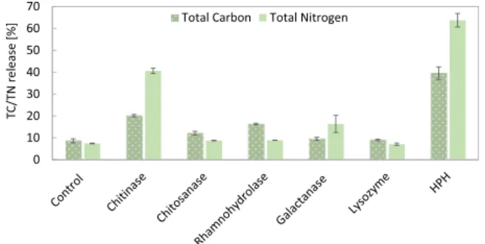
4 the total carbon (TC) and nitrogen (TN) released into the supernatant (Fig. 1).
Among the investigated enzymes, chitinase showed the highest release of TC (20.1% ±0.5%) and TN (40.6% ±1.2%) compared to the control (TC =8.6% ±0.9%, TN =7.4% ±0.1%). Chitosanase showed an increase in TC release up to 12.1% ±0.8%. According to Gerken et al.
(2013), chitinase causes a general decrease in the electron density of the wall, whereas chitosanase accounts for a minor thinning of the outer region of the cell wall. While both have chitosan (poly-β-1,4-D-glucos- amine) as their main substrate, chitinase is additionally able to degrade chitin, hydrolysing this polymer to liberate the amino sugar β-1, 4-D-N-acetylglucosamine (Coelho et al., 2019; Gerken et al., 2013).
Interestingly, lysozyme showed no disruption efficiency. Lysozyme can hydrolyse the linkage between peptidoglycan units in the cell walls.
Specifically, it degrades polymers containing N-acetylglucosamine, which is a derivative of glucosamine, an important component of the cell wall (Gerken et al., 2013; Kapaun & Reisser, 1995). Lysozyme improved the lipid extraction yield from wet samples of Scenedesmus from 4% to 16.6% (Taher et al., 2014). Gerken et al. (2013) showed that lysozyme degrades the outer surface of C. vulgaris cell wall and eliminates the hair-like fibres extending from the outer layer. Moreover, the authors showed that lysozyme was already effective alone but had a synergistic effect in enhancing the cell permeability when in combination with other enzymes. In our study, lysozyme alone did not significantly affect carbon and nitrogen release, probably because its activity is limited to external hair-like fibres.
Rhamnohydrolase increased TN release up to 8.9% ±0.1% and TC release to 16.3% ±0.4%, suggesting the presence in the cell wall of rhamnogalacturonan, which is a complex polymer with a poly- saccharidic backbone of repetitive disaccharide units composed of rhamnose and galacturonic acid. Rhamnogalacturonan rhamnohy- drolase is an enzyme that participates in the exohydrolysis of rhamno- galacturonan oligosaccharides, releasing β-D-rhamnose from the nonreducing end (Nzytech, 2021). This result correlates well with the cell wall composition of C. vulgaris, containing up to 26% rhamnose and 5% uronic acid (Canelli et al., 2021). As the usual ratio rhamnose: gal- acturonic acid in pectin-like structure is between 1:1 and 1:2 (Voragen et al., 2009), the high amount of rhamnose found in the cell wall of C. vulgaris indicates the additional presence of a rhamnose-rich poly- saccharide. Unfortunately, the structural characterization of such a polysaccharide has not been published yet. To the best of our knowl- edge, only a type of sulphated rhamnan has been reported in green algae (Suzuki & Terasawa, 2020). Moreover, there is a lack commercially available enzymes targeting non-pectic rhamnose-rich polysaccharides.
Galactanase showed a clear increase in TN release up to 16.3%. This suggests the presence in the cell wall of galactan or similar polymers that galactanase can hydrolyse, such as type I arabinogalactans.
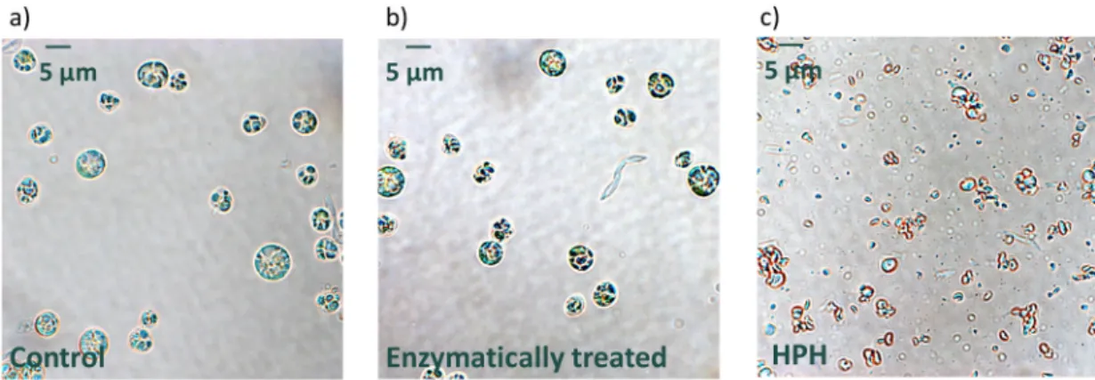
As a positive control, HPH treatment was performed. HPH is a
mechanical disruption treatment that leads to the full disruption of algal cells, as later shown by optical microscopy (Fig. 4c). HPH led to TC and TN releases of 39.6% ±2.9% and 63.8% ±3.1%, respectively. These results indicate that not all material was released into the supernatant.
Indeed, even after HPH, cell debris was still present, as confirmed by microscopy and particle size analysis (section 3.3); thus, not all the cellular material was dissolved into the supernatant.
Based on these results, the combination of the three best performing enzymes (chitinase, rhamnohydrolase, and galactanase) was chosen for further experiments.
3.2. Lipid and protein bioaccessibilities
In this section, we evaluated the effect of enzymatic treatment (combination of chitinase, rhamnohydrolase, and galactanase) on the lipid and protein bioaccessibilities of C. vulgaris cells. The bio- accessibility of enzymatically treated microalgal biomass was compared to that of the untreated control (biomass incubated for 24 h without cell wall-degrading enzymes), as well as that of HPH-treated biomass (Fig. 2).
The protein bioaccessibility of untreated biomass (control, 49.2% ± 3.9%) was similar to that of 51 ±9% described by Muys et al. (2018).
Enzymatic treatment caused an increase in protein bioaccessibility (from 49.2% ±3.9% to 58.7% ±3.5%) but not lipid bioaccessibility (from 24.9% ±3.0% to 21.1% ±1.1%). To date, the protein and lipid bioaccessibilities of enzymatically treated algal biomass have not been investigated yet, therefore no comparison to other studies can be done.
To evaluate whether freeze-drying the biomass prior to the enzy- matic treatment could impact the resulting bioaccessibility, we have assessed the protein and lipid bioaccessibilities of biomass that was freshly harvested and directly treated by enzymes (Fig. S1). A similar increase in protein bioaccessibility after enzymatic treatment was observed while the null effect of the enzymatic treatment on the lipid bioaccessibility was confirmed, indistinctly if the biomass was priorly freeze-dried or not. Noteworthy, freeze-drying caused an increase in lipid bioaccessibility, which explains the higher value of lipid bio- accessibility in the control of Fig. 2 compared to Fig. S1.
The protein bioaccessibility of HPH-treated biomass was 59.8% ± 1.8%, which is comparable to that of enzymatically treated biomass.
HPH caused an increase in lipid bioaccessibility up to 61.8% ±2.6%
(+36.9% compared to the control), showing that mechanical disruption is necessary to enhance lipid bioaccessibility. However, HPH treatment did not lead to full bioaccessibility of either protein or lipids, suggesting that some of these molecules might still be present in the cell debris and difficult to digest. It is important to note that protein bioaccessibility was measured as the ratio between the TN in the micellar phase and the TN in the full digesta. Therefore, part of the nitrogen released during digestion may come from compounds other than protein, e.g.
glucosamine.
Our results correlate with the total tract apparent protein Fig. 1.Release (%) of total carbon (TC, dotted pattern) and total nitrogen (TN,
full pattern) into the supernatant after enzymatic treatment (24 h, 37 ◦C) of C. vulgaris with individual enzymes (chitinase, chitosanase, rhamnohydrolase, galactanase, lysozyme) and after high-pressure homogenization (HPH) treat- ment. Error bars represent the standard deviation of triplicates (n =3).
Fig. 2. Lipid (diagonal line pattern) and protein (full pattern) bioaccessibility of C. vulgaris biomass that was untreated (control) and subjected to enzymatic and high-pressure homogenization (HPH) treatment. Error bars represent the standard deviation of triplicates (n =3).
G. Canelli et al.
digestibility measured by Komaki et al. (1998), which was enhanced by HPH only to a minor extent (from 87.4% to 88.6%). The higher absolute value described in Komaki et al. (1998) compared to our study is probably due to the different methodology used, as the authors measured apparent protein digestibility in rats.
3.3. Particle size
To evaluate cell integrity and debris formation upon disruption treatments, we measured the particle size of untreated cells, cells treated with the enzyme combination (chitinase, rhamnohydrolase, and gal- actanase), and cells treated with HPH. The mean diameter of the volume based droplet size distribution (d43) and the volume density distribution (q3) are reported in Fig. 3a and b, respectively.
HPH led to a reduced mean diameter from 5.1 ±0.1 μm to 2.0 ±0.1 μm, indicating a major disintegration of microalgae cells. This result further supports the theory of HPH being one of the most efficient disruption methods (Safi et al., 2015). In Fig. 3b, HPH-treated cells show a unimodal distribution, confirming the effectiveness of the disruption treatment. Similarly, Carullo et al. (2018) achieved a mean particle size of 2.22 μm in C. vulgaris after 5 passes at 1500 bar.
Enzymatic treatment (5.0 ±0.1 μm) did not lead to any change in particle size compared to the control. This is in accordance with the observation that enzymatic treatment preserves cellular integrity and results in visually intact microalgae cells, as observed with light mi- croscopy (Fig. 4b) (Safi et al., 2020). Indeed, enzymes may hydrolyse some components of the cell wall and modify the cell boundary layer without altering the morphological shape of the cells while allowing products to leach (Günerken et al., 2015).
The small peaks of particles measuring approximately 0.25 μm found in both untreated and enzymatically treated cells (Fig. 3b) probably represent cell debris naturally occurring in reproducing algae cultures.
At the maturation stage, C. vulgaris mother cells break, liberating the daughter cells, while the residual debris of the mother cell will be used up as substrate by the recently formed daughter cells (Safi et al., 2014).
3.4. Oxidative stability
The biomass oxidative stability was assessed by measuring the development of secondary oxidation products during storage over 12 weeks at 40 ◦C (Fig. 5). The production of volatiles was examined in enzymatically treated biomass (chitinase + rhamnohydrolase +
galactanase), HPH-treated biomass and untreated biomass (control).
Hexanal and (Z)-3-hexenal values are shown as targeted degradation products of the most abundant fatty acids in C. vulgaris, named linoleic acid (C18:2,n6) and α-linolenic acid (C18:3,n3), respectively (Rohfritsch et al., 2019; Shahidi, 2001).
The signal of hexanal and (Z)-3-hexenal in untreated biomass did not show an increase over 12 weeks of storage, indicating the remarkable oxidative stability of dried C. vulgaris biomass.
For the first time, the secondary oxidation of microalgae biomass treated with cell wall degrading enzymes was assessed. The volatiles in the enzymatically treated biomass showed similar signals compared to the control. The stability of enzymatically treated biomass over storage confirms that enzymatic treatment is a mild process that preserves the quality of the extractable component, in this case PUFA-rich lipids.
The signal intensity in HPH-treated biomass of hexanal and (Z)-3- hexenal was, respectively, 1.4- and 7.1-fold higher than that in the control after 12 weeks, indicating the harsh effect of HPH on the biomass oxidative stability. Differently, Gheysen et al. (2019) described a similar secondary oxidation in intact and HPH-disrupted Nannochloropsis biomass. This difference may be attributed to the different species or different level of antioxidants.
In our study, a clear decrease in the volatile signal from week 0 to week 2 was observed in the case of HPH-treated biomass, suggesting that extensive oxidation already occurred during the disruption treatment before storage. Indeed, it was shown that by disrupting the cells with HPH treatment, the lipolysis starts directly during the process (Balduyck et al., 2017). During the first two weeks of storage, part of the formed aldehydes might have further reacted with other components released by cell disruption or be degraded into organic acids, which were not detected with the analytical method used.
Blind sniffing was performed on the same samples. Hexanal is characterized by a fishy and grassy flavour, while (Z)-3-hexenal has a cucumber-like aroma (Rohfritsch et al., 2019). The sensory outcomes agreed with the analytical data. The untreated biomass was described as neutral with a slight cheese-like flavour over the entire storage. Enzy- matically treated biomass has a cereal-like, baked flavour at time 0, which turns into a fermented-like aroma over time. Notably, rancidity was not described in enzymatically treated biomass. In contrast, strong rancidity was perceived in HPH-treated biomass at week 0. The intensity of this note decreased over time, supporting the analytical results.
This study confirms the crucial function played by cell integrity in the oxidative stability of C. vulgaris cells, as was also hypothesized for Fig. 3.a) Mean diameter of the volume based droplet size distribution (d43) of C. vulgaris cells that were untreated (control), enzymatically treated (chitinase +rhamnohydrolase +galactanase) and treated with HPH. Error bars represent the standard deviation of triplicates (n = 3). b) Particle size distribution expressed as volume density (q3, μm−1) of C. vulgaris cells that were untreated (control, ●), enzymatically treated (chitinase +rhamnohydrolase +galactanase,
■) and treated with high-pressure homogenization (HPH, ▴).
LWT 143 (2021) 111157
6 Nannochloropsis by Balduyck et al. (2017). In addition to cell integrity, the chemical reaction catalysed by cell wall-degrading enzymes could also explain the remarkable oxidative stability of biomass treated with enzymes. Indeed, studies in plants suggested that carbohydrate-active enzymes may help reduce the complexation of hydrophilic phenolic compounds with polysaccharides and thus increase the amount of free phenolics, favouring a higher antioxidant efficacy (Muniglia et al., 2014). On the other hand, another reason for the extensive oxidation of HPH-treated microalgae, in addition to the loss of cell integrity, could be that cavitation during HPH can lead to the formation of free radicals causing oxidation (Günerken et al., 2015). Overall, the effect that cell disruption treatment has on product quality can be a direct indicator of its mildness (Günerken et al., 2015).
4. Conclusion
Although more work is needed to optimise the enzymatic treatment to maximise the nutrient bioaccessibility, this study showed that enzy- matic treatment is an effective method for the release of cellular material from algae while preserving cell integrity. Most importantly, we showed for the first time that enzymatic treatment of algae biomass could conserve the stability of important nutrients (e.g., PUFAs) and therefore could avoid off-flavour formation, preserving biomass quality. In contrast, mechanical treatment with HPH led to the highest lipid and protein bioaccessibilities but compromised the oxidative stability of such biomass. Future work should aim to optimise lipid bioaccessibility and improve the stability of biomass after HPH (e.g., by optimizing treatment conditions and antioxidant loading). In addition, enzyme immobilization could reduce the needed quantity of enzymes and lower the downstream processing costs.
CRediT authorship contribution statement
Greta Canelli: Conceptualization, Methodology, Formal analysis, Project administration, Writing – original draft, Writing – review &
editing. Patricia Murciano Martínez: Conceptualization, Methodol- ogy, Formal analysis, Writing – review & editing. Billie Maude Hauser:
Methodology, Formal analysis, Writing – review & editing. Isabelle Kuster: Methodology, Formal analysis, Writing – review & editing.
Zhen Rohfritsch: Conceptualization, Methodology, Formal analysis, Writing – review & editing. Fabiola Dionisi: Conceptualization, Writing – review & editing. Christoph J. Bolten: Conceptualization, Writing – review & editing. Lukas Neutsch: Conceptualization, Writing – review
& editing. Alexander Mathys: Funding acquisition, Project adminis-
tration, Conceptualization, Writing – review & editing.
Declaration of competing interest
The authors declare that they have no known competing financial interests or personal relationships that could have appeared to influence the work reported in this paper.
Acknowledgements
The authors gratefully acknowledge the Nestl´e Research VCLB, Lausanne, Switzerland, and the ETH Zurich Foundation, Switzerland, for their support. In addition, the authors sincerely thank Roberta Carpine and Sabrina Tevere from ZHAW, Campus Grüental, W¨adenswil, Switzerland, for support with biomass cultivation. The authors sincerely thank Christina Vafeiadi and Sean Austin, Nestl´e Research VCLB, Lau- sanne, Switzerland, for scientific assistance. The authors thank Lukas B¨ocker, ETH Zurich, for proofreading the article.
Fig. 4. Representative light microscopy images of C. vulgaris cells that were untreated (a, control), enzymatically treated (b, chitinase +rhamnohydrolase +gal- actanase) and treated with high-pressure homogenization (c, HPH).
Fig. 5. Evolution of secondary oxidation products - a) hexanal and b) (Z)-3-hexenal - during 12 weeks of storage at 40 ◦C for C. vulgaris biomass untreated (control,
●), enzymatically treated (chitinase +rhamnohydrolase +galactanase, ■) and high-pressure homogenization treated (HPH,▴). The signal is expressed as the ratio of the area of the targeted compound to the area of the internal standard (ISTD) hexanal-d12. Error bars represent the standard deviation of triplicates (n =3).
G. Canelli et al.
Appendix A. Supplementary data
Supplementary data to this article can be found online at https://doi.
org/10.1016/j.lwt.2021.111157.
References
Balduyck, L., Stock, T., Bijttebier, S., Bruneel, C., Jacobs, G., Voorspoels, S., Muylaert, K.,
& Foubert, I. (2017). Integrity of the microalgal cell plays a major role in the lipolytic stability during wet storage. Algal Research, 25, 516–524. https://doi.org/10.1016/j.
algal.2017.06.013
Batista, A. P., Gouveia, L., Bandarra, N. M., Franco, J. M., & Raymundo, A. (2013).
Comparison of microalgal biomass profiles as novel functional ingredient for food products. Algal Research, 2(2), 164–173. https://doi.org/10.1016/j.
algal.2013.01.004
Baudelet, P. H., Ricochon, G., Linder, M., & Muniglia, L. (2017). A new insight into cell walls of Chlorophyta. Algal Research, 25, 333–371. https://doi.org/10.1016/j.
algal.2017.04.008
Bernaerts, T. M. M., Verstreken, H., Dejonghe, C., Gheysen, L., Foubert, I., Grauwet, T., &
Loey, A. M. Van (2020). Cell disruption of Nannochloropsis sp. improves in vitro bioaccessibility of carotenoids and ω3-LC-PUFA. Journal of Functional Foods, 65 (103770). https://doi.org/10.1016/j.jff.2019.103770
B¨ocker, L., Bertsch, P., Wenner, D., Teixeira, S., Bergfreund, J., Eder, S., Fischer, P., &
Mathys, A. (2020). Effect of Arthrospira platensis microalgae protein purification on emulsification mechanism and efficiency. Journal of Colloid and Interface Science, 584, 344–353. https://doi.org/10.1016/j.jcis.2020.09.067
Buchmann, L., Br¨andle, I., Haberkorn, I., Hiestand, M., & Mathys, A. (2019). Pulsed electric field based cyclic protein extraction of microalgae towards closed-loop biorefinery concepts. Bioresource Technology, 291, 121870. https://doi.org/10.1016/
j.biortech.2019.121870
Canelli, G., Murciano Martínez, P., Austin, S., Ambühl, M., Dionisi, F., Bolten, C. J., … Mathys, A. (2021). Biochemical and morphological characterization of heterotrophic Crypthecodinium cohnii and Chlorella vulgaris cell walls. J. Agric. Food Chem., 69, 2226–2235. https://doi.org/10.1021/acs.jafc.0c05032
Canelli, G., Neutsch, L., Carpine, R., Tevere, S., Giuffrida, F., Rohfritsch, Z., Dionisi, F., Bolten, C. J., & Mathys, A. (2020a). Chlorella vulgaris in a heterotrophic bioprocess:
Study of the lipid bioaccessibility and oxidative stability. Algal Research, 45. https://
doi.org/10.1016/j.algal.2019.101754
Canelli, G., Tarnutzer, C., Carpine, R., Neutsch, L., Bolten, C. J., Dionisi, F., & Mathys, A.
(2020b). Biochemical and nutritional evaluation of Chlorella and auxenochlorella biomasses relevant for food application. Frontiers in Nutrition, 7, 1–9. https://doi.
org/10.3389/fnut.2020.565996
Caporgno, M. P., & Mathys, A. (2018). Trends in microalgae incorporation into innovative food products with potential health benefits. Frontiers in Nutrition, 5, 1–10. https://doi.org/10.3389/fnut.2018.00058
Carullo, D., Abera, B. D., Casazza, A. A., Donsì, F., Perego, P., Ferrari, G., & Pataro, G.
(2018). Effect of pulsed electric fields and high pressure homogenization on the aqueous extraction of intracellular compounds from the microalgae Chlorella vulgaris. Algal Research, 31(January), 60–69. https://doi.org/10.1016/j.
algal.2018.01.017
Cho, H. S., Oh, Y. K., Park, S. C., Lee, J. W., & Park, J. Y. (2013). Effects of enzymatic hydrolysis on lipid extraction from Chlorella vulgaris. Renewable Energy, 54, 156–160.
https://doi.org/10.1016/j.renene.2012.08.031
Coelho, D., Lopes, P. A., Cardoso, V., Ponte, P., Br´as, J., Madeira, M. S., Alfaia, C. M., Bandarra, N. M., Gerken, H. G., Fontes, C. M. G. A., & Prates, J. A. M. (2019). Novel combination of feed enzymes to improve the degradation of Chlorella vulgaris recalcitrant cell wall. Scientific Reports, 9(1), 5382. https://doi.org/10.1038/s41598- 019-41775-0
Galasso, C., Gentile, A., Orefice, I., Ianora, A., Bruno, A., Noonan, D. M., Sansone, C., Albini, A., & Brunet, C. (2019). Microalgal derivatives as potential nutraceutical and food supplements for human health: A focus on cancer prevention and interception.
Nutrients, 11(6), 1226. https://doi.org/10.3390/nu11061226
Gerken, H. G., Donohoe, B., & Knoshaug, E. P. (2013). Enzymatic cell wall degradation of Chlorella vulgaris and other microalgae for biofuels production. Planta, 237(1), 239–253. https://doi.org/10.1007/s00425-012-1765-0
Gheysen, L., Bernaerts, T., Bruneel, C., Goiris, K., Van Durme, J., Van Loey, A., De Cooman, L., & Foubert, I. (2018). Impact of processing on n-3 LC-PUFA in model systems enriched with microalgae. Food Chemistry, 268(December 2017), 441–450.
https://doi.org/10.1016/j.foodchem.2018.06.112
Gheysen, L., Lagae, N., Devaere, J., Goiris, K., Goos, P., Bernaerts, T., Van Loey, A., De Cooman, L., & Foubert, I. (2019). Impact of Nannochloropsis sp. dosage form on the
oxidative stability of n-3 LC-PUFA enriched tomato purees. Food Chemistry, 279 (December 2018), 389–400. https://doi.org/10.1016/j.foodchem.2018.12.026 Goettel, M., Eing, C., Gusbeth, C., Straessner, R., & Frey, W. (2013). Pulsed electric field
assisted extraction of intracellular valuables from microalgae. Algal Research, 2(4), 401–408. https://doi.org/10.1016/j.algal.2013.07.004
Grima, E. G., Aci´en Fern`andez, F. G., & Robles Medina, A. (2013). Downstream processing of cell mass and products. In A. Richmond, & Q. Hu (Eds.), Handbook of microalgal culture. New Jersey, United States: Wiley Blackwell. https://doi.org/
10.1002/9781118567166.
Günerken, E., D’Hondt, E., Eppink, M. H. M., Garcia-Gonzalez, L., Elst, K., &
Wijffels, R. H. (2015). Cell disruption for microalgae biorefineries. Biotechnology Advances, 33(2), 243–260. https://doi.org/10.1016/j.biotechadv.2015.01.008 Kapaun, E., & Reisser, W. (1995). A chitin-like glycan in the cell wall of a Chlorella sp.
(Chlorococcales, Chlorophyceae). Planta, 197, 577–582. https://doi.org/10.1007/
978-3-642-21527-8_15
Kim, J., Yoo, G., Lee, H., Lim, J., Kim, K., Kim, C. W., Park, M. S., & Yang, J. W. (2013).
Methods of downstream processing for the production of biodiesel from microalgae.
Biotechnology Advances, 31(6), 862–876. https://doi.org/10.1016/j.
biotechadv.2013.04.006
Komaki, H., Yamashita, M., Niwa, Y., Tanaka, Y., Kamiya, N., Ando, Y., & Furuse, M.
(1998). The effect of processing of Chlorella vulgaris: K-5 on in vitro and in vivo digestibility in rats. Animal Feed Science and Technology, 70(4), 363–366. https://doi.
org/10.1016/S0377-8401(97)00089-8
Muniglia, L., Claisse, N., Baudelet, P.-H., & Ricochon, G. (2014). Enzymatic aqueous extraction (EAE). In F. Chemat, & M. A. Vian (Eds.), Alternative solvents for natural products extraction (pp. 167–204). Springer. https://doi.org/10.1007/978-3-662- 43628-8_8.
Muys, M., Vermeir, P., Lesueur, C., Vandenheuvel, D., Schwaiger, B., Vlaeminck, S. E., &
Sui, Y. (2018). High variability in nutritional value and safety of commercially available Chlorella and Spirulina biomass indicates the need for smart production strategies. Bioresource Technology, 275, 247–257. https://doi.org/10.1016/j.
biortech.2018.12.059
Nzytech. (2021). Rhamnogalacturonan rhamnohydrolase 106A. https://www.nzytech.
com/products-services/cazymes/glycoside-hydrolases/other-activities/cz0848/.
Rohfritsch, Z., Schafer, O., & Giuffrida, F. (2019). Analysis of oxidative carbonyl compounds by UPLC-High-Resolution Mass Spectrometry in milk powder. Journal of Agricultural and Food Chemistry, 67(12), 3511–3520. https://doi.org/10.1021/acs.
jafc.9b00674
Safi, C., Frances, C., Ursu, A. V., Laroche, C., Pouzet, C., Vaca-Garcia, C., &
Pontalier, P. Y. (2015). Understanding the effect of cell disruption methods on the diffusion of Chlorella vulgaris proteins and pigments in the aqueous phase. Algal Research, 8, 61–68. https://doi.org/10.1016/j.algal.2015.01.002
Safi, C., Olivieri, G., Engelen-Smit, N., Spekking, W., Veloo, R., den Broek, L. A. M. va, &
Sijtsma, L. (2020). Effect of growth conditions on the efficiency of cell disruption of Neochloris oleoabundans. Bioresource Technology, 300. https://doi.org/10.1016/j.
biortech.2019.122699
Safi, C., Zebib, B., Merah, O., Pontalier, P. Y., & Vaca-Garcia, C. (2014). Morphology, composition, production, processing and applications of Chlorella vulgaris: A review.
Renewable and Sustainable Energy Reviews, 35, 265–278. https://doi.org/10.1016/j.
rser.2014.04.007
Scherer, D., Krust, D., Frey, W., Mueller, G., Nick, P., & Gusbeth, C. (2019). Pulsed electric field (PEF)-assisted protein recovery from Chlorella vulgaris is mediated by an enzymatic process after cell death. Algal Research, 41, 101536. https://doi.org/
10.1016/j.algal.2019.101536
Sevenich, R., & Mathys, A. (2018). Continuous versus discontinuous ultra-high-pressure systems for food sterilization with focus on ultra-high-pressure homogenization and high-pressure thermal sterilization: A review. Comprehensive Reviews in Food Science and Food Safety, 17, 646–662.
Shahidi, F. (2001). Headspace volatile aldehydes as indicators of lipid oxidation in foods.
In R. L. Rouseff, & K. R. Cadwallader (Eds.), Headspace analysis of food and flavors:
Theory and practice (pp. 113–123). Kluwer Academic/Plenum Publishers. https://
doi.org/10.1007/978-1-4615-1247-9_9.
Sierra, L. S., Dixon, C. K., & Wilken, L. R. (2017). Enzymatic cell disruption of the microalgae Chlamydomonas reinhardtii for lipid and protein extraction. Algal Research, 25, 149–159. https://doi.org/10.1016/j.algal.2017.04.004
Suzuki, K., & Terasawa, M. (2020). Biological activities of rhamnan sulfate extract from the green algae Monostroma nitidum (Hitoegusa). Marine Drugs, 18(4). https://doi.
org/10.3390/md18040228
Taher, H., Al-Zuhair, S., Al-Marzouqi, A. H., Haik, Y., & Farid, M. (2014). Effective extraction of microalgae lipids from wet biomass for biodiesel production. Biomass and Bioenergy, 66, 159–167. https://doi.org/10.1016/j.biombioe.2014.02.034 Voragen, A. G. J., Coenen, G. J., Verhoef, R. P., & Schols, H. A. (2009). Pectin, a versatile
polysaccharide present in plant cell walls. Structural Chemistry, 20(2), 263–275.
https://doi.org/10.1007/s11224-009-9442-z