DISSERTATION
zur Erlangung des akademischen Grades doctor rerum naturalium
(Dr. rer. nat.) im Fach Biologie eingereicht an der
Mathematisch-Naturwissenschaftlichen Fakult¨ at I Humboldt-Universit¨ at zu Berlin
von
Herr Dipl.-Biol. (technisch orientiert) Uwe Anzenberger geboren am 22.03.1976 in Singen a. Htwl.
Pr¨ asident der Humboldt-Universit¨ at zu Berlin:
Prof. Dr. Christoph Markschies
Dekan der Mathematisch-Naturwissenschaftlichen Fakult¨ at I:
Prof. Thomas Buckhout, Phd Gutachter:
1. Prof. Dr. Thomas Willnow 2. Prof. Dr. Michael Bader 3. Prof. Dr. Wolfgang Lockau
Tag der m¨ undlichen Pr¨ ufung: 22. September 2006
Abstract
LRP2 is a member of the low-density lipoprotein receptor gene family that is mainly expressed in the yolk sac and in the neuroepithelium of the early embryo.
Deficiency for this 600 kDa protein in mice results in holoprosencephaly, indicating an important yet unknown role for LRP2 in forebrain development.
In this study, mice with a complete or a conditional loss of lrp2 function were used to further elucidate the consequences of the lack of LRP2 expression. This study shows that the presence of LRP2 in the neuroepithelium but not in the yolk sac is crucial for early forebrain development. Lack of the receptor resulted in an increase of Bone morphogenic protein (Bmp) 4 signaling in the rostral telencephalon at E9.5. As a consequence, sonic hedgehog (shh) expression at E10.5 was lost completely in a ventral region of the telencephalon termed anterior entopeduncular area (AEP). The absence of Shh activity in this area subsequently led to the loss of ventrally induced oligodendroglial and interneuronal cell populations in lrp2 deficient mice. Similar dorsalizing effects have also been observed in mice with increased Bmp4 signaling. Taking into account that Bmp4 was found to bind to LRP2in vitro and in vivo in this study, these results suggest a previously unknown role for LRP2 in patterning the rostral ventral neural tube, possibly by acting as a clearance receptor for Bmp4.
The underlying molecular mechanisms by which LRP2 patterns the ventral forebrain were then further analyzed in the zebrafish, a model organism that is amenable for various experimental manipulations. The cytoplasmic tail, the transmembrane domain and a short extracellular part of the zebrafish LRP2 were identified by searching the Sanger Zebrafish Zv4.0 genomic database.
LRP2-deficient animals were generated by injecting Morpholino oligonucleotides that interfered with the splicing of the lrp2-pre-mRNA leading to a deletion of the transmembrane-exon. Injected animals suffered from impaired renal clearance processes, demonstrating the functional conservation of LRP2 in the larval zebrafish pronephros and in the mammalian kidney.
Brain structures were not affected in these animals and the expression patterns of marker genes for early forebrain development that were changed in the mouse were not changed in the zebrafish.
Apparently, Morpholino mediated interfering with the splicing of the lrp2-pre- mRNA did not affect the early forebrain formation because properly processed
Keywords:
LRP2, Megalin, Holoprosencephaly, Shh
Abstract
LRP2 geh¨ort zu einer Gruppe funktionell und strukturell eng verwandter Proteine, die in derLow Density Lipoprotein Rezeptor (LDLR) Genfamilie zusammengefasst werden. LRP2 wird w¨ahrend der fr¨uhen Embryonalentwicklung haupts¨achlich im Dottersack und im Neuroepithel exprimiert. Der funktionelle Verlust dieses 600 kDa großen Proteins in M¨ausen f¨uhrt zu schweren Fehlbildungen bei der Vorderhirnentwicklung, die als Holoprosenzephalie bezeichnet werden. LRP2 scheint daher eine wichtige, aber bisher unbekannte Funktion w¨ahrend der Vorderhirnentwicklung auszu¨uben.
Um die zu Grunde liegenden Mechanismen dieser Fehlbildungen zu analysieren, wurden f¨ur diese Arbeit M¨ause verwendet, bei denen lrp2 im gesamten Tier oder nur in bestimmten Geweben inaktiviert war. Es konnte gezeigt werden, dass die Expression von LRP2 im Neuroepithel, nicht aber im Dottersack wichtig f¨ur die korrekte Vorderhirnentwicklung ist. Das Fehlen des Proteins f¨uhrte am Tag 9.5 der Embryonalentwicklung zu einer ¨Uberaktivierung des Bone morphogenic protein (Bmp) 4 Signalweges im rostralen Telencephalon. Am Tag 10.5 war ein Verlust der sonic hedgehog (shh) Expression in einem begrenzten Bereich des ventralen, rostralen Telencephalons zu sehen, der als anteriore entopedunculare Zone (AEP) bezeichnet wird. Das Fehlen von Shh in der AEP f¨uhrte zum Verlust von ventralen Oligodendrozyten und Interneuronen, deren Bildung normalerweise in diesem Bereich von Shh induziert wird. ¨Ahnliche Defekte wurden ebenfalls in M¨ausen beschrieben, bei denen der Bmp4 Signalweg verst¨arkt ist. Es konnte in dieser Arbeit gezeigt werden, dass Bmp4 sowohlin vitro als auchin vivo an LRP2 bindet und dem lysosomalen Abbau zugef¨uhrt wird. Diese Ergebnisse deuten darauf hin, dass LRP2 maßgeblich an der Entwicklung des ventralen Telencephalons beteiligt ist - m¨oglicherweise indem es die verf¨ugbare Menge an Bmp4 durch Endozytose reguliert.
Die Signalwege, in die LRP2 eingebunden ist, sollten im Zebrafisch weiter unter- sucht werden, da dieser Organismus leichter als die Maus experimentell manipuliert werden kann. Durch Datenbankanalysen (Sanger Zebrafish Zv 4.0) konnte die codierende Sequenz des zytoplasmatischen Bereiches, der Transmembran-Region, sowie eines kurzen extrazellul¨aren Bereiches von LRP2 im Zebrafisch identifiziert werden. Tiere, denen funktionelles LRP2 fehlte, wurden durch die Injektion von Morpholino-Oligonukleotiden generiert. Die Morpholino-Oligonukleotide
von lrp2 defizienten M¨ausen beschrieben wurde. Diese Ergebnisse zeigen, dass die Funktion von LRP2 im Zebrafisch Pronephros und in der Niere der S¨augetiere konserviert ist.
Die Gehirnstrukturen waren bei diesen Tieren allerdings normal entwickelt und auch die Expressionsmuster von Markergenen f¨ur die Vorderhirnentwicklung waren normal.
Vermutlich war die St¨orung derlrp2-pr¨a-mRNA Prozessierung nicht ausreichend, um hier einen Effekt hervorzurufen, da bereits im 1-Zell Stadium gen¨ugend kom- plett prozessierte maternale lrp2-mRNA f¨ur die Translation von LRP2 vorhanden war.
Schlagw¨orter:
LRP2, Megalin, Holoprosenzephalie, Shh
This document was created using L
ATEX
1 Introduction . . . 1
1.1 The low-density lipoprotein receptor gene family . . . 1
1.2 Holoprosencephaly . . . 13
1.3 Forebrain development . . . 15
1.3.1 The sonic hedgehog pathway . . . 16
2 Aim of this study . . . 19
3 Material and Methods . . . 21
3.1 Animal Experiments . . . 21
3.1.1 Mouse husbandry . . . 21
3.1.2 Zebrafish husbandry . . . 21
3.1.3 Morpholino Injections . . . 22
3.1.4 Dye filtration experiments . . . 22
3.1.5 Acridine orange staining . . . 23
3.2 Microbiological Methods . . . 23
3.2.1 Culture media . . . 23
3.2.2 Preparation of electrocompetent bacteria . . . 23
3.2.3 Cryopreservation of bacteria . . . 24
3.2.4 Transformation of bacteria with DNA . . . 24
3.3 Molecular biology methods . . . 24
3.3.1 Isolation of plasmid DNA from bacteria . . . 24
3.3.2 Isolation of genomic DNA from tissue samples . . 25
3.3.3 Isolation of total RNA from tissue samples . . . . 25
3.3.4 DNA and RNA concentration determination . . . 25
3.3.5 Enzymatic digest of DNA . . . 26
3.3.6 Agarose gel electrophoresis of DNA and RNA . . 26
3.3.7 Isolation of DNA from agarose gels . . . 26
3.3.8 Ligation of PCR-products in the pGEM-T Easy Vector . . . 26
3.3.9 Polymerase chain reaction . . . 27
3.3.10 Primer sequences . . . 27
3.3.11 Reverse transcription . . . 28
3.3.12 In vitro transcription of digoxigenin labeled RNA . 29 3.3.13 In situ probes . . . 29
3.3.14 ISH on whole-mount mouse embryos . . . 31
3.3.15 ISH on whole-mount zebrafish embryos . . . 32
3.3.16 Genotyping of mice . . . 33
3.3.17 Membrane extracts . . . 34
3.3.18 Protein concentration determination . . . 34
3.3.19 SDS polyacrylamide gel electrophoresis of proteins 34 3.3.20 Western blotting . . . 35
3.3.21 Coomassie brilliant blue staining of SDS-PAGE-gels 36 3.3.22 In vitro analysis of protein-protein binding . . . . 36
3.3.23 In vivo uptake and degradation studies . . . 36
3.4 Histology . . . 37
3.4.1 Paraffin sections . . . 37
3.4.2 Plasticsections . . . 37
3.4.3 Cryosections . . . 38
3.4.4 Counterstaining of sections . . . 38
3.4.5 Immunohistchemistry on cryosections . . . 38
4 Results I: Mouse . . . 41
4.1 Forebrain defects in mice with epiblast-specific lrp2 gene disruption . . . 41
4.2 Absence of shh expression in the AEP of lrp2-deficient em- bryos . . . 41
4.3 Loss of ventral cell fates in the telencephalon of lrp2- deficient embryos . . . 44
4.4 Enhanced Bmp4 signaling and aberrant fgf8 expression in the dorsal midline of lrp2-deficient mice . . . 46
4.5 LRP2 acts as an endocytic receptor for Bmp4 . . . 48
5.2 Identification of the LRP2 ortholog in the zebrafish . . . 53 5.3 The expression pattern of zf-lrp2 and mouse-lrp2 is conserved 55 5.4 Functional analysis of zf-LRP2 protein . . . 56 5.5 Generation of lrp2-deficient zebrafish embryos . . . 57 5.6 Zebrafish-lrp2 is essential for tubular clearance of metabo-
lites via receptor-mediated endocytosis . . . 65 5.7 Tracer accumulates in vesicles that are positive for the early
endosomal marker Rab4. . . 68 5.8 Mechanisms of receptor mediated endocytosis are function-
ally conserved between the zebrafish larval pronephros and the adult mammalian kidney. . . 69 5.9 Dab2 is essential for tubular clearance of metabolites via
receptor-mediated endocytosis . . . 70 5.10 Analyzing new components for their role in endocytic pro-
cesses in the zebrafish pronephros . . . 73 5.11 Zf-LRP2 and forebrain development . . . 76 5.12 Expression of early forebrain marker genes is unchanged . . 77 5.13 Zf-lrp2 mRNA is provided maternally . . . 82 6 Discussion . . . 83 6.1 Analysis of LRP2 function in the mouse model . . . 83
6.1.1 The role of lrp2 in forebrain development: yolk sac or neuroepithelium? . . . 83 6.1.2 Lrp2-deficiency impairs Shh-dependent ventral
cell fate . . . 83 6.1.3 Lrp2-deficiency increases Bmp4 activity in the
rostral dorsal neural tube . . . 85 6.1.4 A role for LRP2 in patterning the rostral neural
tube: a working model . . . 86 6.2 Analysis of LRP2 function in the zebrafish model . . . 88
6.2.1 Expression of the LRP2 ortholog in the zebrafish pronephros . . . 88 6.2.2 Functional analysis of zf-LRP2 in the zebrafish
pronephros . . . 89
6.2.3 Role of the LRP2 ortholog in kidney and forebrain
development of the zebrafish . . . 91
7 Appendix . . . 114
7.1 Abbreviations . . . 114
7.2 Danksagung . . . 116
7.3 Selbstst¨andigkeitserkl¨arung . . . 118
1 The LDL receptor gene family members. . . 2 2 Cranofacial and brain malformations in lrp2-deficient newborn mice. 11 3 Analysis of E8.5 and E9.5lrp2 mutant mouse embryos. . . 12 4 Neuroanatomy and analysis of cell proliferation and apoptosis of
lrp2-deficient embryos. . . 13 5 Ligands and functions of the mammalian LDL receptor gene family
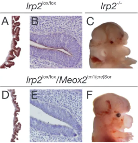
members. . . 14 6 Forebrain abnormalities and LRP2 expression pattern in E14.5 em-
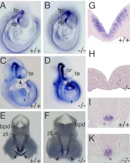
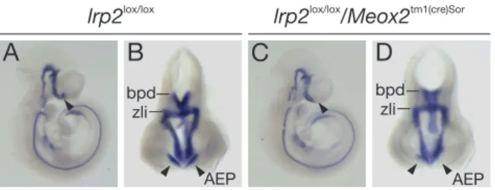
bryos with conditional lrp2 gene inactivation. . . 42 7 Shh expression in wildtype and lrp2 -/- embryos. . . 43 8 Shhexpression inlrp2 lox/lox and lrp2 lox/lox/Meox2 tm1(cre)Sor E10.5
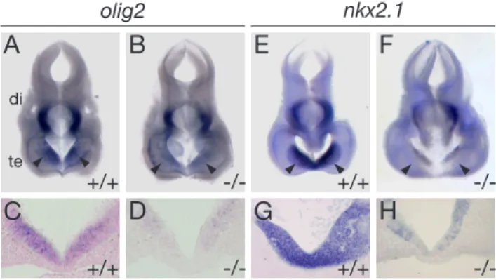
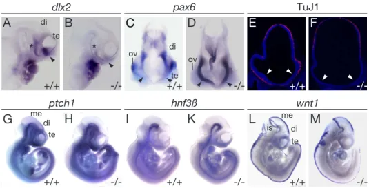
embryos. . . 44 9 Expression of olig2 and nkx2.1 in the forebrain at E10.5. . . 45 10 Expression of marker genes of early forebrain development in E10.5
embryos. . . 46 11 Analysis of Fgf8 and Bmp4 pathways in E9.5 and E10.5 embryos. . 48 12 Increased Bmp4 signaling phosphorylates Smad proteins and in-
duces downstream targets. . . 49 13 LRP2 mediates binding and cellular catabolism of Bmp4. . . 50 14 Chemical differences between Morpholino and RNA oligonucleotides. 52 15 Amino acid sequence alignment of human LRP2 with the zebrafish
ortholog. . . 54 16 Expression pattern of zf-lrp2 at 48hpf and 72hpf in wildtypes. . . . 55 17 Expression oflrp2,cubilin,dab2,wt1 andpax2.1 in the developing
pronephros. . . 60
18 Renal clearance of tracers within the proximal pronephric duct is restricted to the LRP2 expression domain. . . 61 19 Renal clearance of tracers occurs within the proximal pronephric duct. 61 20 Molecular characterization of the lrp2 morphant embryos. . . 62 21 Sequence analysis of RT-PCR products of Morpholino injected em-
bryos. . . 63 22 Lrp2 morphant embryos are phenotypically normal. . . 63 23 The renal system is developed normally in zf-lrp2 morphants. . . . 64 24 Tubular clearance defects in zf-lrp2 morphants. . . 65 25 Statistical analysis of tubular clearance defects in zf-lrp2 morphants. 67 26 Lrp2 morphant embryos are negative for Rab4 positive early endo-
somes. . . 68 27 Dab2 morphants are phenotypically normal. . . 70 28 Tubular clearance defects in dab2 morphants. . . 71 29 Statistical analysis of tubular clearance defects in dab2 morphants. 71 30 Dab2 morphant embryos are negative for Rab4 positive early en-
dosomes. . . 72 31 Localization of zf-LRP2 in dab2 morphants is not changed. . . 73 32 Lack of renal clearance in Tg(cmlc2:has) mutants. . . 75 33 Lack of Rab4-positive early endosomes in Tg(cmlc2:has) mutants. . 76 34 Mosaic clonal analysis ofprkci function in tubular endocytic processes. 76 35 Brain morphology of 48hpf and 96hpf wildtype and morphant animals. 78 36 Expression of shh in wildtype and lrp2 morphant animals at 48hpf
and 72hpf. . . 79 37 Expression of nkx2.1 in wildtype and lrp2 morphant animals at
48hpf and 72hpf. . . 80 38 Expression ofolig2 in wildtype andlrp2 morphant animals at 48hpf
and 72hpf. . . 80 39 RT-PCR for zf-lrp2 transcripts at various stages of development. . 82 40 Schematic working models of a possible role for LRP2 in patterning
of the rostral neural tube. . . 87 41 Phylogenetic tree of different species illustrating evolutionary dis-
tance. . . 92
1 Introduction
1.1 The low-density lipoprotein receptor gene family
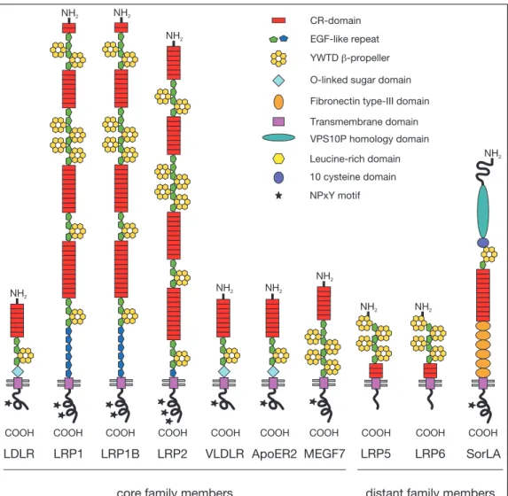
The low-density lipoprotein receptor (LDLR) gene family is composed of ten mem- bers of structurally closely related cell-surface proteins (Figure 1). In addition to humans, members of this family have been identified in different taxa including invertebrates like the fruitfly (Wehrli et al., 2000), and vertebrates like the african clawfrog (Houston, 2002), the house mouse (Gafvels et al., 1994; Brown et al., 1998) and the rat (Lee et al., 1989). So far, no members of the LDLR gene family have been identified in eukaryotic unicellular organisms such as baker’s yeast.
The receptors are homologous among species, even receptors of two evolutionary distant organisms display a high grade of conservation at the amino acid level (Houston, 2002). This high degree of evolutionary conservation suggests that the LDLR gene family early developed roles that are critical for the functional integrity of multicellular organisms.
The core of the gene family (Figure 1) in mammals consists of the low- density lipoprotein receptor (LDLR), the LDLR related proteins 1, 1B and 2 (LRP1, LRP1B, LRP2), the very low-density lipoprotein receptor (VLDLR), the apolipoprotein E receptor-2 (ApoER2) and the multiple epidermal growth factor repeat containing protein 7 (MEGF7). Members of the family that are not included in the core because of their differing domain structure (Figure 1) are the low-density lipoprotein receptor related protein 5, 6 (LRP5/LRP6) and the sorting protein related receptor containing LDLR class A repeats (SorLA). A ta- ble summarizing important ligands, functions and sites of expression for the gene family members can be found at the end of this section on page 17.
All of these receptors belong to the superfamily of type I cell-surface receptors, featuring a large amino-terminal extracellular domain, a single transmembrane seg- ment and a short carboxy-terminal cytoplasmic tail region. The extracellular part is mainly composed of motifs responsible for binding ligands and of motifs important for releasing ligands (Brown et al., 1997) upon internalization in the endosomes in a pH-dependent manner (Rudenko et al., 2002).
1. INTRODUCTION
CR-domain
YWTD -propellerb O-linked sugar domain Fibronectin type-III domain Transmembrane domain VPS10P homology domain
NPxY motif EGF-like repeat
Leucine-rich domain 10 cysteine domain
ApoER2
NH2
COOH
VLDLR
COOH NH2
LRP6
COOH
NH2
NH2
LDLR
COOH NH2
MEGF7
NH2
NH2
COOH
LRP2
NH2
COOH
LRP1B
NH2
COOH
LRP1
NH2
COOH
LRP5
NH2
COOH NH2
NH2
NH2
COOH
SorLA
core family members distant family members
Figure 1: Depicted are the seven core family members of the LDL receptor gene family and the three more distantly related family members. All share common motifs, includ- ing a single transmembrane segment, complement-type repeats and epidermal growth factor (EGF) precursor homology domains. The NPxY-motif(s)in the cytoplasmic tail of the receptor mediate the clustering into clathrin-coated pits. O-linked sugar domains are found in the LDLR, VLDLR and ApoER2 only. Abbreviations: LDLR, low-density lipoprotein receptor; LRP1, 1B, 2, low-density lipoprotein receptor related protein 1, 1B, 2; VLDLR, very low-density lipoprotein receptor; ApoER2, apolipoprotein E receptor 2; MEGF7, multiple EGF-repeat-containing protein 7; LRP5, 6, low-density lipoprotein receptor related protein 5, 6; SorLA, sorting protein related receptor containing LDLR class A repeats.
The motifs responsible for binding ligands are termed complement-type repeat (CR)-domains and consist of 40 amino acids containing six cysteine residues that are disulphide linked in the pattern one to three, two to five, and four to six. Each CR-domain harbors a Ca2+ binding site that is important for the correct folding and stabilization of this protein module. Typically, an acidic motif (asp-any amino acid-ser-asp-glx) is located between cysteine residues five and six (Bieri et al., 1995; Fass et al., 1997). The difference in ligand binding-specificity is mainly due to the variability of amino acid sequences linking the CR-domains.
The motif responsible for releasing ligands is termed epidermal growth factor pre- cursor homology (EGFP) domain and is composed of three epidermal growth factor (EGF)-like repeats with a stretch of 260 amino acids intersected between EGF-like repeat two and three. This additional stretch of amino acids contains six repeats of the amino acids tyr-trp-thr-asp (YWTD) and folds into a structure called ß- propeller. At low pH in the endosmes the ß-propeller undergoes conformational changes and becomes an alternate ligand for the CR-domains, thereby triggering the release of lipoprotein ligands (Rudenko et al., 2002).
The LDLR, VLDLR and ApoER2 contain an O-linked sugar domain that immedi- ately preceeds the membrane-spanning segment (Iijima et al., 1998). Carbohydrate chains of variable length are linked to this domain preventing the proteolytic cleav- age of the extracellular part of the receptor (Kozarsky et al., 1988). The VLDL and ApoER2 receptors can be synthesized with or without this sugar domain in a tissue specific manner. A single trans-membrane domain anchors the receptors in the plasma membrane of the cell. The cytoplasmic tail is highly variable in the different receptors and contains one or more asn-pro-any amino acid-tyr (NPxY) motifs (Figure 1). The NPxY motifs localize the receptor to specialized regions (clathrin coated pits) of the cell surface for endocytosis (Chen et al., 1990; Bansal and Gierasch, 1991) and bind phosphotyrosine binding (PTB) domain containing adaptor proteins.
Although a plethora of different ligands has been identified for the various mem- bers of the LDLR gene family, one ligand that is common to all members of the LDLR gene family is the receptor associated protein (RAP). RAP binds to the nascent amino acid chain in the endoplasmatic reticulum helping the protein to fold correctly. Furthermore it prevents the premature binding of ligands that are often synthesized in the same cell type (Willnow, 1998). RAP binds all members of the LDL receptor gene family except LDLR with high affinity and is commonly used in binding studies as a specific receptor antagonist.
1. INTRODUCTION
LDLR
The low-density lipoprotein receptor was the first member of the family to be iden- tified by Brown and Goldstein 20 years ago. They could show that the physiological role of the receptor is to take up cholesterol-carrying lipoproteins into target cells (Brown and Goldstein, 1986). Cholesterol serves many important functions within the organism: it is an important component of the cell membrane and the major precursor for the synthesis of vitamin D and steroid hormones, including cortisol and aldosterone in the adrenal glands, the sex hormones progesterone, estrogen, and testosterone.
Cholesterol is a steroid-derived lipid and not soluble in water. Because of its hy- drophobic nature cholesterol is transported in the bloodstream surrounded by a monolayered phospholipid shell. The shell also contains protein molecules that mediate binding to the receptor, leading eventually to endocytosis of the package.
There are two known ligands for the LDLR. The main ligand is the low-density lipoprotein (LDL) that binds to the receptor by apolipoprotein B-100 (apoB). The second ligand is the very low-density lipoprotein (VLDL) that also binds to the receptor with its protein part, apolipoprotein E (apoE) (Havel, 1989).
Basically every cell type in the adult organism expresses the LDLR at variable levels, suggesting an important function of this receptor. Heterozygous carriers of mutations in the gene encoding the LDLR that disable the receptor’s function have a disturbed lipid metabolism. The cholesterol level in these individuals is elevated due to the inability of the cells to clear LDL respectively VLDL particles from the bloodstream. As a consequence, patients suffer from premature atherosclero- sis and coronary artery disease. Individuals carrying a homozygous mutation for LDLR have dramatically elevated cholesterol levels and usually suffer fatal heart attacks within the first two decades of life (Havel, 1989), a condition referred to as familial hypercholesterolemia.
Although the LDL receptor is expressed in the embryonic and adult nervous sys- tem, LDLR-knockout mice surprisingly do not show defects in embryonic neuronal development (Ishibashi et al., 1993).
LRP1
The LDLR related protein 1 was the second member of the family to be discov- ered (Herz et al., 1988). Its main site of expression are hepatocytes and neurons, but it is expressed at low levels in many other cell types (Moestrup et al., 1992).
Because LRP1 is expressed in hepatocytes and binds ApoE (Beisiegel et al., 1989, 1991; Willnow et al., 1994), it was initially thought to be mainly involved in lipid metabolism. Subsequent studies revealed that LRP1 binds to proteases and their inhibitors, coagulation factors, lipases and the amyloid precursor protein (APP) (Gliemann, 1998; Kounnas et al., 1995), implying a much broader biological func- tion for this receptor.
A unique feature of LRP1 is the fact that this receptor is cleaved by the protease furin in an 85 kDa and a 515 kDa fragment in the late secretory compartment. The 85 kDa fragment comprises the transmembrane domain and the cytoplasmic tail, whereas the 515 kDa fragment encompasses the extracellular region that remains noncovalently linked to the smaller fragment.
Mice homozygous for a disruption in the LRP1 gene die around day 12.5 of embry- onic development (Herz et al., 1993), indicating a critical role for the receptor in embryogenesis. Convincing evidence that LRP1 is involved in signal transduction came from Boucher et al. (2003). By generating a smooth muscle cell (SMC) spe- cific LRP1 conditional knockout mouse, they were able to show that LRP1 forms a complex with the platelet derived growth factor receptor (PDGFR) thereby in- hibiting PDGF signaling. Inactivation of LRP1 in vascular SMCs of mice causes abnormal activation of PDGFR signaling, resulting in a marked susceptibility to cholesterol-induced atherosclerosis. Importantly, atherosclerosis was not caused by an abnormal lipid metabolism since cholesterol levels were not elevated in the knockout animals compared to the wildtype animals.
Further proof that LRP is part of the PDGF signaling pathway was obtained by treating conditional knockout mice with a synthetic inhibitor (Gleevec) of the PDGF signaling pathway. Animals receiving the inhibitor were rescued and did not develop atherosclerosis (Boucher et al., 2003) because Gleevec was mimicking the inhibitory function of the missing LRP1 protein.
LRP1B
The LDLR related protein 1B was the last of the LDL receptor gene family to be discovered by Liu et al. (2001). In their study the authors found homozygous deletions or abnormal transcripts of the LDLR related protein 1B (LRP1B) gene in nearly 50% of the non-small cell lung cancer cell lines they analyzed. Similarly, alterations of the LRP1B gene have been described in high-grade urothelial can- cer (Langbein et al., 2002). LRP1B, originally termed LRP-DIT (LRP-deleted in tumors) is therefore considered a candidate tumor suppressor gene.
1. INTRODUCTION
On the amino acid level, LRP1B is 60% identical to LRP1 and has an overall struc- ture nearly identical to LRP1, with the exception of two additional exons. One (exon 68) codes for an additional repeat in the fourth ligand-binding repeat cluster and another (exon 90) encodes a 33 amino acid sequence within the cytoplasmic tail that shows no homology to other known proteins (Marschang et al., 2004).
Animals lacking functional LRP1B show no obvious phenotype, probably due to functional compensation by other family members. Unexpectedly, knockout an- imals are not prone to develop tumors suggesting that the role of LRP1B as a tumor suppressor gene is depending on preceding events to malignant transfor- mation and that loss of LRP1B alone is not sufficient to induce the formation of tumors (Marschang et al., 2004).
VLDLR
The very low-density lipoprotein receptor was identified by Takahashi et al. (1992) in a screen for novel LDL receptor-like cDNAs. The newly discovered receptor displays a close structural similarity to the LDLR (Figure 1) and binds, like every core member of the LDL receptor gene family, apoE, mediating its endocytosis.
Therefore, the receptor has been proposed to function in the VLDL metabolism.
However, VLDLR is not expressed in the liver, the main site of VLDL catabolism.
The highest level of expression of VLDLR in adults is found in heart and skeletal muscle and in the endothelial cells lining the major blood vessels (Willnow et al., 1996b; Wyne et al., 1996).
Additional proof that the receptor only plays a minor role in the VLDL metabolism came from the analysis of VLDLR-deficient mice in which the lipoprotein profile is completely normal compared to wildtypes (Frykman et al., 1995). However, these mice tend to be leaner because they are partially resistant to diet-induced obesity (Goudriaan et al., 2001). The mechanism by which the VLDLR protects from obesity remains to be elucidated.
ApoER2
The human homologue of the apolipoprotein E receptor-2 was identified in a genome-wide search for receptors that are homologuous to the LDLR by Kim et al. (1996). Like VLDLR, ApoER2 binds to ApoE and is also not expressed in the liver. Instead, expression is restricted to the brain and testes in adults (Novak et al., 1996; Stockinger et al., 1998). Mice lacking the receptor do not show any
major disturbance in lipid homeostasis (Trommsdorff et al., 1999), but the males are virtually infertile, implying a role for ApoER2 in the production or survival of sperm (Andersen et al., 2003).
A major step in understanding the function of the VLDLR, respectively the ApoER2 was the generation of the compound knockout by Trommsdorff et al. (1999). Mice that are deficient for both receptors are characterized by the inability to properly coordinate limb movement. Further analysis of these animals showed that the or- der of neuronal layering of the cerebellum was reversed, leading to the coordination defects mentioned above (Trommsdorff et al., 1999; Hiesberger et al., 1999).
Interestingly, the phenotype of the compound knockout animals strikingly resem- bled that of a naturally occuring mutation (termed reeler) in the gene for the protein Reelin, described by Falconer (1951). Reelin is a secreted protein produced by a specialized population of neurons on the surface of the developing neocor- tex. The protein confers positional information to neurons that are migrating from their birthplace in the ventricular zone along the radial glial guidance fibers to their final positions in the cortex. Mice lacking functional Reelin protein display the same layering defect seen in the VLDLR/ApoER2 compound knockout. An- other naturally occuring mutation in the gene for disabled1 (dab1) causes the same neurological disorders as in reeler mice (Sweet et al., 1996). This phenotype was not compounded in mice lacking both Reelin and Dab1 (Howell et al., 1999), suggesting that the two proteins function in a linear pathway that controls cortical development. Since Reelin is an extracellular signaling molecule and Dab1 is a cytoplasmic adaptor protein, these studies imply that specific receptors for Reelin are present on the cell surface of migrating neurons.
The finding that VLDLR/ApoER2 compound knockout animals mimick the phe- notype of mice lacking Reelin or Dab1 and that both receptors bind Dab1 on their cytoplasmic tails suggested a function as receptors for Reelin (Trommsdorff et al., 1999). Hiesberger et al. (1999) and D’Arcangelo et al. (1999) showed that both receptors indeed bind Reelin with high affinity. Together, these findings suggest that VLDLR and ApoER2 participate in transmitting the extracellular Reelin signal to intracellular signaling processes initiated by Dab1.
MEGF7
The multiple epidermal growth factor repeat containing protein 7 was found during a screen for cDNAs that are expressed in the central nervous system and contain multiple EGF-, respectively CR-domains (Nakayama et al., 1998). The biolog-
1. INTRODUCTION
ical role of the receptor is not completely understood so far, but homozygous MEGF7-deficient mice are growth-retarded and display abnormalities in the fore- and hindlimb development (Johnson et al., 2005; Simon-Chazottes et al., 2006).
These findings may reflect a possible involvement of MEGF7 in cellular signaling similar to what has been reported for other family members.
LRP5 and LRP6
The low-density receptor related proteins 5 and 6 are closely related to each other in structure and in amino acid sequence. The extracellular part of these recep- tors contains CR- as well as EGF-domains but they are arranged in a different manner than in the core members of the LDLR gene family (Figure 1). The cyto- plasmic tail region does not contain an NPxY-motif, which is present in all other core family members. Both receptors are expressed in a variety of tissues, the main sites of expression being heart and skeletal muscle, liver and kidney (Brown et al., 1998; Hey et al., 1998; Dong et al., 1998). The loss of LRP5 function in humans leads to decreased bone mineral density and skeletal fragility, a disorder termed Osteoporosis-Pseudoglioma syndrome (OPPG) (Gong et al., 2001). Mouse embryos lacking functional LRP6 die at birth and exhibit a variety of severe devel- opmental abnormalities including a truncation of the axial skeleton, limb defects, malformation of the urogenital tract and small eyes (microphtalmia) (Tamai et al., 2000; Pinson et al., 2000). The observed phenotypes in LRP6-deficient animals strongly resemble phenotypes seen in animals with defects in the wingless-type protein (Wnt) signaling pathway that is important during embryogenesis. It was shown by Pinson et al. (2000) and Tamai et al. (2000) that LRP5 and LRP6 act as co-receptors for signaling proteins of the Wnt-family. Without these co-receptors the Wnt proteins can not bind to their authentic receptor, the signal is not relayed and target genes are not activated. Lack of functional LRP6 mimicks the defects observed in mice that are devoid of a functional Wnt signaling pathway, empha- sizing again the importance of members of the LDL gene receptor family in signal transduction pathways.
SorLA
The sorting protein related receptor containing LDLR class A repeats was identified independently by biochemical purification of RAP binding proteins (Jacobsen et al., 1996) and by a genetic screen for novel LDL receptor family members (Yamazaki
et al., 1996; Morwald et al., 1997). The extracellular domain consists of motifs that are shared with other members of the family, e.g. CR-domains and a ß- propeller (Jeon et al., 2001), whereas other motifs are only present in sorLA. For example, a unique motif with homology to proteins involved in vacuolar protein sorting (VPS) is present at the amino-terminus of the receptor suggesting a role for SorLA in intracellular sorting mechanisms. Indeed, the receptor has been shown to interact with the amyloid precursor protein (APP) and to regulate its intracellular trafficking (Andersen et al., 2006; Offe et al., 2006).
1. INTRODUCTION
LRP2
The low-density receptor related protein 2 was identified by Kerjaschki and Far- quhar in 1982. In their original work they purified a large glycoprotein (gp) with a molecular weight of 330 kDa from brush border membranes of the rat kidney.
Rats immunized with this purified protein (gp330) developed anti-brush border antibodies and displayed inflamed glomeruli in the kidney leading eventually to renal failure (Kerjaschki and Farquhar, 1982). The disease pattern seen in these rats strikingly resembled a specific kind of kidney inflammation in humans termed Heymann nephritis (HN).
In the initial study, Kerjaschki and Farquhar isolated a degradation product of LRP2, further studies showed that the molecular weight of the intact protein was 600 kDa and the protein was renamed to gp600 (Hjalm et al., 1996; Saito et al., 1994). Since gp600 it is the biggest member of the LDLR gene family, a widely used designation is Megalin.
Like the other receptors of the gene family, LRP2 is a multi-ligand endocytic recep- tor expressed in a variety of tissues. In the adult organism, LRP2 is expressed mainly in the proximal tubules of the kidney, in type II pneumocytes and in the Clara cells of the lung, minor expression sites are the ependyma of the brain, endometrium of the uterus, principal cells of the epididymis, inner ear and lining cells of the ileum (Zheng et al., 1994; Kounnas et al., 1994; Hermo et al., 1999). In the mouse embryo it is expressed in the neuroepithelium as well as in the visceral endoderm of the yolk sac at midgestation. At later stages it can be found most prominently in the choroid plexus, ependyma, metanephric tubules, ear, thyroid, pericardium, and intestine (Kounnas et al., 1994). In all these tissues LRP2 is expressed on the apical surface of the epithelial cell layer. A large number of ligands for LRP2 has been identifiedin vitro including proteases, protease inhibitor complexes, plas- minogen, aminoglycosides, vitamin carrier proteins, lactoferrin, lipoprotein-lipase and apolipoprotein B (Stefansson et al., 1995b,a; Willnow et al., 1992; Kounnas et al., 1993)
Basic insight into the receptors function in vivo was gained when Willnow et al.
(1996a) generated mice that lacked functional LRP2 protein by targeted gene disruption. Mice that are heterozygous for the defective gene are phenotypically indistinguishable from their littermates whereas animals homozygous for the defec- tive gene show a variety of abnormalities. The knockout animals are characterized by fused brain hemispheres, lacking olfactory bulbs and facial abnormalities such as a shortened snout (Figure 2). Most animals are blind or have abnormally developed
+/+
-/-
A B C
1 2 3
+/+ -/-
Figure 2: Cranofacial and brain malformations in lrp2-deficient newborn mice. (A) Head profiles of wildype (+/+) andlrp2-deficient (-/-) newborn mouse. (B) brains of +/+ and -/- mice after removal of the skin and bone. Arrowheads indicate olfactory bulbs. (C) Alizarin Red S/Alcain Blue staining of bone (red) and cartilage (blue) of a wild type (specimen 3) and two variably affected lrp2 -/- mouse fetuses at day 18.5 (specimens 1 and 2). Modified from Willnow et al. (1996a)
eyes (Willnow et al., 1996a). Because of the severity of these defects most of the embryos homozygous for the gene defect die shortly after birth due to respiratory insufficiency (Willnow et al., 1996a).
Little is known about the exact mechanism by which LRP2 contributes to fore- brain formation in the developing embryo. The various defects mentioned above are the typical hallmarks of a disease termed holoprosencephaly (HPE) in humans and indicate an essential function of the protein in establishing normal forebrain structures. The first defects in forebrain morphology are visible at day 8.5 of em- bryonic development (Figure 3). Embryos that were obtained from matings of lrp2 +/- animals were analyzed by scanning electron microscopy. One fourth of these embryos showed distinct dysplastic abnormalities primarily around the region of the forebrain neural folds (fnf). The telencephalic vesicles were found to be reduced in size and in some cases the epithelial cohesion was impaired (Willnow et al., 1996a).
First evidence that the forebrain defect is probably a consequence of a reduction of proliferation in a specific area of the rostral neural tube came from Spoelgen et al.
(2005). He showed that the neuroepithelium wall thickness in the rostral forebrain is reduced inlrp2 -/- animals compared to wildtype animals. This reduction in wall thickness was found to be most pronounced in a region termed anterior entope- duncular area (AEP) and in the medial ganglionic eminece (MGE) (Figure 4 F, G, H). He could also observe a decrease of proliferation in this restricted area of the forebrain (Figure 4 D, I), whereas the number of apoptotic cells in that region was not changed (Figure 4E, K).
In adults, LRP2 is heavily expressed in the renal proximal tubule cells of the kidney
1. INTRODUCTION
A B C
D E F
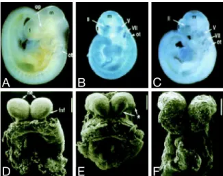
Figure 3: Analysis of E8.5 and E9.5 lrp2 mutant mouse embryos. (A-C) Nile blue staining of E9.5 wild-type (A) andlrp2 -/-mouse embryos (B and C). op, Optic vesicle;
ot, otic vesicle; m, midbrain; t, telencephalon; II, perioptic neural crest and mesoderm;
V, trigeminal ganglion; VII, facio-acoustic ganglion. (D-F) Scanning electron microscopy of forebrain development in E8.5 embryos. The asterisk (E) indicates dysplastic forebrain neural folds (fnf); ne, neuroepithelium (Bars in D-F = 200 um). Modified from Willnow et al. (1996a).
where low molecular weight metabolites (<70 kDa) that have passed glomerular filtration are reabsorbed. The urine oflrp2 knockout mice is abundant in these low molecular weight metabolites (small plasma proteins, plasma vitamin carrier pro- teins, peptide hormones like insulin or parathyroid hormone and lysozyme) because they are apparently not resorbed by the proximal tubules of the kidney (Leheste et al., 1999; Willnow and Herz, 1995). This condition is called low molecular weight proteinuria. Because vitamins are lost together with their carrier proteins in the urine, these animals also suffer from a disturbed vitamin homeostasis (Leheste et al., 2003; Nykjaer et al., 1999; Moestrup et al., 1996). These findings show that LRP2 plays an important role in reuptake of metabolites from the glomerular filtrate in the renal proximal tubules.
A B C D E
F G H I K
+/+
-/- +/+
-/-
+/+
-/-
+/+
-/-
+/+
-/-
E12.5 E10.5
MGE AEP
MGE AEP
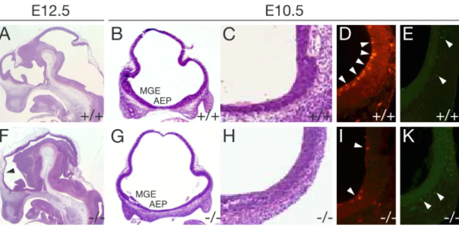
Figure 4: Neuroanatomy and analysis of cell proliferation and apoptosis oflrp2-deficient embryos. (A, F) Sagittal (B-E, G-K) coronal forebrain sections from wild-type (A-E) and lrp2 -/- (F-K) embryos subjected to H+E staining (A-C, F-H), or detection of cell proliferation (D, I) (anti-phosphohistone H3 immunofluorescence), or detection of apop- tosis (E, K) (TUNEL assay). Lrp2 -/- animals suffer from a reduction in neuroepithelial wall thickness (F, arrowhead) compared with controls (A). (B, G) Coronal forebrain sections , indicating a decrease in thickness of the ventral neuroepithelium that is most pronounced in the AEP and the MGE oflrp2 -/-embryos (G) compared with wild-types.
(D, I) Coronal sections through the rostroventral neural tube analyzed for cell prolif- eration, indicating a reduction of mitotic cells in the lrp2 -/- embryos (I, arrowheads) compared with wild-types (D, arrowheads). No difference is seen in the number of apoptotic nuclei in both genotypes (E, K, arrowheads). Abbreviations: AEP, anterior entopeduncular area; MGE, medial ganglionic eminence. Modified from Spoelgen et al.
(2005).
1.2 Holoprosencephaly
The failure of the forebrain (prosencephalon) to divide into bilateral cerebral hemi- spheres is a defect known as holoprosencephaly (HPE). As a consequence the development of facial as well as of brain structures is affected. In humans, the disease manifests itself in a spectrum of defects (Matsunaga and Shiota, 1977).
The most severe cases involve malformations of the brain that lead to perinatal death. Mildly affected individuals display facial defects where the eyes, nose or upper lip are affected, but where the brain is developed almost normally. The least severe of the facial anomalies is the median cleft lip (premaxillary agenesis). The most severe is cyclopia, an abnormality characterized by a single eye located in the area normally occupied by the root of the nose, and a missing or tubular-shaped
1. INTRODUCTION
Receptor Expression Biological functions Ligands LDLR ubiquitous, e.g. hepato-
cytes, macrophages, cen- tral nervous system
cholesterol homeostasis apoE, apoB
LRP1 almost all cell types, e.g.
hepatocytes, neurons, vascular smooth mus- cle cells, macrophage, trophoblast, embryonic tissues
endocytosis of a broad range of ligands, regula- tion of calcium currents, phagocytosis of apoptotic cells, embryonic develop- ment
apoE, amyloid precur- sor protein, lipopro- tein lipase, alpha2- macroglobulin, pro- tease/protease inhibitor complexes, platelet derived growth factor LRP1B central nervous system unknown synaptotagmin, laminin
receptor precursors, apoE LRP2 apical plasma membrane
of absorptive and secre- tory epithelia, thyroid and parathyroid gland, tro- phectoderm, visceral yolk sac, neuroectoderm
vitamin/nutrient supply, calcium homeostasis, recovery of filtered low-molecular weight proteins, uptake and transcytosis of thyroglob- ulin
apoB, apoE, apoJ, apoH, albumin, cubilin, retinol- binding protein, vitamin D binding protein, sonic hedgehog, bone morphogenic protein 4 VLDLR developing and adult
brain, heart and en- dothelial cells, adipose tissue
neuronal migration, synaptic transmission
apoE, Reelin, lipoprotein lipase, tissue factor path- way inhibitor
ApoER2 developing and adult brain, testis
neuronal migration, synaptic transmission, male fertility
apoE, Reelin
MEGF7 restricted expression pat- tern in the embryo, adult central nervous system
limb development, body growth
apoE
LRP5 macrophages, smooth muscle cells, CNS neu- rons, developing lung, osteoblasts, osteoclasts
Regulation of bone for- mation, Wnt signal trans- duction
Wnt proteins
LRP6 smooth muscle cells, eye primordium, osteoblasts, osteoclasts
limb development, mid- and hindbrain develop- ment
Wnt proteins
SorLA neurons, hepatocytes, spleen, lung
intracellular sorting of proteins
APP, ApoE, lipoprotein lipase, head activator pro- tein
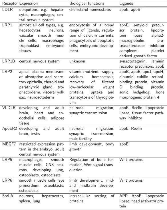
Figure 5: Ligands and functions of the mammalian LDL receptor gene family members.
The table highlights a selection of biological functions and ligands of the listed receptors.
nose located above the eye. Based on the severity of the defects, three forms of holoprosencephaly are distinguished in humans:
1. Alobar holoprosencephaly is the most severe form. The brain fails to separate and affected individuals display facial abnormalities.
2. Semilobar holoprosencephaly is an intermediate form of the disease. The brain hemispheres display a slight tendency to separate.
3. Lobar holoprosencephaly is the least severe form. The hemispheres are al- most completely separated, the brain appears physiologically nearly normal.
In humans, holoprosencephaly is the most common brain abnormality, affecting 1 in 16,000 live-born children (Roach et al., 1975) and 1 in 250 miscarried foetuses (Matsunaga and Shiota, 1977). Holoprosencephaly can have many reasons. Ge- netic disorders can lead to HPE, but also exposure of the embryo to teratogenic chemicals like ethyl alcohol or retinoic acid during pregnancy can result in HPE (Cohen and Shiota, 2002). The molecular mechanisms leading to HPE are not completely understood at present. Studies in the mouse show that the defect is likely a result of defective patterning during the development of the area giving rise to the forebrain.
1.3 Forebrain development
The forebrain (prosencephalon) is the rostral-most portion of the brain. Together with the midbrain (mesencephalon) and the hindbrain (rhombencephalon) these three primary portions of the brain are formed during early development. Later in development, the forebrain separates into the diencephalon and the telencephalon.
The diencephalon can be further subdivided into the epithalamus, thalamus and hypothalamus. Functionally, the diencephalon is responsible for linking the nervous to the endocrine system and for relaying sensory information to the cerebral cor- tex. The telencephalon can also be divided into subregions: limbic system, cerebral cortex, basal ganglia, corpus striatum and olfactory bulbs.
In humans, the telencephalon serves the following major functions: attributing motor function to the body, sensing of smell, formation of memory and emotional sensation. In the mouse, the structures giving rise to the central nervous system start forming approximately at day 8.5 of embryonic development. At this time a specialized region of the ectoderm on the dorsal surface of the embryo termed
1. INTRODUCTION
neural plate starts folding in upon itself to form the neural tube. The process of the flat neural plate forming into the cylindrical neural tube is termed neurulation (Sadler, 2003). The rostral part of the neural tube is giving rise to brain structures, whereas the caudal part develops into the spinal chord. Pathways that are involved in neural tube patterning have also been implicated in the etiology of HPE.
In particular, alterations in the pathways that specify the dorsoventral axis of the rostral neural tube may cause this syndrome. For example, increased dorsal signal- ing through bone morphogenic proteins (Bmps) or through wingless type proteins (Wnts) results in HPE (Golden et al., 1999). Also, loss of sonic hedgehog (Shh) expression, a factor that specifies ventral cell fates in the neural tube (Rubenstein et al., 1998; Inoue et al., 2000) leads to holoprosencephalic syndrome in humans and mice (Chiang et al., 1996; Roessler et al., 1996). Recent findings suggest a likely involvement of LRP2 in the Shh pathway in forebrain development, e. g.
McCarthy et al. (2002) showed that LRP2 binds Shh with high affinity and medi- ates endocytosis of the morphogen. Internalized Shh was able to bypass lysosomal degradation, implicating LRP2 as a new regulatory component of the Shh signaling pathway (McCarthy et al., 2002).
1.3.1 The sonic hedgehog pathway
Sonic hedgehog is a secreted protein expressed in the notochord and in the floor- plate that undergoes autocatalytic cleavage into a 19 kDa amino terminal (Shh-N) and a 25 kDa carboxy terminal (Shh-C) domain. During cleavage, a cholesterol moiety becomes covalently attached to Shh-N (Porter et al., 1996), the active fragment that binds the receptor Patched (Ptch) (Ingham and McMahon, 2001).
This cholesterol modification is crucial for the proper activity of Shh-N (Porter et al., 1996).
In the absence of sonic hedgehog, Ptch constitutively inhibits activity of the path- way by binding to another transmembrane protein termed Smoothened (Smo).
Binding of Shh-N to Ptch results in the release of Smo and activation of down- stream Shh target genes through glioma-associated oncogene (Gli) proteins (Ing- ham and McMahon, 2001).
Expression of LRP2 in the yolk sac and in the neuroepithelium of the developing embryo and its ability to take up lipoproteins suggest a possible function for the receptor in delivery of cholesterol-rich lipoproteins from the maternal circulation to the embryo for Shh activation (Farese and Herz, 1998). The idea that a lack of cholesterol can lead to forebrain malformations is supported by experiments
where pregnant rats received an inhibitor for cholesterol biosynthesis and were fed a cholesterol free chow. Embryos, where the mother was subjected to this treat- ment and that were depleted of cholesterol indeed displayed HPE (Lanoue et al., 1997). A known genetic disorder in humans (Smith-Lemli-Opitz syndrome) that affects cholesterol synthesis also leads to HPE (Kelley et al., 1996). Alternatively, expression of LRP2 in the neuroepithelium and its ability to bind Shh indicates a more direct effect as a co-receptor in Shh signaling (McCarthy et al., 2002).
1. INTRODUCTION
2 Aim of this study
During the last years, our understanding of the members of the low-density lipopro- tein receptor gene family has changed substantially. It was initially assumed that the family members act mainly in the lipid metabolism but now it has become clear that most members of this protein family are playing essential roles in more biological processes than merely endocytosis.
Especially the generation of mouse models lacking specific receptors has proved that most family members are part of important signaling cascades. The genera- tion of LRP2-deficient mice by Willnow et al. in 1996 provided insights into this receptor’s physiological function. Whereas the receptor’s function in the kidney has been well characterized, the forebrain defects have only been described histo- logically so far, the underlying mechanism remaining to be elucidated.
The aim of this study was to determine the role of LRP2 during early forebrain development. The receptor is heavily expressed in the visceral endoderm of the yolk sac and in the neuroepithelium of the developing embryo. In the yolk sac it is considered to be important for the supply of the early embryo with lipids and vitamins from the maternal circulation.
A role for the receptor in the neuroepithelium besides the described proliferative defects seen in the AEP and MGE of lrp2 -/- embryos has not been established yet. Theoretically, both sites of expression could be important for forebrain de- velopment. To answer the question to what extent LRP2 in the embryo proper respectively in the yolk sac contributes to the observed forebrain malformations, the receptor’s function in these tissues should be dissected. This should be achieved by conditional lrp2 gene inactivation in the embryo proper.
In addition to that, different signaling pathways important for forebrain develop- ment should be analyzed by comparing the expression patterns of marker genes in lrp2 knockout and wildtype embryos.
This study should provide new insights on how LRP2 contributes to the proper formation of forebrain structures in the developing embryo.
2. AIM OF THIS STUDY
3 Material and Methods
3.1 Animal Experiments
3.1.1 Mouse husbandry
Mice were kept at standard conditions according to the German animal protection act. The used wildtype mice were of mixed genetic background (129SvEmcTer and C57BL/6N). The generation of mice with a targeted disruption of the lrp2 gene has been described (Willnow et al., 1996a). The lrp2 gene defect was an- alyzed in receptor-deficient and somite-matched wildtype littermates on a hybrid (129SvEmcTer x C57BL/6N) background.
For conditional inactivation of the lrp2 gene in the epiblast, animals homozy- gous for a loxP-modified allele of the lrp2 gene (lrp2lox/lox) (Leheste et al., 2003) were crossed with mice carrying a knock-in of the Cre transgene into the mes- enchyme homeobox 2 gene locus Meox2tm1(cre)Sor obtained from Jackson Labora- tories (www.JAX.org) (Tallquist and Soriano, 2000). Embryos from breeding of lrp2lox/wt/Meox2tm1(cre)Sor mice with lrp2lox/lox animals were used for phenotypic analysis following genotyping of embryo- or yolk sac-derived tissues.
Timed matings were set up in the evening to obtain embryos at different stages of development. The presence of a vaginal plug in the morning was considered as day 0.5 post coitum (dpc). The embryos were harvested by sacrificing pregnant mice according to the German animal protection act, staged by counting the somites and further processed.
3.1.2 Zebrafish husbandry
Zebrafish were kept at standard conditions according to the German animal protec- tion act. The used wildtype line was of the AB background. Embryos were kept in egg water (60 µg/ml Instant Ocean Sea Salts, Aquarium Systems Inc, USA) with or without 0,003% PTU (1-Phenyl-2-thiourea, Sigma, USA) to suppress pigmen- tation and were staged by hours post fertilization (hpf) at 28.5◦C or by counting somites (Kimmel et al., 1995).
3. MATERIAL AND METHODS
3.1.3 Morpholino Injections
Morpholino antisense oligonucleotides were purchased from GENE TOOLS, LLC, USA. Sequences were chosen to target an exon splice donor site resp. the ATG re- gion of the corresponding mRNA. This resulted in aberrant splicing of the mRNA, resp. blocking of the translation initiation site. The Morpholinos were injected at a concentration of 100 mM in sterile ddH2O using glass micropipettes and a microinjector/micromanipulator setup (MPPI-2 Pressure Injector/ BP15 Back Pressure Unit, Applied Scientific Instrumentation, USA; MM33 Micromanipula- tor, Maerzhaeuser, Germany). One- to two-cell stage embryos were used for the injection of approximately 4 nl Morpholino-solution. The effect of the splice- variant-Morpholinos was verified by RT-PCR on total embryo RNA. MO sequences (5’→3’):
lrp2MO1 AATCAGTGCTTGTGGTTTACCTGGG lrp2MO2 GTACGAGTGTGTTCACCTGTGCCAG dab2MO TTCTGCTTCAGGTGACTGTGACATG
3.1.4 Dye filtration experiments
1 mg/ml solutions of lysine-fixable 10 kDa dextran labeled with rhodamine (10 kDa-RD; Molecular Probes, USA) and lysine-fixable 70 respectively 500 kDa dextran labeled with fluoresceine (70 resp. 500 kDa-FD; Molecular Probes, USA) were prepared in phosphate buffered saline (PBS; 137 mM NaCl, 2.7 mM KCl, 12.5 mM Na2HPO4, 1.7 mM KH2PO4, pH 7.4). Recombinant His-tagged RAP (39 kDa) was custom-labeled with the Cy2TM bisfunctional reactive dye kit (Cy2- RAP; Amersham, UK) according to manufacturer’s instructions.
The tracers were injected into the common cardinal vein (CCV) of embryos anaes- thetized with 0.2 mg/ml tricaine (3-amino benzoic acid ethyl ester, Sigma, USA) solution in egg water using the same setup as for injecting morpholinos. Uptake of filtered tracer was evaluated 1 to 1.5 hrs after injection on whole mounts using a fluorescent dissecting stereomicroscope (Leica MZ16F, Leica, Germany). Embryos were then fixed in 4% paraformaldehyde (PFA), embedded in either Technovit 7100 (Heraeus Kulzer, Germany) or Tissue-TekR (Sakura, Japan) and sectioned at 5 µm on a Leica RM 2155 or Leica Cryocut 3000 rotary microtome (Leica, Germany).
3.1.5 Acridine orange staining
Acridine orange (AO) is a vital dye that stains apoptotic cells, but not necrotic cells (Barrallo-Gimeno et al., 2004). Zebrafish embryos were incubated in 5µg/ml AO solution for 1 hr in the dark at RT. The embryos were then washed three times for 10 min with egg water at RT and analyzed using a fluorescence dissecting stereomicroscope (Leica MZ16F, Leica, Germany).
3.2 Microbiological Methods
3.2.1 Culture media
Medium Composition
LB 10 g/l bacto-tryptone, 5 g/l bacto-yeast extract,10 g/l NaCl;
pH 7.2
SOC 20 g/l bacto-peptone, 5 g/l bacto-yeast extract, 0.5 g/l NaCl, 0.17 g/l KCl, 0.95 g/l MgCl2, 3.6 g/l glucose; pH 7.0
LB-Agar LB-medium containing 15 g/l agar
3.2.2 Preparation of electrocompetent bacteria
A single colony ofE. coli HB101 picked from a LB agar plate was used to inoculate 20 ml of LB medium in a 250 ml sterile flask. The cells were grown overnight with vigorous aeration at 37◦C. The next day 10 ml of the cell suspension was diluted into 1 l of LB medium and grown at 37◦C until the OD600 reached 0.5. The cell suspension was then incubated on ice for 20 min and the cells were harvested by centrifugation (4000xg; 15 min; 4◦C). The pellet was resuspended in 1 l of icecold sterile 10% glycerol. Cells were again collected by centrifugation (4000xg; 15 min;
4◦C) and the pellet was resuspended in 500 ml of icecold sterile 10% glycerol.
The suspension was pelleted again (4000xg; 15 min; 4◦C) and resuspended in 20 ml icecold sterile 10% glycerol. After collecting (4000xg; 15 min; 4◦C) and resuspending in 2 ml of 10% icecold sterile glycerol once more, the cells were stored at -80◦C in 100 µl aliquots.
3. MATERIAL AND METHODS
3.2.3 Cryopreservation of bacteria
1 ml of an overnight culture ofE. coli HB101 was mixed with 1 ml of 50% glycerol and immediately frozen at -80◦C.
3.2.4 Transformation of bacteria with DNA
ElectrocompetentE. coli HB101 cells were transformed with purified plasmid DNA or directly with DNA-ligation reactions. An aliquot of electrocompetent HB101 cells was thawed on ice. 10 ng of plasmid DNA (or 1/3 of the ligation reaction) were mixed with 40 µl of electrocompetent HB101 cell suspension and electroporated at 1.8 kV.
The cell suspension was transferred from the cuvette to a 2.0 ml tube, mixed with 1 ml of SOC medium and incubated at 37◦C for 30 min. Cells were then collected (2500xg; 5 min; RT) and resuspended in 100 µl of LB medium and plated on a LB agar plate containing the appropriate selective agent.
3.3 Molecular biology methods
3.3.1 Isolation of plasmid DNA from bacteria
5 ml of LB medium were inoculated with a single colony ofE. coli HB101 picked from a LB agar plate containing the appropriate selection marker. The LB culture was grown overnight at 37◦C with vigorous shaking.
The next day the cells were collected by centrifugation (14000xg; 5 min; RT). The pellet was resuspended in resuspension buffer (50 mM Tris-HCl, 10 mM EDTA, 100 µg/ml RNase A; pH 8.0) and subsequently lysed by adding an equal volume of lysisbuffer (200 mM NaOH, 1% SDS (w/v)). The solution was mixed cau- tiously with an equal volume of neutralization buffer (3.0 M K-acetate; pH 5.5) and incubated on ice for 15 min. Cellular debris and genomic DNA were re- moved by centrifugation of the solution (14000xg; 20 min; 4◦C). Remaining proteins were removed by extracting the supernatant with an equal volume of phenol/chloroform/isoamylalcohol (25:24:1) followed by centrifugation (14000xg;
5 min; RT) to separate the phases. The upper, aqueous DNA containing phase was mixed with 2.5 volumes of 100% ethanol and the DNA was precipitated by centrifugation (14000xg; 20 min; 4◦C). The pellet was washed once with 70%
ethanol and resuspended in 50 µl of TE-buffer (10 mM Tris-HCl, 1 mM EDTA;
pH 8.0). The purified plasmid DNA was stored at -20◦C.
3.3.2 Isolation of genomic DNA from tissue samples
Tissue for isolating genomic DNA was obtained by subjecting adult mice to a tail biopsy, respectively adult zebrafish to a tailfin biopsy. Yolk sac tissue was used to isolate DNA for genotyping mouse embryos. DNA was isolated by incubating the tissue with Protease K in tail buffer (10 mM Tris-HCl, 0.3 M Na-Acetate, 0.1 mM EDTA, 1% SDS (w/v); pH 7.0) at a final concentration of 0.5 mg/ml for several hours to overnight at 52◦C. Proteins were removed by extracting with an equal volume of phenol/chloroform/isoamylalcohol (25:24:1) followed by centrifugation (14000xg; 5 min; RT) to separate the phases. The upper, aqueous DNA containing phase was mixed with 2.5 volumes of 100% ethanol. The precipitate was collected by centrifugation (14000xg, 10 min, 4◦C) washed once with 70% ethanol and redissolved in TE-buffer (10 mM Tris-HCl, 1 mM EDTA; pH 8.0). Isolated genomic DNA was stored at 4◦C.
3.3.3 Isolation of total RNA from tissue samples
Total RNA was isolated with the TRIZOLR reagent from Invitrogen, USA. Tis- sue samples were homogenized in 1 ml of TRIZOLR reagent per 50-100 mg of tissue and incubated for 5 min at RT. 0.2 ml of chloroform were added to the homogenate per 1 ml of TRIZOLR reagent. Samples were shaken vigorously by hand for 15 seconds and incubated at RT for 3 min. The phases were separated by centrifugation (12000xg; 15 min; RT). Following centrifugation, the upper aque- ous phase was transferred to a fresh tube and precipitated by adding 0.5 ml of isopropyl alcohol per 1 ml of TRIZOLR. The RNA was collected by centrifugation (12000xg, 10 min, 4◦C) and washed once with 1 ml of 70% ethanol per 1 ml TRIZOLR. The pellet was air-dried for 10 min redissolved in RNase-free water and stored at -80◦C.
3.3.4 DNA and RNA concentration determination
The concentration of DNA and RNA samples was determined spectrophotometri- cally at a wavelength of 260 nm (OD260) since the concentration of DNA and RNA is a direct function of the optical density at this wavelength. For DNA, an OD260 of 1.0 equals a concentration of 50 µg/ml of double stranded DNA, for RNA an OD260 of 1.0 equals a concentration of 40µg/ml of RNA.
DNA quality measurement was done by measuring the OD280because proteins ab- sorb UV-light maximally at this wavelength. Pure DNA solutions have an OD260:
3. MATERIAL AND METHODS
OD280 ratio of 1.8. A lower ratio indicated contamination of the sample with proteins.
3.3.5 Enzymatic digest of DNA
The appropriate amount of DNA was incubated with the corresponding restric- tion enzyme(s) and buffer at a ratio of 0.5 U emzyme/µg DNA. The digest was incubated at 37◦C for 2 hrs to overnight. All restriction enzymes were obtained from New England Biolabs, USA. After incubation, the digest was either column purified or subjected to gel electrophoresis to isolate the fragment of interest.
3.3.6 Agarose gel electrophoresis of DNA and RNA
DNA and RNA fragments were separated according to their molecular weight on 0.8-2.0% agarose gels. Ethidium bromide was added to the gel at a final concen- tration 0.5µg/ml to facilitate visualization of the DNA resp. RNA fragments after electrophoresis.
3.3.7 Isolation of DNA from agarose gels
Polymerase chain reaction (PCR) products or DNA digests were resolved on 0.8- 1.2% agarose gels containing ethidium bromide. By exposing the agarose gel to UV-light, the DNA was visualized and bands of interest were cut from the gel. The DNA was extracted using the High Pure PCR Product Purification Kit from Roche, Switzerland. The gel slice was incubated with binding buffer (3 M guanidine- thiocyanate, 10 mM Tris-HCl, 5% ethanol (v/v); pH 6.6) at a ratio of 300 µl buffer/100 mg agarose at 52◦C until the gel slice was completely dissolved. The sample was transferred to a filter column and subjected to centrifugation (14000xg, 1 min, RT). The filter column was washed two times with 500 µl washing buffer (2 mM Tris-HCl, 20 mM NaCl, 80% ethanol (v/v); pH 7.5) and the DNA was eluted with 50µl elution buffer (10 mM Tris-HCl; pH 8.5) and stored at -20◦C.
3.3.8 Ligation of PCR-products in the pGEM-T Easy Vector
The PCR-reaction-mix was separated electrophoretically on a 0.8-1.2% agarose-gel, and the band of interest cut from the gel and column purified. Ligation of PCR- Products with the pGEM-TR Easy Vector (Promega, USA) was done according to
the manufacturers instructions: 5-10 ng of the PCR-product were incubated with 5 µl 2X Rapid Ligation BufferR, 30 ng of pGEM-T Easy Vector and 3 U of T4 DNA-Ligase. The volume of the reaction setup was adjusted to 10 µl with H2O and incubated at 16◦C overnight. The next day 1/3 of the ligation reaction was used to transform electrocompetentE. coli HB101.
3.3.9 Polymerase chain reaction
If not stated otherwise PCR was carried out according to the following parameters:
50 ng of template DNA were incubated in the presence of 250µM dNTPs, 300 nM forward primer, 300 nM reverse primer, 2 units AmpliTaq Gold DNA-Polymerase (Perkin-Elmer, USA), 0.01% Triton X-100 (v/v), 50 mM KCl, 10 mM Tris-HCl, 1.5 mM MgCl2 and 0.001% (w/v) gelatin; pH 8.3 in a thermocycler. During the first cycle the reaction mix was denatured at 95◦C for 10 min, primers were allowed to anneal at 60◦C for 1 min and DNA synthesis was carried out at 72◦C for 1 min.
For the following 35 cycles the reaction mix was denatured at 95◦C for 45 sec, annealed at 60◦C for 45 sec and elongated at 72◦C for 45 sec. This was followed by a final elongation step at 72◦C for 10 min. The reaction mix was then cooled down to 4◦C and further processed.
3.3.10 Primer sequences
3. MATERIAL AND METHODS
Mouse specific primer
Primer identifier Sequence (5’→3’)
BPA GATTGGGAAGACAATAGCAGGCATGC
g15 CTGAGGTACCCAGTCTCCTGTCAG
g20 GACCATTTGGCCAGCCAAGG
g21 CATATCTTGGAAATAAAGCGAC
LAKA ACCTTGCGTGAATTCTGGG
morecre CCAGATCCTCCTCACAAATCAGC
morefor GGGACCACCTTCTTTTGGCTT
morerev AAGATGTGGAGAGTTCGG
TKP2 TGAAAACCACACTGCTCGATCCGGAAC
Zebrafish specific primer
Primer identifier Sequence (5’→3’)
Dab2 For CCCCCTCCCACACTTTTTCTTTCA
Dab2 Rev TGCCATTTCCTTCACCCTTTTGTC
LRP2 For1.1 GCAGATGCATGAATGGAGGAACG
LRP2 For AATGGGCAAGTCTAGAGGAGC
LRP2 Rev2 TTTGGGAGGAACAAACGGA
LRP2 Rev3 TCCACTGCCTGCTGATATGT
LRP2seq For GGATGGATGGATGGATGGATA
LRP2seq Rev ATTGGTGACTATACAAGGGCG
Nkx2.1 For GACCTGCCGGCCTATCAAGACG
Nkx2.1 Rev CGGAGCGCCACCCCAGTT
Olig2 For TGAACGCCATGGACTCTGACACGA
Olig2 Rev CCTGGCACATGCTACACGGACAAG
3.3.11 Reverse transcription
Generation of cDNA from RNA was done using the SensiscriptR Reverse Tran- scription Kit (Qiagen, Germany). 50 ng of total RNA were incubated with 2.0µl 10x Buffer RT, 2.0 µl dNTP Mix (5 mM each dNTP), 1 µM Oligo-dT primer, 10 U RNase inhibitor and 1.0µl SensiscriptR Reverse Transcriptase. The volume of the reaction setup was adjusted to 20 µl with H2O and incubated at 37◦C for 60 min. The reaction mix was then stored at -20◦C.