ATP measurement as method to monitor the quality of reprocessing flexible endoscopes
ATP-Bestimmung als Methode zur Qualitätskontrolle der Endoskopaufbereitung
Abstract
Insufficient performance of cleaning and disinfection of flexible endo- scopes can pose an infection risk to patients. Actually quality of repro-
Dorothea Hansen
1Daniel Benner
1cessing is checked by performing microbiological cultures. Unfortunately,
Martin Hilgenhöner
1their results are not available on the same day so that more rapid
Therese Leisebein
1methods are desirable. We compared the ATP (adenosine triphosphate) bioluminescence for hygiene checking of the reprocessing procedures
Andreas Brauksiepe
1of 108 flexible endoscopes with routine microbiological culture technics.
Walter Popp
1Sensitivity and specifity of ATP bioluminescence was calculated. 28 endoscopes showed bacterial growth of at least one sample. Depending
on the applied threshold of bioluminescence between 67 and 28 endo- 1 Krankenhaushygiene, Universitätsklinikum Essen, Germany
scopes were positive. Sensitivity varied between 0.46 and 0.75 and specifity between 0.43 and 0.81. ATP bioluminescence does not replace routine microbiologic methods but it can indicate the need of immediate check of reprocessing.
Zusammenfassung
Von unzureichender Reinigung und Desinfektion flexibler Endoskope können Infektionsrisiken für Patienten ausgehen. Die Endoskopaufbe- reitung wird derzeit durch mikrobiologische Untersuchungen kontrolliert.
Der Nachteil ist, dass die Ergebnisse oft erst nach Tagen vorliegen, so dass ein Schnelltest wünschenswert wäre. Wir verglichen die ATP(Ade- nosintriphosphat)-Biolumineszenz-Bestimmung zur hygienischen Kon- trolle der Endoskopaufbereitung mit konventionellen mikrobiologischen Kulturen bei 108 flexiblen Endoskopen und bestimmten die Sensitivität und Spezifität der ATP-Biolumineszenz. 28 Endoskope zeigten mindes- tens in einer mikrobiologischen Kultur Bakterienwachstum. Abhängig vom zugrunde gelegten Grenzwert der Biolumineszenz waren zwischen 67 und 28 der Endoskope bei der ATP-Bestimmung positiv. Die Sensiti- vität der ATP-Bestimmung lag zwischen 0,46 und 0,75, die Spezifität zwischen 0,43 und 0,81. Die ATP-Bestimmung ersetzt nicht die regel- mäßigen mikrobiologischen Untersuchungen, sie kann jedoch die Not- wendigkeit einer sofortigen Überprüfung der Aufbereitungsmodalitäten anzeigen.
Introduction
Infections by endoscopes have been described as a consequence of insufficient cleaning and disinfection [1], [2]. Reports regard duodenoscopes [3], coloscopes [4], and bronchoscopes [5], [6] and concern exists regarding the possible transmission of infective agents like hepatitis B-/C- virus, HIV, Mycobacterium tuberculosis and Helico- bacter pylori [7]. An investigation in southern Germany has shown that 50% of endoscopes were still contamin- ated by bacteria after reprocessing [8]. American [9], [10], [11] and European [7], [12], [13], [14], [15] recom-
mendations for the reprocessing of endoscopes have been published. Whereas in Germany and various other countries control of cleaning and disinfection by microbi- ological methods is recommended the use of microbiolo- gical cultures to routinely check the reprocessing process is discussed controversely in USA [7], [16]. Unfortunately, there are many disadvantages of microbiological cultures:
Getting results lasts for days, so the endoscopes are used with other patients. Additionally, viruses, Helicobacter and M. tuberculosis are not at all included and slowly growing organisms only if the time of incubation is long
enough. So a more rapid method for checking the repro- cessing of endoscopes is needed.
ATP (adenosine triphosphate) measurement is used as indicator of cleaning control [17], [18] in food and kitchen hygiene. ATP is as well an indicator of organic as of micro- biological contamination. It is a simple method measuring the amount of light which is emitted when the enzyme luciferase comes into contact with molecular ATP and which is directly proportional to the amount of ATP [19].
ATP measurement may be a suitable method to control the quality of endoscope reprocessing as it is measuring cleaning effectiveness which may indicate the reduction of infection risk.
We did an investigation to compare the ATP biolumines- cence for hygiene checking of reprocessing with routine microbiological cultures.
Methods
Between January and December 2003 we examined 108 endoscopes (40 gastroscopes, 18 coloscopes, 8 duoden- oscopes and 42 bronchoscopes) after reprocessing.
Sterile swabs were moistened with sterile 0.9% NaCl. We took swabs of distal end and rinsing valve and rinsed the operating channel with 20 ml sterile 0.9% NaCl. We did not use neutralizers. Swabs and 0.5 ml of rinsing fluid were inoculated on blood agar, MacConkey agar and Sa- bouraud agar and incubated for 48 hours at 37° C and 7 days at 22° C respectively. Bacterial species identific- ation followed routine microbial laboratory proceedings (API biomerieux). Every bacterial growth was considered microbiological positive regardless of species or number of cfu (colony forming unit). ATP and AMP (adenosine monophosphate) bioluminescence was determined using Lumitester PD 10 (Scil Diagnostics). The assay was car- ried out according to the manufacturer´s instructions immediately after swabbing. Reagent blanks were ob- tained using sterile swabs moistened with sterile 0.9%
NaCl, instrument disinfectant and endoscope cleaner used for endoscope reprocessing. Bioluminescence readings were expressed as relative light units (RLU).
Thresholds for bioluminescence were chosen between 30 and 100 RLU according to manufacturer's personal recommendation and after determining RLU of disinfect- ant and cleaner below 10 RLU. Sensitivity and specifity of ATP bioluminescence compared with microbiological culture as a gold standard were calculated for all types of examined endoscopes together and displayed in a ROC curve for various threshold values. The area under the ROC curve, which is usually chosen as the summary measure of diagnostic accuracy, was also computed.
Results
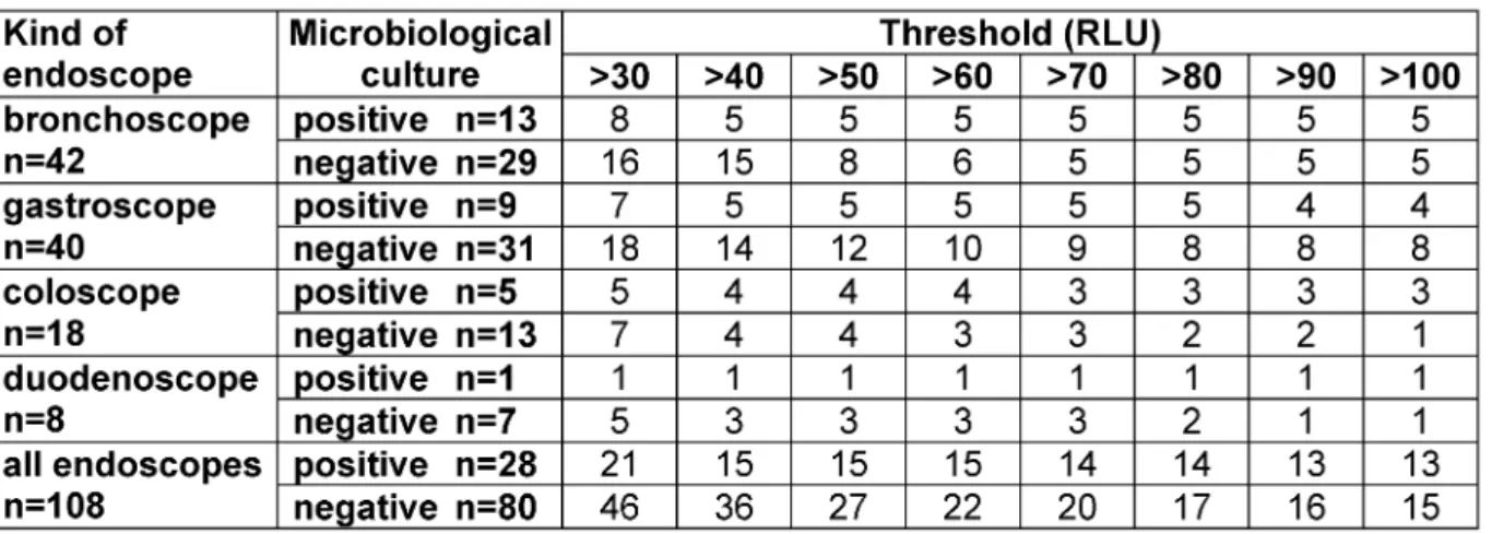
Results of microbiological culture and ATP biolumines- cence are shown in Table 1. Microbiological cultures of 28 endoscopes (26%) showed bacterial growth. 13% of
checked duodenoscopes, 28% of coloscopes, 23% of gastroscopes and 31% of bronchoscopes were bacterially contaminated. The detected organisms were Pseudomo- nas aeruginosa, other non fermenting gramnegative rods, Enterobacteriaceae, Staphylococcus aureus, koagulase negative Staphylococci, Corynebacteriae, Bacilli, Candida and moulds. Dependent on the chosen threshold between 28 (26%) and 67 (62%) endoscopes were positive for ATP bioluminescence. ATP bioluminescence of 75% of duodenoscopes, 67% of coloscopes, 63% of gastroscopes and 57% of bronchoscopes was above a threshold of 30 RLU. There were still 25% of duodenoscopes, 50% of co- loscopes, 30% of gastroscopes and 24% of broncho- scopes above a threshold of 100 RLU. ATP biolumines- cence of 5 bronchoscopes and 2 gastroscopes was below 30 RLU despite being microbiologically contaminated.
Choosing 30 RLU as threshold 21 endoscopes (8 bron- choscopes, 7 gastroscopes, 5 coloscopes and 1 duoden- oscope) were positive for ATP bioluminescence and mi- crobiological culture and 34 endoscopes (13 broncho- scopes, 13 gastroscopes, 6 coloscopes and 2 duodeno- scopes) were negative. 46 endoscopes (16 broncho- scopes, 18 gastroscopes, 7 coloscopes and 5 duodeno- scopes) had an ATP bioluminescence above 30 RLU despite negative microbiological result. Above a threshold of 100 RLU 65 endoscopes (24 bronchoscopes, 23 gastroscopes, 12 coloscopes and 6 duodenoscopes) had concordant negative and 13 endoscopes (5 broncho- scopes, 4 gastroscopes, 3 coloscopes and 1 duodeno- scope) concordant positive results. Microbiological cul- tures of 15 endoscopes (5 bronchoscopes, 8 gastro- scopes, 1 coloscope and 1 duodenoscope) were negative despite ATP bioluminescence above 100 RLU. The ROC curve of sensitivity and specifity for thresholds between 30 and 100 RLU is presented in Figure 1. The area under the ROC curve is 0.63. Compared with microbiological culture as a gold standard sensitivity of bioluminescence varied between 0.75 for a threshold of 30 RLU (95%
confidence interval 0.55-0.89) and 0.46 for 100 RLU (95% confidence interval 0.28-0.66) and specifity between 0.43 (95% confidence interval 0.32-0.54) and 0.81 (95% confidence interval 0.71-0.89) respectively (Table 2).
Discussion
In our study, 26% of tested endoscopes showed microbi- ological contamination. The number of contaminated endoscopes in our investigation is in accordance with the results of Moses and Lee [20], who found between 12%
and 24% positive cultures during a 10-year study period.
It is much lower than that of the HYGEA study [8]. Moses and Lee examined only endoscopes used in a clinical in- stitution and reprocessed in an automated washer whereas in half of endoscopy facilities of the HYGEA study endoscopes were reprocessed manually. In our study 92 endoscopes (85%) were reprocessed in an automated washer.
Table 1: Number of endoscopes with a bioluminescence value above the threshold compared with microbiological culture
Table 2: Sensitivity and specifity of bioluminescence as compared with microbiological culture and 95% confidence interval
Figure 1: ROC curve of bioluminescence compared with microbiological culture as gold standard for thresholds between 30 and 100 RLU
Dependent on the chosen threshold between 62% and 26% of the tested endoscopes had a positive biolumines- cence result indicating possible organic contamination.
In order to calculate sensitivity and specifity of ATP biolu- minescence there must be another method which truly indicates contamination of reprocessed endoscopes. The only established method for checking endoscope repro- cessing is microbiological culture. Microbiological culture may fail in indicating all contaminated endoscopes. There may be non viable organisms or organisms which cannot be cultured on conventional culture medium and other than bacterial contaminations are possible. Because of the absence of other methods for checking endoscopes we calculated sensitivity and specifity of ATP biolumines- cence compared to microbiological culture as gold standard. Sensitivity and specifity of bioluminescence differ dependent on the chosen threshold. In our study sensitivity was only 0.75 even when the chosen threshold of RLU was low. The ROC curve of ATP bioluminescence presented in Figure 1 with an area under the curve of 0.63 indicates that there is no strong concordance between ATP bioluminescence and microbiological cul- ture. Our results are similar to those found by Murphy et al. [18] for testing food contact surfaces. Murphy et al.
suspected that conventional microbiology is more sensi- tive than ATP bioluminescence when total ATP is low [18].
Bacterial ATP content may be below the limit of ATP bio- luminescence. Different bacterial specimen can contain different amounts of ATP and the amount of ATP also depends on the metabolism of the organisms [19]. ATP bioluminescence may also be influenced by the number of viable bacteria present. We did not differ between kind of specimen and number of cfu cultured. The number of cfu found on most swabs was very low and this may ex- plain the low sensitivity of ATP bioluminescence compared to routine microbiology in our study. Additionally the low specifity of ATP bioluminescence may be explained by the fact, that not only viable bacteria but also other organ- ic contamination is detected. Similar to our study Poulis et al. [21] could not find a clear relationship between ATP bioluminescence measurements and number of cfu on surface plates under practical conditions on surfaces in a factory. Alfa et al. [22] reported that the presence of high residual soil (protein, carbohydrate, hemoglobin and endotoxin) did not correlate with microbiological contam- ination of reprocessed endoscopes. Thus measurable ATP bioluminescence may indicate contamination of en- doscopes without presence of cultivable microorganisms.
Reprocessed endoscopes should be clean. A clean endo- scope should not only show a less amount of viable or- ganisms but also a less amount of all organic contamin- ation and ATP sources. The presence of any ATP source may indicate an infectious risk for consecutively examined patients and should be avoided irrespective of cultivable bacteria.
We conclude that ATP bioluminescence does not replace routine microbiologic methods but it should be applied additionally to check endoscope reprocessing. In contrast to microbiologic methods results of ATP bioluminescence
are available at once and can indicate the need for checking the reprocessing practice immediately.
References
1. Nelson DB, Barkun AN, Block KP, Burdick JS, Ginsberg GG, Greenwald DA, Kelsey PB, Nakao NL, Slivka A, Smith P, Vakil N.
Technology status evaluation report. Transmission of infection by gastrointestinal endoscopy. May 2001. Gastrointest Endosc.
2001;54(6):824-8.
2. Cowen AE. The clinical risks of infection associated with endoscopy. Can J Gastroenterol. 2001;15(5):321-31.
3. Langenberg W, Rauws EA, Oudbier JH, Tytgat GN. Patient-to- patient transmission of Campylobacter pylori infection by fiberoptic gastroduodenoscopy and biopsy. J Infect Dis.
1990;161(3):507-11.
4. Bronowicki JP, Venard V, Botte C, Monhoven N, Gastin I, Chone L, Hudziak H, Rihn B, Delanoe C, LeFaou A, Bigard MA, Gaucher P. Patient-to-patient transmission of hepatitis C virus during colonoscopy. N Engl J Med. 1997;337(4):237-40.
5. Bronchoscopy-related infections and pseudoinfections--New York, 1996 and 1998. MMWR Morb Mortal Wkly Rep.
1999;48(26):557-60.
6. Ramsey AH, Oemig TV, Davis JP, Massey JP, Torok TJ. An outbreak of bronchoscopy-related Mycobacterium tuberculosis infections due to lack of bronchoscope leak testing. Chest.
2002;121(3):976-81.
7. Anforderungen an die Hygiene bei der Aufbereitung flexibler Endoskope und endoskopischen Zusatzinstrumentariums.
Empfehlung der Kommission für Krankenhaushygiene und Infektionsprävention beim Robert Koch-Institut (RKI).
Bundesgesundheitsbl Gesundheitsforsch Gesundheitsschutz.
2002;45(4):395-411.
8. Bader L, Blumenstock G, Birkner B, Leiss O, Heesemann J, Riemann JF, Selbmann HK. HYGEA (Hygiene in der
Gastroenterologie - Endoskop-Aufbereitung): Studie zur Qualität der Aufbereitung von flexiblen Endoskopen in Klinik und Praxis.
Z Gastroenterol. 2002;40(3):157-70.
9. Nelson DB, Jarvis WR, Rutala WA, Foxx-Orenstein AE, Isenberg G, Dash GR, Alvarado CJ, Ball M, Griffin-Sobel J, Petersen C, Ball KA, Henderson J, Stricof RL; Society for Healthcare Epidemiology of America. Multi-society guideline for reprocessing flexible gastrointestinal endoscopes. Society for Healthcare Epidemiology of America. Infect Control Hosp Epidemiol. 2003;24(7):532-7.
10. Reprocessing of flexible gastrointestinal endoscopes. American Society for Gastrointestinal Endoscopy. Gastrointest Endosc.
1996;43(5):540-5.
11. Alvarado CJ, Reichelderfer M. APIC guideline for infection prevention and control in flexible endoscopy. Association for Professionals in Infection Control. Am J Infect Control.
2000;28(2):138-55.
12. Cleaning and disinfection of equipment for gastrointestinal endoscopy. Report of a Working Party of the British Society of Gastroenterology Endoscopy Committee. Gut. 1998;42(4):585- 93.
13. Systchenko R, Marchetti B, Canard JN, Palazzo L, Ponchon T, Rey JF, Sautereau D; French Society of Digestive Endoscopy.
Guidelines of the French Society of Digestive Endoscopy:
recommendations for setting up cleaning and disinfection procedures in gastrointestinal endoscopy. Endoscopy.
2000;32(10):807-18.
14. Bundesärztekammer. Empfehlung zur Qualitätssicherung in der gastrointestinalen Endoskopie. Dtsch Ärztebl. 2000;97(8):A475- A477.
15. Anforderungen der Hygiene an die baulich-funktionelle Gestaltung und apparative Ausstattung von Endoskopieeinheiten.
Empfehlung der Kommission für Krankenhaushygiene und Infektionsprävention beim Robert Koch-Institut.
Bundesgesundheitsbl Gesundheitsforsch Gesundheitsschutz.
2002;45(4):412-4.
16. Leung J, Vallero R, Wilson R. Surveillance cultures to monitor quality of gastrointestinal endoscope reprocessing. Am J Gastroenterol. 2003;98(1):3-5.
17. Seeger K, Griffiths MW. Adenosine triphosphate bioluminescence for hygiene monitoring in health care institutions. J Food Prot.
1994;57(6):509-12.
18. Murphy SC, Kozlowski SM, Bandler DK, Boor KJ. Evaluation of adenosine triphosphate-bioluminescence hygiene monitoring for trouble-shooting fluid milk shelf-life problems. J Dairy Sci.
1998;81(3):817-20.
19. Baumgart J. Möglichkeiten und Grenzen moderner Schnellverfahren zur Prozesskontrolle von Reinigungs- und Desinfektionsverfahren. Zentralbl Hyg Umweltmed. 1996;199(2- 4):366-75.
20. Moses FM, Lee J. Surveillance cultures to monitor quality of gastrointestinal endoscope reprocessing. Am J Gastroenterol.
2003;98(1):77-81.
21. Poulis JA, de Pijper M, Mossel DA, Dekkers PP. Assessment of cleaning and disinfection in the food industry with the rapid ATP- bioluminescence technique combined with the tissue fluid contamination test and a conventional microbiological method.
Int J Food Microbiol. 1993;20(2):109-16.
22. Alfa MJ, Olson N, DeGagne P, Jackson M. A survey of reprocessing methods, residual viable bioburden, and soil levels in patient- ready endoscopic retrograde choliangiopancreatography duodenoscopes used in Canadian centers. Infect Control Hosp Epidemiol. 2002;23(4):198-206.
Corresponding author:
Dorothea Hansen
Krankenhaushygiene, Universitätsklinikum Essen, Hufelandstr. 55, 45122 Essen, Deutschland dorothea.hansen@uni-essen.de
Please cite as
Hansen D, Benner D, Hilgenhöner M, Leisebein T, Brauksiepe A, Popp W.
ATP measurement as method to monitor the quality of reprocessing flexible endoscopes.Ger Med Sci. 2004;2:Doc04.
This article is freely available from
http://www.egms.de/en/gms/2004-2/000014.shtml
Received:2004-01-19 Published:2004-04-26
Copyright
©2004 Hansen et al. This is an Open Access article distributed under the terms of the Creative Commons Attribution License
(http://creativecommons.org/licenses/by-nc-nd/3.0/deed.en). You are free: to Share — to copy, distribute and transmit the work, provided the original author and source are credited.