New insights into the FLS2 trafficking and signaling pathway revealing a role for late defense responses in Arabidopsis immunity
Inaugural-Dissertation zur
Erlangung des Doktorgrades
der Mathematisch-Naturwissenschaftlichen Fakultät der Universität zu Köln
vorgelegt von Thomas Spallek aus Germersheim
Köln, September 2011
II Die vorliegende Arbeit wurde am Max-Planck-Institut für Pflanzenzüchtungsforschung in Köln in der Abteilung für Molekulare Phytopathologie (Direktor: Prof. Dr. P. Schulze-Lefert) angefertigt.
Berichterstatter: Prof. Dr. Paul Schulze-Lefert Prof. Dr. Marcel Bucher Dr. Matthieu Joosten
Prüfungsvorsitzender: Prof. Dr. Martin Hülskamp
Tag der Disputation: 10.11.2011
III
PUBLICATIONS
Spallek T, Robatzek S, Göhre V (2009) How microbes utilize host ubiquitination Cellular Microbiology 11: 1425-1434. Review.
.
Göhre V, Spallek T, Häweker H, Mersmann S, Mentzel T, Boller T, de Torres M, Mansfield JW, Robatzek S (2008)
Plant Pattern-Recognition Receptor FLS2 Is Directed for Degradation by the Bacterial Ubiquitin Ligase AvrPtoB
Current Biology 18: 1824-1832.
.
IV
TABLE OF CONTENTS
Publications ... III Table of contents ... IV Abbreviations ... VI Summary ... IX Zusammenfassung ... X
1. INTRODUCTION ... 1
1.1. The plant immune system ... 1
1.2. Defense signaling ... 1
1.3. Cellular defense ... 6
1.4. Aim of my thesis ... 10
2. MATERIALS AND METHODS ... 11
2.1. Materials ... 11
2.1.1. Plant material and growth conditions ... 11
2.1.2. Bacterial strains ... 12
2.1.3. Oomycete strains ... 12
2.1.4. Plasmids ... 13
2.1.5. Oligonucleotides ... 13
2.1.6. Chemicals ... 15
2.1.7. Enzymes ... 15
2.1.8. Antibodies ... 15
2.1.9. Growth media and antibiotics ... 16
2.2. Methods ... 17
2.2.1. Seed sterilization ... 17
2.2.2. Generation of Arabidopsis thaliana F
1,F
2and F
3progeny ... 17
2.2.3. Stable transformation of Arabidopsis thaliana ... 17
2.2.4. Transient transformation of Nicotiana benthamiana ... 18
2.2.5. Pathogen infections ... 18
2.2.6. Plant assays ... 20
V
2.2.7. Molecular biological methods... 21
2.2.8. Cell biological methods ... 29
2.2.9. Data processing and statistical analysis ... 30
3. RESULTS... 32
3.1. Ubiquitination-pattern influences FLS2 function ... 32
3.2. FLS2 undergoes ESCRT-mediated sorting ... 37
3.3. FLI1 regulates late flg22 responses ... 46
4. DISCUSSION ... 66
4.1. Ubiquitination and FLS2 trafficking ... 66
4.2. FLI1 regulates late PAMP responses in plant immunity ... 71
5. REFERENCES ... 75
APPENDIX ... VI
Appendix A – Supplement data ... VI
Appendix B – Figure and table lists ... XI
List of figures ... XI
List of tables ... XII
List of supplement figures ... XII
List of supplement tables ... XII
ACKNOWLEDGEMENTS ... XIII
ERKLÄRUNG ... XIV
LEBENSLAUF ... XV
VI
ABBREVIATIONS
°C degree Celsius
µ micro
∅ diameter
α anti
A. thaliana Arabidopsis thaliana A. tumefaciens Agrobacterium tumefaciens
AA amino acid
ABA abscisic acid
ACO 1-aminocyclopropane-1-carboxylate oxidase
ADP adenin diphosphate
AP alkaline phosphatase
APS ammonium persulfate
ARA Arabidopsis Rab-like GTPase
Arabidopsis Arabidopsis thaliana
ATP adenosine triphosphate
BF bright field
BSA bovine serum albumin
CamR chloramphenicol resistance
CBB Coomassie Brilliant Blue
cDNA complementary DNA
CFP cyan fluorescent protein
cfu colony forming units
Chr chromosome
cm centimeter
Col-0 Colambia-0
CTAB Cetyltrimethylammonium bromide
ddH20 double-distilled water
DMSO dimethyl sulfoxide
DNA deoxyribonucleic acid
dNTP deoxyribonucleotide triphosphate
dpi days post infection
DTT dithiothreitol
E. coli Escherichia coli
e.g. exempli gratia
EDS ENHANCED DISEASE SUSCEPTIBILITY
EDTA ethylenediaminetetraacetic acid
EE early endosomes
engl. englisch
ER endoplasmatic reticulum
ESCRT Endosomal Sorting Complex Required for Transport
et al. et alii
F1 first filial generation after crossing two different parental lines F2 second filial generation after crossing two different parental lines
VII
F3 third filial generation after crossing two different parental linesFC fold change
fli flg22-Insensitive
FLS2 FLAGELLIN SENSITIVE 2
FLS2CD cytosolic domain of FLS2
FW fresh weight
g gram
GentR Gentamycin resistance
GFP green fluorescent protein
GST Glutathione S transferase
GTP guanosine triphosphate
GW Gateway
Hpa Hyaloperonospora arabidopsidis
hpi hours post infection
IPTG Isopropyl β-D-1-thiogalactopyranoside
K lysine
kb kilo base pair
kDa kilo Dalton
l Liter
Le Lycopersicon esculentum
Ler Landsberg erecta
m milli
m meter
M molar
M5 fifth filial generation after mutagenesis
mA Milli Ampere
Mb mega base pairs
MEKK MAP KINASE KINASE KINASE
min minutes
MKK MAP KINASE KINASE
MS Murashige and Skoog
ms milli seconds
MS/MS tandem mass spectrometry
MVB Multivesicular body
NaCl sodium chloride
N. benthamiana Nicotiana benthamiana
Nb Nicotiana benthamiana
nm nano meter
NTA mitrilotriacetic acid
N-Terminus amino terminus
o/n over night
OD600 optical density measured at 600 nm
Os Oryza sativa
p promotor fragment
P probability value
p35S promoter of Cauliflower mosaic virus promoter 35S
VIII
PAGE polyacrylamide gel electrophoresis
PCR polymerase chain reaction
pERK phosphorylated Extracellular signal- Regulated Kinase
pH negative logarithm of proton concentration
PM plasma membrane
PMSF phenylmethanesulfonyl fluoride
PRR Pattern Recognition Receptor
Pto DC3000 Pseudomonas syringe pv. syringae DC3000
pv. pathovar
PVDF polyvinylidene fluoride
qPCR quantitative PCR
R arginine
Ref reference
Re-seq re-sequencing
RFP red fluorescent protein
RifR Rifampicilin resistance
RLK Receptor Like Kinase
RLP Receptor Like Protein
RNA ribonucleic acid
RT-PCR real-time polymerase chain reaction
SD standard deviation
SDM site directed mutagenesis
SDS sodium dodecyl sulfate
sec seconds
SNP short nucleotide polymorphisms
SYP SYNTAXIN OF PLANTS
T1 first filial generation after transformation
T2 second filial generation after transformation
TBE Tris-Borate-EDTA
TBS-t Tris-Buffered Saline and tween 20
TEMED N,N,N',N'-Tetramethylethylenediamine
TGN Trans-Golgi Network
U Unit
Ub Ubiquitin
UTR untranslated region
UV ultra violet
V Volt
v/v volume of solute per volume of solvent
vs. versus
w/v weight of solute per volume of solvent
Ws Wassilewskija
wt wild-type
x fold
YFP yellow fluorescent protein
λ lambda
χ chi
IX
SUMMARY
Pathogen-associated molecular pattern (PAMP) triggered immunity (PTI) enables plants to efficiently defend themselves against most pathogens. PTI is initiated by plasma membrane pattern recognition receptors like Flagellin Sensitive 2 (FLS2), which detects flg22 peptides derived from bacterial flagellin. Flg22 triggers cellular trafficking of FLS2 to endosomal compartments and several defense responses, including early responses such as closure of stomatal apertures or gene activation and late responses such as seedling growth arrest or callose deposition.
Cellular trafficking is often regulated by ubiquitination. In order to find factors modulating FLS2 endosomal trafficking, we used the Pseudomonas syringe pv. tomato (Pto) DC3000 effector AvrPtoB to identify residues in FLS2, which are ubiquitinated by AvrPtoB. Using tandem mass-spectrometry we identified one ubiquitination site in the juxtamembrane domain of FLS2. FLS2 mutated in all juxtamembrane located lysines showed reduced ubiquitination in vitro, enhanced resistance to Pto DC3000 and increased FLS2 endosome numbers upon flg22 treatment. Ubiquitinated plasma membrane proteins are targeted to late endosomal compartments by the Endosomal Sorting Complex Required for Transport (ESCRT) in yeast and animals. Using confocal microscopy we observed flg22-dependent co-localization of FLS2 with ESCRT-1 positive vesicles. Furthermore, ESCRT-1 mutants vps28-2 and vps37-1 showed reduced flg22-triggered defense gene activation, loss of flg22-dependent stomatal closure and decreased numbers of FLS2 endosomes. Both mutants showed higher susceptibility to biotrophic pathogens, indicating a role of ESCRT-1 components in plant immunity.
A second approach aimed to genetically dissect flg22 responses by analyzing a previously isolated flagellin insensitive 1 (fli1) mutant. Fli1 mutants were similar susceptible to Pto DC3000 than fls2-17 receptor mutants. Increased susceptibility of fli1 to Pto DC3000 correlated with higher expression of sugar starvation responsive genes during infections and reduced late flg22 responses. Early flg22 responses and transcriptional profiles three hours post infection resembled wild-type plants, suggesting a positive role of late PAMP responses in plant immunity. Genetic analysis showed that fli1 is recessive inherited and co-segregated with markers on the upper arm of chromosome 5.
Sequence differences in fli1 predicted by whole genome sequence analysis were, however, shared with Ler wild-type plants, leaving the designation of fli1 to one gene open.
In conclusion, these data provide good evidences for a role of late FLS2 endosomal trafficking and
late flg22-repsonses as critical components of plant immunity against Pto DC3000.
X
ZUSAMMENFASSUNG
Die Erkennung von Pathogenen wird über diverse Wirtsrezeptoren gewährleistet. Diese so genannten Mustererkennungsrezeptoren (engl. Pattern Recognition Receptors, PRR) erkennen pathogen-assoziierte molekulare Muster (engl. Pathogen Associated Molecular Pattern, PAMP). In Arabidopsis thaliana wird flg22, ein konservierter Bestandteil des bakteriellen Flagellums, von FLS2 (Flagellin Sensitive 2) Rezeptoren erkannt. Flg22 Perzeption führt zur Endozytose von FLS2 und zur Initiierung von früheren und späten PAMP Antworten. Frühe PAMP Antworten sind zum Beispiel die transkriptionelle Aktivierung von Genen und das Schließen von Stomataöffnungen. Späte Antworten dagegen beinhalten Kalloseablagerungen und die Reduktion des Keimlingswachstums.
Endozytose und endosomaler Transport wird oft über Ubiquitinierung reguliert. FLS2 wird von endogenen Ubiquitinligasen und AvrPtoB, einem Effektorportein aus Pseudomonas syringae pv.
tomato (Pto) DC3000, ubiquitiniert. Unsere Experimente zeigen, dass FLS2 an mindestens zwei unterschiedlichen Positionen von AvrPtoB in vitro ubiquitiniert wurde. Eine Position konnte dabei mittels Massenspektrometrie einem Lysin in der Juxtamembranregion von FLS2 zugeordnet werden.
FLS2 Varianten, in denen mögliche Ubiquitinierungsstellen mutiert wurden, zeigten eine reduzierte Ubiquitinierung durch AvrPtoB in vitro, eine erhöhte Resistenz gegenüber Pto DC3000, sowie eine erhöhte Anzahl von FLS2 Endosomen im Vergleich zu untersuchten Wildtypkonstrukten.
Studien in Hefe und tierischen Zellen zeigen, dass ubiquitinierte Plasmamembranproteine über den endosomalen Sortierung Komplex für Transport (ESCRT) zu spätendosomalen Kompartimenten transportiert werden. Mittels konfokaler Mikroskopie beobachteten wir, dass in transgenen Arabidopsis Pflanzen, FLS2 Endosomen mit ESCRT-1 Vesikeln partiell kolokalisieren. Darüberhinaus zeigten ESCRT-1 Mutanten vps28-2 und vps37-1 reduzierte flg22 induzierte Genaktivierung, Verlust von flg22 vermittelter Schließung der Stomataöffnungen, und eine geringere Anzahl an FLS2 Endosomen als Wildtyppflanzen. Beide Mutanten zeigten zudem eine erhöhte Anfälligkeit gegenüber biotropher Pathogene, was auf eine Rolle von ESCRT-1 Proteinen in der Pflanzenimmunität hindeutet.
In einer zweiten Studie, wurde die Rolle später flg22 Antworten genetisch untersucht. Dabei wurde eine zuvor isolierte flagellin insensitive 1 (fli1) Mutante genauer analysiert. Fli1 Mutanten zeigten eine ähnliche erhöhte Anfälligkeit gegenüber Pto DC3000 wie fls2-17 Rezeptormutanten.
Transkriptionsanalysen zeigten, dass fli1 Mutanten besonders in späten Infektionsphasen deutlich anders als Wildtyppflanzen reagieren. So wurde einer erhöhten Expression von Gene gefunden, denen eine Rolle im Zuckermangel zugeschrieben wird. Auch andere späte Immunantworten waren in fli1 Mutanten deutlich verändert. Es wurden deutlich weniger flg22 induzierte Kalloseablagerungen und eine geringere flg22 Sensitivität in Keimlingswachstumsexperimenten gefunden. Frühe transkriptionelle Antworten dagegen glichen denen in Wildtyppflanzen. Auch in frühen PAMP Antworten unterschieden sich fli1 Mutanten nicht signifikant von Wildtyppflanzen.
Zusammenfassend deuten unsere Daten daraufhin, dass späte PAMP Antworten eine wichtige Rolle
XI
in der pflanzlichen Immunität spielen. Genetische Analysen zeigten, dass fli1 rezessiv vererbt wird
und mit Markern auf dem oberen Arm von Chromosom 5 kosegregiert. Sequenzunterschiede in fli1,
ermittelt durch Genomsequenzierung, wurden jedoch auch in Wildtyppflanzen gefunden, so dass fli1
keinem Gen zu geordnet werden konnte.
1
1. INTRODUCTION
1.1. THE PLANT IMMUNE SYSTEM
The plant immune system is based on two layers of active defense responses (Jones and Dangl, 2006). The first layer senses pathogen-associated molecular patterns (PAMPs, also referred to as MAMPs for microbe-associated molecular patterns). PAMPs are conserved across large classes of microbial species, e.g. bacterial flagellin or fungal chitin, and detected by cognate pattern recognition receptors (PRRs). The second layer of plant immunity employs recognition of Avirulence gene products from one particular pathogen strain. In this case, recognition occurs often intracellular, when pathogen-derived Avirulence gene products or effectors, are injected into host cells. Effector proteins interact either directly or indirectly with their corresponding plant resistance proteins. Following pathogen recognition defense signaling is initiated, leading to several defense responses and subcellular re-organization (Boller and Felix, 2009; Frey and Robatzek, 2009).
1.2. DEFENSE SIGNALING
Plant PRRs can be grouped based on their modular structure into two major classes: Receptor-like kinases (RLKs) include rice Xa21 (Song et al., 1995), FLAGELLIN SENSITIVE 2 (FLS2) (Gómez-Gómez and Boller, 2000), ELONGATION FACTOR-TU RECEPTOR (EFR) (Zipfel et al., 2006), or CHITIN ELICITOR RECEPTOR KINASE1 (CERK1) (Miya et al., 2007). RLKs are built-up by a ligand binding extracellular domain, a single transmembrane domain and an intracellular kinase. Transmembrane domains of RLKs are flanked by regulatory juxtamembrane regions, which can modulate protein-protein interactions and kinase activity (Chen et al., 2010c; Chen et al., 2010b).
The second class of PRRs lacks an intracellular kinase domain: Receptor-like proteins (RLPs) include
Chitin Elicitor-Binding Protein (CEBiP) or tomato LeEIX2 (Kaku et al., 2006). LeEIX2 recognizes fungal
ETHYLENE-INDUCED-XYLANASE (EIX) (Ron and Avni, 2004) and shows high homology to tomato Cf
disease resistance proteins (Ron and Avni, 2004). Cf proteins confer resistance to certain
2 Cladosporium fulvum strains by detecting their cognate Avr protein (Stergiopoulos and de Wit, 2009).
Arabidopsis FLS2 is a major model to study PRR function (Boller and Felix, 2009). FLS2 directly binds a conserved 22 amino acid peptide (flg22) of bacterial flagellin (Gómez-Gómez and Boller, 2000).
FLS2 is wide-spread in plant genomes with orthologues found in tomato, Nicotiana benthamiana and rice (Hann and Rathjen, 2007; Robatzek et al., 2007; Takai et al., 2008). In contrast to FLS2, EFR is restricted to Brassicaceae and binds elf18, a conserved peptide derived from bacterial elongation factor Tu (Kunze et al., 2004; Zipfel et al., 2006).
FLS2 and EFR require the BRI1-ASSOCIATED-KINASE1 (BAK1) for efficient activation of downstream responses (Chinchilla et al., 2007). BAK1 belongs to the family of SOMATIC-EMBRYOGENESIS RECEPTOR-LIKE KINASES (SERK1-5) and interacts with brassinosteroid receptor, BRASSINOSTEROID INSENSITIVE, BRI1 (Li et al., 2002). SERK1-5 share partially redundant functions in brassinosteroid signaling (Albrecht et al., 2008) and were recently also identified in active PRR complexes (Roux et al., 2011). FLS2 and EFR associate with BAK1 (SERK3) seconds after ligand binding (Chinchilla et al., 2007; Schulze et al., 2010). The resulting protein complex triggers trans-phosphorylation events, which are essential for proper PAMP signaling (Chinchilla et al., 2007; Schulze et al., 2010;
Schwessinger et al., 2011). Following the formation of this receptor complex cytosolic BOTRYTIS- INDUCED KINASE 1 (BIK1) dissociates from FLS2 to activate downstream responses (Zhang et al., 2010). Within in seconds or minutes after initiation of PRR signaling several cellular responses can be measured, including ion fluxes across the plasma membrane (Jeworutzki et al., 2010), generation of reactive oxygen species (ROS) (Gómez-Gómez and Boller, 2000), activation of mitogen-activated protein kinases (MAPKs) (Asai et al., 2002) and activation of calcium-dependent protein kinases (CDPKs) (Boudsocq et al., 2010). These responses are followed by transcriptional reprogramming of more than 1000 flg22 and elf18 responsive genes (Zipfel et al., 2004).
This first wave of detected immune responses is collectively referred to as early PAMP responses
(Boller and Felix, 2009). By contrast, late responses appear hours or days after PAMP stimulus and
include callose deposition, and inhibition of seedling growth (Boller and Felix, 2009). Callose
synthases PMR4 (POWDERY MILDEW RESISTANT 4, also referred to as GLUCAN SYNTHASE-LIKE 5,
GSL5) is the major source of flg22-induced callose in Arabidopsis (Clay et al., 2009). Analyses of
pmr4-1 loss of function mutants are hampered by elevated salicylic acid (SA), resulting in enhanced
resistance to biotrophic pathogens (Nishimura et al., 2003). The relevance of callose deposition in
3 restricting bacterial growth is under debate (Boller and Felix, 2009) and alternative roles of callose in detoxification of antimicrobial compounds are discussed (Luna et al., 2011). If PAMP perception continues over days, Arabidopsis seedlings reduce growth and display severe stress symptoms (Boller and Felix, 2009). PAMP-induced hormones like ethylene, jasmonic acid or SA account only marginally for flg22-induced growth inhibition (Tsuda et al., 2009). Alternatively, down-regulation of auxin signaling by flg22 induced micro RNAs and gibberellin signaling via DELLA protein is linked to flg22-induced growth phenotypes (Navarro et al., 2006; Navarro et al., 2008). To date, most identified components of PTI signaling affect early PAMP responses, whereas genetic control of late response and PAMP-triggered immunity remains largely unknown.
The contribution of one single PRR to plant immunity is often difficult to estimate. Fls2 mutants for example are only immuno-compromised in the pre-invasive infection phase, when pathogenic Pseudomonas syringae pv. tomato DC3000 (Pto DC3000) are spray-inoculated, but not during the post-invasive phase, when bacteria are infiltrated into the leaf tissue (Zipfel et al., 2004). Flg22- induced stomatal closure is discussed to account for FLS2’s predominate role in pre-invasive immunity (Zhang and Zhou, 2010). In contrast to eukaryotic plant pathogens, bacteria are unable to actively penetrate the plant epidermis. To access the nutrient-rich apoplastic space, they rely on natural openings like stomata or wounds. Flg22 induces stomatal closure within one hour of PAMP perception (Melotto et al., 2006) and requires PAMP-induced ROS signaling via Respiratory burst oxidase homolog D (RbohD), (Mersmann et al., 2010), MAPK signaling (Gudesblat et al., 2009) and several components which are shared with abscisic acid (ABA) - mediated stomatal closure (Melotto et al., 2006). Plant pathogens in turn evolved strategies to re-open stomata, for example with the help of the jasmonate mimic coronatine (Melotto et al., 2006).
Besides the phytotoxin coronatine, several of the 28 type-three secreted effectors encoded by Pto DC3000 genome target PTI components (Cunnac et al., 2009): AvrPto and HopF2 interfere with PRR complex formation (Shan et al., 2008; Xiang et al., 2011), whereas AvrPtoB directly interacts with FLS2 (Göhre et al., 2008). AvrPtoB structurally mimics eukaryotic ubiquitin ligases (Rosebrock et al., 2007). Ubiquitin ligases covalently attach ubiquitin moieties to lysine residues of substrate proteins, a common post-translational protein modification in eukaryotes. AvrPtoB ubiquitinates several plant kinases including FLS2 and CERK1 and promotes thereby their degradation (Rosebrock et al., 2007;
Göhre et al., 2008; Gimenez-Ibanez et al., 2009). FLS2 ubiquitination was recently also shown by two
4 endogenous PLANT U-BOX PROTEINS (PUBs), PUB12 and PUB13 (Lu et al., 2011). Even though enhanced FLS2 turn-over by ubiquitination through PUB12, 13 or AvrPtoB appears analogous and requires flg22 activation in vivo, both interactions differ in their mode of action. Whereas PUB12 and PUB13 are recruited by BAK1, AvrPtoB binds directly to FLS2 to facilitate FLS2 ubiquitination and degradation.
In conclusion, the progression of an infection is balanced between efficient activation of host immunity and deactivation by invading pathogens. This explains, why flg22 pre-treatment renders Arabidopsis plants more resistant to Pto DC3000 even in the post-invasive infection phase, a phenomenon termed flg22-induced resistance, (Zipfel et al., 2004). It also explains, why PTI component FLS2 is the major quantitative trait locus (QTL) for Arabidopsis non-host resistance to Pseudomonas syringae pv. phaseolicola 1448A (Forsyth et al., 2010). Vice versa, adapted pathogens partially loss their virulence, when exposed to PRRs of non-host plants as shown by expression of Arabidopsis EFR in Solanaceas or transgenic citrus expressing rice Xa21 (Mendes et al., 2009;
Lacombe et al., 2010).
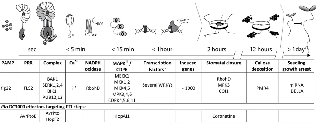
Table 1 summarizes key components in PTI signaling on the basis of FLS2-flg22 pathway, including
steps which are targeted by Pto DC3000 effectors.
5
Table 1 Illustration of key steps in the flg22-FLS2 signaling pathway
PAMP PRR Complex Ca
2+NADPH
oxidase MAPK
b/ CDPK
Transcription Factors
cInduced genes
Stomatal closure Callose deposition
Seedling growth arrest
flg22 FLS2
BAK1 SERK1,2,4
BIK1, PUB12,13
?
aRbohD
MEKK1 MKK1,2 MKK4,5 MPK3,4,6 CDPK4,5,6,11
Several WRKYs > 1000
RbohD MPK3
COI1 PMR4 miRNA
DELLA
Pto DC3000 effectors targeting PTI steps:
AvrPtoB AvrPto
HopF2 HopAI1 Coronatine
a
Channels conducting flg22-induced calcium (Ca2+) influxes are currently not known, but inhibitor studies suggest a role ionotropic glutamate receptor -like channels (Kwaaitaal et al., 2011).
bTwo MAPK cascades are activated by flg22 (Rodriguez et al., 2010). HopAI1 inactivates MAPK3 and MAPK6 by removing phosphor groups (P) from activated MAPKs (Zhang et al., 2007a).
cWRKY transcriptions play a major role in regulating transcriptional defense responses (Rushton et al., 2010).
6 1.3. CELLULAR DEFENSE
Plant immunity involves the interplay of different subcellular compartments and vesicle transport pathways (Frey and Robatzek, 2009; Wang and Dong, 2011). PRRs for example need to be secreted from the endoplasmic reticulum (ER) via golgi compartments to the plasma membrane to perceive PAMPs. The importance of secretory and ER quality control pathway in PRR function was discovered in forward genetic screens for mutants impaired in elf18 signaling (Nekrasov et al., 2009; Saijo et al., 2009). Interestingly, identified mutants were only slightly or not at all affected in flg22 and chitin responses, illustrating specific requirements for ER quality control proteins in different PRR maturation processes (Nekrasov et al., 2009; Saijo et al., 2009). EFR protein accumulation and functions are thereby differently compromised in ER quality control mutants. Radial swelling 3 (rsw3, glucosidase 2) mutants show for example only slight reduction in elf18 binding and maintain wildtype-like early PAMP responses and callose depositions upon elf18 treatment, but fail to maintain wildtype-like defense gene expression and elf18-induced immunity during infections with Pto DC3000 (Lu et al., 2009).
Post-golgi vesicle trafficking plays a major role in plant defense against non-host pathogens.
Resistance of Arabidopsis thaliana against barley powdery mildew fungus Blumeria gramminins hordie (Bgh) is mediated by a protein complex consisting of the syntaxin PENETRATION 1 (PEN1, AtSYP121), SNAP33 (SOLUBLE N-ETHYLMALEIMIDE-SENSITIVE FACTOR ADAPTOR PROTEIN 33) and VESICLE-ASSOCIATED MEMBRANE PROTEIN (VAMP) 721/722 (Collins et al., 2003; Kwon et al., 2008).
The PEN1-SNAP33-VAMP721/722 complex accumulates at fungal penetration sites and mediates
exocytosis of cell-wall enhancing compounds (Kwon et al., 2008). SYP132 of N. benthamiana is
essential for secretion the of PATHOGEN-RELATED protein 1 (PR1) and required for resistance to
non-host Pto DC3000 (Heese et al., 2001). Alternatively to golgi-derived vesicles, PEN2 and PEN3
dependent secretion of anti-microbial compounds employs trafficking of peroxisomes to fungal
infection sites (Lipka et al., 2005; Stein et al., 2006; Bednarek et al., 2009).
7 The trans-golgi network (TGN) serves not only as secretory compartment, but also partially merges with plasma membrane derived early endocytic vesicles in plants (Richter et al., 2009; Viotti et al., 2010). The TGN – early endosome compartment is highly dynamic and sorts vesicles back to the plasma membrane or to late endosomes (Viotti et al., 2010). Endomembrane systems are classified by associated proteins like Rab GTPase, which regulate vesicle formation, mobility and fusion (Stenmark and Olkkonen, 2001). Whereas Rab5 related proteins define early endosomes in animals, it also associated with late endosomes in plants or yeasts (Robinson et al., 2008; Ebine et al., 2011).
Early endosome are the first endomembrane compartment of internalized plasma membrane proteins. Endocytosis of several plant plasma membrane proteins was shown, including auxin carrier proteins, metal transporters, BRI1, but also for PRRs like FLS2 and LeEIX2 (Russinova et al., 2004;
Dhonukshe et al., 2007; Kasai et al., 2010; Barberon et al., 2011).
Endocytosed plasma membrane proteins are often marked by ubiquitination (Sorkin and von Zastrow, 2009). Different types of ubiquitination have been reported. Attachment of one single ubiquitin (Ub) to lysines of substrate proteins is referred to as mono-ubiquitination. Mono- ubiquitination and poly-ubiquitin chains linked via lysine 63 of ubiquitin (Ub
K63) are often associated with endocytic trafficking and protein-protein interactions. By contrast, Ub
K48-linked poly- ubiquitination leads mainly to proteasomal degradation of substrate proteins (Mukhopadhyay and Riezman, 2007). In planta ubiquitination of two metal transporters was recently described (Kasai et al., 2010; Barberon et al., 2011). Controversially, ubiquitination is essential for internalization of IRON-REGULATED TRANSPORTER 1 (IRT1)(Barberon et al., 2011), but not for boron transporter REQUIRES HIGH BORON 1 (BOR1)(Kasai et al., 2010). Instead BOR1 ubiquitination is required for late endosomal trafficking to lytic vacuoles. Although FLS2 is ubiquitinated in an flg22-dependent manner (Lu et al., 2011), the implementation of ubiquitination in FLS2 endocytosis remains elusive. Inhibitor studies suggest that ubiquitination is required for receptor internalization (Robatzek et al., 2006). In addition, a FLS2
ΔPESTvariant is incapable to undergo endocytosis, but maintains a wild type-like ROS burst after flg22 treatment (Salomon and Robatzek, 2006). PEST-sequences act as ubiquitination and phosphorylation signals in yeast and plants (Rogers et al., 1986; Camborde et al., 2010). Notably,
FLS2
ΔPESTmutant variants do not escape ubiquitination by PUB12, 13 and AvrPtoB in vitro, which
could point at an ubiquitination independent role of the FLS2 PEST domain in regulating endocytosis
(Göhre et al., 2008; Lu et al., 2011).
8 After internalization BOR1 endosomes mature to late endosomes and co-localize with ARA6 and ARA7 (Takano et al., 2005; Ebine et al., 2011). Late endosomes are characterized by the presence of small internal vesicles, and are therefore also referred to as multivesicular bodies (MVBs) (Robinson et al., 2008). MVBs terminate in lytic plant vacuoles in plants and yeasts or fuse with lysosomes in animals (Stenmark and Olkkonen, 2001; Richter et al., 2009).
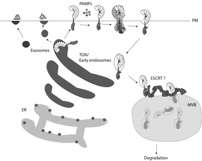
Figure 1 Vesicle trafficking in plant immunity. PRR transport form the ER via the TGN to the plasma membrane (PM) is required for PRR function. PAMPs mediate PRR endocytosis from PM to TGN - early endosomal compartments, followed by maturation to late endosomes, MVBs, and receptor degradation. TGN dirved vesicles are recruited to fungal penetration sites in order to release cell wall enhancing compounts. Anti-microbial defense products are alternatively exocytosed from TGN-indepenent compartments.
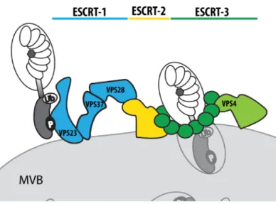
The presence of the Endosomal Complex Required for Transport (ESCRT) is a hallmark of MVBs.
ESCRT proteins are directly involved in MVB biogenesis by mediating vesicle invagination and cargo
recognition (Hurley and Hanson, 2010). The ESCRT machinery consists of three sub-complexes,
9 ESCRT-1-3, which are conserved in all eukaryotes (Figure 2) (Field and Dacks, 2009). Whereas ESCRT-1 initiates cargo recruitment, ESCRT-2 and ESCRT-3 are mainly involved in membrane scission (Hurley and Hanson, 2010). ESCRT-1 is composed of three different proteins: VACULAR PROTEIN SORTING 23 (VPS23) mediates ubiquitin binding, VPS28 bridges ESCRT complex 1 to complex 2 and VPS37 interacts with acidic membrane lipids (Hurley and Hanson, 2010). Arabidopsis carries at least two VPS23 homologous (ELC, and ELC-like), two copies of VPS28 (VPS28-1 and VPS28-2) and two copies of VPS37 (VPS37-1 and VPS37-2) (Spitzer et al., 2006). ESCRT-3 accessory proteins CHARGED MULTIVESICULAR BODY PROTEIN1 (CHMP1) and CHMP2 were previously reported to target auxin carriers PIN-FORMED 1 (PIN1), PIN2 and AUXIN RESISTANT 1 (AUX1) to MVBs and to be essential for embryo and seedling development (Spitzer et al., 2009). It is currently not known, if ESCRT proteins also participate in PRR trafficking.
Figure 2 The ESCRT machinery consists of three major sub-complexes. ESCRT complex 1 (blue), 2 (yellow) and 3 (green) constitute together the ESCRT machinery, which is essential for MVB biogenesis. ESCRT-1 consists of VPS23 (ELC in Arabidopsis), VPS37 and VPS28 and recruits early endosomes via interaction with ubiquitinated cargos to MVBs. Vesicle fusion is mediated by ESCRT-2 and ESCRT-3. VPS4 disassembles ESCRT complexes after endosomal sorting.
The complexity of endocytic pathways and its integration in different signaling networks known from
animals (Sorkin and von Zastrow, 2009) is likely to find its equivalent in plant cells. Many key
components along endocytic trafficking pathways are conserved between animals and plants, others
seem to be plant specific (Robinson et al., 2008). Although several plant receptors are endocytosed,
a detailed understanding of their cellular routes and possible connections to signaling is still pending
and focus of current and future research (Frey and Robatzek, 2009; Hicks and Raikhel, 2010).
10 1.4. AIM OF MY THESIS
PAMP-triggered immunity comprises plasma membrane-derived signaling, endocytic trafficking and a series of cellular immune responses. To which extent these cell responses are linked is not well understood. My work aims to dissect spatio-temporal flg22 signaling and to contribute new insights to the two following questions:
1. What controls FLS2 endocytic trafficking?
2. Which genes regulate temporal flg22 responses?
To address the first question, conserved features of endocytosis known from animals were
exploited: Receptor ubiquitination and the Endosomal Sorting Complex Required for Transport
(ESCRT) of ubiquitinated cargos. The second question is addressed by analysis of a late PAMP
response-defective mutant, which was previously identified in a forward genetic screen for flagellin
insensitivity (Salomon, 2009).
11
2. MATERIALS AND METHODS
2.1. MATERIALS
2.1.1. Plant material and growth conditions
Arabidopsis thaliana plants used in this study are listed in Table 2. Unless otherwise indicated, plants were grown on soil (Arabidopsis mix, John Ines Centre, Norwich) or sterile on Murashige and Skoog medium (recipe in Table 6) under 10 hours or 16 hours of light at 20 - 22
oC and 65 % humidity. Seeds were stratified in dark at 4°C for two days in 0.1 % (w/v) agarose or directly on agar plates. Nicotiana benthamiana plants were grown under 16 hours of light at approximately 24°C with a relative humidity of 45 - 65 %.
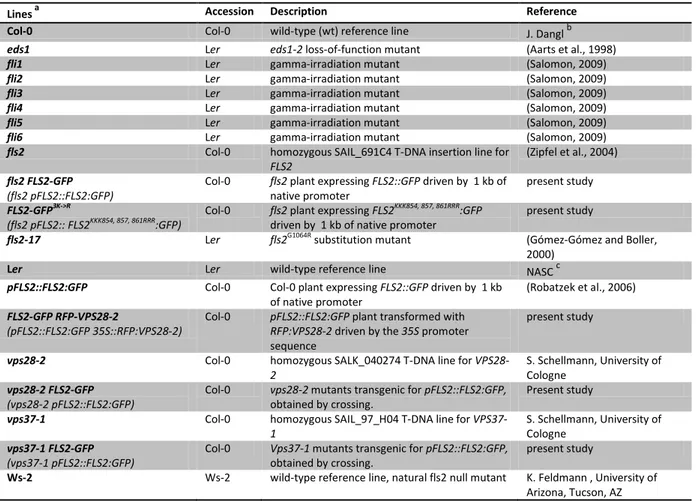
Table 2 Description of plant material presented in this study.
Lines a
Accession Description Reference
Col-0 Col-0 wild-type (wt) reference line J. Danglb
eds1 Ler eds1-2 loss-of-function mutant (Aarts et al., 1998)
fli1 Ler gamma-irradiation mutant (Salomon, 2009)
fli2 Ler gamma-irradiation mutant (Salomon, 2009)
fli3 Ler gamma-irradiation mutant (Salomon, 2009)
fli4 Ler gamma-irradiation mutant (Salomon, 2009)
fli5 Ler gamma-irradiation mutant (Salomon, 2009)
fli6 Ler gamma-irradiation mutant (Salomon, 2009)
fls2 Col-0 homozygous SAIL_691C4 T-DNA insertion line for
FLS2
(Zipfel et al., 2004) fls2 FLS2-GFP
(fls2 pFLS2::FLS2:GFP) Col-0 fls2 plant expressing FLS2::GFP driven by 1 kb of
native promoter present study
FLS2-GFP3K->R
(fls2 pFLS2:: FLS2KKK854, 857, 861RRR:GFP) Col-0 fls2 plant expressing FLS2KKK854, 857, 861RRR:GFP
driven by 1 kb of native promoter present study
fls2-17 Ler fls2G1064R substitution mutant (Gómez-Gómez and Boller,
2000)
Ler Ler wild-type reference line NASCc
pFLS2::FLS2:GFP Col-0 Col-0 plant expressing FLS2::GFP driven by 1 kb
of native promoter (Robatzek et al., 2006) FLS2-GFP RFP-VPS28-2
(pFLS2::FLS2:GFP 35S::RFP:VPS28-2)
Col-0 pFLS2::FLS2:GFP plant transformed with RFP:VPS28-2 driven by the 35S promoter sequence
present study
vps28-2 Col-0 homozygous SALK_040274 T-DNA line for VPS28-
2
S. Schellmann, University of Cologne
vps28-2 FLS2-GFP (vps28-2 pFLS2::FLS2:GFP)
Col-0 vps28-2 mutants transgenic for pFLS2::FLS2:GFP, obtained by crossing.
Present study
vps37-1 Col-0 homozygous SAIL_97_H04 T-DNA line for VPS37-
1 S. Schellmann, University of
Cologne vps37-1 FLS2-GFP
(vps37-1 pFLS2::FLS2:GFP)
Col-0 Vps37-1 mutants transgenic for pFLS2::FLS2:GFP, obtained by crossing.
present study
Ws-2 Ws-2 wild-type reference line, natural fls2 null mutant K. Feldmann , University of Arizona, Tucson, AZ a
Names written in bold are used abbreviation in this work.
b
University of North Carolina, Chapel Hill, NC, USA; c
Nottingham Arabidopsis Stock Centre
12 2.1.2. Bacterial strains
E. coli DH5α, E. coli Rosetta and Agrobacterium tumefaciens and were used for cloning and heterologous gene expression, respectively. Plant pathogenic Pseudomonas syringe pv. tomato DC3000 and its non-virulent hrcC mutant (Yuan and He, 1996) were tested on selected Arabidopsis mutants to investigate potential immune deficiencies. All strains with corresponding antibiotic resistances and growth conditions are listed in Table 3.
Table 3 Use of pathogenic and non-pathogenic bacterial strains
Strain Used for Resistance Media
Escherichia coli DH5α Cloning none L-medium
Escherichia coli Rosetta
heterologous gene expression
CamR L-medium
Agrobacterium tumefaciens GV3101
heterologous gene expression
RifR + GentR L-medium
Pseudomonas syringe
pv. tomato DC3000 Pathogenicity test RifR NYBA, King's B Pseudomonas syringe
pv. tomato DC3000 hrcC (Yuan and He, 1996)
Pathogenicity test RifR NYBA, King's B
2.1.3. Oomycete strains
Waco 9 isolate of Hyaloperonospora arabidopsidis (Hpa) was used in infection assays and microscopy. Isolates were maintained as previously described in Toer et al., 2002 (Toer et al., 2002).
Both strains originated from stocks maintained by Professor Dr. Jonathan Jones, Norwich.
13 2.1.4. Plasmids
Plasmids were constructed by either classical cloning with appropriate restriction enzymes or using Gateway technology (Invitrogen, UK). Table 4 describes used vectors.
Table 4 Vector used to generate transgenic plants or for heterologous gene expression.
Name Insert Backbone type of vector Reference
35S::CFP:ELC cds of ELC pAM-PAT-GW (Weinl et al., 2005)
binary vector present study
35S::YFP:VPS28-2 cds of VPS28-2 pEARLY104 (Earley et al., 2006)
binary vector, present study
35S::RFP:VPS37-1 cds of VPS37-1 pGWB555 (Nakagawa et al., 2007)
binary vector present study
pFLS2::FLS2:GFP pFLS2::FLS2:GFP pGREEN binary vector (Robatzek et al., 2006) pFLS2::FLS2KjmR:GFP pFLS2::FLS2KKK854,
857, 861RRR:GFP
pGREEN binary vector present study FLS2CD-His cds of FLS2CD pET42 E. coli
expression
(Göhre et al., 2008) FLS2CD KKK854, 857,
861RRR-His
cds of
FLS2CDKKK854, 857, 861RRR:GFP
pET42 E. coli expression
present study
GST-AvrPtoB AvrPtoB pGEX-2TM-
GW
E. coli expression
(Göhre et al., 2008)
UBC9-His cds UBC9 pET42 E. coli
expression
(Göhre et al., 2008)
2.1.5. Oligonucleotides
Oligonucleotides were synthesized by Sigma-Aldrich (St. Louis, MI, USA), diluted with ddH
20 to 100
µM stock solutions and 10 µM working solution. Table 5 lists used oligonucleotides and their
corresponding targets.
14
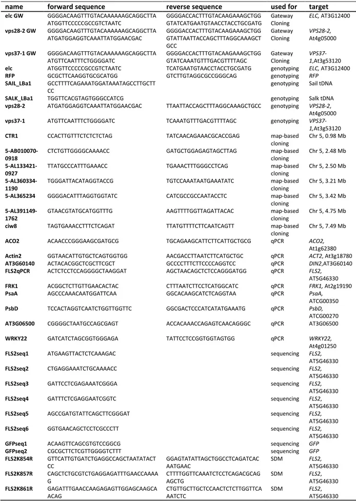
Table 5 Overview of cloning, genotyping and qPCR primers with corresponding targets and sequences
name forward sequence reverse sequence used for target
elc GW GGGGACAAGTTTGTACAAAAAAGCAGGCTTA
ATGGTTCCCCCGCCGTCTAATC GGGGACCACTTTGTACAAGAAAGCTGG
GTATCATGAATGTAACCTACCTGCGATG Gateway
Cloning ELC, AT3G12400 vps28-2 GW GGGGACAAGTTTGTACAAAAAAGCAGGCTTA
ATGATGGAGGTCAAATTATGGAACGAC
GGGGACCACTTTGTACAAGAAAGCTGG GTATTAATTACCAGCTTTAGGCAAAGCT GCC
Gateway Cloning
VPS28-2, At4g05000 vps37-1 GW GGGGACAAGTTTGTACAAAAAAGCAGGCTTA
ATGTTCAATTTCTGGGGATC
GGGGACCACTTTGTACAAGAAAGCTGG GTATCAAATGTTTGACGTTTTAGC
Gateway Cloning
VPS37- 1,At3g53120 elc ATGGTTCCCCCGCCGTCTAATC TCATGAATGTAACCTACCTGCGATG genotyping ELC, AT3G12400
RFP GCGCTTCAAGGTGCGCATGG GTCTTGTAGGCGCCGGGCAG genotyping RFP
SAIL_LBa1 GCCTTTTCAGAAATGGATAAATAGCCTTGCTT
CC genotyping Sail tDNA
SALK_LBa1 TGGTTCACGTAGTGGGCCATCG genotyping Salk tDNA
vps28-2 ATGATGGAGGTCAAATTATGGAACGAC TTAATTACCAGCTTTAGGCAAAGCTGCC genotyping VPS28-2, At4g05000
vps37-1 ATGTTCAATTTCTGGGGATC TCAAATGTTTGACGTTTTAGC genotyping VPS37-
1,At3g53120
CTR1 CCACTTGTTTCTCTCTCTAG TATCAACAGAAACGCACCGAG map-based
cloning
Chr 5, 0.98 Mb 5-AB010070-
0918 CTCTGTTGGGGCAAAACC GATGCTGGAGAGTAGCTTAG map-based
cloning Chr 5, 2.48 Mb 5-AL133421-
0927
TTATGCCCATTTGAAACC TGAAACTTTGGGCCTCAG map-based
cloning
Chr 5, 2.50 Mb 5-AL360334-
1190
TGGGATTACATAGGTACCG TGTCCAAATAATGAAATATC map-based
cloning
Chr 5, 3.21 Mb
5-AL365234 GGGGACATTTAGGTGGTATC CATCGCCGCCAATACCTC map-based
cloning
Chr 5, 3.42 Mb 5-AL391149-
1762 GTAACGTATGCATGGTTTG AAGTTTTGGTTAGATTACAC map-based
cloning Chr 5, 4.75 Mb
ciw8 TAGTGAAACCTTTCTCAGAT TTATGTTTTCTTCAATCAGTT map-based
cloning
Chr 5, 7.49 Mb
ACO2 ACAACCCGGGAAGCGATGCG TGCAGAAGCATTCTTCATTGCTGCG qPCR ACO2,
At1g62380
Actin2 GGTAACATTGTGCTCAGTGGTGG AACGACCTTAATCTTCATGCTGC qPCR ACT2, At3g18780
AT3G60140 ACTACACGGCTCGCTTCGCT GCCCCTTTCTTCCCCAGGTCC qPCR DIN2,AT3G60140
FLS2qPCR ACTCTCCTCCAGGGGCTAAGGAT AGCTAACAGCTCTCCAGGGATGG qPCR FLS2,
AT5G46330
FRK1 ACGGCTCTTGTTGAACACTAC CTTTAATCTTCCTCATGGCATC qPCR FRK1, At2g19190
PsaA AGCCCAAACAATGGATTCAA GGCACAAGCATCTCAGGTAA qPCR PsaA,
ATCG00350
PsbD TCCACTAGGTCAATCTGGTTGGTTC GGCGACTCCCATCATATGAAATG qPCR PsbD,
ATCG00270
AT3G06500 CGGGGCTAATGCCAGCGAGT ACCACAAACCAGAGTCAACAGGGC qPCR AT3G06500
WRKY22 GATCATCTAGCGGTGGGAGA TATTCCTCCGGTGGTAGTGG qPCR WRKY22,
At4g01250
FLS2seq1 ATGAAGTTACTCTCAAAGAC sequencing FLS2,
AT5G46330
FLS2seq2 CTGAGGAAATCTGCAAAACC sequencing FLS2,
AT5G46330
FLS2seq3 GATTCCTCGAGAAATCGGGA sequencing FLS2,
AT5G46330
FLS2seq4 GATTTCTCGAGGAATCGGTC sequencing FLS2,
AT5G46330
FLS2seq5 AGCCGATGTATTCAGCTTCGGGAT sequencing FLS2,
AT5G46330
FLS2seq6 GGTGAACAGCTCCTCGCCCTT sequencing FLS2,
AT5G46330
GFPseq1 ACAAGTTCAGCGTGTCCGGCG sequencing GFP
GFPseq2 CGCGCTTCTCGTTGGGGTCTTT sequencing GFP
FLS2K854R GTTCATTGTGATCTGAGGCCAGCTAATATACT CC
GGAGTATATTAGCTGGCCTCAGATCAC AATGAAC
SDM FLS2,
AT5G46330 FLS2K857R CAGCTCTGCGTCTGAGGAGATTTGAACCAAAA
G
CTTTTGGTTCAAATCTCCTCAGACGCAG AGCTG
SDM FLS2,
AT5G46330 FLS2K861R GAGATTTGAACCAAGAGAGTTGGAGCAAGCA
ACAG CTGTTGCTTGCTCCAACTCTCTTGGTTCA
AATCTC SDM FLS2,
AT5G46330
15 2.1.6. Chemicals
If not stated otherwise, standard chemicals were purchased from Sigma-Aldrich (St. Louis, MI, USA), Merck (Whitehouse Station, NJ, USA), Invitrogen (Carlsbad, CA, USA), VWR (Radnor, PA, USA) or Helena Bioscience (Gateshead, UK). EZBiolab Inc. (Westfield, IN, USA) synthesized the flg22 peptide used in this study and previous work (Göhre et al., 2008).
2.1.7. Enzymes
Genotyping PCRs were performed with Taq DNA polymerase from New England Biolabs (Ipswich MA, USA). DNA amplification for cloning purposes was done the Expand High Fidelity PCR system (Roche Diagnostics Co., UK). RT-PCRs were carried out with Superscript II (Invitrogen Carlsbad, CA, USA), and SybrGreen master mix (Sigma-Aldrich, St. Louis, MI, USA). Restriction enzymes were commonly purchased from New England Biolabs (Ipswich MA, USA), Gateway Cloning Enzymes from Invitrogen (Carlsbad, CA, USA).
2.1.8. Antibodies
Anti (α)-FLS2 antibodies were raised in rabbits and purified by Eurogentec (Seraing, Liège, Belgium).
1:5000 deletions in 1 % (v/w) milk powder (Premier International Food, St Albans, UK) TBS-t (10 mM Tris-HCl, pH 7.4, 14 mM NaCl, 2.5 mM KCl , 0.05 % (v/w) Tween 20) were sufficient for specific detection of FLS2 as described in Göhre et al., 2008 (Göhre et al., 2008). αPhospho-p44/42 MAPK (α- pERK, rabbit polyclonal antibody from Cell Signaling, Boston, MA, USA) were diluted 1:1000 in 5 % BSA (w/v) TBS-t. In both cases goat α-rabbit IgG coupled to alkaline phosphatase (Sigma-Aldrich (St.
Louis, MI, USA) was used as secondary antibody. GFP as probed with αGFP from Roche Applied
Science (Penzberg, Germany) 1:1000 dilutions, followed by goat α-mouse IgG- alkaline phosphatase
(Sigma-Aldrich, St. Louis, MI, USA) detection. Ubiquitin conjugated were detected by αUbiquitin
(mouse monoclonal, P4D1, Santa Cruz Biotechnology, Santa Cruz, CA, USA) in deletions of 1:2000.
16 2.1.9. Growth media and antibiotics
Growth media were autoclaved for 20 min at 121°C, cooled down to approximately 50°C before adding antibiotics. Detailed recipes are listed in Table 6 and final concentrations of antibiotics in Table 7.
Table 6 Growth media recipes
Name Species Recipe
L-Medium E.coli and
A.tumefaciens
10 g/l 5 g/l 5 g/l 1 % (w/v) pH 7.2
bacto-peptone yeast extract NaCl
bacto-agar (optional, (Duchefa, Haarlem, Netherlands)
King’s B-Medium Pseudomonas 20 g/l 1.5g/l 1.5g/l 10 % (v/v) 1 % (w/v) pH 7.2
bacto-peptone
heptahydrated magnesium sulfate potassium hydrogen phosphate glycerol
bacto-agar (optional)
NYG-Medium Pseudomonas 5.0 g/l 3 g/l 20 % (v/v) 1.5 % (w/v) pH 7.2
bacto-peptone yeast extract glycerol
bacto-agar (optional)
MS (Murashige and Skoog)
Arabidopsis thaliana
4.4 g/l 10.0 g/l pH 5.8
MS powder Sucrose
Table 7 Used antibiotics
Name Abbreviation Final Concentration [µg/ml]
Carbenicillin Carb 100 in ddH20
Chloramphenicol Cam 35 in Ethanol
Gentamicin Gent 30 in ddH20
Hygrommycin Hygr 30 in ddH20
Kanamycin Kan 50 in ddH20
Phosphinothricin PPT 15 in ddH20
Rifampicin Rif 50 in DMSO
Spectinomycin Spec 100 in ddH20
17 2.2. METHODS
2.2.1. Seed sterilization
Arabidopsis thaliana seeds were sterilized for 10 min in 70 % (v/v) ethanol 0.05 % (v/v) SDS, followed by three short washings with 98 % (v/v) ethanol. Seeds were then dried on sterile Whatman paper before sowing on MS agar plates.
2.2.2. Generation of Arabidopsis thaliana F
1,F
2and F
3progeny
Plants were crossed according to Weigel and Glazbrook (Weigel and Glazebrook, 2002). Basically, flowers with well-developed stigma were emasculated and pollinated with one single donor stamen.
Developing siliques were then carefully wrapped in paper envelopes and harvest once they turned yellow. The resulting F
1generation was tested for heterozygosity by PCR and allowed to self- pollinate to generate F
2progeny. With the exception of fli1 F
2families used for map-based cloning, F
2individuals were screened for the aimed genotypes and propagated to the next generation for further analyzes.
2.2.3. Stable transformation of Arabidopsis thaliana
Arabidopsis plants were stably transformed using floral dip method (Clough and Bent, 1998).
Flowering plants were dipped for 30 sec in a 0.8 OD
600-dense solutions of Agrobacteria (5 % sucrose, 0.025 % Silwet L-77 (Lehle Seeds, Round Rock, TX, USA), kept in a dark environment for 24 hours and further propagated under standard long-day plant growth conditions. Successfully transformed seeds were recovered in the following T
1generation on selective MS agar plates or by spraying BASTA (Duchefa, Haarlem, Netherlands) three-times on 4-5 –week-old plants at weekly intervals.
18 2.2.4. Transient transformation of Nicotiana benthamiana
Agrobacteria were grown over night (o/n) at 28°C in L-medium containing the appropriate antibiotics, centrifuged for 5 min at 5000 x g and washed once with 10 mM MgCl
2. A final OD
600= 0.1 of agrobacterial suspensions were then infiltrated underneath the lower epidermis of three to four week-old N. benthamiana leaves using a 1 ml needless syringe. Microscopically analysis followed two to three days post infiltration.
2.2.5. Pathogen infections
2.2.5.1. Pseudomonas syringae patho-phenotyping
Arabidopsis plants were grown for two weeks on Jiffy pellets (Jiffy Products International AS, Grorud,
Norway) under controlled environment and 10 hours of photoperiod before inoculation. To prepare
the inoculum, Pseudomonas was streaked out on King’s B plates with appropriate antibiotics from
glycerol stocks (30 % (v/v) glycerol, -80°C) and incubated for two days at 28°C. Single colonies were
then transferred to 50 ml NYG broth containing 50 µg/ml of Rifampicin and grown under constant
shaking for 12 to 13 hours. Bacterial cultures were then centrifuged for 15 min at 1000 g (RC8,
Sorvall, Thermo Fisher Scientific, Waltham, MA, USA) resuspended in 5 ml 10 mM MgCl
2and
adjusted to a final density of OD
600= 0.02. 0.04 % Silwet (Lehle Seeds, Round Rock, TX, USA) was
added before spraying the inoculum with an airbrush system (model AS18-2, Ningbo Haosheng
Pneumatic Machinery Ningbo, China) on each seedling for approximately 2 seconds. Inoculated
plants were kept under elevated humidity for 2.5 hours and disease symptoms were assessed 5 days
post infection. Disease symptoms were grouped into four classes, where class 0 showed no
symptoms, class 1 mild symptoms on maximal one leaf, class 2 infected plants had at least two
infected leaves but showed no necrosis and class 3 with plants displaying severe disease symptoms
including beginning necrotic lesions. Plants were randomly distributed on different trays.
19
2.2.5.2. Pseudomonas syringae growth quantificationBacterial growth curves assays followed the infection procedure described above (2.2.5.2), but using four-week-old plants. To quantify bacterial replication, two leaf disks (∅ = 0.5 cm) from two different surface-sterilized leaves (30 sec in 70 % (v/v) ethanol, followed by 30 sec in sterile ddH
2O) and at least four plants per genotype were sampled 4 days after inoculation. Leaf disks were grinded in 10 mM MgCl
2with an electric drill (Heidolph, Schwabach, Germany), diluted 1:10 serially and plated on selective NYG agar plates. Two dates after incubation at 28°C colonies forming units were counted and statistically analyzed.
2.2.5.3. Flg22-induced resistance to Pseudomonas syringae
According to the protocol published by Zipfel et al., 2004 (Zipfel et al., 2004), four-week-old plants were either pre-infiltrated with ddH
2O or 1 µM flg22 using a needle-less syringe. 24 hours later the same leaves were syringe-inoculated with Pseudomonas syringe pv. tomato DC3000 (OD
600= 0.001).
Bacterial growth was quantified as described above.
2.2.5.4. Hyaloperonospora arabidopsidis (Hpa) spore counting
Hpa spore suspensions of 5 x 10
4spores/ml were spray-inoculated onto 14-day-old seedling and
incubated at high humidity at 18°C. 6 days post-inoculation, spores of 12 seedlings per genotype (in
pools of 3) were washed from infected leaves by vortexing in 1 ml ddH
2O and quantified in relation
to seedling fresh weight. Spores were counted with an improved Neubauer haemocytometer (Brand,
Wertheim, Germany).
20 2.2.6. Plant assays
2.2.6.1. Measurements of root growth
Response to salt stress was studied according to Achard et al., 2006 (Achard et al., 2006). Seedlings were grown sterile on MS plates for 5-7 days and then transferred to MS plates containing 0, 50 mM or 100 mM NaCl. Plates were orientated vertically, and primary root growth of at least 20 individuals measured 3 days later. Glucose stress was applied similar to Zhou et al., 1998 (Zhou et al., 1998).
Seven-day-old seedlings grown on MS plate were exposed for additional seven days to 0 %, 4 % (w/v) and 6 % (w/v) glucose. Primary root growth of at least 20 seedlings was measured and statistically analyzed. Root growth arrest upon flg22 stress was examined on seedlings grown in dark on vertical MS plates supplemented with 0, 100 nM or 500 nM flg22. At least 20 individual primary roots were measured per genotype and experiment.
2.2.6.2. Flg22-induced callose deposition
Flg22-induced callose deposition was stained as described in (Lu et al., 2009). Briefly, ten-day-old seedlings were transferred to liquid MS media with and without 1 µM flg22, destained after 24 hours in acetic acid - ethanol (1:3) for four hours, washed twice with ddH
2O, and incubated o/n in aniline blue solution (150 mM KH
2PO
4, 0.01 % (w/v) aniline blue, pH 9.5). Stained callose was then visualized ultraviolet epifluorescence microscopy (Zeiss Axiophot, Carl Zeiss AG, Oberkochen, Germany).
2.2.6.3. Measurements of stomatal apertures
According to Mersmann et al., 2010 (Mersmann et al.) Arabidopsis seedlings were transferred after
one week growth on solid MS plates to liquid MS media. One week later MS media was exchanged
with 5 µM flg22 or ddH
2O respectively. Mild vacuum was applied for 10 min and after 2 hours of
recovery seedlings were mounted on glass slides. Stomatal apertures were imaged with a Zeiss
21 Axiophot microscope (Carl Zeiss AG, Oberkochen, Germany). In total at least 20 stomata were measured per genotype and condition. High-throughput stomatal apparatus measurements ( > 100 stomata) were conducted with a fully automated confocal imaging system Opera (Perkin Elmer, Germany) and subsequent computational analyzes (Dr. Gildas Bourdais, The Sainsbury Laboratory, Norwich).
2.2.6.4. Measurements of reactive oxygen production (ROS)
16 leaf discs (∅ = 0.5 cm) per genotype of four-week-old plants were incubated o/n in ddH
2O. Water was then exchanged with 100 µl of 20 μM luminol (Sigma-Aldrich (St. Louis, MI, USA), 1 μg horseradish peroxidase (Sigma-Aldrich, St. Louis, MI, USA) and 100 nM flg22. Light emission was read with a Varioskan Flash multiplate reader (Thermo Fisher Scientific, Waltham, MA, USA) for 35 min.
2.2.7. Molecular biological methods
2.2.7.1. Isolation of genomic plant DNA
A modified version of the Edward’s DNA extraction protocol (Sambrook and William Russell, 2001) was used to extract genomic DNA. Approximately 10-20 mg a leaf tissue was grinded in 180 µl Edward’s buffer (200 mM Tris-HCl, pH 7.5, 250 mM, NaCl, 25 mM EDTA, 0.5 % SDS (v/v)), centrifuged at maximum speed for 5 min (table centrifuge 5415D, Eppendorf, Hamburg Germany) and 120 µl of supernatant transferred into a fresh tube. One volume of isopropanol was added to precipitate DNA and centrifuged at maximum speed for 5 min. The supernatant was discarded and the pellet was washed once with 70 % (v/v) ethanol. Air-dried DNA pellets were dissolved in 100 μl sterilized ddH
2O and 3 μl used in 50 μl PCR reactions.
To extract high quality DNA for re-sequencing potential SNPs 10-20 mg leaf tissue was grinded in 200
µl of CTAB extraction buffer (1 % CTAB (w/v), 100 mM Tris-HCl, pH 7.5, 1 M NaCl, 50 mM EDTA, 5 %
N-Sarcosylate (v/v)), incubated for 10 min at 65°C followed by 5 minutes at room temperature. 200
µL of 24:1 (v/v) chloroform-isoamylacohol was added, and centrifuged at 15000 g for 5 minutes at
22 4°C (table centrifuge 5415R, Eppendorf, Hamburg Germany). The upper phase was transferred into a new tube and one volume of cold (-20°C) isopropanol was added. Tubes were inverted 3 times and centrifuged at 15,000 g for one minute at 4°C. The precipitated DNA was washed once with 70 % (v/v) ethanol, air-dried and resuspended 100 μl sterilized ddH
2O.
2.2.7.2. Isolation of plasmid DNA and PCR products
Plasmid DNA was purified with QIAprep Spin Miniprep Kit (Qiagen, Hilden, Germany) and PCR product were isolated with QIAquick PCR purification and gel extraction kits (Qiagen, Hilden, Germany) according to manufacturer's recommendations.
2.2.7.3. PCR protocols
Standard DNA amplification, which did not require sequence accuracy and used mainly for genotyping, was performed with 0.2 mM dNTPs (Promega, Fitchburg, WI, USA), 0.2 μM of each primer, 15 mM MgCl
2and 1 U Taq polymerase (New England Biolabs) in Peltier Thermal Cycler PTC- 225 (GMI Inc., Ramsey, USA). 4 min of initial denaturation at 96°C proceeded 20-30 cycles of denaturation at 96°C, 30 sec of primer annealing at 55°C to 60°C, and 1 min per 1 kb DNA amplification at 72°C. A final elongation step at 72°C for 3 min was added at the end of the protocol.
Standard PCR
In cases where sequence accuracy was necessary (e.g. cloning of genes or introducing mutation, Expand High Fidelity PCR system (Roche Applied Science, Penzberg, Germany) was used according to manufactory’s instructions. The same thermal profile was used as in standard PCRs with the exception of 68°C instead of 72°C and 2 sec per kb during elongation steps. The Expand High Fidelity PCR system was also used for site-directed mutagenesis according to the protocol and primer design described for QuikChange Site-Directed Mutagenesis Kit (Startagene, Santa Clara, CA, USA).
High Fidelity PCR and site-directed mutagenesis
23 20 µl PCR product was mixed with 5 µl of DNA-loading dye (5 M Betain, 0.05 % (w/v) Orange G, Sigma-Aldrich (St. Louis, MI, USA) and separated on 1 -2 % agarose gels (Melford Laboratories, Ipswich, UK) by electrophoresis (80-110 V, Biorad, UK). Agarose gels were prepared by boiling agarose in TBE buffer (45 mM Tris, 45 mM boric acid, 1 mM EDTA, pH 8.3). 1 μl/ml ethidium bromide (Sigma-Aldrich St. Louis, MI, USA) was added before pouring gels and visualized by UV excitation (ChemiDOC XRS, Bio-Rad Laboratories, Hercules, CA, USA). 2-log DNA ladder (New England Biolabs) was used as a reference.
Visualizing PCR products
2.2.7.4. Sanger-Sequencing
Sanger method (Sanger et al., 1977) was used to verify DNA sequences. Reactions were carried out in final volumes of 10 μl containing 80-100 ng template DNA, 1 μM primer, 0.05 % (v/v) DTT, 1 μl 5x sequencing buffer and 1 μl Big Dye Terminator Ready Reaction Mix (Perkin Elmer, Waltham, MA, USA). The PCR program started with an initial denaturation step at 96°C for 1 min, followed by 35 cycles of denaturation at 96°C for 10 sec, annealing at 50°C for 5 sec and elongation at 60°C for 4 min. Read analysis was carried out with Dye-Deoxy Terminator Cycle Sequencer (Perkin Elmer, Waltham, MA, USA) in the The Genome Analysis Centre (TGAC, Norwich, UK).
2.2.7.5. Illumina-Sequencing