Mechanisms involved in regulation of MHC class I molecules in murine embryonic stem cells
Inaugural-Dissertation
Zur
Erlangung des Doktorgrades
der Mathematisch-Naturwissenschaftlichen Fakultät der Universität zu Köln
Vorgelegt von
Manoj Kumar Gupta
(aus Bulandshahr, Indien)
Köln , 2011
Die Untersuchungen zur vorliegenden Arbeit wurden in der Zeit von August 2006 bis Mai 2011 am Institut für Neurophysiologie der Medizinischen Fakultät der Universität zu Köln unter der Leitung von Dr.
Dr. Tomo Šarić durchgeführt.
Tag der mündlichen Prüfung : 18.10.2011
Vorsitzende des Promotionsausschusses: Prof. Dr. Angelika A. Noegel
Erstgutachter: Prof. Dr. Guenter Plickert
Zweitgutachter: Prof. Dr. Guenter Schwarz
Datum der Promotion:
Contents
Contents ………
3Abbreviations ………
6Abstract ………
8Zusammenfassung
………10I. Introduction ………..
12I.1. Organization of the immune system I.2. Major histocompatibility complex (MHC) molecules and their role in immunorecognition I.3. Interferon gamma (IFNγ) and regulation of MHC molecules I.4. Role of STAT1 and STAT3 signaling in biological systems I.5. Allorecognition of embryo and expression of MHC molecules in the course of embryonic development I.6. Embryonic stem (ES) cells I.7. Immunological properties of ES cells (ESCs)
II. Aims ……….
.35III. Results ………..
36 III.1. Regulation of MHC class I molecules in ESCsIII.1.1. Expression of MHC class I molecules on ESCs
III.1.2. Expression of components of MHC class I processing machinery in ESCs and their differentiated derivatives
III.1.3. Expression of IFNγ receptors in ESCs and their differentiated derivatives
III.1.4. Expression and activity of STAT1 in ESCs
III.1.5. Expression of regulatory components of STAT1 signaling pathway and their activation by IFNγ in murine ESCs
III.1.6. The role of LIF in control of MHC class I expression in murine ESCs and its differentiated derivatives.
III.1.7. The role of STAT3 in regulation of MHC class I molecules in undifferentiated ESCs
III.2. Epigenetic regulation of MHC class I molecules in murine ESCs
III.2.1. STAT3 regulates the expression of epigenetic modifier genes in ESCs
III.2.2. Dnamt knockout (KO) murine ES cell lines show enhanced MHC class I expression
III.2.3. Epigenetic modifiers 2,5-azacytidine and Trichostatin A increase the expression of MHC class I molecules on ESCs
III.3. Biological consequences of STAT3 KD in ESCs
III.3.1. Downregulation of STAT3 in murine ESCs reduces their lysis by syngenic NK cells
III.3.2. STAT3 partially inhibits the lysis of murine ESCs by CTLs
III.4. Regulation of MHC class I expression in ES cell-derived cardiomyocytes (ES-CMs)
III.4.1. STAT3 KD in ES-CMs does not induce the expression of MHC class I molecules on their surface
III.4.2. Effect of IFNγ on expression of components of MHC class I antigen processing machinery in ES-CMs
III.4.3. ES-CMs are partially lysed by CTLs only after induction of MHC class I molecules by IFNγ
IV. Discussion ………
71V. Materials and methods
………84 V.1. MaterialsV.1.1. Cell lines V.1.2. Mouse strains
V.1.3. Molecular biology reagents V.1.4. Cell culture reagents V.1.5. Radioactive material
V.1.6. Common reagents V.1.7. Primers
V.1.8. Antibodies used for immunoblotting V.1.9. Antibodies used for flow cytometry
V.1.10. STAT3 gene short hairpin RNA (shRNA) sequence V.1.11. Glassware and Plastics
V.1.12. Instruments and devices
V.2. Methods
V.2.1. Culture of Undifferentiated ES/iPS cells and other cell lines V.2.2. RT-PCR and quantative RT-PCR
V.2.3. STAT3 KD and its validation in ESCs V.2.4. Immunoblotting
V.2.5. Flow cytometric analyses V.2.6. Luciferase reporter assay
V.2.7. Teratoma formation by ES cells in mice
V.2.8. Activation and isolation of ovalbumin-specific CTLs V.2.9. Activation and isolation of syngeneic NK cells V.2.10. 51Cr-release cytotoxicity assay
V.2.11. Interferon-γ enzyme-linked immunosorbent assay V.2.12. Generation of ES cell-derived cardiomyocytes
VI. References ……….
101VII. Acknowledgements ………
114VIII. Statement (Erklärung) ..………...
116Abbreviations
°C degree Celsius
α alpha
β beta
γ gamma
μL microliter
μg microgram
bp base pair
BSA bovine serum albumin
bFGF basic fibroblast growth factor cpm counts per minute
cDNA complimentary deoxyribonucleic acid
CTLs cytotoxic T lymphocytes
CTSB cathepesin B
CMs cardiomyocytes D day
DNA deoxyribonucleic acid
DMSO dimethyl sulpfoxide
DEPC diethyl pyrocarbonate
DMEM Dulbecco’s modified Eagle’s medium DTT dithiothreitol
DPBS Dulbecco’s phosphate buffered saline Dnmt DNA methyl transferase
ESCs embryonic stem cells
ESCMs ES cell derived cardiomyocytes
EBs embryoid bodies
FBS fetal bovine serum GFP green fluorescent protein
GMEM Glasgow minimal essential medium h hour
IMDM Iscove’s modified Dulbecco’s medium
INDO Indoleamine 2,3-deoxygenase
IRES Internal ribosomal entry site IRF interferon regulatory factor IFNγ interferon gamma
iPSCs induced pluripotent stem cells JAK Janus activating kinase kb kilobase L liter LIF leukemia inhibitory factor mg miligram min minute ml mililiter mM milimolar
MACS magnetically assisted cell sorting mRNA messenger ribonucleic acid
MEFs murine embryonic fibroblasts MHC major histocompatibility complex NEAA non-essential amino acids NK cells natural killer cells
OD optical density
Ova ovalbumin
PCR polymerase chain reaction
PBS phosphate buffer saline
PE phycoerythrin PTP1B protein-tyrosine phosphatase 1B
αPIG alpha-myosin heavy chain-puromycin-IRES-GFP
RT room temperature
RNA ribonucleic acid
rpm revolution per minute
rcf relative centrifugal force
RPMI Roswell Park memorial Institute RT-PCR reverse transcription-PCR SOCS suppressor of cytokine signaling
STAT signal transducers and activators of transcription S8L SIINFEKL
SIINFEKL Ser-Ile-Ile-Asn-Phe-Glu-Lys-Leu sec second
TCR T cell receptor
TSA trichostatin A
2,5-aza 2,5-azacytidine
KD knock down
KO knock out
PI(3)K phosphatidylinositol 3-kinase
Con-A Concanavalin- A
NKT Natural killer T cells
Abstract
Major histocompatibility complex (MHC) is at the center of immune responses that support survival, fitness and adaptation of mammalian species to the environment. These molecules are not only crucial for adaptive and innate immune responses against microorganisms and cancer cells but also play an important role in reproduction process and development of embryo during the preimplantation period. In the present study, we use murine embryonic stem cells (ESCs) as model to dissect the molecular mechanism involved in the regulation of MHC class I molecules during differentiation in vitro. MHC class I molecules are expressed at very low levels on murine ESCs and they are not induced by the immunomodulatory cytokine interferon gamma (IFNγ) despite the presence of IFNγ receptors on their cell surface. First, we showed that removal of leukemia inhibitory factor (LIF), a standard component of murine ES cell culture media required for the maintenance of a pluripotent state, did not result in up regulation of MHC class I expression in murine ESCs, presumably due to incomplete inactivation under these experimental conditions of STAT3 signaling pathway, which is used by LIF. However, the addition of LIF to differentiated cells in embryoid bodies strongly suppressed the expression of these molecules. Down regulation of STAT3 in undifferentiated ESCs cultured in the presence of LIF significantly increased the expression of MHC class I molecules and this was further enhanced by IFNγ treatment. Flow cytometric analysis revealed that STAT1 is phosphorylated by IFNγ in STAT3 knockdown (KD) ESCs, whereas there was only weak or no phophorylation detected in mock siRNA 647-treated ESCs exposed to IFNγ. Luciferase reporter assay also indicated that GAS promoter responded to IFNγ much strongly in STAT3- depleted ESCs than in intact cells, suggesting that ESCs do not respond to IFNγ at least partially due to inhibitory effects of STAT3-signaling components on STAT1-phosphorylation. Moreover, the down regulation of suppressor of cytokine signaling 3 (SOCS3) in STAT3 KD ESCs increases the possible regulation of STAT1 phosphorylation by SOCS3. No effect in
MHC class I molecules induction was observed in STAT3 KD ES-derived cardiomyocytes (ESCMs) indicating the differential regulation of these molecules during the course of differentiation from undifferentiated stage (ESCs) to differentiated stage (ESCMs).
An additional mechanism by which STAT3 regulates MHC class I expression may involve epigenetic modification of MHC class I gene expression since Dnmt knockout (KO) murine ESCs showed upregulation of MHC class I molecules. Chromatin modifying gene Eed1 but not Dnmt1 and Jmjd1 was significantly downregulated in STAT3 KD murine ESCs.
Additionally, murine ESCs showed increased MHC class I expression and enhanced response to IFNγ after treatment with the DNA- methyltransferase inhibitor 2,5-azacytidine and histone deacetylase inhibitor Trichostatin A.
Modulation of MHC class I expression by STAT3 KD in ESCs reduced their lysis by activated syngeneic NK cells and increased their lysis by cytotoxic T cells compared to mock siRNA 647-treated ESCs. These data indicate that STAT3 pathway plays a dual role in modulating the MHC class I expression in ESCs. Interfering with the inhibitory pathways that suppress MHC class I expression in pluripotent ESCs may help to control teratoma formation from contaminating ESCs in therapeutic cell transplants and may also help to eradicate cancer cells and virus-infected cells that are known to frequently evade immune recognition by down regulating the MHC class I expression.
Zusammenfassung
Der Haupthistokompatibilitätskomplex (MHC) ist der Mittelpunkt der Immunantwort, welche Überleben, Fitness und Anpassung der Säugetiere an die Umgebung gewährleistet. Die MHC Moleküle sind nicht nur für die adaptive und die angeborene Immunantwort verantwortlich, sondern spielen außerdem eine wichtige Rolle im Reproduktionsprozess und der Entwicklung des Embryos während der Präimplantationsphase. In der vorliegenden Arbeit nutzen wir murinen embryonale Stammzellen (mESCs), um die molekularen Mechanismen, die an der Regulation der MHC Klasse I Moleküle während der in vitro Differenzierung der Stammzellen beteiligt sind, zu untersuchen. MHC Klasse I Moleküle werden in murinen ESCs nur schwach exprimiert und werden trotz entsprechender Rezeptoren auf ihrer Zelloberfläche nicht vom immunmodulatorischen Zytokin Interferon Gamma (IFNγ) induziert.
Zunächst zeigten wir, das in Abwesenheit vom Leukemia inhibierenden Faktor (LIF), welcher als Standardmediumkomponente muriner ESC Kultur zur Erhaltung der Pluripotenz eingesetzt wird, nicht zur Hochregulierung der MHC Klasse I Molekülexpression führte. Dieser Umstand könnte auf eine unvollständige Inaktivierung des STAT3 Signalweges, über welchen LIF reguliert, unter den verwendeten experimentellen Konditionen zu erklären sein. Allerdings führte die Zugabe von LIF zu differenzierenden embryonalen Körpern (EB) zu einer starken Unterdrückung der MHC- Moleküle. Abregulierung von STAT3 in undifferenzierten ESCs, welche in Anwesenheit von LIF kultiviert wurden, erhöhten ihre MHC Klasse I Molekülexpression signifikant. IFNγ-Behandlung verstärkte diese Expression. Mittels Durchflusszytometrie konnte gezeigt werden, dass STAT1 in ESCs von IFNγ phosphoryliert wird, wenn STAT3 in der Zelle ausgeschaltet wird. Im Gegensatz dazu war nur eine schwache bis keine Phosphorylierung in ESCs unter IFNγ-Einfluss detektierbar, welche mit Kontroll-siRNA-647 behandelt wurden. Der Luziferasetest ergab ebenfalls, dass der GAS promotor in Zellen ohne STAT3-Expression weitaus stärker auf IFNγ reagierte, als in Zellen mit STAT3-Expression. Diese Daten
lassen darauf schließen, dass ein Grund, warum ESCs nicht auf IFNγ reagieren, auf inhibierende Effekte von Komponenten aus dem STAT3- Signalweg auf die STAT1-Phosphorylierung zurückzuführen sind.
Darüberhinaus führte die Abregulierung des Unterdrückerzytokinsignal 3 (SOC3) in STAT3-defizienten ESCs zu einem Anstieg der möglichen Regulation der STAT1-Phosphorylierung durch SOCS3. In STAT3- defizienten Kardiomyozyten, welche aus ESCs generiert wurden (ESCMs), war keine Induktion der MHC Klasse I Moleküle zu beobachten. Dies weist auf eine verschiedene Regulierung dieser Moleküle vom undifferenzierten zum differenzierten Zustand hin. Der Mechanismus durch den STAT3 direkt auf die MHC Klasse I Expression wirkt mag auch epigenetische Modifikationen mit einschließen, da murine DNMT Knockout (KO) ESCs ebenfalls eine Hochregulierte MHC Klasse I Expression zeigten. In STAT3 KD murinen ESCs wurde das Gen Eed1 signifikant herabreguliert, während die Expreession von Dnmt1 und Jmjd1 nicht signifikant verändert wurde. Außerdem zeigten murine ESCs erhöhte MHC Klasse I Expression und eine verstärkte Antwort auf IFNγ nach Behandlung mit dem DNA- methyltransferase Inhibitor 2,5-azacytidine und dem Histondeacetylase Inhibitor Trichostatin A. Im Vergleich zu mock siRNA 647-treated ESCs wurden STAT3 KD ESCs auch weniger effizient von aktivierten sygenen NK Zellen und effizienter von zytotoxischen T-Zellen lysiert. Diese Daten weisen darauf hin, dass STAT3 eine duale Rolle bei der Modulation der MHC Klasse I Expression in ESCs spielt.
Die Auseinandersetzung mit den inhibierenden Signalwegen, welche die MHC Klasse I Molekülexpression in ESCs unterdrücken, kann hilfreich sein bei der Kontrolle von Tumorbildung durch kontaminierende ESCs in therapeutischen Zelltransplantationen, sowie bei der Vernichtung von Krebs- und virusinfizierten Zellen, die bekannt dafür sind der Immunerkennung durch Abregulierung der MHC Klasse I Moleküle zu entkommen.
I. Introduction
I.1. Organization of the immune system
The major role of the immune system is to protect against disease by recognizing and killing pathogens such as bacteria, viruses, fungi and parasites as well as by eliminating abnormal, transformed somatic cells. If immune system is defective, the organism will die due to infection by microorganisms or development of cancer. For protection of our body, various immune mechanisms evolved that recognize and kill infectious agents. The primitive forms of the immune system in bacteria, invertebrates, plants and insects are based on antimicrobial enzymes, toxins and peptides called – defensins. Humans have typical vertebrate immune system that is composed of many types of proteins, cells, tissue and organs that work and interact in a more complex and sophisticated way to defend against various types of pathogens (Beck et al. 1996). The human immune system is divided into two main parts: humoral and cellular. Humoral immunity deals with the infectious agents in blood and body tissues and is mediated by the action of different soluble factors such as lysosome, complement system, cytokines, acute phase proteins and antibodies. The cellular part of the immune system is responsible for phygocytosis, processing of pathogens, secretion of soluble mediators of immune reactions and elimination of body cells that have been infected with intracellular pathogens. Both humoral and cellular branches of the immune system have their unspecific (innate, native) and specific (adaptive, acquired) components (Table1).
Table 1. The characteristics of innate and adaptive immune responses Characteristic Innate (native) Adaptive (acquired) Species distribution Nearly all forms of life Only in jawed vertebrates
Pathoden specificity Low Very high
Diversity Limited High (∼ 109)
Memory None Yes
Secondary response None Yes
Clonality None Yes
Kinetic of response Immediate Delayed
Innate immunity
The innate immune system refers to the first line of defense against infection that a species possesses as basic resistance to disease. The responses in this system are non-specific and have no memory or long lasting protective immunity. These responses are phylogenetically old (found in plants, fungi, insects and primitive multicellular organisms) and have a limited repertoire of recognition molecules (Litman et al. 2005).
They encounter possible pathogens and destroy in daily life and the disfunctions of specific components in this system lead to rare diseases such as leukocyte adhesion deficiencies, congenital neutropenia, chronic granulomatous disease, various complement deficiencies and others.
The cells of the innate immune system include natural killer cells, basophils, eosinophils including phagocytic cells such as neutrophils, dendritic cells and macrophages. The main function of these cells is to recognize and eliminate the pathogens from the body. (Guermonprez et al.
2002; Middleton et al. 2002; Kariyawasam et al. 2006; Krishnaswamy et al.
2006). In addition, professional antigen-presenting cells (macrophages, dendritic cells and B cells) brake down pathogens in a process of antigen processing and presentation, which is required for activation of cells of the adaptive immune system.
Adaptive immunity
Adaptive immunity is acquired in jawed vertebrates including human and is activated by the innate immune system. When an immune system encounters foreign molecules (antigens), the cells and other components of adaptive immunity attack and process each antigen. Intracellular antigens (e.g. foreign viral proteins) are presented by MHC class I molecules to CD8+ T cells. Extracellular antigens are processed in the endolysosomal system and presented by MHC class II molecules to CD4+ T cells (more details about antigen presentation are found in next chapter).
This type of immunity takes time to develop after exposure to a new antigen. But once memory is formed, the immune response is mounted in a more effective and rapid way against previously encountered antigens (Pancer et al. 2006).
The fundamental characteristics of the acquired immune system are its specificity, diversity, ability to learn and, by retaining the memory of a previous encounter with a pathogen, prepare the body against the future challenges by the same pathogen. Four distinct but related cell-membrane molecules are responsible for a highly specific antigen recoginition. These are membrane-bound antibodies on B cells, T-cell receptors, class I MHC molecules and class II MHC molecules. These molecules play a unique role in antigen recoginition, ensuring that the immune system can recognize and respond to the various types of antigen that it encounters (see detailed information in the text below).
The main cells of acquired immunity are lymphocytes which are a type of white blood cells. They enable body’s immune system to discriminate self from non-self and to remember antigens. Lymphocytes migrate without any restriction in the blood stream and lymphatic system and infiltrate into tissues as needed. Lymphocytes are divided into two classes: B cells and T cells.
B cells develop in the adult bone marrow or the fetal liver. The antigenic specificity of each B cell is determined by the membrane-bound antigen- receptor (antibody) expressed by the cell. The B cell antigen receptor (BCR) is composed of membrane immunoglobulin (mIg) molecules (antibody) and associated Igα/Igβ (CD79a/CD79b) heterodimers (α/β). The mIg subunits bind antigen, resulting in receptor aggregation, while the α/β subunits transduce signals to the interior of the cell. The complexity of BCR signaling permits many distinct outcomes, including survival, tolerance or apoptosis, proliferation, and differentiation into antibody- producing cells or memory B cells. The outcome of the response is determined by the maturation state of the cell, the nature of the antigen, the magnitude and duration of BCR signaling, and signals from other receptors such as CD40 and BAFF-R. The antibody on a B cell can recognize epitopes on macromolecules with incredible precision. The random gene rearrangements during B-cell maturation in the bone marrow generate an enormous number of different antigenic specificities. Other
functions for B cells include antigen presentation, cytokine production and lymphoid tissue organizations.
T cells develop in the thymus where they learn how to differentiate self from non-self (Zinkernagel 1978). T cells which ignore self antigen molecules are allowed to mature and leave the originating place. T cells are abundantly present in lymphatic system and migrate to secondary lymphoid organs such as spleen, lymph nodes, tonsils, appendix and Payer’s patches in the small intestine. There are several classes of T cells that can be distinguished by expression of specific molecules on their cell surface: CD8+-expressing cytotoxic T cells, CD4+-expressing helper T cells, and CD4+CD25+-expressing regulatory T cells. CD8+ T cells are involved directly in the killing of infected cells, while helper T (Th) cells augment the responses of other lymphocytes by secreting various cytokines (Abbas et al. 1996; Harty et al. 2000; Radoja et al. 2006). CD4 helper T cells produce two types of cytokines - Th1-type and Th2-type.
Th1-type cytokines mainly include IFNγ which tend to produce the proinflammatory responses responsible for killing intracellular pathogens and for perpetuating auoimmune responses. The Th2-type cytokines include interleukins 4, 5 and 13, which are associated with promotion of IgE and eosinophilic responses in atopy, and also interleukin-10, which has an anti-inflammatory properties. In excess, Th2 responses will counteract the Th1 mediated microbicidal action. Regulatory T cells produce molecules that help to end the immune responses (Sakaguchi 2005). An additional type of T cells are gamma/delta (γδ) T cells which share the characteristics of helper T cells, cytotoxic T cells and NK cells.
They are considered to be linker of adaptive and innate immunity (Holtmeier et al. 2005). T cells are especially important in cell-mediated immunity, which is the defense against tumor cells and pathogenic organisms inside body cells and are also involved in graft rejection reaction.
I.2. Major Histocompability Complex (MHC) and their role in immunorecognition
All vertebrate cells except erythrocytes express histocompatibility molecules on their surfaces. They are mostly responsible for immune responses to protein antigens are encoded by a region of highly polymorphic genes, called ther major histocomaptibility complex. Human MHC molecules are known as human leckocyte antigens (HLA) and mouse as histocompatibility 2 (H-2) antigens (Gill et al. 1978).
Their genetic loci H-2k, H-2d and H-2L are present on chromosome 17 in mice while HLA-A, HLA-B and HLA-C are present on chromosome 6 in humans. Each MHC class I gene encodes a single transmembrane polypeptide chain known as alpha (α), which is folded into three extracellular globular domains (α1, α2, α3), of which the domains α1 and α2 are highly polymorphic. Each α-chain is non-covalently associated with a protein called β2-microglobulin. MHC class I molecules present peptide fragments of endogenous protein antigens to CD8+ T cells. These MHC class I/peptide complex is recognized by T cell receptor (TCR). The random rearrangement of the TCR genes is capable of generating about 109 unique antigenic specificities. The TCR is composed of two different heterodimeric protein chains. The α/β receptor chains are present in 95%
of T cells (CD4 and CD8 T cells) while γ/δ receptor chains consist of only 5% of T cells (γδ T cells). CD3 molecules (CD3-γ, δ, ε, and ζ), which are assembled together with the TCR heterodimer, possess a characteristic sequence motif for tyrosine phosphorylation, known as ITAMs (Immunoreceptor Tyrosine-based Activation Motifs). The TCR polypeptides themselves have very short cytoplasmic tails, and all proximal signaling events are mediated through the CD3 molecules. The interaction of an antigenic peptide with the TCR-CD3 complex generates the initial signal 1. TCR activation is regulated by various costimulatory receptors/molecules such as CD28, B7-1, B7-2 and CD45. CD28 provides an essential co-stimulatory signal (signal 2) during T cell activation, which augments the production of interleukin-2 (IL-2) cytokine, increases T cell proliferation and prevents the induction of anergy and cell death. On the
other hand, engagement of CTLA-4, a close relative of CD28, by B7 inhibits T-cell activation. Engagement of the TCR with MHC class I/peptide complex initiates positive (signal-enhancing) and negative (signal- attenuating) cascades that ultimately result in cellular proliferation, differentiation, cytokine production, and/or activation-induced cell death.
MHC molecules play a role in intercellular recognition and in discrimination between self and nonself. They determine whether transplanted tissue is accepted as self (histocompatible) or rejected as foreign (histoincompatible). The MHC plays a critical role in antigen recognition by T cells. The phenomenon that a given T cell recognizes a peptide antigen only when it is bound to a host own MHC molecules, is known as the MHC restriction phenomenon. MHC restriction is due to positive selection of thymocytes bearing receptors capable of binding self-MHC molecules. On the other side, negative selection by elimination of thymocytes bearing high-affinity receptors for self-MHC molecules alone or self-antigen presented by self-MHC results in self-tolerance. Both MHC restriction and self-tolerance are necessary to generate mature T cells that are self-MHC restricted and self-tolerant.
Allorecognition refers to the T-cell recognition of genetically encoded polymorphisms within the members of the same species. T-cell recognition involves both the MHC molecules and its associated peptide ligand. The polymorphic residues located within the peptide binding groove of the MHC and inaccessible to the T cell receptor can greatly affect selection and recognition of bound peptides. The antigenic impact of MHC polymorphisms is profoundly amplified by these peptide differences between histoincompatible individuals and result in the high frequency of alloreactive cells. Moreover, the frequency of T cells responding to any conventional foreign antigen presented on self-MHC molecules usually amonts to 0.01-0.001% of all T cells. However, T cell-mediated allorecognition is a rapid and vigorous process because up to 1% of peripheral T cells in each individual can cross-react with allogeneic MHC antigens (Lindahl et al. 1977; Suchin et al. 2001).
MHC molecules are highly polymorphic. Each individual has multiple genetic loci encoding MHC molecules, so it is very rare for two individuals
to have same set of MHC proteins leading to the problem of tissue matching. But on the other hand, MHC molecule polymorphism is very important from the point of view of disease resistance because allelic forms of MHC genes may encode molecules that serve as receptors for pathogens including viruses and bacterial toxins. The MHC genes are divided into three classes: the MHC class I and class II genes, which encode the antigen-presenting MHC molecules, and the class III genes, which encode the genes with both known or unknown immune functions (1999; Shiina et al. 2004; Trowsdale et al. 2004; Deakin et al. 2006).
Antigens bound by MHC class I molecules are generated by proteolytic degradation of pathogen-encoded (e.g. by viruses) or normal host proteins in the cytosol or nucleus of cells. The major protease involved in this process is the proteasome, which cleaves all intercellular proteins into short peptides (Figure 1). Peptides generated in the cytosol are further cleaved by aminopeptidases to a length of 8-9 amino acid residues optimal for binding to MHC class I molecules. Longer proteasome products as well as correctly trimmed antigenic peptides are transported into rough endoplasmic reticulum (ER) by heterodimeric transmembrane protein channel called transporter associated with antigen processing (TAP). In the ER, longer peptides can be further edited by ER-aminopeptidases ERAP1 and ERAP2 (Saric et al. 2002). Peptides of 8-9 residues in length are then captured by empty MHC heterodimers in the lumen of the ER with the help of several ER resident proteins such as calnexin, tapasin and protein disulfide isomerase. Finally, the stabilized MHC class I-antigenic peptide complex is carried out to the cell membrane via Golgi complex and is recognized on the cell surface by CD8+ T cells (Li et al.; Rock et al.
1999; Hansen et al. 2009).
MHC class II molecules are mainly present on professional antigen- presenting cells (APCs) such as dendritic cells, macrophages and B lymphocytes. They are also expressed on thymic epithelial cells. In mice, their genetic loci are called I-A and I-E and in humans HLA-DP, HLA-DQ and HLA-DR. Each MHC class II gene encodes heterodimeric chains (α and β) with two conserved immunoglobulin-like domains (α2 and β2) close
to the membrane and two antigen-binding polymorphic amino-terminal domains (α1 and β1) farthest from the membrane. MHC class II molecules bind and present peptide fragments of exogenous, extracellular protein antigens to CD4+ helper T cells. Exogenous antigens that are engulfed by APCs by means of phagocytosis or endocytosis are processed in the endosomal-lysosomal antigen processing pathway (Sant et al. 1994;
Lennon-Dumenil et al. 2002; Gelin et al. 2009) (Figure 1).
Figure 1. The antigen presentation pathways in dendritic cells. “All dendritic cells (DCs) have functional MHC class I and MHC class II presentation pathways. MHC class I molecules present peptides that are derived from proteins degraded mainly in the cytosol, which in most DC types comprise almost exclusively endogenous proteins (synthesized by the cell itself). MHC class II molecules acquire peptide cargo that is generated by proteolytic degradation in endosomal compartments. CD8+ DCs have a unique ability to deliver exogenous antigens to the MHC class I (cross-presentation) pathway, although the mechanisms involved in this pathway are still poorly understood. The bifurcated arrow indicates that the MHC class II and the MHC class I cross-presentation pathways may compete for exogenous antigens in CD8+ DCs, or that the endocytic mechanism involved in internalization of a given antigen may determine whether it is preferentially delivered to MHC class II pathway or the MHC class I cross-presentation pathway. TAP, transporter associated with antigen processing”. (Villadangos and Schnorrer, Nature Reviews Immunology, 2007)
I.3. Interferon gamma (IFNγ) and regulation of MHC molecules
Interferons (IFNs) are proteins made and released by host cells in response to the presence of pathogens or tumor cells. They are typically divided into type I (alpha and beta) and type II (gamma) classes. IFNs are
multifunctional secreted proteins having similar functions activating partly overlapping pattern of induced genes related to cell growth regulation and anti-viral and immune defense (Stark et al. 1998). Interferon-gamma (IFNγ), a 34 KDa protein, was discovered in 1965 as an antiviral protein (Boehm et al. 1997). IFN activity may be divided into acid stable and acid labile activity. All cells produce the acid stable activity while acid labile is only produced by lymphocytes, and this is known as IFNγ. Besides antiviral function, IFNs have broader range of anti-proliferation and pro- inflammatory activities. IFNγ exposure to cells can up-regulate MHC class I expression on their cell surface and enhance cytotoxic T-lymphocytes (CTLs) recognition (Rosa et al. 1988). In this way, CTLs destroy bacteria and virus infected cells via CTL epitope/MHC class I complexes on the surface of target cells (Boehm et al. 1997). The components of MHC class I antigen processing machinery are up-regulated by IFNγ through JAK/STAT signal transduction pathway (Kohlhuber et al. 1997; Wu et al.
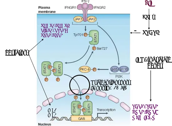
1997; Fruh et al. 1999). IFNγ performs its biological functions through binding to IFNγ-receptor (IFNGR). IFNGR is composed of two subunits (α and β) and is present on all nucleated cells. IFNGR-α chain associates with Janus active kinase-1 (JAK1) and signal transducer and activator of transcription 1 (STAT1) while IFNGR-β chain binds to JAK2 (Figure 2).
IFNγ bound to its receptor leads to the trans-phosphorylation and reciprocal activation of JAKs which subsequently activate STAT1 by phosphorylation at tyrosine 701 (Y701) and serine 727 (S727) DNA- binding site. After activation the phosphorylated STAT1 dimerizes in the cytosol and is translocated into the nucleus to activate IFNγ-inducible genes by binding to the gamma interferon activation site (GAS) elements in their promoter regions. The nuclear transport of STAT1 dimers occurs through nuclear pore complexes and is dependent on heterodimeric importin receptors, which, on one side, bind to the nuclear localization signal of the cargo (importin α subunit) and, on the other side, enable docking of the protein cargo on the cytoplasmic site of the nuclear pore complex (importin β subunit). There are six different α-importin (α1-α6) and one β-importin (β1). Various α-importins are differentially expressed in
different tissues and are responsible for nuclear import of different cargo proteins. Nuclear import of activated STAT1 occurs through importin-α1 (McBride et al. 2002). In the nucleus, phosphorylated STAT1 dimers activate IFNγ inducible target genes including MHC class I heavy chain and light chain molecules and several components of the MHC class I antigen processing and presentation pathway (Fruh et al. 1999). IFNγ also activates some important transcription factors from interferon regulatory factor (IRF) family (especially IRF-1 and IRF-8). They have a capacity to bind to IFNγ regulated response elements in promoter region of genes and stimulate the transcription of IFNγ stimulated genes (ISGs) such as LMP2, LMP7, TAP1, TAP2 and tapasin (Taniguchi et al. 2002). IRF-1 expression is induced by IFNγ through STAT1-dependent fashion over a period of hours (Harada et al. 1994; Pine et al. 1994) and it is not directly activated by IFNγ. IRF-1 cooperates with STAT1 to transcribe ISGs which require intact IRF-1 and STAT1 binding sites to be optimally transcribed. IRF-1 plays an important role in regulating MHC class I gene expression (Mori et al. 1999). However, other IFNγ -induced factors such as p48, also up- regulate MHC class I gene expression in STAT1 independent manner.
This describes the diversity of regulation of MHC class I expression mediated by IFNγ (Bluyssen et al. 1996; Kimura et al. 1996; Majumder et al. 1998).
MHC class II trans-activator (CIITA) is a transcriptional factor that regulates the expression of MHC class II (Chang et al. 1996). CIITA is also efficiently induced by IFN-γ through a complex consisting of upstream factor 1 (USF-1) and STAT1 (Muhlethaler-Mottet et al. 1997; Muhlethaler- Mottet et al. 1998). This is an additional example of co-operation of STAT1 with other transcriptional factors. In contrast, the transcription factors STAT3, STAT5, NF-κB and AP-1 are induced by IFN-γ without the cooperation of STAT1. In some cases, STAT3 and STAT5 are activated by IFNγ to compensate the absence of STAT1 (Meinke et al. 1996; Ramana et al. 2000; Ramana et al. 2005) but during anti-viral response, STAT3 can not compensate the loss of STAT1 (Horvath et al. 1996). Because, biologically active STAT1 must be full length and phosphorylated on both
tyrosine and serine residues for full IFNγ antiviral activity. Furthermore, these closely related STAT proteins (STAT1 and STAT3) are not functionally redundant in their ability to establish the antiviral effect.
Previous reports demonstrated that the expression and phophorylation of STAT3 was increased in cells deficient of STAT1 (Ramana et al. 2005).
But, this was not true in all STAT1-deficient strains (Gough et al. 2007).
The reason for this discrepancy is still unclear and might be dependent on animal strain.
Figure 2. The IFNγ signaling pathway in all nucleated cells. IFNγ binds to its receptor IFNGR1/R2 and leads to activation of JAK1/2 which phosphorylates STAT1 on Tyr701 or Ser727 residues. Active STAT1 dimerizes and goes into the nucleus by nuclear import proteins importin α1/β. Active STAT1 binds to the IFNγ-activated site (GAS) in promoter region leading to the induction of IFNγ-inducible genes including TAP1,TAP2, LMP2, LMP7, MHC I heavy chain and β2M. SHP1, IRF1 and IRF2 act as positive regulators while SOCS1, PIAS1, PTP1B and SHP2 act as negative regulators of this pathway.
LIF/IL-6 inhibits through STAT3 pathway the IFNγ induced STAT1 signaling. (Leonidas C.
Platanias, Nature Reviews Immunology, 2005)
The significance of the NF-κB pathway in IFNγ signaling is not known so far. However, IFNγ can induce DNA binding of NF-κB in the absence of STAT1 in some primary fibroblasts. This cell restricted phenomenon reveals that this may not be the main mechanism of STAT1 independent
TAP1, TAP2 LMP2,LMP7 MHC I, β2M SOCS1,SOCS3
PIAS1, PTP1B SHP1,SHP2
IRF1 IRF2
STAT3
-
ESCs
LIF
Regulators
IFNγ-inducible genes
Nuclear transport Importin-α1/α5/β
TAP1, TAP2 LMP2,LMP7 MHC I, β2M SOCS1,SOCS3
PIAS1, PTP1B SHP1,SHP2
IRF1 IRF2
STAT3
-
ESCs
LIF
Regulators
IFNγ-inducible genes
Nuclear transport Importin-α1/α5/β
transcriptional effects of IFNγ (Deb et al. 2001). The activator protein 1 (AP-1) is the family of dimeric transcription factors, which are rapidly activated by IFNγ and required for transcription of several IFNγ stimulated genes (Clarke et al. 2003; Gough et al. 2007). AP-1 DNA binding activity is increased rapidly by IFNγ independently of JAK1/2 or STAT1. AP-1 activity is also required for transcription of several ISGs with or without co- operating with STAT1 (Gough et al. 2007).
I.4. Role of STAT1:STAT3 signaling in biological systems
Interferons and gp130 family of cytokines play a major role in regulating immune responses and in mediating cellular decisions during growth and development. Signal transducer and activators of transcription, especially STAT1 and STAT3, are the main targets of type I and type II interferons and cytokines belonging to the gp130 family such as interleukin 6 (IL-6) and leukemia inhibitory factor (LIF). STAT3 promotes cell proliferation/survival and immune tolerance and inhibits inflammation, while STAT1 counteracts proliferation and supports innate and adaptive immune responses. IFNγ mainly activates prolonged STAT1 activation through JAK1 and JAK2 while type I IFNs activate the distinct transcriptional complex including STAT1, STAT2 and ISGF3. The cytokines of IL-6 family activate JAK1, JAK2 and TYK2 through specific leukemia inhibitory factor receptor (LIFR) leading to the prolonged phosphorylation of STAT3. LIF acts through a high-affinity receptor complex composed of a low affinity LIF binding chain (LIFR, also known as CD118) and high-affinity converter subunit gp130.
STAT1 pathway activates the expression of apoptotic genes like caspases, death receptors and ligands, and nitric oxide synthase (Allione et al. 1999; Fulda et al. 2002). On the other side, it negatively regulates the expression of prosurvival genes such as Bcl-xL, and Bcl-2 (Stephanou et al. 2000). STAT3 target genes differ according to cell type and environmental circumstances. In general, STAT3 induces anti-apoptotic genes of Bcl family to prevent apoptosis and it promotes proliferation
through the induction of oncogenes and cell cycle regulatory genes such as cyclin D1, c-myc and pim-1 (Chin et al. 1996; Dimberg et al. 2003; Xiao et al. 2006). STAT1 and STAT3 exert opposing roles in inflammation. IFN- mediated STAT1 activation acts as a proinflammatory factor by inducing tissue apoptosis and a number of genes that favor activation and recruitment of immune cells to the side of inflammation. STAT1 activation by type I and II IFNs advocates antigen presentation by enhancing the expression of MHC class I and MHC class II molecules and the components of antigen-processing machinery (Lee et al. 1996; Brucet et al. 2004; Marques et al. 2004; Rouyez et al. 2005). In contrast, STAT3 mediates the functions of major anti-inflammatory cytokine IL-10 which maintains the balance between activation and deactivation of mononuclear cells and downregulates the surface expression of MHC molecules (Donnelly et al. 1999). STAT3 activated IL-10 also directly inhibits IFN- induced gene transcription partly by downregulating STAT1 activation due to inhibition of its tyrosine phosphorylation (Ito et al. 1999). Interestingly, STAT1 is usually considered as a tumor suppressor while STAT3 is known as an oncogene since it is constitutively expressed in almost 70% of solid and hematological tumors (Turkson et al. 2000; Turkson 2004; Kim et al.
2007). STAT1 directly controls tumor cell expansion by up regulating many pro-apoptotic and anti-proliferative genes in cancer cells. In contrast, STAT3 constitutive activity is necessary for survival and proliferation of many different kinds of established or primary cancer cells (Turkson et al.
2004; Kim et al. 2007) and STAT3 allows cancer cells to escape from cell- mediated immune system by enhancing the secretion of dendritic cell inhibitors such as IL-10 and VEGF (Wang et al. 2004). Therefore, the balance between STAT3 and STAT1 activity is important for homeostasis and physiology of normal cells (Figure 3).
Although STAT1 and STAT3 play opposing roles in cell survival, proliferation, apoptotic death or inflammation, the counterbalance between STAT1 and STAT3 may decide the result of cytokine treatment and pathological responses. In fact, previous reports demonstrated that STAT1 or STAT3-deficient cells have reciprocal STAT1:STAT3 regulatory mechanisms and relative affluence of STAT3 or STAT1 may play a role in
shaping the biological effects of their main activating cytokines (Costa- Pereira et al. 2002; Qing et al. 2004; Gimeno et al. 2005; Tanabe et al.
2005). For example, in absence of STAT3, gp130 cytokines strongly modify STAT1 activation profiles and trigger prolonged phosphorylation of STAT1 upregulating multiple IFNγ inducible genes in a number of cell types (Costa-Pereira et al. 2002).
Figure 3. The importance of balanced expression/activation of STAT1 and STAT3 in tumor setting. When STAT3 activation and/or expression overwhelm, tumor development and maintenance are favored. The presence of soluble factors such as IL- 10 induces tolerance in immune cells; tumor cell proliferation and survival are favored not only directly, but also indirectly, by the enhancement of angiogenesis. On contrary, the prevelance of STAT1 activation directly blocks cell cycle progression and induces apoptosis of cancer cells. Moreover, STAT1 favors the generation of an adequate immune response against the tumor. (Regis et al, Seminars in Cell and Developmental Biology, 2008)
These findings indicate that STAT3 downregulates STAT1 activity (IFNγ responses) and allow LIF/IL-6 specific responses in normal cells. Each specific cell type has characteristic STAT protein levels. The proteins of the suppressor of cytokine signaling (SOCS) family are induced as immediate early genes downstream of different STATs and inhibit STATs phosphorylation as a negative feedback mechanism (Chen et al. 2000).
Fascinatingly, both SOCS1 and SOCS3 work not only as feedback mechanisms to quantitatively regulate STAT1 and STAT3 activation
respectively, but they can also finely adjust STAT1 or STAT3 mediated responses. For example, STAT3-dependent SOCS3 induction can prevent STAT1 activation and vice versa by inhibiting the JAKs in Con-A induced heapatitis in hepatocytes (Hong et al. 2002; Wormald et al. 2006).
I.5. Allorecognition of embryo and expression of MHC molecules in the course of embryonic development
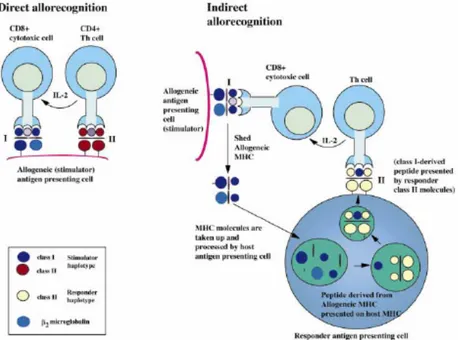
Allorecognition attributes to T-cell recognition of genetic polymorphisms between members of same species. MHC molecules are the main targets of the immune responses to allogeneic tissues which are present on the donor cells. There are two pathways of allorecognition: direct and indirect (Lechler et al. 1982). Direct recognition involves the recognition by recipient T cells of intact donor MHC molecules complexed with peptide on donor antigen presenting cells. On contrary, the requirement of indirect recognition is that recipient antigen presenting cells (APCs) process the donor-MHC antigen prior to presentation to recipient T cells in a self- restricted manner. Therefore, the direct and indirect alloresponses are governed by different APCs and differ in their cellular mechanisms (Figure 4).
Allorecognition is a basic system that animals use to preserve individuality.
Although embryos are usually semiallogeneic with their mother, viviparous animals including human beings are required to allow these embryos to develop inside the mother’s body eliminating an ‘invasion’ by nonself. The embryo is directly exposed to the maternal immune system during pregnancy and embryonic cells can be found in maternal organs and blood in mice and humans (Liegeois et al. 1981; Guetta et al. 2003). This provides ample opportunities for the maternal immune system to recognize fetal alloantigens. However, embryo is not destroyed indicating that the immune system is tolerant of the fetal presence. It has been shown that maternal tolerance of the embryo in allogeneic pregnancy is dependent upon CD25+ regulatory T (Treg) cells but CD25+ Treg cells play no role during late stages of pregnancy (Shima et al. 2010; Aluvihare et al. 2004), indicating the involvement of other mechanisms. Interestingly, maternal
and fetal cells are involved in the establishment of the tolerogenic environment having overlapping molecular mechanisms. The maternal endometrium expresses the immunosuppressive factors TGF-β, Galectin-1 (GAL-1) and thymic stromal lymphopoietin (TSLP) (Simpson et al. 2002;
von Wolff et al. 2005). They induce Treg cells and Th2 responses and inhibit Th1 response providing tolerogenic environment. Both the maternal decidua and fetal trophoblast express indoleamine 2,3-dioxygenase (IDO) (Kamimura et al. 1991) which induces Treg cells and inhibits the activation of T cells and NK cells (Frumento et al. 2002). Crry is also expressed by fetal and maternal cells and it stops deposition of the complement proteins C3 and C4 to stop the formation of the cytosolic membrane attack complex (Weigle et al. 1983; Molina et al. 1992; Kim et al. 1995). Human villous cytotrophoblast cells also express non-classical HLA class Ib HLA-G molecules which help to evade cytotoxic NK activity and induce the tolerogenic phenotype of dendritic cells. Fas-L expression on these cells also helps to kill activated T cells via apoptosis (Le Bouteiller et al. 1999;
Fournel et al. 2000; Hviid 2006; Carosella et al. 2008).
In mammals the expression of MHC genes is developmentally programmed. After fertilization, a hierarchical order of gene expression takes place including the MHC genes. In order to understand how the developing embryo avoids rejection, it is essential to know if, and when, MHC class I expression occurs during embryonic development. It is well postulated that a possible route of escape of the embryo from maternal rejection is by down-modulating the MHC. This down-modulation of MHC genes occurs either at the feto-maternal interface and/or on the surface of other embryo cells. HLA class I and II are absent while HLA-G and β2M are present in human blastocyts and preimplantation embryos (Roberts et al. 1992; Jurisicova et al. 1996). Human trophoblasts are resistant to NK cell attack, possibly as a consequence of the presence of HLA-G, which ensures that NK cells detect the trophoblast as normal self. It is possible, that expression of certain MHC products is advantageous for the survival of the early embryo. The selective expression of MHC products during development is potentially subject to modulation, by a fluctuating pattern of
Th1-type and Th2-type cytokines, maternal hormones and other cellular interactions. Unexplained human reproductive failure is quite common and has often associated with MHC products (Christiansen et al. 1997). If MHC determinants were to be expressed in vivo, then cytotoxic T-cell attack could occur, resulting in spontaneous abortion.
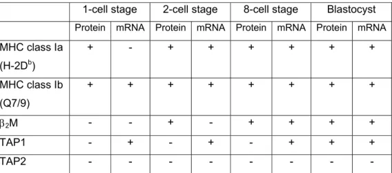
The onset of expression of MHC class Ia, class Ib, β2M, TAP1 and TAP2 in murine embryonic development is shown in Table 2 (Sprinks et al. 1993;
Cooper et al. 1998).
Table 2. Detection of MHC and transporter associated with antigen processing (TAP) mRNA and protein products during preimplantation mouse embryonic development
1-cell stage 2-cell stage 8-cell stage Blastocyst Protein mRNA Protein mRNA Protein mRNA Protein mRNA MHC class Ia
(H-2Db)
+ - + + + + + +
MHC class Ib (Q7/9)
+ + + + + + + +
β2M - - + - + + + +
TAP1 - + - + - + + +
TAP2 - - -
Note: symbols in parentheses indicate presence (+) or absence (-) of mRNA transcripts, as detected by reverse transcription-polymerase chain reaction, and protein products, as detected by indirect immunofluoresence. Based on data from: Sprinks et al, (1993), sprinks (1994), Cooper et al. (1998) and Cooper (1998).
Taken together these data suggest that, at least for mice, MHC class I complexes composed of heavy chain and β2M are present at the surface of embryos at very early stages of development. However, they are unlikely to be functional in the normal sense of antigen presentation, due to the lack of a functional peptide transporter. Therefore, during pregnancy the developing fetus is protected from immunological attack potentially by both a physical barrier, such as negatively charged, sialic acid-rich mucopolysaccharide, and also an immunologically inert layer of placental
trophoblast tissue, on which MHC class I antigens are very weakly distributed and from which MHC class II molecules are absent.
Figure 4. Diagrammatic representation of direct and indirect allorecognition In direct allorecognition pathway, intact donor Class-I and –II MHC molecules present on the surface of donor antigen presenting cell (APC) are recognized directly by recipient CD8+ and CD4+ T cells, respectively. In the indirect allorecognition pathway, donor Class-I and –II MHC molecules are taken up, processed and presented in the context of recipient MHC molecules to recipient CD8+ and CD4+ T cells, respectively. (Rogers and Lechler, American Journal of Transplantation, 2001).
I.6. Embryonic stem (ES) cells
Embryonic stem cells (ESCs) are derived from the inner cell mass of blastocysts (Evans et al. 1981; Martin 1981); (Thomson et al. 1998).
Human ESCs are maintained in undifferentiated stage in the presence of basic fibroblasts growth factor (bFGF). The human ESC colonies have typical round shape compact morphology and cells express SSEA-4, Tra- 1-60 and Tra-1-81 pluripotent markers on their cell surface. They are passaged every 5-7 days either by mechanical cutting or by using collagenase IV or dispase in the form of small clumps. Single cell passaging of these ESCs can be done in presence of Rho kinase inhibitor (ROCKi), thiazovivin or neurotrophins using trypsin-EDTA (Pyle et al.
2006; Watanabe et al. 2007). Murine ESCs are maintained in undifferentiated stage in the presence of LIF. In comparison to human
ESCs, the colonies are smaller in size having round or oval shape structures. They express SSEA-1 as pluripotency marker on their cell surface but not the SSEA4 or Tra antigens.They are passaged as single cells every 2-3 days using trypsin-EDTA. The characteristics of murine and human ESCs are summarized in Table 3.
Table 3. Characteristics of murine and human ESCs
Marker Mouse ESCs Human ESCs
Oct-4 + +
Alkaline
Phosphatase + +
SSEA-1 + -
SSEA-4 - +
Tra-1-60 - +
Tra-1-81 - +
Telomerase
activity + +
Factors aiding in self-renewal
LIF+MEFs or LIF+gelatin-coated
plastic MEFs+serum or MEFs+bFGF
Morphology
Tight, rounded, multilayer colonies, high nucleus to
cytoplasm ratio
Flatter colonies with tight border, high nucleus to cytoplasm ratio Rate of
division Fast, passage every 2-3 days Slower, passage every 3-7 days Teratoma
formation + +
Germ line
competent + Unknown
Note: Presence (+) and absence (-), SSEA = stage specific embryonic antigen, LIF = leukemia inhibitory factor, bFGF = basic fibroblast growth factor; MEFs = murine embryonic fibroblasts. Table adapted from NIH Stem Cell report (NIH, 2006).
I.7. Immunological properties of ES cells (ESCs)
ESCs express MHC molecules on their cell surface but the constitutive expression of MHC I molecules as well as in their response to IFNγ differ greatly in human and murine ESCs. MHC class I molecules are present on the cell surface of undifferentiated human ESCs at low levels and are
inducible by IFNγ (Drukker et al. 2002; Grinnemo et al. 2006). There was no MHC induction by IFN-α and IFN-β due to the missing expression of these cytokine receptors on human ES cells (Drukker et al. 2002). On other side, the expression of MHC class I molecules was not detectable by flow cytometry on undifferentiated murine ES cells (Tian et al. 1997;
Magliocca et al. 2006; Abdullah et al. 2007; Nussbaum et al. 2007).
However, transcripts of MHC class I molecules, β2-microglobulin and several components of antigen presentation pathway were present in detectable amount in murine ESCs (Magliocca et al. 2006; Abdullah et al.
2007). Using a highly sensitive and specific method for detection of rare peptide-MHC class I complexes utilizing the lacZ-inducible, antigen/MHC- specific T cell hybridomas it was possible to provide the evidence that murine ES cells express MHC class I molecule but at very low levels that are not detectable by flow cytometry (Abdullah et al. 2007). The murine ESCs do not respond to IFN-γ and only after differentiation, they respond to IFNγ and strongly up regulate the expression of MHC class I molecules on a significant fraction of cells and components of antigen presenting pathway (Abdullah et al. 2007). Interestingly, MHC class II transcripts were present in human (Grinnemo et al. 2006) and murine (Magliocca et al.
2006) ESCs that were inducible by IFNγ.
Previous reports showed that murine as well as human ES cells may possess immune-privileged properties. Bonde and Zavazava have demonstrated that murine ESCs can not be lysed by naïve allogenic NK (Bonde et al. 2006) or polyI:C activated NK cells (Koch et al. 2008).
However, other reports showed that murine ESCs can be lysed by NK cells in presence of IFNγ (Bonde and Zavazava 2006). The recognition of ESCs by NK cells appears to be mediated by ligands of activating natural- killer group 2 member D (NKG2D) molecules and intercellular adhesion molecule-1 (ICAM-1) expressed on ESCs (Bonde et al. 2006; Frenzel et al. 2009). Human ES cells also showed limited lysis by NK cells possibly due to low expression of activating NK cell ligands such as NKp46 and CD16 on these ES cells (Drukker et al. 2002). Similarly, murine ES cells were not lysed by in vitro generated alloreactive CTLs (Bonde et al. 2006).
Additionally, lymphocytic choriomeningitis (LCM) virus-infected or peptide–
loaded murine ES cells were also resistant to killing by activated LCM virus-specific syngeneic CTLs (Abdullah et al. 2007). The expression of the granzyme B inhibitor serine protease inhibitor 6 (SPI-6) in undifferentiated murine ES cells appears to protect ESCs from CTL killing in vitro (Abdullah et al. 2007). This study also provided the evidence for functional recognition of ES cells by cytotoxic CD8+ T cells in vitro. Another study showed that ES cells suppress T cell proliferation in a contact- independent manner by secreting TGF-β and expressing Fas-L on their surface to block alloreactive T cell apoptosis (Koch et al. 2008).
Induced pluripotent stem cells (iPSCs) are ES-like cells made first time by retroviral reprogramming of murine somatic cells using combination of four transcription factors Oct3/4, Sox2, Klf4 and c-Myc (Takahashi et al. 2006).
These iPSCs have highly similar transcriptional and epigenetic features to those of ESCs (Gupta et al. 2010; Takahashi et al. 2007; Wernig et al.
2007; Yu et al. 2007). After this major findings, lot of different groups reprogrammed different murine and human somatic cells into iPSCs using viral and non-viral methods (Takahashi et al. 2007; Wernig et al. 2007;
Stadtfeld et al. 2008; Kaji et al. 2009; Yu et al. 2009). In first stance, iPS technology opened new avenues for autologus transplantation but there are still lots of challenges of immunogenicity regarding the use of iPSCs and its derivatives in regenerative medicine. Additionally, researchers also found the epigenetic differences between ES and iPS cells (Kim et al.
2010; Polo et al. 2010; Doi et al. 2009).
A recent study demonstrated that few cells derived from iPSCs can be immunogeneic even in syngeneic settings (Zhao et al. 2011).This group reprogrammed C57BL6 (B6) mouse embryonic fibroblasts (MEFs) into iPSCs using either retroviral approach (ViPSCs) or an episomal approach (EiPSCs) that causes no permanent genomic integration of vector sequences. Later on, they transplanted B6 ESCs, ViPSCs and EiPSCs into the B6 recipients. Teratomas were formed by ESCs while teratomas formed by B6 ViPSCs were mostly immune-rejected by B6 recipients mice.
Additionally, B6 EiPSCs teratomas were also immunogenic showing tissue
damage and regression with T cell infiltration. Since iPSCs and ESCs have shown to have subtle yet apparent epigenetic differences, this could be the basis for the expression of abnormal expression of antigens (minor antigens) on iPSCs but not on ESCs who have normal development and differentiation (Kim et al. 2010; Polo et al. 2010; Chin et al. 2009; Doi et al.
2009). The expression of minor antigens on iPSCs makes them susceptible for T-dependent immune rejection. Moreover, iPSCs have shown mutations in their coding sequences that could further contribute to the immunogenicity of iPSCs derivetives (Gore et al. 2011). Recently, iPSCs have generated from patients with genetically inherited as well as sporadic diseases for in vitro disease modeling, gene corrections of defected gene and transplantation of corrected cell types to patients (Howden et al. 2011; Liu et al. 2011; Zou et al. 2011). However, successful replacement of cell types may elicit rejection of the grafted cells themselves enduring to recognition of the processed gene product in a MHC-restricted manner, likewise to recognition of minor histocompatibility antigens (Figure 5). To circumvent these limitations, one can capitalize the pluripotency of iPSCs to provide a source of immature dendritic cells (DCs) expressing the alloantigens to which tolerance is required (Fairchild 2010).
Pearl and coworkers reported that short time blockade treatment of costimulatory molecules is enough to induce engraftment of allogeneic mouse ESCs and iPSCs as well as xenogeneic human ESCs and iPSCs (Pearl et al. 2011). They showed that short term blockade of three costimulatory receptors: CTL-associated antigen 4 (CTLA4)-Ig, anti-CD40 ligand (anti-CD40L), and anti-lymphocyte function-associated antigen 1 (anti-LFA-1) - could induce long-term allogeneic and xenogeneic ESC engraftment by decreasing the expression of proinflammatory cytokines and increasing the establishment of a proapototic phenotype. These results indicated that short term blockade of costimulatorly molecules may also overcome the problem of minor histocompatibilty antigens in cellular engraftment of iPSCs.
Figure 5. Induction of tolerance to the products of corrected genes. “The generation of induced pluripotent stem cells (iPSCs) from individuals with diseases that are under monogenic control might enable correction of the genetic defect. Successful replacement of cell types in vivo could, however, induce neutralizing immune responses to products of the corrected gene or provoke rejection of the grafted cells themselves owing to recognition of the processed gene product in an MHC-restricted manner, similarly to the recognition of minor histocompatibility antigens. One approach to circumventing such immunogenicity might be the directed differentiation of immature dendritic cells (DCs) from the disease-corrected iPSCs, which endogenously express the gene product to which tolerance is required. Administration of such DCs in advance of cell replacement therapy in a non-inflammatory, tolerogenic context that does not promote DC maturation could enable the induction of a repertoire of regulatory T cells specific for the therapeutic gene product. Tolerance can subsequently be reinforced by the allograft itself, which functions as a continuous source of antigen”. (Fairchild,PJ, Nature Reviews Immunology, Dec. 2010).
II. Aims
In mammals the expression of MHC genes are developmentally programmed. The MHC class I molecules play crucial role not only in fertilization and development of embryos but also in reinforcement of innate and adaptive immune responses against infectious agents or tumor cells. Since ESCs are excellent models to study development and differentiation into various types of cells, we used it to analyze the regulation of MHC class I molecules in these cells. Our group has previously shown that murine ESCs express very low levels of MHC class I molecules, which are not inducible by IFNγ. I, therefore, wished to investigate the reason of low expression of MHC class I molecules in ESCs and their unresponsiveness to IFNγ.
The specific aims of this thesis are:
1. To elucidate the mechanisms responsible for low expression of MHC class I molecules in undifferentiated murine ESCs
2. To determine the mechanism for IFNγ unresponsiveness in ESCs.
3. To determine whether same mechanisms are involved in regulation of expression of MHC class I molecules in differentiated cells
4. To determine whether modulation of expression of MHC class I molecules affects the susceptibility of ESCs to immune cells in vitro.
III. Results
III.1. Regulation of MHC class I molecules in ESCs
III.1.1. Expression of MHC class I molecules on ESCs
Human and murine ESCs grow on feeders and need, respectively, bFGF or LIF as crucial additives in culture media to maintain their pluripotency.
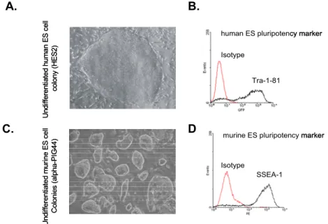
Human ESCs show typical flat, large and compact colony expressing Tra- 1-81 as a pluripotency marker on their surface (Figure 6A,B). Murine ESCs are round, oval shaped, smaller in size and express SSEA1 as a pluripotency marker on their surfaces (Figure 6C,D).
A. B.
C. D
Figure 6. Characterization of human and murine ESCs. Typical morphology of human (A) and murine (C) ESCs. The expression of pluripotency markers Tra-1-81 (B) and SSEA1 (D) on human and murine ES cells, respectively, was determined by flow cytometry.
To set conditions for our further mechanistic studies we used flow cytometry to determine expression of MHC class I molecules on ESCs and validate previously published results. We showed that human ESCs stained with pan-HLA-ABC antibodies W6/32 express MHC class I molecules on their surface (93.2% of positive cells, MFI=103.9) and that their expression is enhanced 7.8-fold after stimulation with IFNγ (98.4% of positive cells, MFI=812.3) (Figure 7A). In contrast, staining for H-2Kb MHC
Undifferentiated human ES cell colony (HES2)Undifferentiated murine ES cell Colonies (alpha-PIG44)
Tra-1-81
SSEA-1 Isotype
Isotype
human ES pluripotency marker
murine ES pluripotency marker Undifferentiated human ES cell colony (HES2)Undifferentiated murine ES cell Colonies (alpha-PIG44)
Tra-1-81
SSEA-1 Isotype
Isotype
human ES pluripotency marker
murine ES pluripotency marker