• Keine Ergebnisse gefunden

Three dimensional electron microscopy and in silico tools for macromolecular structure determination

N/A
N/A
Protected

Academic year: 2021

Aktie "Three dimensional electron microscopy and in silico tools for macromolecular structure determination"

Copied!
12
0
0

Wird geladen.... (Jetzt Volltext ansehen)

Volltext

(1)

Review article:

THREE DIMENSIONAL ELECTRON MICROSCOPY AND IN SILICO TOOLS FOR MACROMOLECULAR STRUCTURE DETERMINATION Subhomoi Borkotoky, Chetan Kumar Meena, Mohammad Wahab Khan, Ayaluru Murali*

Centre for Bioinformatics, School of Life Sciences, Pondicherry University, Puducherry-605014, India

* Corresponding author: Dr. A. Murali, Centre for Bioinformatics, School of Life Sciences, Pondicherry University, Puducherry-605014, India; E-Mail - murali@bicpu.edu.in

ABSTRACT

Recently, structural biology witnessed a major tool – electron microscopy – in solving the structures of macromolecules in addition to the conventional techniques, X-ray crystallo- graphy and nuclear magnetic resonance (NMR). Three dimensional transmission electron mi- croscopy (3DTEM) is one of the most sophisticated techniques for structure determination of molecular machines. Known to give the 3-dimensional structures in its native form with liter- ally no upper limit on size of the macromolecule, this tool does not need the crystallization of the protein. Combining the 3DTEM data with in silico tools, one can have better refined structure of a desired complex. In this review we are discussing about the recent advance- ments in three dimensional electron microscopy and tools associated with it.

Keywords: cryo-EM, single particle analysis, CTF correction, segmentation

INTRODUCTION

Recent past witnessed the emergence of new tool in structural biology in the form of electron microscopy and single particle analysis. It complemented the currently ex- isting tools namely, X-ray crystallography and nuclear magnetic resonance (NMR).

This tool has several advantages over the conventional structural biology tools (X-ray crystallography and NMR) such as, struc- ture elucidation without crystallization, solving the structure in physiological condi- tions, and with literally no upper limit on size of the protein. Though greater purity is of high demand, this technique needs low concentrations of the protein (typically of the order of 1 ng/μl for a protein of

~100 kDa molecular weight for negative stain). However, despite of proving its abil- ity in all corners of structural biology, three dimensional transmission electron micros-

copy (3DTEM) has relatively fewer entries in RCSB compared to deposits from crys- tallography and NMR. The present review article was taken up to give an outline of the single particle analysis with few exam- ples where 3DTEM was found to be unique. Also this review article presents a brief account on other promising online and offline tools that will complement the 3DTEM technique.

As per advancements of more sophisti-

cated instruments and in silico tools over

recent years, electron microscopy is getting

a greater push to the arena of solving mo-

lecular structures, along with other methods

like X-ray crystallography and nuclear

magnetic resonance (NMR). As compared

to the prominent structure exploration

methods like X-ray crystallography and

NMR, 3DTEM techniques are the structural

biologists’ best choice for larger macromo-

(2)

lecular assemblies (having molecular weights greater than 60 kDa) and other larger cellular components. There is no need for growing 2D crystals for 3DTEM methods; it mainly relies on the symmetry properties of particle (Jonic et al., 2008).

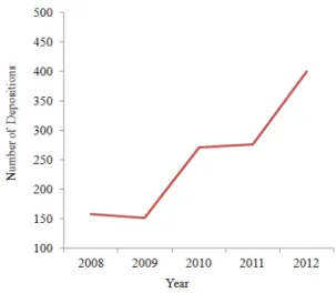
The increase in the number of electron mi- croscopy (EM) solved structures is evident from the collaboration of the ftp archives of Protein Data Bank (PDB) and EM Data Bank (EMDB) to facilitate user access to EM maps and models (Berman et al., 2013). According to EM databank statistics, 1749 entries were deposited in its database (http://www.emdatabank.org/recententries.html) till February, 2013 and with a gradual in- crease in data submission in EMDB in the last five years (Figure 1).

Figure 1: Deposition of EM Structures in EMDB in last five years

Since the electron microscopy technique is a classic technique and known to scien- tists in all branches of life sciences, a huge collection of literature exists and it is not possible to include all of it. We focus on single particle analysis (SPA) – a tool that works with electron microscopy, which has laid its foundation in 1970s and started gaining the attention of more researchers since 1995 (Frank, 2009). In view of the space restriction, the present review consid- ers only the developments in the recent years. Readers might refer to the earlier re- views that appeared on similar topics for complete coverage of the literature on this

subject (Jiang and Ludtke, 2005; Llorca, 2005; Ruprecht and Nield, 2001).

The present review mainly focuses on the specimen preparation approaches for electron microscopy with an introduction of single particle analysis, some important tools that are available to complement the 3DTEM and concludes with some notable reports to prove the uniqueness of 3DTEM.

Specimen preparation methods for electron microscopy

The most commonly used staining methods in electron microscopy are nega- tive staining, CryoEM and Cryo-negative staining. Each of these three methods plays a considerable role in resolution of the data and contrast of the images. A detailed ac- count of these methods is listed below.

Negative staining

Among all the staining methods availa-

ble, negative staining (NS) is the simplest

one. First reported in 1959 (Brenner and

Horne), it was revisited by several re-

searchers. Generally, the aqueous solutions

of heavy metal salts are used as stains

which make the sample to appear darker

than the background, so the term “negative

staining” is attributed to this process. Most

commonly used negative stain is uranyl ac-

etate which gives high contrast. The other

compounds that are in use are sodi-

um/potassium phosphotungstate, uranyl

formate, ammonium molybdate (Bremer et

al., 1992), as well as patented stains, such

as NanoVan® and NanoW® (Hainfeld et

al., 1994). Negative staining can reveal the

true solvent-excluded surface and shape of

a biological molecule. Proper identification

of intra-molecular information by negative

staining, such as alpha-helices and/or beta-

sheets are quite theoretical, which relies

upon the relatively large mass-thickness

difference between the biological material

and the surrounding stain (De Carlo and

Harris, 2011). The high vacuum that is re-

quired to be maintained in a typical TEM

column can deform the biological samples

(3)

by collapsing/flattening effect which is mainly due to the vacuum drying.

Cryo-freezing

It is well known that the negative stain- ing approach always results in a low resolu- tion data (typically of the order of 1.5 – 2 nm). CryoEM (also known as Cryo- freezing) is an alternative where the nativity of the macromolecule is preserved and con- siderably high resolution is achieved. Also, Cryo-freezing eliminates many of the arti- facts that may disturb the structural integri- ty of the biological macromolecules. It al- lows vitrification of biological samples in their native state and the freezing process is done so rapidly that no ice crystals will be formed during the sample preparation (Dubochet et al., 1982). There are different methods of preparing frozen, hydrated bio- logical specimens such as – sandwich be- tween folding grids, sandwich between two carbon films, sandwich between pretreated behenic acid film and poly lysine coated film, sandwich between grids of different meshes (Jaffe and Glaeser, 1984; Talmon et al., 1979; Taylor and Glaeser, 1973) in which manual freezing is done at pretreated carbon films in liquid nitrogen. Other methods like thin film freezing (Adrian et al., 1984) use pretreated carbon films or holey grids for support and liquid ethane as coolant. This approach is known to give higher resolution structural details but suf- fers from poor contrast and high radiation damage. The poor contrast that is inherent in the cryoEM sets a lower limit (of about 200 kDa) on the protein size.

However, there are some attempts to work-around by generating focal pairs (Ludtke and Chiu, 2003). In this approach, two micrographs were taken for the same image – one with true focus (poor or no contrast) and one with greater defocus (hav- ing better contrast with almost loss of reso- lution of the image). The images from the true focus, as identified by transferring the coordinates of the images from micrographs of greater defocus, will be used for analysis.

Cryo-negative staining

Cryo-negative staining combines the advantages of both negative staining and cryo-EM. It uses stains like ammonium mo- lybdate just before vitrification (Adrian et al., 1998). It has many advantages over tra- ditional techniques such as better signal to noise ratio, better stability of the specimen, rapid collection and analysis of images, re- duced radiation damage and lower range of sample size. Recent studies on Limulus SAP-like pentraxin (Shrive et al., 2009) and human Pol II complex (Kassube et al., 2012) uses the cryo-negative staining ap- proach and reported structures to the tune of 14 Å and 25 Å resolution respectively.

There are however, some limitations of cryo-negative staining. Few biological as- semblies may show sensitivity to the satu- rated stain, or it may overemphasize the contrast of low-resolution features (De Carlo et al., 2002; Jawhari et al., 2006).

Single particle analysis

Single particle analysis (SPA) is one of the novel approaches for reconstructing theoretical models of large proteins and be- came popular in recent years. The term

“single particles” refers to the unique views of the projection images that are isolated and unordered (Frank, 2002). The main aim of single particle reconstruction is to gener- ate a 3D model from 2D images. The com- bination of SPA and EM is capable of re- vealing information related to conforma- tional changes between various states (Rouiller et al., 2002), morphological char- acterization (Ludwig et al., 2003), DNA break repair (Spagnolo et al., 2006) etc.

There is also evidence for combining nano- technology and SPA to study important bio- logical problems like self-assembly of vi- rus-like particles (Sun et al., 2007), and the influence of charge and size variables on the templated assembly of virus capsids (Daniel et al., 2010).

The single particle reconstruction is an

iterative process that can be divided into

several common steps which include selec-

tion of individual projections of the parti-

(4)

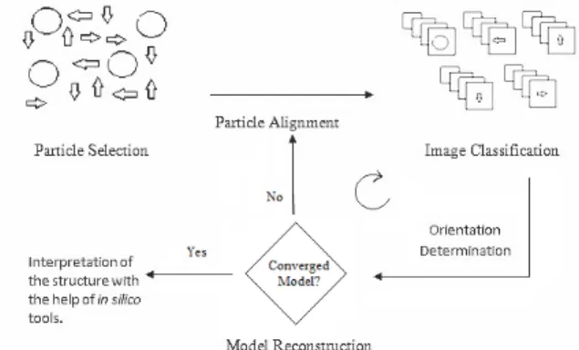
cles from the electron micrograph, classifi- cation of particles with identical views and their alignment, orientation determination by comparing each particle to a set of refer- ence projections, and finally reconstruction of an initial three dimensional model. The model thus generated in the first iteration (initial model or density map), can be used as a reference for the reconstruction process by repeating the above mentioned steps, until one gets a useful stable final model.

Occasionally, one would also like to apply contrast transfer function (CTF) correction (see next section) in order to improve the resolution of the model. With the final model in hand, one can use various in silico tools for visualization and structure inter- pretation (Thuman-Commike, 2001). A graphical workflow of this process is given in Figure 2.

Figure 2: Major steps involved in single particle analysis

IN SILICO TOOLS FOR 3DTEM DATA ANALYSIS

In this section we will discuss some of the in silico tools and packages available for 3DTEM data analysis.

Tools for generating the electron density maps

Software packages available for single particle analysis are mainly EMAN1/

EMAN2 (Ludtke et al., 1999), IMAGIC (van Heel et al., 1996), SPIDER (Frank et al., 1996), SPARX (Hohn et al., 2007), XMIPP an X-Window-based Microscopy Image Processing Package (Sorzano et al.,

2004), SUPRIM (Schroeter and Bretau- diere, 1996).

Apart from these, one can use the pipe- line, APPION (Lander et al., 2009), which includes the packages ranging from single particle analysis, tomography to image analysis tools. Few of them are discussed below:

Single particle analysis [EMAN, SPI- DER, FREALIGN (Grigorieff, 2007), IMAGIC and XMIPP], and

Tomography [IMOD (Kremer et al., 1996) and ProTomo (Winkler, 2007)].

Particle picker: In general, one has to pick-up several thousands of well- isolated particles from electron micro- graphs for reconstruction purpose. Few automated tools such as FindEM (Roseman, 2004), DoGPicker and Tilt- Picker (Voss et al., 2009) are available which will be useful when handling a huge number of electron micrographs.

CTF correction: Contrast transfer func- tion (Yi et al., 2004), also known as CTF correction is an important step in the SPA that one has to pay attention to, in order to get a high resolution model.

This becomes very important especially when one deals with CryoEM data.

Tools such as ACE (Mallick et al., 2005), CTFFind and CTFTilt (Mindell and Grigorieff, 2003) are available for arriving at the right CTF.

Resolution measurement: The most common practice for measurement of resolution of the refined model is 0.5 cut-off in the Fourier Shell correlation (FSC), meaning to take the point at which the FSC drops below 0.5 (Booth et al., 2004). Almost all major SPA packages listed above, including EMAN, provide inbuilt tools to generate these FSCs. Apart from these, Sousa and Grigorieff (2007) designed a novel tool – RMEASURE – to estimate the resolution of the refined maps by calculating the correlation between neighbouring pixels of the model in Fourier space.

Bfactor correction: Bfactor, also known

as temperature factor is another im-

(5)

portant parameter in context to protein structure as it indicates relative vibra- tional motion of different parts of the structure. EM-BFACTOR (Fernandez et al., 2008; Rosenthal and Henderson, 2003) is one of the tools that allows au- tomatic determination of the Bfactor for maps at a resolution higher than 10 Å, and allows to sharpen the map and com- pensate the decay of the amplitudes.

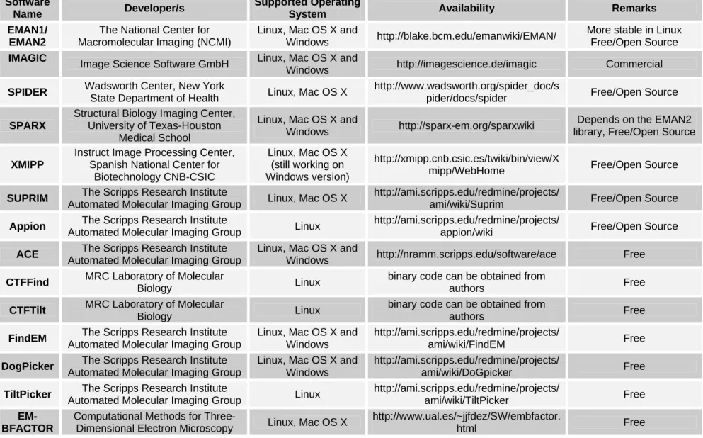
A brief summary of these tools is given in Table 1.

Software/packages for working with the electron density maps generated by SPA

Having the model of a macromolecule (or macromolecular complex) successfully generated using SPA, one would like to work around the model in several aspects.

For visualization of electron density maps one can use Chimera (Pettersen et al., 2004), Amira (Pruggnaller et al., 2008), Avizo (Westenberger, 2008), IMOD (Kremer et al., 1996) and VMD (Humphrey et al., 1996). Among these, Amira and Avi- zo are commercial packages.

At times one may have to deal with the density map of a macromolecule in its oli- gomeric form. In such case, segmentation of individual monomers is of utmost im- portance. Manual segmentation of density maps can be done with Amira, Avizo and Chimera, while automatic segmentation can be done with EMAN, CoDiv (Volkmann, 2002), VolRover (Baker et al., 2006) and Segger (Pintilie et al., 2010) plug-in im- plemented with Chimera. Segmentation procedure is mainly done for extracting a particular region or density of interest from a map of macromolecular assembly. In most of the tools (e.g., Chimera, CoDiv), the segmentation of maps is done by water- shed transform (Beucher and Lantuejoul, 1979; Vincent and Soille, 1991). One can validate the individual subunits identified from automated segmentation by fitting them with their corresponding high- resolution crystal structures and/or manual- ly segmented subunits using foldhunter (Jiang et al., 2001).

When both the Cryo-EM map of a ma- cromolecule and its homology model or high resolution crystal structure are availa- ble, one can use fitting studies for getting a more detailed view of structural characteri- zation of a molecular assembly. MODEL- LER, which is a homology modelling tool commonly used with X-ray and NMR structures as templates, also incorporates two fitting modules – Mod-EM for rigid fitting (Topf et al., 2005) and Flex-EM for flexible fitting (Topf et al., 2008). Similar- ly, one can also use the SITUS package for fitting crystal structures by both rigid body and flexible way (Wriggers et al., 1999).

Other tools that can be used for docking atomic models into EM maps are Dock-EM (Roseman, 2000), EMFit (Rossmann et al., 2001), Foldhunter (Jiang et al., 2001), Fit in Map and MultiFit module in UCSF Chime- ra (Goddard et al., 2007; Tjioe et al., 2011), ADP_EM (Garzon et al., 2007) etc. Segger plug-in for the UCSF Chimera also facili- tates rigid-body docking of models into density maps using segmented regions and it is reported (Pintilie and Chiu, 2012) to be faster than other fitting methods, such as ADP_EM, SITUS, Foldhunter and EMFit.

With the current advancements of elec-

tron microscopes and computational tools,

subnanometer resolution (5-10 Å) struc-

tures are now easily obtainable. At these

resolutions secondary structure elements

(SSE) can be identified with help of com-

ponent crystal structures or comparative

models and the secondary structure ele-

ments α-helices and β-sheets appear as

straight rods and curved plates respectively

and loops as curved rods (Abeysinghe et al.,

2008; Ludtke et al., 2008). Helixhunter and

foldhunter are the computational methods

which facilitate the quantitative identifica-

tion of structural features in terms of known

folds in three-dimensional density maps at

different resolutions. Helixhunter is used to

analyze a three-dimensional map for alpha

helix content at intermediate resolutions,

while foldhunter can be used to localize

(6)

Table 1: Software used for single particle analysis Software

Name Developer/s Supported Operating

System Availability Remarks

EMAN1/

EMAN2

The National Center for Macromolecular Imaging (NCMI)

Linux, Mac OS X and

Windows http://blake.bcm.edu/emanwiki/EMAN/ More stable in Linux Free/Open Source IMAGIC

Image Science Software GmbH Linux, Mac OS X and

Windows http://imagescience.de/imagic Commercial SPIDER Wadsworth Center, New York

State Department of Health Linux, Mac OS X http://www.wadsworth.org/spider_doc/s

pider/docs/spider Free/Open Source SPARX

Structural Biology Imaging Center, University of Texas-Houston

Medical School

Linux, Mac OS X and

Windows http://sparx-em.org/sparxwiki Depends on the EMAN2 library, Free/Open Source

XMIPP

Instruct Image Processing Center, Spanish National Center for

Biotechnology CNB-CSIC

Linux, Mac OS X (still working on Windows version)

http://xmipp.cnb.csic.es/twiki/bin/view/X

mipp/WebHome Free/Open Source

SUPRIM The Scripps Research Institute

Automated Molecular Imaging Group Linux, Mac OS X http://ami.scripps.edu/redmine/projects/

ami/wiki/Suprim Free/Open Source Appion The Scripps Research Institute

Automated Molecular Imaging Group Linux http://ami.scripps.edu/redmine/projects/

appion/wiki Free/Open Source

ACE The Scripps Research Institute Automated Molecular Imaging Group

Linux, Mac OS X and

Windows http://nramm.scripps.edu/software/ace Free CTFFind MRC Laboratory of Molecular

Biology Linux binary code can be obtained from

authors Free

CTFTilt MRC Laboratory of Molecular

Biology Linux binary code can be obtained from

authors Free

FindEM The Scripps Research Institute Automated Molecular Imaging Group

Linux, Mac OS X and Windows

http://ami.scripps.edu/redmine/projects/

ami/wiki/FindEM Free

DogPicker The Scripps Research Institute Automated Molecular Imaging Group

Linux, Mac OS X and Windows

http://ami.scripps.edu/redmine/projects/

ami/wiki/DoGpicker Free

TiltPicker The Scripps Research Institute

Automated Molecular Imaging Group Linux http://ami.scripps.edu/redmine/projects/

ami/wiki/TiltPicker Free

EM- BFACTOR

Computational Methods for Three-

Dimensional Electron Microscopy Linux, Mac OS X http://www.ual.es/~jjfdez/SW/embfactor.

html Free

(7)

known or predicted folds or domains within larger macromolecular assemblies at lower resolutions (< 20 Å). Helixhunter and fold- hunter are available within EMAN package (Jiang et al., 2001). SSEHunter is also used for detection of alpha helices and beta sheets in subnanometer resolution Cryo-EM structures; SSEBuilder is a tool for the rap- id identification and annotation of SSEHunter results. These are packaged within EMAN (Baker et al., 2007). UCSF Chimera also includes these tools under analysis of intermediate resolution struc- tures (AIRS) toolkit (Goddard et al., 2007) (to which support is currently discontin- ued). Another recent tool for analyzing pro- tein structures at near atomic resolution is Gorgon (Baker et al., 2011), which is aimed at de novo model building in cryo-EM with the help of density skeletonization, SSE identification, building and correspondence, Cα placement and model optimization. The above mentioned SSEHunter is also incor- porated into Gorgon to identify SSEs, but it includes more features than the EMAN ver- sion of SSEHunter.

For identifying conserved regions in cryo-EM maps of large macromolecular assemblies one can use MOTIF-EM (Saha et al., 2010). SPI-EM can be used for pre- dicting CATH superfamilies in 3D-EM Maps (Velazquez-Muriel et al., 2005).

Apart from these techniques, Russel et al. (2012) introduced a new method called the integrative modeling platform (IMP), a software package which combines the data from various individual experimental meth- ods like X-ray crystallography, NMR spec- troscopy, electron microscopy, small angle X-ray scattering etc. to generate new mod- els and to overcome the barriers of each in- dividual techniques. The modular structure of the 26S proteasome holocomplex is a recent example of the use of integrative ap- proach (Lasker et al., 2012).

Some of the recently studied structures by three dimensional electron microscopy and single particle analysis

26S proteasome: One of the recent structures solved by 3DTEM is that of 26S proteasome (2.5 MDa) from Saccharomy- ces cerevisiae, at a resolution of 7.4 Å (Fourier-Shell Correlation cut-off of 0.5).

The molecular machine of the 26S pro- teasome is built from 31 different subunits, which catalyzes protein degradation. This high resolution structure was generated from 2.4 million individual particles. This map was used in conjunction with molecu- lar dynamics-based flexible fitting to build a near-atomic resolution model of the holo- complex. They also determined the archi- tecture of the lid complex subunits Rpn8/Rpn11. The map was released to the EM databank in 2012 (Beck et al., 2012).

Native LDL particles: Low-density lipoprotein (LDL) particles are the major carriers of cholesterol in the human circula- tion. Kumar et al. (2011) studied the three- dimensional structure of native LDL parti- cles at 16 Å resolution at physiological hu- man body temperature (37

o

C). In the single particle reconstruction of LDL particles 52 micrographs at 6

o

C and 23 micrographs at 37

o

C were used, from which datasets of 71,521 and 29,844 images were resulted at 6

o

C and 37

o

C respectively and they found some noticeable differences like, the com- pact molecular packing of the core and or- der in a lipid-binding domain of apoB-100 were observed at 6

o

C, but not at 37

o

C and the features in the LDL particles that were not clearly separable in 3D maps at 6

o

C, they were able to highlight them at 37

o

C.

They achieved a resolution of 16 Å for the final reconstructions of LDL at both tem- peratures using a Fourier Shell Correlation cutoff of 0.5. The density map was released in EM databank with an EMD id 2180.

Turnip crinkle virus: Bakker et al.

(2012) studied the 3D structures of both

native and expanded forms of turnip crinkle

virus (TCV), using cryo-electron microsco-

py, to visualize the encapsulated single-

stranded RNA and coat protein (CP) N-

(8)

terminal regions not seen in the high- resolution X-ray structure of the virion. Fit- ting studies were done with the crystal structure of the TCV S and P domains. To study the expansion in TCV, the native vi- rions were incubated in low-ionic-strength buffer containing EDTA at pH 8.5. The re- sulted particles were used to determine an icosahedrally averaged, 3-D reconstruction for the expanded TCV virion at ∼17 Å resolution. The cryo-EM density maps for both native (11.5 Å) and expanded (17Å) TCVs determined by FSC at 0.5 cut-off were submitted to EM databank on with EMD ids 1863 and 1864 respectively.

SUMMARY

As we have seen so far, three dimen- sional electron microscopy stands as an outstanding tool for macromolecular struc- ture determination for molecules like mem- brane proteins and heterogeneous assem- blies, apart from other conventional tech- niques. These studies are very much im- portant to understand the structure-function relationships of the macromolecules. It also has a unique feature like visualization of structures in their native state. Though there is a resolution limit for structures achieved by these techniques, sub-nanometer resolu- tion is now easily achievable on mid-range microscopes (Cong and Ludtke, 2010).

With further improvements of data acquisi- tion and processing techniques and in silico tools one can have high resolution and more informative structure in near future.

ACKNOWLEDGEMENT

Ayaluru Murali thanks University Grants Commission, New Delhi (India) for financial support.

REFERENCES

Abeysinghe SS, Baker M, Chiu W, Ju T.

Segmentation-free skeletonization of gray- scale volumes for shape understanding.

IEEE International Conference on Shape Modeling and Applications 2008;63-71.

Adrian M, Dubochet J, Lepault J, McDowall AW. Cryo-electron microscopy of viruses. Nature 1984;308:32-6.

Adrian M, Dubochet J, Fuller SD, Harris JR. Cryo-negative staining. Micron 1998;

29:145-60.

Baker ML, Yu Z, Chiu W, Bajaj C. Auto- mated segmentation of molecular subunits in electron cryomicroscopy density maps. J Struct Biol 2006;156:432-41.

Baker ML, Ju T, Chiu W. Identification of secondary structure elements in inter- mediate-resolution density maps. Structure 2007;15:7-19.

Baker ML, Abeysinghe SS, Schuh S, Coleman RA, Abrams A, Marsh MP et al.

Modeling protein structure at near atomic resolutions with Gorgon. J Struct Biol 2011;174:360-73.

Bakker SE, Ford RJ, Barker AM, Robottom J, Saunders K, Pearson AR et al. Isolation of an asymmetric RNA uncoating inter- mediate for a single-stranded RNA plant virus. J Mol Biol 2012; 417:65-78.

Beck F, Unverdorben P, Bohn S, Schweitzer A, Pfeifer G, Sakata E et al.

Near-atomic resolution structural model of the yeast 26S proteasome. Proc Natl Acad Sci USA 2012;109:14870-5.

Berman HM, Kleywegt GJ, Nakamura H,

Markley JL. The future of the protein data

bank. Biopolymers 2013;99:218-22

(9)

Beucher S, Lantuejoul C. Use of water- sheds in contour detection. In: Proceedings of the International Workshop on Image Processing, Real-Time Edge and Motion Detection/Estimation (pp 17-21). Rennes, France, 1979.

Booth CR, Jiang W, Baker ML, Zhou ZH, Ludtke SJ, Chiu W. A 9 angstrom single particle reconstruction from CCD captured images on a 200 kV electron cryo- microscope. J Struct Biol 2004;147:116-27.

Bremer A, Henn C, Engel A, Baumeister W, Aebi U. Has negative staining still a place in biomacromolecular electron microscopy? Ultramicroscopy 1992;46:85- 111.

Brenner S, Horne RW. A negative staining method for high resolution electron microscopy of viruses. Biochim Biophys Acta 1959;34:103-10.

Cong Y, Ludtke SJ. Single particle analysis at high resolution. Methods Enzymol 2010;

482:211-35.

Daniel MC, Tsvetkova IB, Quinkert ZT, Murali A, De M, Rotello VM et al. Role of surface charge density in nanoparticle- templated assembly of bromovirus protein cages. ACS Nano 2010;4:3853-60.

De Carlo S, Harris JR. Negative staining and cryo-negative staining of macro- molecules and viruses for TEM. Micron 2011;42:117-31.

De Carlo S, El-Bez C, Alvarez-Rúa C, Borge J, Dubochet J. Cryo-negative staining reduces electron-beam sensitivity of vitrified biologi-cal particles. J Struct Biol 2002;138:216-26.

Dubochet J, Lepault J, Freeman R, Berri- man JA, Homo J-C. Electron microscopy of frozen water and aqueous solutions. J Mi- crosc 1982;128:219–37.

doi: 10.1111/j.1365-2818.1982.tb04625.x (electronically published in 2011).

Fernandez JJ, Luque D, Caston JR, Carrascosa JL. Sharpening high resolution information in single particle electron cryo- microscopy. J Struct Biol 2008;164:170-5.

Frank J. Single-particle imaging of macro- molecules by cryo-electron microscopy.

Annu Rev Biophys Biomol Stuct 2002;31:

303-19.

Frank J. Single-particle reconstruction of biological macromolecules in electron microscopy - 30 years. Quart Rev Biophys 2009;42:139-58.

Frank J, Radermacher M, Penczek P, Zhu J, Li Y, Ladjadj M et al. SPIDER and WEB:

processing and visualization of images in 3D electron microscopy and related fields. J Struct Biol 1996;116:190-9.

Garzon JI, Kovacs J, Abagyan R, Chacon P.

ADP_EM: fast exhaustive multi-resolution docking for high-throughput coverage.

Bioinformatics 2007;23:427-33.

Goddard TD, Huang CC, Ferrin TE.

Visualizing density maps with UCSF Chimera. J Struct Biol 2007;157:281-7.

Grigorieff N. FREALIGN: high-resolution refinement of single particle structures. J Struct Biol 2007;157:117-25.

Hainfeld JF, Safer D, Wall JS, Simon M,

Lin B, Powell RD . Methylamine vanadate

(NanoVan) negative stain. In: Proceedings

of the 52nd Annual Meeting, Microscopy

Society of America (pp 132-2). San

Francisco, CA: San Francisco Press, 1994.

(10)

Hohn M, Tang G, Goodyear G, Baldwin PR, Huang Z, Penczek PA et al. SPARX, a new environment for Cryo-EM image processing. J Struct Biol 2007; 157:47-55.

Humphrey W, Dalke A, Schulten K. VMD:

visual molecular dynamics. J Mol Graph 1996;14:33-8, 27-8.

Jaffe JS, Glaeser RM. Preparation of frozen-hydrated specimens for high resol- ution electron microscopy. Ultramicroscopy 1984;13:373-7.

Jawhari A, Uhring M, De Carlo S, Crucifix C, Tocchini-Valentini G, Moras D et al.

Structure and oligomeric state of human transcription factor TFIIE. EMBO Rep 2006;7:500-5.

Jiang W, Ludtke SJ. Electron cryo-micro- scopy of single particles at sub-nanometer resolution. Curr Opin Struct Biol 2005;15:

571-7.

Jiang W, Baker ML, Ludtke SJ, Chiu W.

Bridging the information gap: Computat- ional tools for intermediate resolution structure interpretation. J Mol Biol 2001;

308:1033-44.

Jonic S, Sorzano CO, Boisset N. Compar- ison of single-particle analysis and electron tomography approaches: an overview. J Microsc 2008;232:562-79.

Kassube SA, Fang J, Grob P, Yakovchuk P, Goodrich JA, Nogales E. Structural insights into Transcriptional Repression by non- coding RNAs that bind to Human Pol II. J Mol Biol 2012, Sep 4. pii: S0022- 2836(12)00701-2.

doi: 10.1016/j.jmb.2012.08.024 [Epub ahead of print].

Kremer JR, Mastronarde DN, McIntosh JR.

Computer visualization of three-dimen- sional image data using IMOD. J Struct Biol 1996;116:71-6.

Kumar V, Butcher SJ, Öörni K, Engelhardt P, Heikkonen J, Kaski K et al. Three- dimensional cryoEM reconstruction of native LDL particles to 16Å resolution at physiological body temperature. PLoS One 2011;6(5):e18841.

Lander GC, Stagg SM, Voss NR, Cheng A, Fellmann D, Pulokas J et al. Appion: an integrated, database-driven pipeline to facilitate EM image processing. J Struct Biol 2009;166:95-102.

Lasker K, Forster F, Bohn S, Walzthoeni T, Villa E, Unverdorben P et al. Molecular architecture of the 26S proteasome holo- complex determined by an integrative approach. Proc Natl Acad Sci USA 2012;

109:1380-7.

Llorca O. Introduction to 3D reconstruction of macromolecules using single particle electron microscopy. Acta Pharmacol Sin 2005;26:1153-64.

Ludtke SJ, Chiu W. Focal pair merging for contrast enhancement of single particles. J Struct Biol 2003;144:73-8.

Ludtke SJ, Baldwin PR, Chiu W. EMAN:

semiautomated software for high-resolution single-particle reconstructions. J Struct Biol 1999;128:82-97.

Ludtke SJ, Baker ML, Chen D-H, Song J-L, Chuang DT, Chiu W. De novo backbone trace of GroEL from single particle electron cryomicroscopy. Structure 2008;16:441-8.

Ludwig K, Yan S, Fan H, Reutter W, Bottcher C. The 3D structure of rat DPPIV/

CD26 as obtained by cryo-TEM and single particle analysis. Biochem Biophys Res Commun 2003;304:73-7.

Mallick SP, Carragher B, Potter CS,

Kriegman DJ. ACE: automated CTF

estimation. Ultramicroscopy 2005;104:8-

29.

(11)

Mindell JA, Grigorieff N. Accurate determination of local defocus and specimen tilt in electron microscopy. J Struct Biol 2003;142:334-47.

Pettersen EF, Goddard TD, Huang CC, Couch GS, Greenblatt DM, Meng EC et al.

UCSF Chimera - a visualization system for exploratory research and analysis. J Comput Chem 2004;25:1605-12.

Pintilie G, Chiu W. Comparison of Segger and other methods for segmentation and rigid-body docking of molecular com- ponents in Cryo-EM density maps. Bio- polymers 2012;97:742-60.

Pintilie GD, Zhang J, Goddard TD, Chiu W, Gossard DC. Quantitative analysis of cryo-EM density map segmentation by watershed and scale-space filtering, and fitting of structures by alignment to regions.

J Struct Biol 2010;170:427-38.

Pruggnaller S, Mayr M, Frangakis AS. A visualization and segmentation toolbox for electron microscopy. J Struct Biol 2008;

164:161-5.

Roseman AM. Docking structures of domains into maps from cryo-electron microscopy using local correlation. Acta Crystallogr D Biol Crystallogr 2000;56:

1332-40.

Roseman AM. FindEM - a fast, efficient program for automatic selection of particles from electron micrographs. J Struct Biol 2004;145:91-9.

Rosenthal PB, Henderson R. Optimal determination of particle orientation, absolute hand, and contrast loss in single- particle electron cryomicroscopy. J Mol Biol 2003; 333:721-45.

Rossmann MG, Bernal R, Pletnev SV.

Combining electron microscopic with X- ray crystallographic structures. J Struct Biol 2001;136:190-200.

Rouiller I, DeLaBarre B, May AP, Weis WI, Brunger AT, Milligan RA et al.

Conformational changes of the multi- function p97 AAA ATPase during its ATPase cycle. Nat Struct Biol 2002;9: 950- 7.

Ruprecht J, Nield J. Determining the structure of biological macromolecules by transmission electron microscopy, single particle analysis and 3D reconstruction.

Prog Biophys Mol Biol 2001;75:121-64.

Russel D, Lasker K, Webb B, Velazquez- Muriel J, Tjioe E, Schneidman-Duhovny D et al. Putting the pieces together: integrative modeling platform software for structure determination of macromolecular assemb- lies. PLoS Biol 2012;10:e1001244.

Saha M, Levitt M, Chiu W. MOTIF-EM: an automated computational tool for identi- fying conserved regions in CryoEM structures. Bioinformatics 2010;26:i301-9.

Schroeter JP, Bretaudiere JP. SUPRIM:

easily modified image processing software.

J Struct Biol 1996;116:131-7.

Shrive AK, Burns I, Chou HT, Stahlberg H, Armstrong PB, Greenhough TJ. Crystal structures of Limulus SAP-like pentraxin reveal two molecular aggregations. J Mol Biol 2009;386:1240-54.

Sorzano CO, Marabini R, Velazquez- Muriel J, Bilbao-Castro JR, Scheres SH, Carazo JM et al. XMIPP: a new genera-tion of an open-source image processing package for electron microscopy. J Struct Biol 2004;148:194-204.

Sousa D, Grigorieff N. Ab initio resolution

measurement for single particle structures. J

Struct Biol 2007;157:201-10.

(12)

Spagnolo L, Rivera-Calzada A, Pearl LH, Llorca O. Three-dimensional structure of the human DNA-PKcs/Ku70/Ku80 com- plex assembled on DNA and its impli- cations for DNA DSB repair. Mol Cell 2006;22:511-9.

Sun J, DuFort C, Daniel MC, Murali A, Chen C, Gopinath K et al. Core-controlled polymorphism in virus-like particles. Proc Natl Acad Sci USA 2007;104:1354-9.

Talmon Y, Davis HT, Scriven LE, Thomas EL. Cold-stage microscopy system for fast- frozen liquids. Rev Sci Instrum 1979;50:

698.

Taylor KA, Glaeser RM. Hydrophilic support films of controlled thickness and com-position. Rev Sci Instrum 1973;44:

1546-7.

Thuman-Commike PA. Single particle macromolecular structure determination via electron microscopy. FEBS Lett 2001;505:

199-205.

Tjioe E, Lasker K, Webb B, Wolfson HJ, Sali A. MultiFit: a web server for fitting multiple protein structures into their electron microscopy density map. Nucleic Acids Res 2011;39:W167-70.

Topf M, Baker ML, John B, Chiu W, Sali A. Structural characterization of compon- ents of protein assemblies by comparative modeling and electron cryo-microscopy. J Struct Biol 2005;149:191-203.

Topf M, Lasker K, Webb B, Wolfson H, Chiu W, Sali A. Protein structure fitting and refinement guided by cryo-EM density.

Structure 2008;16:295-307.

van Heel M, Harauz G, Orlova EV, Schmidt R, Schatz M. A new generation of the IMAGIC image processing system. J Struct Biol 1996;116:17-24.

Velazquez-Muriel JA, Sorzano CO, Scheres SH, Carazo JM. SPI-EM: towards a tool for predicting CATH superfamilies in 3D-EM maps. J Mol Biol 2005;345:759-71.

Vincent L, Soille P. Watersheds in digital spaces: an efficient algorithm based on immersion simulations. IEEE Trans Patt Anal Machine Intell 1991;13:583-98.

Volkmann N. A novel three-dimensional variant of the watershed transform for segmentation of electron density maps. J Struct Biol 2002;138:123-9.

Voss NR, Yoshioka CK, Radermacher M, Potter CS, Carragher B. DoG Picker and TiltPicker: software tools to facilitate particle selection in single particle electron micros-copy. J Struct Biol 2009;166:205- 13.

Westenberger P. Avizo-3d visualization framework. Geoinformatics Conference 2008;1-11.

Winkler H. 3D reconstruction and process- ing of volumetric data in cryo-electron tomography. J Struct Biol 2007;157:126- 37.

Wriggers W, Milligan RA, McCammon JA.

Situs: A package for docking crystal struc- tures into low-resolution maps from electron microscopy. J Struct Biol 1999;

125:185-95.

Yi W, Wah C, Zhou ZH. Full contrast

transfer function correction in 3D cryo-EM

reconstruction. IEEE Proc. of International

Conference on Communications, Circuits

and Systems (ICCCAS) 2004;2:960–4.

Abbildung

Figure 1: Deposition of EM Structures in EMDB  in last five years
Figure 2: Major steps involved in single particle  analysis
Table 1: Software used for single particle analysis  Software

Referenzen

ÄHNLICHE DOKUMENTE

1.2 Techniques for structure determination ray diffraction implies that the phase information of the Fourier transform is not recorded, and has to be estimated by other methods such

Kinematic diffraction decomposes the scattering

• Chapter 3 reports the development of a sample conditioning method used to prepare nanoliter volumes of protein particles, protein nanocrystals, and single-cell lysate for

We foresee that in combination with novel grid preparation techniques, the use of single binding site affinity proteins and new class of detectors in electron microscopy, this

Limited proteolysis, secondary structure and biochemical analyses, mass spectrometry and mass mea- surements by scanning transmission electron microscopy were combined

The project is funded by the German Research Foundation (Deutsche Forschungsgemeinschaft) under Ge 841/16 and Sch 424/11. Figure 1: a) (left) Phase of the original object

The possibility of operating from a remote site a transmission or a scanning electron microscope appears realistic considering that in all the instruments of the last generation,

To obtain a distribution map of a certain element one can acquire (i) a sequential series of EELS spectra from different positions on the specimen (EELS spectrum imaging)