From the laboratory into the field:
Testing defense mechanisms of bacterial biofilms against protozoan grazing
Inaugural-‐Dissertation
zur Erlangung des Doktorgrades
der Mathematisch-‐Naturwissenschaftlichen Fakultät der Universität zu Köln
Vorgelegt von Martina Erken
aus Köln
Köln, im April 2011
Berichterstatter: Prof. Dr. Markus Weitere Prof. Dr. Hartmut Arndt
Tag der mündlichen Prüfung: 8. April 2011
Danksagung
Als erstes möchte ich mich ganz besonders bei Markus Weitere für die gute Betreuung der Arbeit bedanken. Auch wenn es mitunter etwas kompliziert war, Köln, Sydney und Magdeburg unter einen Hut zu bringen, so hat das im Endeffekt doch prima geklappt. Ich sag’ nur: Coolcool!
Mein Dank geht auch an Hartmut Arndt – dafür, dass die Arbeit unter seinem Dach angefangen hat, und er diese nun als zweiter Gutachter bewertet.
Prof. Dr. Schneider danke ich für die Übernahme des Vorsitzes der Disputation.
Dem DAAD danke ich für die finanzielle Unterstützung während des erstes Jahres in Sydney im Rahmen eines DAAD Jahresstipendiums für Doktoranden, und der Deutschen Forschungsgemeinschaft für die finanzielle Unterstützung in Deutschland im Rahmen des Projektes zur Kontrolle von Biofilmen durch Grazing (WE 3545/4-‐1). Dem Centre for Marine Bio-‐Innovation, University of New South Wales, Sydney, Australien, danke ich für das APA Stipendium, das mir einen zweiten Aufenthalt am CMB ermöglicht hat.
A big Thank you goes also to Australia:
Thanks to Diane McDougald for the trust, guidance and help in working with Vibrio cholerae, and that she sparked my interest in these fascinating organisms. Also a big thanks for proofreading the chapters and papers.
I thank Staffan Kjelleberg for the opportunity to take my work with protozoans all the way to Sydney in 2007/2008. I also would like to thank him for the chance to come back for another 6 months in 2009.
Anamaria, Nidhi and Lakshmi: thank you so much for the countless hours in the lab
and for your patience with my endless stream of questions regarding bacteria.
I sure miss our cappuccino discussions. Thanks to Leesia Tan for the help with the axenisation of the protozoans.
Thanks to all the people of CMB who were always helpful and fun to be around.
Ein ganz besonderer Dank geht auch an meine Mit-‐Exil-‐Kölner in Sydney, Rüdiger und vor allem Nicole! Ohne die Abende in Cafe Otto, und Samstage in Glebe mit fachlichen und nicht fachlichen Gesprächen wäre die Zeit Down Under nicht gewesen, was sie war.
An der Uni Köln geht mein Dank vor allem an Anja Scherwass, Bärbel Jendral, Rosita Bieg, und Brigitte Gräfe für Rat und Tat in jeder Lage. Die Expertenrunde darf dabei nicht vergessen werden. Danke!
Nicole Farrenschon und Sophia Speckmann danke ich für die Arbeit und die Hilfe auf dem Boot während der Video Versuche. Da zählt es sich direkt leichter bei 11°C Raumtemperatur! Auch Jennifer Wey ein großes Danke für die Hilfe bei der Taxonomie der Protozoen!
Danke an die Exil-‐Kölner in Magdeburg: Hanna, Helge, Marian, Steffi, Jennifer und Timm. Bleibt wie ihr seid! Ist ein Vergnügen mit euch zu arbeiten!
Dem UFZ Magdeburg und seinen Mitarbeitern danke ich für ein lehrreiches und spannendes letztes Jahr.
Und last but not least – meine Familie
Meinem Bruder Thomas danke ich vor allem dafür, dass ich bei ihm unterkommen konnte als ich aus Australien nach Kölle kam. Danke!
Meinen Eltern danke ich dafür, dass sie mich immer unterstützt haben, egal wohin der Weg mich geführt hat. Ohne Euch wäre ich nie soweit gekommen. Danke.
No worries!
Contents
CONTENTS ... 1
ABSTRACT ... 5
KURZZUSAMMENFASSUNG ... 7
GENERAL INTRODUCTION ... 9
Effects of protozoans on bacterial biofilms – caged in the laboratory ... 10
Getting out there – development of methods to investigate protozoa effects on bacterial biofilms in the field ... 11
CHAPTER 1 ... 13
LITERATURE REVIEW ... 13
B
IOFILMS:
AN INTRODUCTION... 14
Biofilm life cycle ... 15
Quorum sensing ... 16
P
ROTOZOA... 18
Recent Protozoan Taxonomy and Systematics ... 19
Global Distribution versus Endemism ... 19
Functional Roles and Ecology ... 21
Morphological groups ... 21
Feeding types ... 24
Feeding process & behaviour ... 27
Protozoan adaptations for life on biofilms ... 27
Biofilm succession ... 29
B
ACTERIA-‐P
ROTOZOAI
NTERACTIONS... 30
Protozoan Grazing ... 30
Taxonomic Composition... ... 31
...of the bacterial prey community ... 31
...of the protozoan community ... 31
Morphological Adaptations of the bacterial prey ... 32
Non-‐morphological bacterial defences ... 33
Microbial loop & nutrient recycling ... 34
V
IBRIO CHOLERAE... 36
Discovery ... 36
Epidemology ... 37
Biology ... 38
Virulence ... 39
Clinical disease ... 40
Ecology ... 40
V. cholerae and protozoa ... 41
CHAPTER 2 ... 43
IMPACT OF TETRAHYMENA GRAZING ON ACINETOBACTER SP. STRAIN C6 BIOFILMS . 43
A
BSTRACT... 44
I
NTRODUCTION... 45
M
ATERIAL ANDM
ETHODS... 46
Organisms and culture conditions ... 46
Laboratory setup ... 47
Confocal laser scanning microscopy and quantitative analysis ... 47
R
ESULTS... 48
D
ISCUSSION... 51
CHAPTER 3 ... 55
PREDATION OF TETRAHYMENA PYRIFORMIS ENHANCES VIBRIO CHOLERAE BIOFILM FORMATION ... 55
A
BSTRACT... 56
I
NTRODUCTION... 57
M
ATERIAL ANDM
ETHODS... 58
Strains and culture conditions ... 58
Grazing assays ... 59
Grazing assays investigating effect of the physical presence of the grazer on biofilms ... 59
Quantification of live vs. dead bacterial cells in V. cholerae biofilms ... 60
Statistical analyses ... 60
R
ESULTS... 61
Grazing by T. pyriformis on V. cholerae A1552 wild type and hapR mutant biofilms ... 61
The role of the physical presence of grazers versus chemical cues ... 63
Grazing by T. pyriformis increases the ratio of live versus dead V. cholerae cells ... 65
D
ISCUSSION... 68
Involvement of hapR in the grazing resistance of V. cholerae biofilms ... 68
Grazing by T. pyriformis on V. cholerae biofilms results in a ‘reverse grazer effect’ ... 69
Nutrient recycling, chemical communication and physical presence of T. pyriformis facilitate the formation of V. cholerae biofilms ... 70
C
ONCLUSIONS... 71
CHAPTER 4 ... 73
IN SITU GRAZING RESISTANCE OF VIBRIO CHOLERAE IN THE MARINE ENVIRONMENT ... 73
A
BSTRACT... 74
I
NTRODUCTION... 75
M
ATERIAL ANDM
ETHODS... 77
Strains and culture conditions ... 77
Environmental chamber set-‐up ... 77
Grazing assays performed under field-‐like conditions in the laboratory ... 78
Effect of glucose concentration on persistence of V. cholerae under grazing pressure ... 79
Statistical analyses ... 79
R
ESULTS... 80
Protozoan abundance on V. cholerae and E. coli biofilms in the marine environment ... 80
Abundance of R. nasuta and C. roenbergensis on V. cholerae biofilms under semi-‐field conditions ... 84
Effect of glucose concentration on persistence of V. cholerae biofilms under grazing by R.
nasuta ... 85
D
ISCUSSION... 86
Protozoan abundance varied on biofilms of different V. cholerae strains in the field as well as under semi-‐field conditions ... 86
Grazing resistance increases as carbon levels decrease ... 88
CHAPTER 5 ... 91
QUANTIFICATION OF INDIVIDUAL PROTOZOA -‐ BACTERIA INTERACTIONS WITHIN SEMI-‐NATURAL BIOFILM ... 91
A
BSTRACT... 92
I
NTRODUCTION... 93
M
ATERIAL ANDM
ETHODS... 94
Study site and facilities ... 94
Experimental set-‐up ... 95
Quantification of the protozoan and bacterial communities ... 96
Organisms and video microscopic analysis ... 96
Statistical analysis ... 97
R
ESULTS... 97
Feeding strategies on single bacteria ... 97
Functional response ... 99
Bacterial colonies, filaments and prey preferences ... 101
D
ISCUSSION... 104
Different feeding strategies among surface-‐feeding flagellates ... 104
HFs select for larger cells on the biofilm ... 105
Microcolonies are being contacted by HFs but not ingested ... 105
Conclusions: River bypass system and video microscopy as tools for observation of microbial food web interactions on the individual level ... 106
GENERAL DISCUSSION ... 109
Effects of protozoans on bacterial biofilms – caged in the laboratory ... 109
Getting out there – development of methods to investigate protozoa effects on bacterial biofilms in the field ... 110
C
ONCLUSION... 112
REFERENCES ... 113
ERKLÄRUNG ... 133
CURRICULUM VITAE ... 135
P
UBLIKATIONEN... 136
Eingereichte Manuskripte ... 136
Abstract
Protozoan grazing on bacteria is among the oldest predator-‐prey interactions in nature. While bacteria developed different defence strategies such as toxicity and microcolony formation to prevent grazing losses, protozoa developed different feeding mechanisms to compass these strategies. One important mode of grazing protection is biofilm formation. Its characteristics such as high bacterial densities and thus possible toxin production, as well as excretion of an extracellular matrix provide bacteria in biofilms with advantages in grazing protection compared to suspended bacteria. However, despite its importance, studies of protozoan grazing on biofilms are rare. This is partly due to the lack of appropriate methods to test mechanisms under complex field conditions. Here, different laboratory as well as field experiments were developed to investigate defence mechanisms of bacterial biofilms against protozoan grazers.
The first part of this thesis demonstrates the impact of the ciliate Tetrahymena pyriformis on biofilms of the microcolony forming bacterial strain Acinetobacter sp.
C6 and toxigenic and non-‐toxigenic strains of Vibrio cholerae, respectively. The grazer had a strong impact on the morphology of Acinetobacter sp. biofilms grown under various nutrient conditions. Microcolony formation did not protect the biofilms as such. However, biofilm biovolume of the grazed treatments stayed the same or increased during the course of the experiment indicating possible nutrient recycling. In a comparative study with T. pyriformis grazing on a toxigenic wild-‐type Vibrio cholerae strain A1552 and a genetically modified, non-‐toxigenic V. cholerae strain hapR it could be demonstrated that biofilms of the toxic V. cholerae A1552 supported less ciliates than biofilms of the non-‐toxic V. cholerae hapR. Microcolony abundances and active bacterial cells within the biofilms of V. cholerae A1552 increased compared to non-‐grazed control biofilms arguing for a mutual benefit for grazer and bacteria possibly due to nutrient recycling and chemical cues.
In the second part of this thesis two new tools for environmental biofilm experiments are presented. (i) Diffusion chambers were successfully modified to expose toxigenic and non-‐toxigenic V. cholerae strains into the natural environment.
The toxicity of wild-‐type V. cholerae A1552 for the flagellate Rhynchomonas nasuta
could be verified. However, in comparison with the natural hapR mutant strain V.
cholerae N16961, the level of toxicity impact on the flagellate varied dependent on seasonal background. The importance of nutrient concentration on V. cholerae toxicity could be demonstrated in subsequent laboratory experiments. This suggested a separate toxicity pathway beside the beforehand known hapR pathway.
(ii) Two established methods of biofilm and protozoa observation were combined to quantify grazing interactions. The coupling of natural biofilm establishment in flow cells and video microscopic analysis of individual flagellate feeding revealed inter-‐
as well as intra-‐specific differences and similarities in feeding behaviour and food preferences in three flagellate species. Whereas the three species showed distinct feeding behaviour, individuals of all species were only able to ingest single prey cells. Although microcolonies were contacted no cells were ingested. Thus, microcolony formation did protect bacteria against flagellate grazing.
Taken together these experiments demonstrate the complex interactions of protozoa and bacteria on biofilms. Nutrient recycling, chemical and structural defence strategies of the bacterial community and the physical presence of the grazer have a major impact on biofilms. The presented methods such as the modified diffusion chambers and video microscopy in combination with the flow cell system are powerful tools to unravel the dynamics of predator-‐prey interactions on biofilms.
Kurzzusammenfassung
Der Fraß von Bakterien durch Protozoen gehört zu den ältesten Räuber-‐Beute Interaktionen in der Natur. Während Bakterien unterschiedliche Strategien als Schutz gegen Fraß von Protozoen entwickelten, (wie zum Beispiel die Produktion von Toxinen oder die Ausbildung von Mikrokolonien), entwickelten Protozoen unterschiedliche Fraßstrategien um Bakterien trotz Verteidigung konsumieren zu können. Obwohl die Interaktionen zwischen Protozoen und Bakterien im Plankton sehr gut untersucht sind, wurden die Dynamiken in Biofilmen bisher vernachlässigt.
Die Ausbildung von bakteriellen Biofilmen, das heißt die Anheftung der Bakterien an ein Substrat, ist ein wichtiger Mechanismus zum Schutz vor Protozoenfraß. Biofilme sind charakterisiert durch eine hohe Dichte an Bakterien und die Produktion von extrazellulärer Matrix. Durch die hohe Dichte an Organismen und die Vielfalt an Verteidigungs-‐ und Fraßstrategien sind die Interaktionen zwischen Bakterien und Protozoen ungleich komplexer als im Plankton. Trotzdem gibt es nur wenige Studien die sich mit diesem Thema beschäftigen. Ein grundlegendes Problem ist ein Mangel an Methoden, welche die Schutzmechanismen unter natürlichen Bedingungen testen könnten. Diese Arbeit stellt sowohl Labor-‐ als auch Feldexperimente vor, die entwickelt wurden, um Verteidigungsmechanismen bakterieller Biofilme gegen den Fraßdruck von Protozoen zu testen.
Der erste Teil der Arbeit zeigte, dass der Ciliat Tetrahymena pyriformis einen starken Einfluss auf die Morphologie der Biofilme von Acinetobacter sp. hatte. Das Biovolumen der Biofilme blieb während der Experimente kontant, beziehungsweise nahm zu, was auf ein mögliches Nährstoffrecycling durch die Ciliaten hindeutet.
Dieses Nährstoffrecycling zeichnet sich dadurch aus, dass Nährstoffe durch Fraß
planktischer Bakterien der bakteriellen Biofilmgemeinschaft zugeführt werden. Bei
einer Herabsetzung der Nährstoffzufuhr, respektive der Nährstoffqualität,
veränderte die Anwesenheit von T. pyriformis die Morphologie noch deutlicher. In
einer vergleichbaren Studie wurde der gegenseitige Einfluss von T. pyriformis und
den toxischen Vibrio cholerae A1552 (Wildtyp), beziehungsweise den genetisch
modifizierten, nicht-‐toxischen V. cholerae hapR (Mutation im Toxin regulierenden
hapR Gen) untersucht. Hier konnte gezeigt werden, dass die Abundanzen des Ciliaten auf Biofilmen des toxischen V. cholerae A1552 signifikant geringer waren als auf den Biofilmen des nicht-‐toxischen V. cholerae hapR. Die Anzahl der Mikrokolonien und der aktiven Bakterienzellen im V. cholerae A1552-‐Biofilm stieg im Vergleich zu Biofilmen, welche sich in Anwesenheit des Ciliaten entwickelten, an.
Die Bedeutung von indirekten Effekten der Protozoen auf Bakteriengemeinschaften, beispielsweise durch Nährstoffrecycling, wird hierdurch erneut hervorgehoben.
Im zweiten Teil dieser Arbeit wurden zwei neue Methoden zur Untersuchung von naturnahen Biofilmen etabliert. (i) Diffusionskammern wurden erfolgreich modifiziert um toxische und nicht toxische V. cholerae-‐Stämme in ihrer natürlichen Umgebung zu exponieren. Die Toxizität des Wildtyps V. cholerae A1552 gegenüber dem Flagellaten Rhynchomonas nasuta konnte für das Freiland bestätigt werden. Im Vergleich mit der natürlichen hapR Mutante V. cholerae N16961 jedoch variierte das Level der Toxizität je nach Jahreszeit. Darauf aufbauend konnte anschließend im Labor die Rolle der Nährstoffkonzentration auf die Toxizität von V. cholerae nachgewiesen werden, was auf einen hapR unabhängigen Pfad der Toxin-‐
Regulierungsmechanismen hindeutet. (ii) Zur Quantifizierung des Fraßverhaltens von Protozoen auf Biofilmen wurden zwei etablierte Methoden, die Fließzelltechnik und die Videomikroskopie, kombiniert. Dabei konnten deutliche Unterschiede im Fraßverhalten von drei oberflächen-‐assoziierten Flagellatenarten nachgewiesen werden. Obwohl Mikrokolonien kontaktiert wurden, wurden ausschließlich Einzelzellen ingestiert. Dies verdeutlicht die Bedeutung von Mikrokolonien als Fraßschutz gegenüber Flagellaten.
Alle Experimente belegen die komplexen Interaktionen von Protozoen und Bakterien in Biofilmen. Zusammenfassend betrachtend können Protozoen durch Nährstoffrecycling, sowie auch die chemischen und morphologischen Verteidigungsmechanismen der bakteriellen Gemeinschaft einen wesentlichen Einfluss auf die Biofilmgemeinschaften haben. Die hier neu oder weiterentwickelten Methoden, Diffusionskammern sowie Videomikroskopie in Verbindung mit dem Fließzellen-‐System, können in Zukunft wichtige Werkzeuge bei der Entschlüsselung von Räuber-‐Beute Dynamiken auf Biofilmen sein.
General Introduction
Biofilms thrive on almost all surfaces in aquatic systems where they harbour high densities of microbial organisms (Carrias and Sime-‐Ngando 2009). These complex communities play an essential role for ecological processes such as bioremediation and self-‐purification of river systems (e.g. Battin et al. 2003). Nevertheless, they also cause severe damage in the industrial and medical sector (e.g. metal corrosion or persistant infections, Hall-‐Stoodley et al. 2004).
Although it has been recognized early that bacteria attach to surfaces in the presence of water (Zobell 1943), this mode of life has long been ignored, mostly due to methodological restrictions. However, with the advancement of new visualization techniques, and biotechnological and molecular methods knowledge on biofilms has increased immensely in the last 30 years. Depending on the field of interest different devices and applications for biofilm research were developed such as reactors (Neu and Lawrence 1997), batch-‐ (Watnick 1999) and flow-‐systems (Wolfaardt et al.
1994). The rise of confocal laser scanning microscopy (CLSM) at the end of the 1980s (first described by Cremer and Cremer 1978) was a major breakthrough in biofilm research. The combination of the capillary flow cell system (Wolfaardt et al.
1994) with the advancement in fluorescent tagging of bacteria (e.g. green-‐
fluorescent protein) and CLSM allowed non-‐invasive observation of biofilms over time and led to major achievements in understanding biofilm structure, development, maturation and dispersal (e.g. Sternberg et al. 1999, Wimpenny et al.
2000, Stoodley et al. 2001, Barraud et al. 2006).
The majority of studies concerning biofilms were run by research groups in the field of medicine, microbiology and engineering (e.g. de Beer et al. 1994, Bradshaw et al.
1999). Thus, focus was mainly on the bacterial fraction of biofilms. Nevertheless,
more and more studies started to investigate the ecological relevance of biofilms and
their persistence in the environment (Huq et al. 2008, Lear et al. 2009). As
mentioned before, biofilms shelter a large fraction of microbial organisms in aquatic
systems. Bacteria, but also protists, fungi and small metazoans are an integral part of
these communities (Wetzel 2001). It is known from planktonic communities that
phagotrophic protozoa exert major grazing pressure on the bacterial community
that is highly selective. But compared to the knowledge on protozoa-‐bacteria interactions in planktonic environments (e.g. del Giorgio et al. 1996, Jürgens and Matz 2002, Pernthaler et al. 2009) only little is known about the interplay of protozoa and bacteria on biofilms until today (e.g. Parry 2004).
The focus of the present thesis was on two different protective mechanisms of bacterial biofilms, (i) microcolony formation and (ii) toxicity, against protozoan grazing under laboratory as well as environmental conditions.
The literature review (chapter 1) summarizes current knowledge on biofilms, protozoa and the bacterium Vibrio cholera. The second part of the thesis presents insights into the effects protozoans can have on bacterial biofilms under different nutrient conditions (chapter 2) as well as the mutual impact protozoa and toxic bacterial prey can have (chapter 3). The third part reports of the challenge to study protozoa bacteria interactions on biofilms in the natural environment and demonstrates the successful application of two different experimental designs for studying biofilms in the natural environment (chapter 4 and 5).
Effects of protozoans on bacterial biofilms – caged in the laboratory Already mentioned methodological limitations in biofilm research made it difficult to study predator-‐prey interactions on biofilms. However, the capillary flow cell system allowed observation of three-‐dimensional biofilm structures under grazing influence. Ecologists started using these techniques and methods developed by medical researchers and microbiologists to investigate basic ecological questions.
One of the earliest interests in biofilm research was biofilm development. Initial
attachment of bacteria on surfaces and subsequent formation of microcolonies are
essential steps in biofilm formation. Former grazing studies demonstrated that
biofilm development is one way of grazing protection and that protozoa even
stimulate microcolony formation on biofilms (e.g. Matz and Kjelleberg 2005, Weitere
et al. 2005). From planktonic communities it is known, that grazing under different
nutrient conditions has different impacts on the bacterial community (e.g. Caron et
al. 1988, Hahn and Höfle 1999). Based on the assumptions that microcolony
formation is stimulated by protozoan grazing and protect bacterial biofilms from
grazing, these effects were tested in the laboratory under different nutrient
conditions. It was tested whether switching to less optimal nutrient supply for
microcolony formation of a bacterial strain might increase biofilm vulnerability towards protozoan grazing and hence, not protect against grazing impact on biofilm structure or biovolume. Biofilms grown under optimal nutrient supply should not experience changes in biofilm morphology or biovolume because they do not benefit from nutrient recycling by protozoans (chapter 2).
In addition to microcolony and biofilm formation some bacterial strains (e.g.
Pseudomonas aeruginosa, Weitere et al. 2005) posses the ability of chemical defense.
V. cholerae wild-‐type strain A1552 produces an anti-‐protozoan compound that prevents the flagellate Rhynchomonas nasuta from growing and thus prevents grazing losses (Matz et al. 2005). This compound is a density dependent, quorum sensing regulated factor that is regulated by the hapR gene (Matz et al. 2005). HapR regulates a number of phenotypes in V. cholerae including virulence and biofilm formation (Hammer and Bassler 2003). In former experiments the ciliate T.
pyriformis was able to reduce biofilm biomass of toxic P. aeruginosa PAO1 (Weitere et al. 2005). Here, we tested whether the anti-‐protozoan compound of V. cholerae A1552 that affected R. nasuta had also implications on T. pyriformis. Since the anti-‐
protozoan compound released by V. cholerae A1552 could be detected in cell free supernatant of V. cholerae A1552 biofilms (Matz et al. 2005), it was expected that T.
pyriformis has no impact on the V. cholerae biofilms in contrast to P. aeruginosa PAO1 biofilms but instead gets affected by the anti-‐protozoan V. cholerae A1552 compound (chapter 3).
Getting out there – development of methods to investigate protozoa effects on bacterial biofilms in the field
Laboratory experiments helped to understand control mechanisms within microbial
communities. However, due to artificial conditions knowledge gained from
laboratory experiments cannot be applied to field conditions without thorough
investigation. For example, intensely discussed mushroom shaped microcolonies of
P. aeruginosa biofilms (Klausen et al. 2003) might be an artifact of laboratory
conditions of flow cell systems and do not necessarily apply to biofilms grown under
environmental conditions where several additional structures such as streamers,
walls and ripples can be seen (Hall-‐Stoodley et al. 2004). Thus, investigation of
biofilms grown under natural conditions is essential.
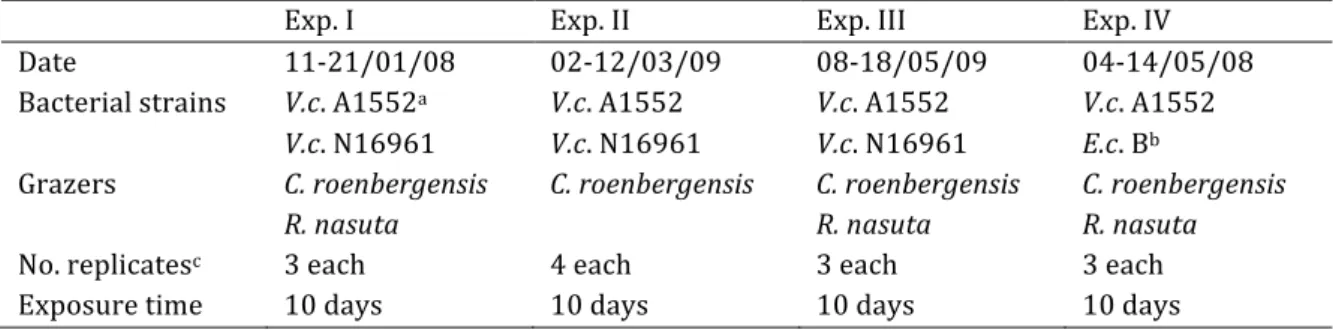
Chapter 4 reports a new method to expose a defined microbial community into the natural environment. One important question was whether the anti-‐protozoan toxicity of V. cholerae against grazers is a laboratory artifact due to the experimental setting or if the toxicity can also be seen in the field. The challenge to expose a community of a defined bacterial strain and grazer was accomplished by modifying diffusion chambers (McFeters and Stuart 1972) to include a biofilm substratum for consequential CLSM and light microscopic analysis of the biofilm community. Strains of toxic and non-‐toxic V. cholerae as well as E. coli strain B were exposed into the marine environment in the presence of the flagellates R. nasuta and Cafeteria roenbergensis. Since the non-‐toxigenic V. cholerae hapR mutant strain is a genetically modified organism it could not be exposed into the natural environment. Thus, V.
cholerae strain N16961 that has a natural frameshift mutation in the hapR gene, was exposed for comparison with the wild type V. cholerae A1552.
To combine the benefits of environmental experiments with the opportunity of daily monitoring of natural biofilm communities, semi-‐natural riverine biofilms were grown in flow cells (Norf et al. 2007) and flagellate-‐bacteria interactions quantified by video microscopy (Boenigk and Arndt 2000). This new approach (chapter 5) allows monitoring of surface-‐associated protozoa on the biofilms and quantification of complex predator-‐prey interactions in microbial biofilm food webs in situ.
Individuals of three typical biofilm associated bacterivorous flagellates were continuously monitored and video recorded. By analyzing the recorded videos regarding different feeding characteristics inter-‐ as well as intraspecific differences and similarities in feeding behavior and food preferences, respectively, were detected in semi-‐natural biofilms for the first time.
Chapter 1 Literature Review
Biofilms: an introduction
Biofilms are everywhere around. These microbial communities are present day and night on almost all surfaces that are in aquatic environments. Bacteria attach to surfaces in the presence of water, e.g. the water air interface, a rock in the water or a medical device such as a medial shunt. Depending on the environment surrounding the biofilm these communities also harbour fungi, protists and small metazoans that rely on these consortia in one way or the other (Costerton et al. 1995, Carrias and Sime-‐Ngando 2009).
The structure of biofilms, a result of multiple complex interactions of different organisms, is very heterogeneous with localised areas containing differing nutrient availabilities, pH and oxygen concentrations (Watnick and Kolter 2000). This gives different organisms with diverse requirements the opportunity to live in a community close to each other and take advantage of resources the neighbouring organisms might supply. Due to the large surface area they cover, biofilms play an important role in the self-‐purification of sediments and water, and the circular flow of nutrients (Hall-‐Stoodley et al. 2004).
Bacteria have three main advantages when living attached to substrate compared to living suspended as single cells: (i) the nutrient availability might be higher for organisms living in mixed species communities. Bacteria that depend on certain metabolites can live in close proximity to bacteria that produce this metabolite and thus gain higher growth rates (e.g. Ylla et al. 2009). (ii) The attachment onto surfaces and enclosure in a matrix protects bacteria from threats such as predation, chemical or biological toxins and UV (e.g. Stewart and Costerton 2001), and (iii) the close proximity of cells enables bacteria to interact on a higher level (e.g. quorum sensing, horizontal gene transfer, Carrias and Sime-‐Ngando 2009).
But, as mentioned above, biofilms are not only beneficial for man but they can also
cause severe damage in industrial and medical settings. Water distribution pipelines
and ship hulls are often covered in biofilms that either hinder the flow or corrode
the material (Beech and Sunner 2004) and removal of these biofouling layers often
lasts only for a short duration. Certain bacteria like Pseudomonas aeruginosa or
Streptococcus sp. can cause contamination of indwelling medical devices and serve as a source of chronic infections (Singh et al. 2000). A feature of biofilms that causes the severe problems is the production of matrix that enables bacteria to resist stresses such as UV light, chemical agents and antibiotics (Stewart and Costerton 2001).
Since biofilms can cause severe damage to humans, research has mainly focused on medically relevant species (e.g. Pseudomonas spp., Staphylococcus spp. or Vibrio cholerae). Most research has been done under well-‐defined laboratory conditions in one or two species experimental set-‐ups in batch cultures, flow-‐cell-‐ or rotating reactor systems. However, studies on biofilms from natural environments such as riverbeds and lakes are scarce. The knowledge gained from these studies has opened the doors to more and more specialized questions: how do bacteria attach, which environmental parameters trigger attachment and detachment, do bacteria communicate and if so, how? What enables bacteria in biofilms to resist antibiotics?
Is communication possible with higher organisms? And can we apply the knowledge gained from simplified laboratory studies to the natural environment?
To summarize the current knowledge on biofilms, (with focus on freshwater biofilms in the natural environment), protozoa and Vibrio cholerae, the following review should give an overview on the aforementioned topics.
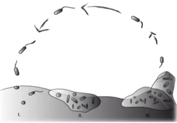
Biofilm life cycle
Bacterial biofilms are well studied in the laboratory and thus knowledge about the
different stages in the development of biofilms is available: in the initiation phase
bacteria attach loosely to the surface followed by a change to irreversible
attachment (Fig. 1.1, I). During the second phase bacteria accumulate through
growth, cell division and recruitment from the bulk phase. Additionally bacteria
start producing extracellular polymeric substances (EPS). This matrix covers the
bacterial cells and is responsible for the high resistance of biofilms (Flemming and
Wingender 2010). First two-‐dimensional growth on surfaces occurs followed by
growth into the three-‐dimensional space (Fig. 1.1, II). Microcolonies, the basic living
structure unit of biofilms (Carrias and Sime-‐Ngando 2009) begin forming. After
some time, detachment of single bacteria cells can be observed (Fig. 1.1, III).
In mature biofilms equilibrium is reached while new bacteria attach to uncolonised areas while biofilm bacteria detach from the biofilm (Stoodley et al. 2002). Grazing or mechanical damage opens space for new bacteria to attach and thus biofilm development is a dynamic, never ending process.
Figure 1.1. Simplified view of the life cycle of a biofilm: (I) loose attachment of bacteria, (II) irreversible attachment and production of EPS, (III) biofilm maturation and dispersal.