© Mary Ann Liebert, Inc.
Selective Transduction of Malignant Glioma by Lentiviral Vectors Pseudotyped with Lymphocytic Choriomeningitis
Virus Glycoproteins
HRVOJE MILETIC,1,* YVONNE HEIDEMARIE FISCHER,2,* HARALD NEUMANN,3VOLKMAR HANS,4 WERNER STENZEL,1TSANAN GIROGLOU,2MANUEL HERMANN,1MARTINA DECKERT,1
and DOROTHEE VON LAER2
ABSTRACT
Malignant gliomas are the most frequent primary brain tumors and have a dismal prognosis due to their in- filtrative growth. Gene therapy using viral vectors represents an attractive alternative to conventional cancer therapies. In a previous study, we established lentiviral vectors pseudotyped with lymphocytic choriomenin- gitis virus (LCMV) glycoproteins (GPs) and demonstrated transduction of human malignant glioma cells in culture. In the current approach, we compared the transduction efficacy of LCMV-GP- and vesicular stom- atitis virus glycoprotein (VSV-G)-pseudotyped lentiviral vectors for malignant glioma cells and normal brain cells
in vitroand in vivo. LCMV-GP pseudotypes transduced almost exclusively astrocytes, whereas VSV-G pseudotypes infected neurons as well as astrocytes. LCMV-GP pseudotypes showed an efficient transduction of solid glioma parts and specific transduction of infiltrating tumor cells. In contrast, VSV-G-pseudotyped lentiviral vectors transduced only a few tumor cells in solid tumor parts and infected mostly normal brain cells in infiltrating tumor areas. In conclusion, lentiviral vectors pseudotyped with LCMV glycoproteins rep- resent an attractive option for gene therapy of malignant glioma.
1091
OVERVIEW SUMMARY
In the present study the level of gene transfer, using lentivi- ral vectors pseudotyped with lymphocytic choriomeningitis virus (LCMV) or vesicular stomatitis virus (VSV) glyco- proteins, into malignant glioma cells and normal brain tis- sue was investigated. VSV-G-pseudotyped vectors trans- duced glial tumor cells with low efficiency in vivo and infected mainly normal brain cells in infiltrating tumor ar- eas. In addition, these vectors exhibited a high tropism for neurons and astrocytes on injection into normal rat brain.
In contrast, LCMV-GP-pseudotyped vectors transduced rat glioma cells with high specificity and efficiency in both solid and infiltrating tumor areas. In normal brain, astrocytes were the predominant cell population transduced by these
vectors in vitroas well as in vivo; infection of neurons was scarce. These data show that LCMV-GP-pseudotyped lentiviral vectors specifically target glial tumor cells in vivo and cause minimal toxicity to normal brain cells in infil- trating tumor areas.
INTRODUCTION
M
ALIGNANT GLIOMAS, comprising the most frequently en- countered primary brain tumors, still have a dismal prog- nosis despite advances in neurosurgery, radiation, and chemo- therapy. Gene therapy based on viral vectors is an attractive alternative therapeutic approach. Vectors derived from the murine leukemia virus (MLV) are the most frequently used1Abteilung für Neuropathologie, Universität zu Köln, D-50931 Cologne, Germany.
2Georg Speyer Haus, D-60596 Frankfurt am Main, Germany.
3Neuroimmunology Unit, European Neuroscience Institute, D-37073 Göttingen, Germany.
4Institut für Neuropathologie, Krankenanstalten Gilead, D-33617 Bielefeld, Germany.
*H.M. and Y.H.F. contributed equally to this work.
retroviruses for gene therapy of brain tumors (Benedetti et al., 1999; Lam and Breakefield, 2001; Tamura et al., 2001; Wang et al., 2003). However, the instability of the glycoprotein impedes clinical application as the vectors cannot be concentrated by ul- tracentrifugation. This is required to achieve vector titers that are sufficient for in vivoapplication. Exchange of the retroviral en- velope protein for the rhabdoviral G protein of the vesicular stom- atitis virus (VSV) improved vector stability and broadened the host range (Emi et al., 1991; Burns et al., 1993; Yee et al., 1994).
Use of these vectors in a rat glioma model has shown high trans- duction and therapeutic efficiency after transfer of the herpes sim- plex virus thymidine kinase gene (HSV-tk) and ganciclovir treat- ment (Galipeau et al., 1999). However, a major disadvantage of the VSV glycoprotein (VSV-G) is its cytotoxicity, thus prevent- ing establishment of stable recombinant packaging cell lines (Burns et al., 1993; Chen et al., 1996). In previous studies, we developed oncoretroviral vectors pseudotyped with glycoproteins (GPs) from the lymphocytic choriomeningitis virus (LCMV) (Miletic et al., 1999; Beyer et al., 2001). These vectors have a broad host range and can be concentrated by ultracentrifugation.
In addition, LCMV-GP is not cytotoxic, and stable recombinant packaging cell lines can be established.
Despite these improvements, the restricted infection of di- viding cells by retroviral vectors still reduces transduction ef- ficacy, as in human glioma most of the tumor cells are not di- viding in a given treatment window. Therefore, a vector system is required for transduction of both dividing as well as nondi- viding tumor cells. In this regard, lentiviral vectors are attrac- tive candidates. These vectors also transduce nondividing cells and can be pseudotyped by VSV-G, thereby increasing both vector stability and host range (Naldini et al., 1996b). In in- fection studies with lentiviral VSV-G pseudotypes in the nor- mal rat brain, neurons were transduced at high levels (Naldini et al., 1996a). Consequently, these vectors may cause serious side effects in gene therapy of malignant glioma.
In one study, we established lentiviral vectors pseudotyped with LCMV-GP and demonstrated transduction of human ma- lignant glioma cells in culture (Beyer et al., 2002). In the cur- rent approach, we determine the tropism of LCMV-GP pseudo- types for rat malignant glioma cells in vitroand in vivo. The transduction efficacy of solid and infiltrating glioma by LCMV- GP- and VSV-G-pseudotyped vectors is compared. Further- more, transduction of normal brain cells in vivoand in vitroby both pseudotypes is analyzed. LCMV-GP pseudotypes show specific and efficient transduction of rat gliomas and infect mostly astrocytes on injection into normal rat brain. In contrast, VSV-G pseudotypes transduce glioma cells with much lower efficiency and infect predominantly normal brain cells in infil- trating tumor areas. In normal rat brain neurons are the major cell population transduced by VSV-G pseudotypes.
MATERIALS AND METHODS Cell lines
9L rat gliosarcoma, 293T human kidney, and TE671 human fibroblast cell lines were obtained from the American Type Cul- ture Collection (Manassas, VA). G62 human glioma cells were kindly provided by M. Westphal (University Hospital Eppen-
dorf, Hamburg, Germany). Cells were grown in Dulbecco’s modified Eagle’s medium (DMEM) and supplemented with 10% fetal bovine serum and penicillin–streptomycin in a hu- midified atmosphere of 5% CO2.
Transduction of 9L cells with DsRed
DsRed in the pMP71 vector backbone was kindly provided by N. Dinauer (Georg-Speyer-Haus, Frankfurt, Germany). For transduction of 9L cells with DsRed, 9L cells were seeded in 24-well plates at a density of 5104cells per well. After 4 hr, retroviral supernatants packaging the pMP71DsRed vector were added. Plates were centrifuged for 1 hr at 1000g. Trans- duction of cells was repeated 16 hr after the first transduction.
DsRed expression was confirmed by fluorescence microscopy (Nikon Eclipse TE300; Nikon, Düsseldorf, Germany). For iso- lation of DsRed-expressing single clones, 102 transduced 9L cells were plated in 10-cm dishes and grown into colonies.
DsRed-positive colonies were identified by fluorescence mi- croscopy and transferred into separate wells of 24-well plates.
DsRed expression levels of isolated clones were determined by flow cytometry on a FACSCalibur (BD Biosciences Immuno- cytometry Systems, San Jose, CA). For in vivotumor implan- tation a clone was selected containing 95% DsRed-positive cells.
Transient production of lentivirus vector pseudotypes
Lentiviral vectors were produced by transient transfection of 293T cells. Volumes of 5106cells were seeded in 10-cm-di- ameter culture dishes 16 hr before transfection in DMEM–FBS.One hour before transfection, the culture medium was changed to DMEM–FBS–PS (10 ml/dish), that is, DMEM–FBS with 25 M chloroquine, penicillin (50 U/ml), and streptomycin (50 g/ml) (GIBCO/Invitrogen Life Technologies, Grand Island, NY). Five micrograms of pRRL.sinCMVeGFPpre, 5 g of pRSV-Rev, 15 g of pMDLg/pRRE (described in Dull et al., 1998), and 1 to 2 g of a pHCMV envelope glycoprotein ex- pression plasmid were then used for the transfection of one cul- ture dish. A combination of 450 l of the plasmids in distilled deionized H2O and 50 l of 2.5 MCaCl2was mixed well and then added dropwise to 500 l of 2HEPES-buffered saline (280 mMNaCl, 100 mMHEPES, 1.5 mMNa2HPO4, pH 7.1).
After vortexing, the precipitate was immediately added to the cul- tures. The medium was changed after 8 hr to 10 ml of DMEM–FBS–PS per dish with 20 mMHEPES. Vector-contain- ing supernatants were collected 24 hr after transfection and every 8 to 16 hr thereafter for a period of 2 days. Cell culture super- natants were pooled and filtered through a 0.22-m-pore size Millex-GP filter (Millipore, Bedford, MA). For in vivoand in vitro(cultured brain cells) applications, vector supernatants were concentrated by ultracentrifugation at 19,500 rpm for 2 hr in an SW28 rotor (Coulter Beckman, Fullerton, CA). For in vitroex- periments on TE671 and various glioma cell lines, vector super- natants were not concentrated.
Vector titration
Lentiviral vector titers were measured by transduction of var- ious cell lines. Serial dilutions of cell supernatants were pre- pared, and 0.5 ml of each dilution was added to 5104cells
and seeded in a well of a 24-well plate 4 hr before transduc- tion. Plates were centrifuged for 1 hr at 1000g. Cells were analyzed 65 hr posttransduction by flow cytometry on a FACS- Calibur (BD Biosciences Immunocytometry Systems) for GFP expression. Titers were calculated from dilutions, which re- sulted in 0.5 to 20% enhanced green fluorescent protein (eGFP)- positive cells, a range of linear relation between vector input and percentage of transduced cells, because multiple vector in- tegrations into the target cell are generally not expected.
Rat hippocampal neuron culture
Primary hippocampal neuronal cell cultures were prepared as described previously (Neumann et al., 1995). Briefly, hip- pocampi were isolated from whole brains of embryonic day 16 Wistar rats, and the meninges were removed. The trimmed tis- sue was dissociated by trituration through a fire-polished Pas- teur pipette. Cells (5104/ml) were plated into four-well chamber slides that had been pretreated with poly-L-ornithine (0.5 mg/ml; Sigma, St. Louis, MO) in 0.15 Mboric acid. Cells were cultured in chemically defined medium containing basal medium Eagle (BME; GIBCO-BRL/Invitrogen Life Technolo- gies, Gaithersburg, MD) with 2% (v/v) B27 supplement (Invit- rogen Life Technologies) and 1% (v/v) glucose (45%; Sigma).
Rat astrocyte-enriched glial cell culture
Hippocampi of embryonic day 16 Wistar rats were isolated and dissociated into single-cell suspensions as described for neuronal hippocampal preparations. Cells were plated in 50-ml tissue culture flasks that had been pretreated with poly-L-lysine (5 g/ml; Sigma). Cells were cultured in serum-containing medium with minimal essential medium (MEM) with D-valine (Invitrogen Life Technologies, Carlsbad, CA), 10% heat-inac- tivated fetal calf serum (FCS; PanSystem, Würzburg, Ger- many), and 1% L-glutamine. Astrocyte-enriched glial cells were cultured for 10–20 days and then plated in BME with B27 sup- plement (2%, v/v; Invitrogen Life Technologies) and 1% (v/v) glucose (45%; Sigma) at a density of 2104/ml in 4-well chamber slides before transduction. In total, 94% (3% SD) of cells were astrocytes as determined by immunolabeling with rabbit antibodies directed against glial fibrillary acidic protein (GFAP, 10 g/ml; DakoCytomation, Glostrup, Denmark).
Transduction of cultured brain cells
Fourteen days after plating, neurons and astrocytes were transduced with lentivirus-pseudotyped vectors carrying the
eGFP marker gene. Two days after transduction, cells were fixed in 4% paraformaldehyde and immunostained with mono- clonal mouse anti--tubulin III antibody (Sigma) for neurons and monoclonal rabbit anti-GFAP antibody (DakoCytomation) for astrocytes. The cells were incubated overnight at 4°C with the primary antibody. Cy3–goat anti-mouse and Cy3–goat anti- rabbit were used as secondary antibodies for 2 hr at room tem- perature. The relative numbers of transduced (eGFP positive) and untransduced (eGFP negative) neuronal (-tubulin III pos- itive) and astrocytic (GFAP positive) cells were determined by fluorescence microscopy and the counting of 10 camera fields for each pseudotype.
Tumor implantation and lentiviral vector delivery
Adult female Fischer 344 rats (Harlan Winkelmann, Borchen, Germany) were anesthetized with an intraperitoneal injection of ketamine (50 mg/kg) and xylazine (2 mg/kg). In- tracranial 9LDsRed tumors were established by injection of 1 1059LDsRed cells (in 5 l of phosphate-buffered saline [PBS]) into the right striatum, using a Hamilton syringe in a stereo- taxic apparatus (Stoelting, Wood Dale, IL). The coordinates used were 4 mm lateral to the bregma and 5 mm below the dural surface. Six days after tumor implantation, rats were anes- thetized, and lentiviral vector pseudotypes with titers ranging from 2106to 1107 transducing units (TU)/ml were in- jected, using the same stereotactic coordinates and 1 mm apart (seven different sites). A total volume of 10 l was injected into each tumor. Fischer rats not receiving tumor cells were anesthetized and lentiviral pseudotypes were injected either into the right striatum or into the right hippocampus. The coordi- nates used for the hippocampus region were 4.5 mm lateral to the bregma, 5.5 mm posterior to the coronal plate, and 3 mm below the dural surface.Analysis of rat brains for lentiviral transduction
Seven (tumor-bearing rats) and 14 days (rats without tumor) after lentiviral vector delivery, animals were killed and perfused with 4% paraformaldehyde. Brains were removed, suspended in 30% sucrose for 3 days, and then snap frozen in isopentane chilled with liquid nitrogen. Coronal sections (12 m) were pre- pared on a cryostat and immunostained with either rabbit anti- GFAP antibodies (DakoCytomation, Hamburg, Germany) for astrocytes or mouse anti-NeuN antibodies (Chemicon, Hofheim, Germany) for neurons. Primary antibodies were incubated overnight at 4°C. Cy3–goat anti-mouse and Cy3–goat anti-rab-TABLE1. VECTORTITERS OFPSEUDOTYPEDLENTIVIRALVECTORS ONTE671 ANDGLIOMACELLLINESa
LCMV-GP VSV-G
Cell line (no. of Pseudotype titer Titer relative to Pseudotype titer Titer relative to
experiments) (TU/ml) TE671 (TU/ml) TE671
TE671 (4) (2.751.07)104 1 (6.086.00)104 1
9L (4) (1.800.78)104 0.65 (0.03) (8.835.81)104 1.91 (0.63)
9LDsRed (2) (1.291.00)104 0.35 (0.21) (8.355.97)104 1.89 (0.07)
aCell lines were transduced with LCMV-GP- or VSV-G-pseudotyped lentiviral vectors packaging eGFP. Titers were measured by FACS analysis. Results represent the means and standard deviations of at least two experiments.
bit (Dianova, Hamburg, Germany) were applied as secondary antibodies for 2 hr at room temperature. The sections were ex- amined under a fluorescence microscope (Zeiss, Jena, Ger- many) and analyzed by confocal scanning laser microscopy (Leica, Bensheim, Germany). Transduction efficiencies in in- fected tumor areas were determined by counting, by three in- dependent investigators in a double-blinded fashion, transduced tumor areas in 10 sections per group.
RESULTS
LCMV-GP- and VSV-G-pseudotyped lentiviral vectors transduce 9L tumor cells in vitro
We compared the transduction efficiencies of both VSV-G- and LCMV-GP-pseudotyped lentiviral vectors in the rat glioma cell lines 9L and 9LDsRed (used for tumor implantation) in vitro. The human epithelial cell line TE671, which could be transduced by both VSV-G- and LCMV-pseudotyped vectors as shown in a previous study (Beyer et al., 2002), was used as infection control. We performed end-point dilutions on 9L, 9L- DsRed, and TE671 cells, and measured the percentage of trans- duced cells by flow cytometry analysis. Both vectors transduced 9L cells, but VSV-G pseudotypes did so with higher efficiency (Table 1). The relative transduction compared with TE671 was 0.65 for LCMV pseudotypes and 1.91 for VSV-G pseudotypes.
In addition, transduction efficiencies for 9L and 9LDsRed tu- mor cells did not differ significantly in vitro(Table 1).
VSV-G pseudotypes transduce cultured neurons and astrocytes more efficiently than do LCMV-GP pseudotypes
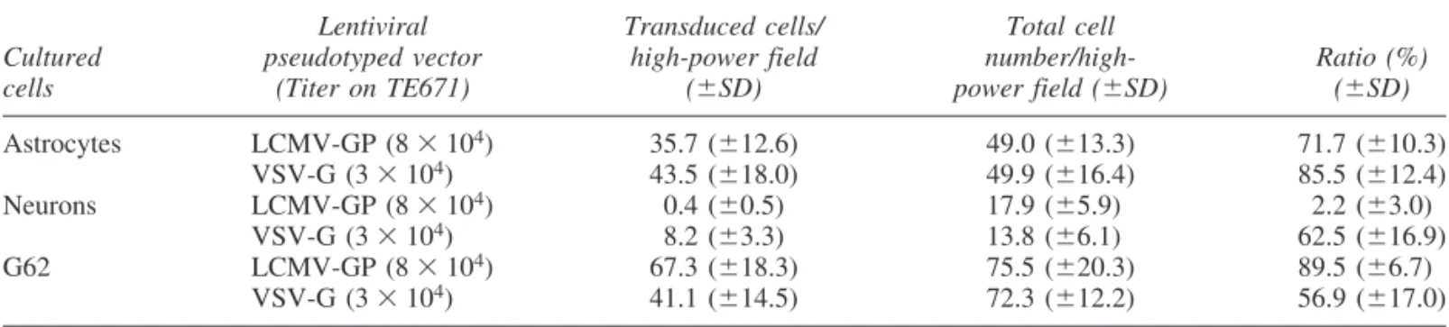
To analyze the tropism of both pseudotyped vectors for nor- mal brain cells in vitro, we infected cultured neurons and astro- cytes derived from embryonic day 16 Wistar rats by end-point di- lutions. The human glioma cell line G62, which could be transduced by both pseudotypes in a previous study, was used as infection control. VSV-G pseudotypes transduced GFAP-positive astrocytes and -tubulin III-positive neurons at a higher level than did LCMV-GP pseudotypes (Table 2). In particular, the trans- duction efficiency for neurons was markedly different: VSV-G pseudotypes transduced 62.5%, whereas LCMV-GP pseudotypes infected only 2.2%, of counted neurons. In addition, LCMV-GP pseudotypes showed a higher transduction efficiency for the hu- man glioma cell line G62 (89.5%) than for cultured astrocytes (71.7%) and neurons (2.2%). In contrast, VSV-G pseudotypes transduced astrocytes (85.5%) and neurons (62.5%) at a higher level than G62 cells (56.9%).
VSV-G and LCMV-GP pseudotypes show a different tropism for normal brain cells
in vivoWe confirmed the in vitroresults by analysis of the in vivo tropism for normal brain cells. For this purpose, we injected LCMV-GP and VSV-G pseudotypes either into the striatum or the hippocampus of Fischer rats. The relative proportion of transduced cell types was analyzed by immunofluorescence staining with cell type-specific markers and confocal mi- TABLE2. TROPISM OFLENTIVIRALPSEUDOTYPEDVECTORS FORCULTUREDBRAINCELLSa
Lentiviral Transduced cells/ Total cell
Cultured pseudotyped vector high-power field number/high- Ratio (%)
cells (Titer on TE671) (SD) power field (SD) (SD)
Astrocytes LCMV-GP (8104) 35.7 (12.6) 49.0 (13.3) 71.7 (10.3)
VSV-G (3104) 43.5 (18.0) 49.9 (16.4) 85.5 (12.4)
Neurons LCMV-GP (8104) 0.4 (0.5) 17.9 (5.9)0 2.2 (3.0)
VSV-G (3104) 8.2 (3.3) 13.8 (6.1)0 62.5 (16.9)
G62 LCMV-GP (8104) 67.3 (18.3) 75.5 (20.3) 89.5 (6.7)0
VSV-G (3104) 41.1 (14.5) 72.3 (12.2) 56.9 (17.0)
aCultured rat astrocytes or neurons were transduced with 3104to 8104eGFP TU of LCMV-GP- or VSV-G-pseudotyped lentiviral vectors. Cells were analyzed by fluorescence microscopy after immunostaining with monoclonal anti-GFAP antibod- ies for astrocytes or with anti--tubulin III antibodies for neurons. Results represent the mean cell numbers and standard devia- tions from 10 randomly selected camera fields (0.75 mm2).
FIG. 1. Neurons and astrocytes were transduced by VSV-G-pseudotyped lentiviral vectors in vivo. Normal rat brain was in- fected with LCMV-GP- or VSV-G-pseudotyped vectors expressing eGFP. Transduction of neurons and astrocytes was analyzed, after staining with antibodies against NeuN and GFAP, by confocal laser scanning microscopy on day 14. (A) Transduction of striatal astrocytes by LCMV-GP-pseudotyped vectors. (B) Striatal astrocytes expressing GFAP. (C) Merge of (A) and (B). (D) In regions with high neuron density (hippocampus) neurons were not transduced, but single astrocytes were. (E) Hippocampal neurons expressing NeuN. (F) Merge of (D) and (E). (G) Transduction of striatal neurons by VSV-G-pseudotyped vectors. (H) Striatal neurons expressing NeuN. (I) Merge of (G) and (H). (K) Tranduction of hippocampal neurons by VSV-G-pseudotyped vectors. (L) Hippocampal neurons expressing NeuN. (M) Merge of (K) and (L). (N) Hippocampal astrocytes transduced by VSV- G-pseudotyped vectors. (O) Hippocampal astrocytes expressing GFAP. (P) Merge of (N) and (O). Original magnification, 40.
croscopy. LCMV-GP pseudotypes transduced almost exclu- sively astrocytes in both brain regions, as shown by staining with antibodies against GFAP (Fig. 1A–C). This observation could be confirmed even in areas with high neuron density (Fig.
1D–F). Transduction of neurons was scarce in the striatum and the hippocampus. In contrast, VSV-G pseudotypes infected neurons with high efficiency. The estimated ratio of cells trans- duced by VSV-G pseudotypes was 3:1 (neurons:astrocytes) in both brain regions (Fig. 1G–I, K–M, and N–P).
LCMV-GP pseudotypes show specific and efficient transduction of glial tumor cells
in vivoWe wanted to exclude the possibility that the established cell line 9LDsRed differed from its parental cell line 9L in vivo.
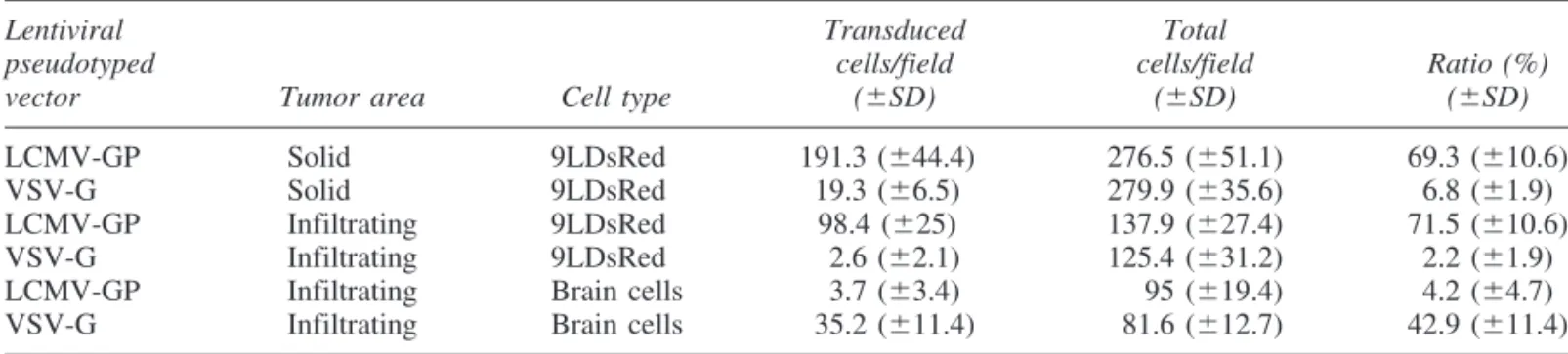
Two weeks after intracerebral tumor cell implantation in Fis- cher rats, established tumors were screened by histology for size and by light microscopy (9L) or fluorescence microscopy (9LDsRed) for their ability to infiltrate into the brain paren- chyma. 9L and 9LDsRed tumors showed no difference in size and contained infiltrating tumor cells. With the DsRed marker even single tumor cells could be detected, migrating into the brain parenchyma. To analyze the transduction of 9LDsRed tu- mors in vivoby both pseudotypes, two groups of female Fis- cher rats, bearing 9LDsRed tumors, were injected with LCMV- GP and VSV-G pseudotypes. To measure the transduction of solid tumor and infiltrating glioma cells, injections were placed in the center of the tumor and 1 mm apart. Transduction effi- ciencies in infected tumor areas were determined quantitatively.
LCMV-GP pseudotypes showed an effective transduction of solid tumor: 69.310.6% of tumor cells were GFP positive in tumor areas injected with vector supernatants (Table 3 and
Fig. 2A–C). In the infiltrating areas of 9LDsRed tumors, LCMV- GP pseudotypes specifically transduced the glioma cells (Fig.
2D–F): whereas 71.510.6% of tumor cells were transduced, only 4.24.7% of normal brain cells were GFP positive (Table 3). Even single infiltrating tumor cells were infected by this vector pseudotype (Fig. 2G–I). Only a few reactive astrocytes around the tumor bed were GFP positive, as revealed by stain- ing with antibodies against GFAP (Fig. 3A–C). Neurons were not infected in infiltrating tumor areas. VSV-G pseudotypes showed a different transduction pattern: solid tumor was trans- duced to a much lower extent (6.81.9%) than with the LCMV-GP pseudotypes (Table 3 and Fig. 2K–M). In contrast, many normal brain cells including neurons and reactive astro- cytes in the infiltration area and surrounding brain parenchyma were GFP positive (42.911.4%), whereas only rarely were tumor cells (2.21.9%) transduced (Table 3 and Fig. 2N–P).
DISCUSSION
In human glioblastoma, recurrent tumors often develop from infiltrating tumor cells, which cannot be eliminated by current therapies. These cells represent a major therapeutic target. In this study, LCMV-GP pseudotypes showed a specific and effi- cient transduction of solid as well as infiltrating rat glioma, whereas VSV-G pseudotypes preferentially transduced normal brain cells. Retroviral vectors based on MLV have shown promising results for gene therapy of brain tumors (Tamura et al., 2001; Wang et al., 2003). However, the low or absent ef- fect in clinical trials (Rainov, 2000) could be due to the re- stricted transduction of dividing tumor cells. In contrast to commonly used retroviral vectors, lentiviral vectors introduce TABLE3. TRANSDUCTIONEFFICIENCY OFLENTIVIRALPSEUDOTYPEDVECTORS FOR9LDsRed CELLSin Vivoa
Lentiviral Transduced Total
pseudotyped cells/field cells/field Ratio (%)
vector Tumor area Cell type (SD) (SD) (SD)
LCMV-GP Solid 9LDsRed 191.3 (44.4)0 276.5 (51.1) 69.3 (10.6)
VSV-G Solid 9LDsRed 19.3 (6.5)0 279.9 (35.6) 6.8 (1.9)
LCMV-GP Infiltrating 9LDsRed 98.4 (25)0 137.9 (27.4) 71.5 (10.6)
VSV-G Infiltrating 9LDsRed 2.6 (2.1) 125.4 (31.2) 2.2 (1.9)
LCMV-GP Infiltrating Brain cells 3.7 (3.4) 00.95 (19.4) 4.2 (4.7)
VSV-G Infiltrating Brain cells 35.2 (11.4) 081.6 (12.7) 42.9 (11.4)
a9LDsRed tumors were transduced with 2106to 1107eGFP TU of LCMV-GP- or VSV-G-pseudotyped lentiviral vec- tor per milliliter. Cells were counted with a fluorescence microscope. Data represent the mean cell numbers and standard devia- tions from 10 microscopic fields (magnification, 200) of solid and infiltrating tumor areas, respectively.
FIG. 2. Solid and infiltrating areas of rat glioma were efficiently transduced by LCMV-GP-pseudotyped lentiviral vectors. In- tracranial 9LDsRed gliomas were infected with LCMV-GP- or VSV-G-pseudotyped lentiviral vector expressing eGFP 7 days af- ter tumor implantation and analyzed by confocal laser scanning microscopy on day 14. (A) Solid tumor (69.310.6%) trans- duced by LCMV-GP-pseudotyped vectors. (B) Solid tumor expressing DsRed. (C) Merge of (A) and (B). (D) Infiltrating glioma parts transduced by LCMV-GP-pseudotyped vectors. (E) Infiltrating glioma parts expressing DsRed. (F) Merge of (D) and (E).
(G) Single infiltrating tumor cells transduced by LCMV-GP-pseudotyped vectors. (H) Single infiltrating tumor cells expressing DsRed. (I) Merge of (G) and (H). (K) Solid tumor (6.81.9%) transduced by VSV-G-pseudotyped vectors. (L) Solid tumor expressing DsRed. (M) Merge of (K) and (L). (N) Infiltrating glioma parts transduced by VSV-G-pseudotyped vectors. (O) In- filtrating glioma parts expressing DsRed. (P) Merge of (N) and (O). Original magnification: (A–F) 20; (G–I) 40; (K–P) 20.
their genome not only into dividing, but also into quiescent, cells. This can be a major advantage with respect to gene ther- apy for tumors in general, as within a short treatment window most tumor cells do not divide. Consequently, we decided to use lentiviral pseudotypes as a new approach.
A major problem regarding lentiviral vectors, however, might be the transduction of nontumoral, quiescent cells. The cells of normal brain, in particular neurons, are in a postmitotic state and can be infected by lentiviral vectors. When lentiviral vectors are used, the specificity for tumor cells should be de- termined by the virus glycoprotein, which is one of the major determinants of virus tropism. Lentiviral vectors are generally pseudotyped with VSV-G, which mediates effective transduc- tion of neurons in vivo(Naldini et al., 1996a). However, the transduction of malignant glioma with these pseudotypes has not been examined yet. In a previous study, we showed that lentiviral VSV-G pseudotypes transduce human glioma cells in culture (Beyer et al., 2002). Also, the 9L rat glioma cell line studied here was highly susceptible to VSV-G pseudotypes. We therefore expected efficient transduction of tumor cells as well as normal brain cells with the VSV-G pseudotypes in the 9L rat glioma model. Surprisingly, the infection of normal brain cells at the tumor border was more efficient than transduction of tumor cells. Also, within solid tumor, transduction of tumor cells was not as efficient as with LCMV pseudotypes. In vitro and in vivowe could demonstrate a strong transduction of neu- rons and astrocytes with VSV-G pseudotypes. Although the transduction of malignant glioma by lentiviral and retroviral VSV-G pseudotypes has not been compared directly, two stud- ies using retroviral VSV-G pseudotypes have shown higher transduction efficiency in experimental brain tumors (Galipeau et al., 1999; Lee et al., 2001) as compared with our study with lentiviral VSV-G pseudotypes. The discrepancy may be ex- plained by different virus strains pseudotyped with VSV-G: as retroviral vectors infect dividing cells, their target in the brain of tumor-bearing animals consists only of tumor cells. Lentivi- ral vectors infect both nondividing as well as dividing cells and, therefore, they can also infect normal brain cells. Moreover, our
data and Naldini et al. (1996b) demonstrated that normal brain cells, in particular neurons, present the major target for VSV- G lentiviral pseudotypes. Altogether, these data show that VSV- G-pseudotyped lentiviral vectors are not suitable vectors for gene therapy of brain tumors.
In contrast, LCMV-GP pseudotypes showed a completely different transduction pattern. The vector infected solid and in- filtrating tumor with high efficiency and specificity. The level of transduction of infiltrating tumor cells was always much higher than that of reactive astrocytes. In vivotropism of lentivi- ral LCMV-GP pseudotypes for normal brain cells has been stud- ied, but not in great detail (Watson et al., 2002; Wong et al., 2004). Both groups showed that VSV-G pseudotypes trans- duced normal brain cells more efficiently than did LCMV-GP pseudotypes. However, the transduced cell populations were not determined precisely. In our experiments, astrocytes were the predominant transduced cell population in the brain of healthy Fischer rats. The same results were achieved with cultured brain cells.
-Dystroglycan was found to be a cellular receptor for LCMV (Cao et al., 1998). In the CNS, -dystroglycan is ex- pressed on neurons and astrocytes (Moukhles and Carbonetto, 2001; Zaccaria et al., 2001). This is in accordance with studies showing that LCMV infects primarily glial cells, from where it spreads to neurons during persistent infection (Joly et al., 1991;
Oldstone and Rall, 1993). In particular, Bonthius et al. (2002) have demonstrated that glial cells are the first cells of the brain parenchyma to be infected with LCMV. These findings are also in accordance with the susceptibility of astrocytes to LCMV- GP pseudotype transduction found in our study. The scarce transduction of neurons, however, is somewhat surprising. A problem may be that the vectors used are replication defective, while spread of virus via direct cell contact between glial and neuronal cells is required for infection of neurons with LCMV.
In addition, a yet unidentified second receptor for LCMV may be required for infection, which is not expressed at sufficient levels on neurons. This hypothesis is supported by two studies showing that some LCMV variants inefficiently interact with FIG. 3. Only a few reactive astrocytes in infiltrating tumor areas were transduced by LCMV-GP pseudotypes. Intracranial 9LDsRed gliomas were infected with LCMV-GP- or VSV-G-pseudotyped lentiviral vector expressing eGFP 7 days after tu- mor implantation. Transduction of neurons and astrocytes in infiltrating tumor areas was analyzed, after staining with anti- bodies against GFAP and NeuN, by confocal laser scanning microscopy on day 14. (A) Tumor cells and single astrocytes (ar- rowheads) transduced by LCMV-GP pseudotypes. (B) Reactive astrocytes expressing GFAP. (C) Merge of (A) and (B). Original magnification, 40.
-dystroglycan and also infect -dystroglycan-negative cells (Sevilla et al., 2000; Smelt et al., 2001).
In previous studies (Miletic et al., 1999; Beyer et al., 2001, 2002), we characterized the major features of both lentiviral and oncoretroviral LCMV-GP pseudotypes: the glycoprotein is stable, allowing concentration by ultracentrifugation for high vector titers. The vector can be frozen and thawed without sig- nificant loss of virus titer. Moreover, stable packaging cell lines producing LCMV-GP-pseudotyped retroviral vectors have been established. This is an important advantage over VSV-G- pseudotyped vectors, because cytotoxicity of the VSV glyco- protein impedes generation of packaging cell lines constitu- tively expressing VSV-G. Stable packaging cell lines, however, are an important tool in gene therapy of solid tumors, as they can be implanted into the tumor bed, which allows more effi- cient delivery of the vector than does direct injection of vector preparations.
Several LCMV strains have been identified that carry gly- coprotein variants with altered tissue tropism (Southern et al., 1984; Fazakerley et al., 1991). Matloubian et al. have shown that single amino acid changes in the LCMV glycoprotein can alter LCMV tropism (Matloubian et al., 1993). By pseudotyp- ing viral vectors with LCMV glycoproteins from these strains, or even by inserting specific mutations into LCMV glycopro- teins, the spectrum of gene therapeutic targets could be broad- ened. In conclusion, although further studies with therapeutic genes are warranted, lentiviral vectors pseudotyped with LCMV glycoproteins represent an attractive option for gene therapy of malignant glioma.
ACKNOWLEDGMENTS
The authors thank M. Carstov and A. Bohl for expert tech- nical assistance and H.U. Klatt for photographic help. This work was supported by the Köln Fortune Program (grant 31/2002) and the Hermann J. Abs Program of the Stiftungsfonds Deutsche Bank.
REFERENCES
BENEDETTI, S., BRUZZONE, M.G., POLLO, B., DIMECO, F., MA- GRASSI, L., PIROLA, B., CIRENEI, N., COLOMBO, M.P., and FINOCCHIARO, G. (1999). Eradication of rat malignant gliomas by retroviral-mediated, in vivodelivery of the interleukin 4 gene. Can- cer Res. 59,645–652.
BEYER, W.R., MILETIC, H., OSTERTAG, W., and VON LAER, D.
(2001). Recombinant expression of lymphocytic choriomeningitis virus strain WE glycoproteins: A single amino acid makes the dif- ference. J. Virol. 75,1061–1064.
BEYER, W.R., WESTPHAL, M., OSTERTAG, W., and VON LAER, D. (2002). Oncoretrovirus and lentivirus vectors pseudotyped with lymphocytic choriomeningitis virus glycoprotein: Generation, con- centration, and broad host range. J. Virol. 76,1488–1495.
BONTHIUS, D.J., MAHONEY, J., BUCHMEIER, M.J., and TAG- GARD, D.A. (2002). A critical role for glial cells in the infection, propagation and spread of lymphocytic choriomeningitis virus in the developing rat brain. J. Virol. 76,6618–6635.
BURNS, J.C., FRIEDMANN, T., DRIEVER, W., BURRASCANO, M., and YEE, J.K. (1993). Vesicular stomatitis virus G glycoprotein pseudotyped retroviral vectors: Concentration to very high titer and
efficient gene transfer into mammalian and nonmammalian cells.
Proc. Natl. Acad. Sci. U.S.A. 90,8033–8037.
CAO, W., HENRY, M.D., BORROW, P., YAMADA, H., ELDER, J.H., RAVKOV, E.V., NICHOL, S.T., COMPANS, R.W., CAMP- BELL, K.P., and OLDSTONE, M.B. (1998). Identification of -dys- troglycan as a receptor for lymphocytic choriomeningitis virus and Lassa fever virus. Science 282,2079–2081.
CHEN, S.T., IIDA, A., GUO, L., FRIEDMANN, T., and YEE, J.K.
(1996). Generation of packaging cell lines for pseudotyped retrovi- ral vectors of the G protein of vesicular stomatitis virus by using a modified tetracycline inducible system. Proc. Natl. Acad. Sci. U.S.A.
93,10057–10062.
DULL, T., ZUFFEREY, R., KELLY, M., MANDEL, R.J., NGUYEN, M., TRONO, D., and NALDINI, L. (1998). A third-generation lentivirus vector with a conditional packaging system. J. Virol. 72, 8463–8471.
EMI, N., FRIEDMANN, T., and YEE, J.K. (1991). Pseudotype for- mation of murine leukemia virus with the G protein of vesicular stom- atitis virus. J. Virol. 65,1202–1207.
FAZAKERLEY, J.K., SOUTHERN, P., BLOOM, F., and BUCH- MEIER, M.J. (1991). High resolution in situhybridization to deter- mine the cellular distribution of lymphocytic choriomeningitis virus RNA in the tissues of persistently infected mice: Relevance to are- navirus disease and mechanisms of viral persistence. J. Gen. Virol.
72,1611–1625.
GALIPEAU, J., LI, H., PAQUIN, A., SICILIA, F., KARPATI, G., and NALBANTOGLU, J. (1999). Vesicular stomatitis virus G pseudo- typed retrovector mediates effective in vivosuicide gene delivery in experimental brain cancer. Cancer Res. 59,2384–2394.
JOLY, E., MUCKE, L., and OLDSTONE, M.B. (1991). Viral persis- tence in neurons explained by lack of major histocompatibility class I expression. Science 253,1283–1285.
LAM, P.Y., and BREAKEFIELD, X.O. (2001). Potential of gene ther- apy for brain tumors. Hum. Mol. Genet. 10,777–787.
LEE, H., SONG, J.J., KIM, E., YUN, C.O., CHOI, J., LEE, B., KIM, J., CHANG, J.W., and KIM, J.H. (2001). Efficient gene transfer of VSV-G pseudotyped retroviral vector to human brain tumor. Gene Ther. 8,268–273.
MATLOUBIAN, M., KOLHEKAR, S.R., SOMASUNDARAM, T., and AHMED, R. (1993). Molecular determinants of macrophage tro- pism and viral persistence: Importance of single amino acid changes in the polymerase and glycoprotein of lymphocytic choriomeningi- tis virus. J. Virol. 67,7340–7349.
MILETIC, H., BRUNS, M., TSIAKAS, K., VOGT, B., REZAI, R., BAUM, C., KUHLKE, K., COSSET, F.L., OSTERTAG, W., LOTHER, H., and VON LAER, D. (1999). Retroviral vectors pseudotyped with lymphocytic choriomeningitis virus. J. Virol. 73, 6114–6116.
MOUKHLES, H., and CARBONETTO, S. (2001). Dystroglycan con- tributes to the formation of multiple dystrophin-like complexes in brain. J. Neurochem. 78,824–834.
NALDINI, L., BLOMER, U., GAGE, F.H., TRONO, D., and VERMA, I.M. (1996a). Efficient transfer, integration, and sustained long-term expression of the transgene in adult rat brains injected with a lentivi- ral vector. Proc. Natl. Acad. Sci. U.S.A. 93,11382–11388.
NALDINI, L., BLOMER, U., GALLAY, P., ORY, D., MULLIGAN, R., GAGE, F.H., VERMA, I.M., and TRONO, D. (1996b). In vivo gene delivery and stable transduction of nondividing cells by a lentiviral vector. Science 272,263–267.
NEUMANN, H., CAVALIE, A., JENNE, D.E., and WEKERLE, H.
(1995). Induction of MHC class I genes in neurons. Science 269, 549–552.
OLDSTONE, M.B., and RALL, G.F. (1993). Mechanism and conse- quence of viral persistence in cells of the immune system and neu- rons. Intervirology 35,116–121.
RAINOV, N.G. (2000). A phase III clinical evaluation of herpes sim-
plex virus type 1 thymidine kinase and ganciclovir gene therapy as an adjuvant to surgical resection and radiation in adults with previ- ously untreated glioblastoma multiforme. Hum. Gene Ther. 11, 2389–2401.
SEVILLA, N., KUNZ, S., HOLZ, A., LEWICKI, H., HOMANN, D., YAMADA, H., CAMPBELL, K.P., DE LA TORRE, J.C., and OLD- STONE, M.B. (2000). Immunosuppression and resultant viral per- sistence by specific viral targeting of dendritic cells. J. Exp. Med.
192,1249–1260.
SMELT, S.C., BORROW, P., KUNZ, S., CAO, W., TISHON, A., LEWICKI, H., CAMPBELL, K.P., and OLDSTONE, M.B. (2001).
Differences in affinity of binding of lymphocytic choriomeningitis virus strains to the cellular receptor -dystroglycan correlate with vi- ral tropism and disease kinetics. J. Virol. 75,448–457.
SOUTHERN, P.J., BLOUNT, P., and OLDSTONE, M.B. (1984). Anal- ysis of persistent virus infections by in situhybridization to whole- mouse sections. Nature 312,555–558.
TAMURA, K., TAMURA, M., IKENAKA, K., YOSHIMATSU, T., MIYAO, Y., NANMOKU, K., and SHIMIZU, K. (2001). Eradica- tion of murine brain tumors by direct inoculation of concentrated high titer-recombinant retrovirus harboring the herpes simplex virus thymidine kinase gene. Gene Ther. 8,215–222.
WANG, W.J., TAI, C.K., KASAHARA, N., and CHEN, T.C. (2003).
Highly efficient and tumor-restricted gene transfer to malignant gliomas by replication-competent retroviral vectors. Hum. Gene Ther. 14,117–127.
WATSON, D.J., KOBINGER, G.P., PASSINI, M.A., WILSON, J.M., and WOLFE, J.H. (2002). Targeted transduction patterns in the mouse brain by lentivirus vectors pseudotyped with VSV, Ebola, Mokola, LCMV, or MuLV envelope proteins. Mol. Ther. 5,528–537.
WONG, L.F., AZZOUZ, M., WALMSLEY, L.E., ASKHAM, Z., WILKES, F.J., MITROPHANOUS, K.A., KINGSMAN, S.M., and MAZARAKIS, N.D. (2004). Transduction patterns of pseudotyped lentiviral vectors in the nervous system. Mol. Ther. 9,101–111.
YEE, J.K., MIYANOHARA, A., LAPORTE, P., BOUIC, K., BURNS, J.C., and FRIEDMANN, T. (1994). A general method for the gen- eration of high-titer, pantropic retroviral vectors: Highly efficient in- fection of primary hepatocytes. Proc. Natl. Acad. Sci. U.S.A. 91, 9564–9568.
ZACCARIA, M.L., DI TOMMASO, F., BRANCACCIO, A., PAGGI, P., and PETRUCCI, T.C. (2001). Dystroglycan distribution in adult mouse brain: A light and electron microscopy study. Neuroscience 104,311–324.
Address reprint requests to:
Dr. Hrvoje Miletic Abteilung für Neuropathologie Universität zu Köln Joseph-Stelzmann-Strasse 9 D-50931 Köln, Germany E-mail:h.miletic@uni-koeln.de Received for publication February 25, 2004; accepted after re- vision September 5, 2004.
Published online: October 26, 2004.