Mitochondrial Dysfunction in Dopaminergic Neurons and the Impact on Neurodegeneration in Parkinson’s disease
I n a u g u r a l – D i s s e r t a t i o n
zur
Erlangung des Doktorgrades
der Mathematisch-Naturwissenschaftlichen Fakultät der Universität zu Köln
vorgelegt von
Thomas Paß aus Coesfeld
Köln, 2020
Berichterstatter/in: Prof. Dr. Peter Kloppenburg Prof. Dr. Aleksandra Trifunovic
Tag der mündlichen Prüfung: 17.07.2020
Index of Contents
Index of Contents
1 Introduction... 1
1.1 Mitochondrial Dysfunction and Parkinson’s disease ... 1
1.1.1 Complex I Deficiency ... 1
1.1.2 Gene Mutations ... 2
1.1.2.1 SNCA ... 2
1.1.2.2 LRRK2 ... 3
1.1.2.3 CHCHD2 ... 5
1.1.2.4 Parkin ... 5
1.1.2.5 PINK1 ... 6
1.1.2.6 ATP13A2 ... 7
1.1.2.7 DJ-1 ... 7
1.1.3 Toxins ... 7
1.1.3.1 MPTP ... 8
1.1.3.2 Rotenone ... 8
1.1.3.3 6-OHDA ... 9
1.1.4 mtDNA Deletions ...10
1.1.5 Oxidative Stress...13
1.2 Dopaminergic Neurons in Parkinson’s disease ...14
1.2.1 Selective Vulnerability ...14
1.2.2 Motor Symptoms...18
1.2.3 Hyposmia ...21
1.2.4 Depression ...22
2 Aim of the Thesis ...24
2.1 Manuscript Declaration ...25
Index of Contents
3 Publications and Manuscripts ...26
3.1 Mitochondrial Dysfunction Combined with High Calcium Load Leads to Impaired Antioxidant Defense Underlying the Selective Loss of Nigral Dopaminergic Neurons ...28
3.2 The Impact of Mitochondrial Dysfunction on Dopaminergic Neurons in the Olfactory Bulb and Odor Detection ...58
3.3 Preserved Striatal Innervation and Motor Function Despite Severe Neurodegeneration of Nigral Dopaminergic Neurons Upon Slow Progressive Impairment of mtDNA Replication ...80
4 Discussion ... 104
4.1 DaN Subpopulations Are Differently Affected Upon TFAM Depletion ... 104
4.2 Comparison between MitoPark and K320E-Twinkle
DaNMice ... 106
4.2.1 Mitochondrial Dysfunction ... 107
4.2.2 Neurodegeneration ... 108
4.2.3 Striatal Fibre Density ... 109
4.2.4 Motor Impairment ... 110
4.3 Clinical Relevance ... 111
5 Conclusion ... 114
6 References ... 115
List of Figures
List of Figures
Fig. 1-1 Mitochondrial dysfunction upon PD-related mutations is linked to a variety of pathways
... 4
Fig. 1-2 Accumulation of mtDNA deletions in SNc DaNs of PD patients ...11
Fig. 1-3 Cell-autonomous factors contributing to the vulnerability of SNc DaNs to mitochondrial dysfunction ...16
Fig. 1-4 Voluntary movement control under physiological conditions and during Parkinson’s disease. ...19
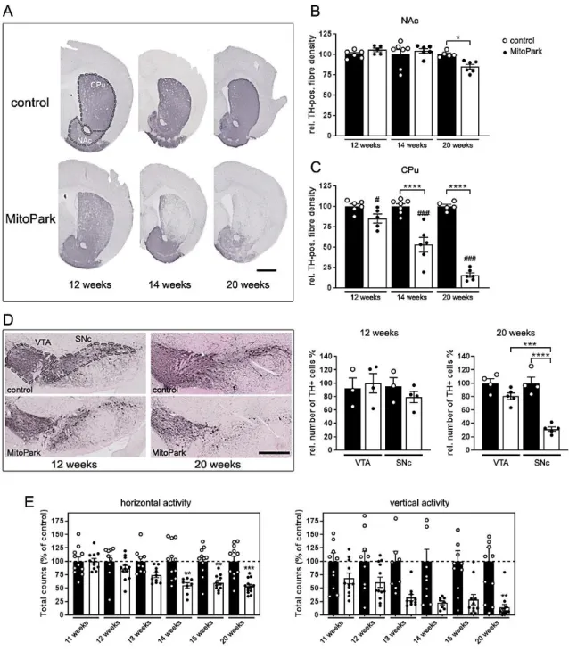
Fig. 3-1 Degeneration of the nigro-striatal system in MitoPark mice. ...38
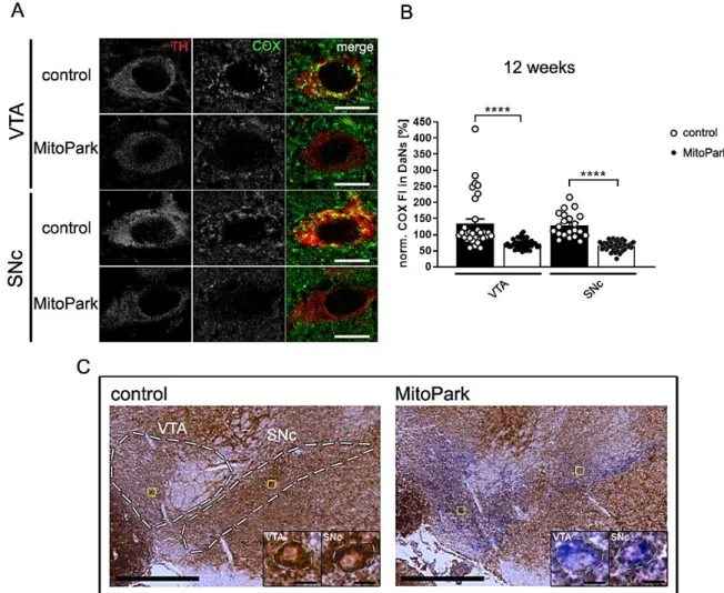
Fig. 3-2 Mitochondrial respiratory chain integrity is lost in dopaminergic neurons of MitoPark mice ...39
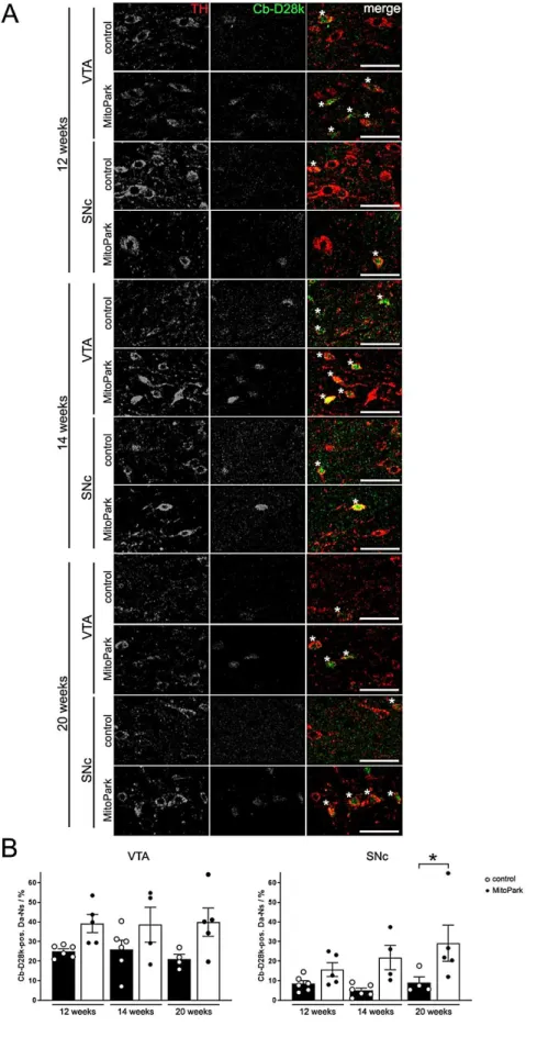
Fig. 3-3 Colocalization of TH and calbindin-D28k (Cb-D28k) in the midbrain of 12-, 14-, and 20-week-old MitoPark mice ...41
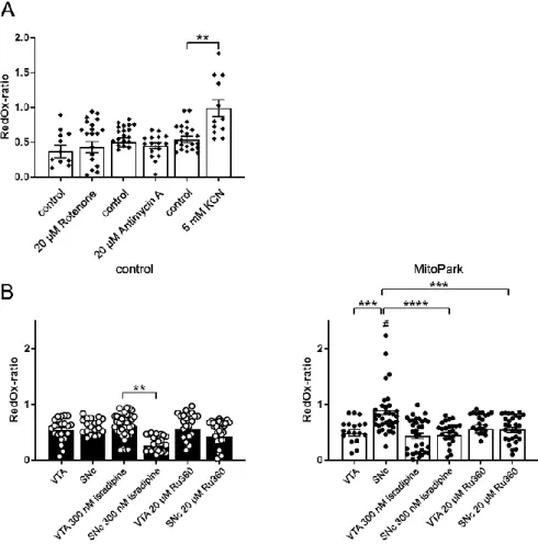
Fig. 3-4 The mitochondrial RedOx-ratio of SNc DaNs is reduced by application of isradipine or Ru360 in MitoPark mice ...43
Fig. 3-5 KCN induces an increase of ΔTMRM-F
KCNin MitoPark SNc DaNs ...45
Fig. 3-4-1 Reducing and oxidizing agents alter the mito-roGFP fluorescence intensity during 2PLSM experiments ...55
Fig. 3-4-2 RedOx-ratio imaging protocol during 2PLSM experiments ...56
Fig. 3-4-3 Oxygen consumption of midbrain sections ...57
Fig. 3-5-1 TMRE fluorescent signal is similarly responding to KCN ...58
Fig. 3-6 Colocalisation of TH and YFP is delayed in OB DaNs ...64
Fig. 3-7 Degeneration of midbrain DaNs and striatal projections in 30-week-old MitoPark mice. ...65
Fig. 3-8 Impaired odor detection in MitoPark mice ...66
Fig. 3-9 Reduced proportion of SCs in the OB of MitoPark mice ...67
Fig. 3-10 No change in the amount of PAX6-expressing DaNs in the OB but in the amount of progenitor cells in the SVZ of MitoPark mice ...69
Fig. 3-8-1 No significant differences in buried food pellet detection between MitoPark and control mice ...79
Fig. 3-11 No differences in spontaneous motor activity between K320E-Twinkle
DaNand control mice ...88
Fig. 3-12 K320E-Twinkle
DaNmice show loss of TH-positive cells in both VTA and SNc ...89
Fig. 3-13 COX-deficient cells in the midbrain of 10-month-old K320E-Twinkle
DaN. ...91
Fig. 3-14 Higher proportion of DaNs with normal complex IV expression in K320E-Twinkle
DaNList of Figures
Fig. 3-16 Decreased sugar consumption but unaltered immobility time of K320E-Twinkle
DaNmice ...95
Fig. 4-1 Schematic summary of the presented manuscripts ... 111
List of Abbreviations
List of Abbreviations
6-OHDA 6-hydroxydopamine α-Syn alpha-Synuclein ATP adenosine triphosphate
ATP13A2 ATPase cation transporting 13A2
bp base pair
Ca2+ calcium
Cav voltage-gated calcium channels Cav1 L-type voltage-gated calcium
channel
Cav1.2 L-type voltage-gated calcium channel subunit 1.2
Cav1.3 L-type voltage-gated calcium channel subunit 1.3
Cav2.3 R-type voltage-gated calcium channel subunit 2.3
CHCHD2 coiled-coil-helix-coiled-coil-helix domain containing 2
ClpP caseinolytic mitochondrial matrix peptidase proteolytic subunit
CNS central nervous system COX cytochrome c oxidase
CPu caudate putamen
CSF cerebrospinal fluid
Cre cre recombinase
DA dopamine
DaN dopaminergic neuron
DAT dopamine transporter DBS deep brain stimulation DHP dihydropyridines DJ-1 parkinsonism associated
deglycase
DNA deoxyribonucleic acid
DOPAL 3,4-dihydroxyphenylacetaldehyde DRP1 dynamin-related protein 1
E embryonic day
ERK2 extracellular-signal-regulated protein kinase 2
ERK extracellular-signal-regulated protein kinase 2
Fig figure
GABA γ-aminobutyric acid
GL glomerular layer
GPi globus pallidus pars interna GPe Globus pallidus pars externa GPx glutathione peroxidase GR glutathione reductase GSH glutathione, reduced form GSSG glutathione, oxidized form H2O2 hydrogen peroxide Ih hyperpolarization-activated
current
iPSC induced pluripotent stem cell K-ATP ATP-sensitive potassium channel
KO knockout
LAC large axonic cell
L-DOPA L-3,4-dihydroxyphenylalanine LRRK2 leucine-rich repeat kinase 2
MAO monoamine oxidase
MAOB monoamine oxidase type B MDVs mitochondrial-derived vesicles
MFN1 mitofusin 1
MFN2 mitofusin 2
MPP+ 1-methyl-4-phenylpyridinium mPTP mitochondrial permeability
transition pore
MPTP 1-methyl-4-phenyl-1,2,3,6- tetrahydropyridine
mt mitochondrial
mtDNA mitochondrial DNA
MUL1 mitochondrial E3 ubiquitin ligase 1
NAcc nucleus accumbens
NADH nicotinamide adenine dinucleotide
NADPH nicotinamide adenine dinucleotide NDP52 nuclear dot protein 52
List of Abbreviations
OB olfactory bulb
OH- hydroxyl radical
OMM outer mitochondrial membrane OPA1 optic atrophy 1
OPTN optineurin
PARIS parkin interacting substrate
PAX6 paired box 6
PCR polymerase chain reaction PET positron emission tomography PD Parkinson’s disease
PGC-1α peroxisome proliferator-activated receptor gamma coactivator 1α PINK1 PTEN-induced kinase 1
POLγ mitochondrial DNA polymerase γ
RN raphe nuclei
RNA ribonucleic acid
ROS reactive oxygen species
SC small anaxonic cell
SDH succinate dehydrogenase SERT serotonin transporter
SNc substantia nigra pars compacta
SNCA synuclein α
SNr substantia nigra pars reticulata STN subthalamic nucleus
SVZ subventricular zone
TFAM mitochondrial transcription factory A TOM translocase of outer membrane TOM20 translocase of outer membrane 20 VPS35 vacuolar protein sorting 35 VTA ventral tegmental area
Abstract
Abstract
Parkinson’s disease (PD) is a neurodegenerative disorder affecting ~1% of the population
above 60 years. It is primarily characterized by severe motor deficits following the progressive
and selective loss of dopaminergic neurons (DaNs) in the substantia nigra pars compacta
(SNc). Non-motor symptoms, such as hyposmia and depression, further arise in a prodromal
manner. Mitochondrial dysfunction plays an essential role for the loss of SNc DaNs, as
evidenced by mitochondrial complex I impairing toxins, an especially high accumulation rate
of mitochondrial DNA (mtDNA) deletions as well as the wide variety of mitochondrial-related
gene mutations in familial forms of PD. Given that neighboring DaNs in the ventral tegmental
area (VTA) are relatively spared from neurodegeneration, however, cell-type specific factors
must additionally contribute to the selective vulnerability in PD. In this work, data of three
manuscripts are presented, confirming that distinct DaN populations respond differently to
mitochondrial dysfunction following inactivation of mitochondrial transcription factor A (TFAM)
in mice. In the olfactory bulb (OB), only small anaxonic DaNs (SCs) are mildly reduced in
numbers, which is however associated with severe olfactory dysfunction and suggests a
putative role for OB DaNs in the development of PD-related hyposmia. The midbrain reveals
progressive and exclusive loss of SNc DaNs, which is accompanied by a severe decline of
motor function. In contrast to the VTA, DaNs in the SNc die through a detrimental imbalance
in the mitochondrial redox system, triggered by enhanced intracellular Ca
2+loads. Whereas
SNc DaNs perish due to the rapid and continuous loss of mtDNA upon TFAM inactivation, they
can adapt to impaired mtDNA replication, demonstrated after the expression of a mutated
variant of the mitochondrial helicase TWINKLE (K320E). Despite similar severe
neurodegeneration of both SNc and VTA DaNs, aged SNc DaNs preserve striatal innervation
and thereby normal motor performance. Conversely, VTA DaN projections are lost, causing
depressive-like behavior in these animals. Thus, identification and stimulation of ongoing
compensatory mechanisms in aged SNc DaNs of K320E-Twinkle
DaNmice host the potential to
help the small number of remaining SNc DaNs in PD patients to escape from cell death and
concurrently to recover motor performance through enhanced striatal innervation.
Zusammenfassung
Zusammenfassung
Die Parkinson-Krankheit (PD) ist eine neurodegenerative Erkrankung und betrifft rund 1% der Bevölkerung über 60 Jahre. Sie äußert sich vor allem durch die motorischen Ausfälle infolge des progressiven und selektiven Verlusts von dopaminergen Neuronen (DaNs) in der substantia nigra pars compacta (SNc). Nichtmotorische Symptome, wie Geruchsverlust und Depression, treten dabei zusätzlich prodromal auf. Die mitochondriale Dysfunktion spielt beim Verlust von SNc DaNs eine entscheidende Rolle, wie durch Toxine, die den mitochondrialen Komplex I beeinträchtigen, einer sehr hohen Akkumulationsrate von mitochondrialen DNA (mtDNA) Deletionen sowie einem breiten Spektrum an mitochondrial-bezogenen Genmutationen bei familiären PD-Fällen gezeigt wird. Die Tatsache, dass benachbarte DaNs im ventralen tegmentalen Areal (VTA) vom Zelltod nahezu verschont bleiben, legt jedoch nahe, dass zusätzliche, zelltypspezifische Faktoren zu der selektiven Vulnerabilität bei PD beitragen.
In dieser Arbeit werden die Daten dreier Manuskripte dargestellt, welche zeigen, dass DaNs
verschiedener Populationen unterschiedlich auf mitochondriale Dysfunktion reagieren, die auf
der Inaktivierung des mitochondrialen Transkriptionsfaktors A (TFAM) in Mäusen beruht. Im
olfaktorischen Bulbus (OB) sind lediglich kleine, anaxonische DaNs (SCs) zahlenmäßig leicht
reduziert, was jedoch mit einer schweren olfaktorischen Dysfunktion assoziiert ist und eine
mutmaßliche Rolle von OB DaNs beim PD-bezogenen Geruchsverlust vermuten lässt. Das
Mittelhirn zeigt hingegen einen progressiven und alleinigen Verlust von SNc DaNs, der mit
einer starken Abnahme der motorischen Leistung einhergeht. Im Gegensatz zum VTA sterben
DaNs der SNc durch ein verheerendes Ungleichgewicht im mitochondrialen Redox-System,
das durch einen zu hohen Ca
2+-Gehalt in ebendiesen Zellen ausgelöst wird. Während SNc
DaNs somit infolge eines schnellen sowie kontinuierlichen Verlusts von mtDNA sterben,
können sie sich an eine Störung der mtDNA Replikation, in Form einer mutierten Variante der
mitochondrialen Helikase TWINKLE (K320E), anpassen. Trotz schwerer Neurodegeneration
sowohl von SNc als auch VTA DaNs schaffen es die überlebenden SNc DaNs ferner die
striatale Innervierung und somit die motorische Aktivität der Mäuse zu erhalten. Auf der
anderen Seite hingegen, verlieren VTA DaNs ihre Projektionen, was schließlich einen
depressiven Phänotyp hervorruft. Somit könnten die kompensatorischen Mechanismen, die in
den SNc DaNs alter K320E-Twinkle
DaNMäuse vonstattengehen, dazu verwendet werden, die
geringe Anzahl überlebender SNc DaNs in PD-Erkrankten vor dem Tod zu bewahren und
zugleich die motorische Leistung durch neue striatale Innervierungen wiederherzustellen.
Introduction
1 Introduction
1.1 Mitochondrial Dysfunction and Parkinson’s disease
Since its first association with the neurodegenerative disease through the discovery of a toxic agent almost 40 years ago, mitochondrial dysfunction has been intensively studied in the context of Parkinson’s disease (PD). Reduced activity of mitochondrial complex I, which is the outcome of toxins related to PD, in the most affected brain area has for a long time been the crucial phenotype at the cellular level besides Lewy bodies. The huge number of mutated genes that are directly linked to mitochondrial function in familial cases, further highlights the essential role of mitochondria in the pathophysiology of PD.
1.1.1 Complex I Deficiency
The mitochondrial enzyme nicotinamide adenine dinucleotide (NADH) ubiquinone oxidoreductase, named complex I, is the first in the mitochondrial electron transport chain, and responsible for both the electron transfer from NADH to Q
10and proton pumping into the mitochondrial intermembrane space in order to ensure ATP production (Wirth et al. 2016).
Reduced activity of mitochondrial complex I has been first described in the substantia nigra pars compacta (SNc) of post mortem brains from patients with idiopathic PD in 1989 (Schapira et al. 1989). The same observations have been made by analyzing isolated mitochondria from the striatum (Mizuno et al. 1989) as well as platelet mitochondria of those patients (Parker, Boyson, and Parks 1989). A reduced complex I activity in PD has been subsequently reported, by a variety of studies, to be a phenomenon that is restricted to the SNc (Hattori et al. 1991;
Mann et al. 1992; Janetzky et al. 1994; Mann et al. 1994). Studies including other brain regions are limited and of contradictory results. Whereas some revealed decreased complex I activity in homogenates of frontal cortices from PD patients (Parker, Boyson, and Parks 1989; Keeney et al. 2006), others could not detect any differences compared to healthy individuals (Schapira, Mann, et al. 1990; Janetzky et al. 1994). A recent study presented unchanged complex I activity in the prefrontal cortex of PD patients, but a reduction in PD patients which additionally suffer from dementia, suggesting altered mitochondrial complex activity in this brain region to be rather linked to the combination of both conditions (Gatt et al. 2016). The analysis of enzyme activity or protein content in the cerebellum and striatum did not show any difference comparing PD patients and controls (Mizuno et al. 1989; Schapira, Mann, et al. 1990; Mann et al. 1992;
Janetzky et al. 1994).
It is out of question that intact complex I activity is of great importance for mitochondrial
functionality, especially for highly energy consuming synaptic processes in the projection area
of SNc dopaminergic neurons (DaNs) (Telford, Kilbride, and Davey 2009; Kilbride, Telford, and
Davey 2020). However, the significance of complex I deficiency in the development of PD is
Introduction
secondary response to disease-related modifications. Flønes et al. recently showed that complex I deficiency is indeed found throughout the PD brain, including the prefrontal cortex, hippocampus and putamen as well as regions which are spared from neurodegeneration, such as the cerebellum (Flones et al. 2018), questioning the pathogenic role of complex I impairment in PD.
1.1.2 Gene Mutations
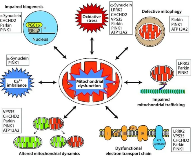
A genetic correlation in PD has been first described almost 100 years ago (Bell and Clark 1926). Today we know that there is a wide range of genetic variants and modifications that is related to PD-like disorders or parkinsonism. In this chapter I will focus on the monogenic origin of PD. With only ~5 – 10% of all patients, only a minority of PD cases is caused by monogenic mutations (Lill 2016), many of them being directly linked to mitochondrial dysfunction (see Fig. 1-1). These mutations occur in an autosomal dominant and recessive manner.
1.1.2.1 SNCA
The first discovered autosomal dominant gene in PD is SNCA (synuclein alpha) (Polymeropoulos et al. 1997). Although rarely found among familial forms of PD (Scott et al.
1999), the discovery of SNCA subsequently led to the identification of its encoded protein α-Synuclein (α-Syn) as the main component of the intracellular Lewy bodies arising also in many cases of idiopathic PD (Spillantini et al. 1997). The function of α-Syn is still not fully understood, but it might be involved in the maintenance, trafficking as well as exocytosis of synaptic vesicle and thereby modulates neurotransmitter release (Bendor, Logan, and Edwards 2013; Lautenschlager, Kaminski, and Kaminski Schierle 2017; Lautenschlager et al.
2018).
In order to unravel its neurotoxic effect in aggregated form, α-Syn and its interactions with mitochondria are intensively studied. Although α-Syn does not possess a canonical mitochondrial targeting sequence, it has been reported to be localized to mitochondria, leading to reduced complex I activity (Devi et al. 2008). Mitochondrial binding is achieved via translocase of outer membrane 20 (TOM20) and impairs mitochondrial protein import (Di Maio et al. 2016). A recent study just showed that α-Syn is involved in the reduction of mitochondrial respiratory activity and neurodegeneration by suppressing the mitochondrial matrix protease ClpP (Hu et al. 2019). When localized to mitochondria-endoplasmic reticulum contact sites, α-Syn is reported to influence mitochondrial morphology (Guardia-Laguarta et al. 2014) as well as calcium (Ca
2+) signaling and thereby reduces energy production (Paillusson et al. 2017).
Aggregates of α-Syn show an increased tendency to bind to mitochondria (Wang, Becker, et
al. 2019) and induce cell death via the oxidation of the ATP synthase and the following
increased opening probability of the mitochondrial permeability transition pore (mPTP)
(Ludtmann et al. 2018). However, recent findings suggest that it is rather the formation of Lewy
Introduction
bodies than solely α-Syn aggregation that causes neurodegeneration through mitochondrial damage (Mahul-Mellier et al. 2020).
1.1.2.2 LRRK2
Another autosomal dominant form is referred to the LRRK2 (leucine-rich repeat kinase 2) gene, in which mutations are most frequently detected in PD (Paisan-Ruiz et al. 2004; Zimprich et al. 2004). Mutations in the LRRK2 gene lead to increased kinase activity and have hence been classified as gain-of-function mutations (West et al. 2005; Greggio et al. 2006; Lu and Tan 2008; Sheng et al. 2012). Although the significance in PD is undisputed, the physiological role as well as the pathological mechanism of LRRK2 is still not fully understood. LRRK2 has been associated with a wide range of cellular processes, with many of them directly impacting mitochondrial functionality. LRRK2 interacts with microtubules and cytoskeleton proteins (Caesar et al. 2013; Beilina et al. 2014; Law et al. 2014) and is important for microtubule assembly (Parisiadou et al. 2009; Lin et al. 2010). In PD patients with LRRK2 mutation, a decrease in tubulin acetylation was detected (Esteves et al. 2015), which is important for organellar trafficking and axonal function (Reed et al. 2006; Esteves, Gozes, and Cardoso 2014). Together with the localization of LRRK2 on the mitochondrial outer membrane (West et al. 2005; Biskup et al. 2006), this raises the question whether in PD, mitochondrial transport might be affected by LRRK2 as well. Indeed, cells expressing a mutant LRRK2 revealed impaired mitochondrial trafficking (Thomas et al. 2016), which could mechanistically be explained by the direct interaction of LRRK2 with Miro, an outer mitochondrial membrane protein anchoring the microtubule motors kinesin and dynein to mitochondria (Hsieh et al.
2016).
Furthermore, mitochondrial morphology is found to be altered in LRRK2 mutants (Yue et al.
2015), including elongated mitochondria (Mortiboys et al. 2010). However, most studies rather reported mitochondrial fragmentation which has been linked to reduced levels of the short form of OPA1 (Stafa et al. 2014) as well as direct interaction of LRRK2 with DRP1 (dynamin-related protein 1), thereupon promoting mitochondrial fission (Wang et al. 2012; Su and Qi 2013;
Smith et al. 2016; Perez Carrion et al. 2018). Eventually, there is evidence that LRRK2 is involved in mitochondrial clearance via autophagy (Ramonet et al. 2011; Cherra et al. 2013;
Hindle et al. 2013; Orenstein et al. 2013; Cookson 2016): While LRRK2 physiologically promotes the removal of Miro, mutated LRRK2 disrupts this function and delays the degradation of damaged mitochondria (Hsieh et al. 2016).
1.1.2.3 VPS35
VPS35 (vacuolar protein sorting 35) encodes the vacuolar protein sorting-associated protein
Introduction
part of the retromer complex, which is involved in intracellular protein trafficking. VPS35 by itself is responsible for binding and sorting of protein cargo (Hierro et al. 2007; Seaman 2012) and contributes to the formation of mitochondrial derived vesicles (MDVs), which shuttle mitochondrial cargo to peroxisomes or lysosomes (Soubannier et al. 2012; Sugiura et al. 2014) in order to degrade a selection of mitochondrial proteins instead of the entire organelle (McLelland et al. 2014). Regarding mitochondrial function, VSP35 has been primarily linked to fusion and fission. A mutant VSP35 caused mitochondrial fragmentation, which is explained by either decreased Mitofusin 2 (MFN2) levels in response to mitochondrial E3 ubiquitin ligase 1 (MUL1) degradation (Tang et al. 2015), or increased turnover of DRP1 complexes by MDVs and lysosomal degradation (Wang et al. 2016).
Fig. 1-1 Mitochondrial dysfunction upon PD-related gene mutations is linked to a variety of pathways.
Monogenic mutations associated with PD can affect mitochondrial function, among others, via impaired mitochondrial biogenesis, enhanced ROS production, defective mitophagy, disrupted mitochondrial trafficking, deficient respiratory chain, modification of mitochondrial dynamics, imbalanced Ca2+ homeostasis and the combination of those, respectively (from Park et al. 2018).
Introduction
1.1.2.3 CHCHD2
CHCHD2 (coiled-coil-helix-coiled-coil-helix domain containing 2) mutations have been recently identified to be responsible for autosomal dominant, late-onset PD in three Japanese families (Funayama et al. 2015). However, other studies, including screening of Caucasians, south Italians (Jansen et al. 2015; Gagliardi et al. 2017) and Brazilians (Voigt et al. 2019), could not confirm this finding. It is believed that the localization of CHCHD2 to the mitochondrial intermembrane space maintains cristae structure and respiratory chain integrity, whereas impaired CHCHD2 function caused a reduced activity of complex III and IV and oxidative damage (Bannwarth et al. 2014; Meng et al. 2017), which could additionally be shown in fibroblasts from CHCHD2 mutant PD patients (Lee et al. 2018).
1.1.2.4 Parkin
With over 120 identified mutations so far (Lill 2016), Parkin (PARK2) mutations are the most common cause of autosomal recessive PD (Kitada et al. 1998) with predominantly early disease onset (Klein et al. 2003; Hedrich et al. 2004; Kilarski et al. 2012). Parkin is a cytosolic E3 ubiquitin ligase that regulates the degradation of mitochondria via mitophagy. It ubiquitinates a wide range of cytosolic as well as outer mitochondrial membrane (OMM) proteins upon mitochondrial damage, such as mitochondrial depolarization (Chan et al. 2011;
Sarraf et al. 2013). The accumulation of polyubiquitin chains on mitochondria subsequently leads to the recruitment of autophagosomes and lysosomes (Geisler et al. 2010; Matsuda et al. 2010; Narendra et al. 2010). Under basal condition, the activity of Parkin is repressed (Chaugule et al. 2011; Riley et al. 2013; Trempe et al. 2013; Wauer and Komander 2013), probably via K6-linked ubiquitination-mediated degradation (Durcan et al. 2014).
Despite its conserved role in mediating mitophagy in diverse mammalian cell lines, the relevance of Parkin in neurons and especially in PD pathology has been questioned. Some studies showed an only moderate Parkin contribution to the degradation of damaged mitochondria in primary neurons (Cai et al. 2012; Grenier, McLelland, and Fon 2013), whereas others confirmed the importance of Parkin in mitophagy in these cells (Grenier, Kontogiannea, and Fon 2014) and especially axons (Ashrafi et al. 2014). Intriguingly, Parkin-deficient mice (Perez and Palmiter 2005) as well as rats (Dave et al. 2014) revealed no PD-related phenotype. However, in the context of mitochondrial dysfunction upon impaired replication of mitochondrial DNA (mtDNA), loss of Parkin caused DaN degeneration and L-3,4- dihydroxyphenylalanine (L-DOPA) reversible motor symptoms (Pickrell et al. 2015), highlighting the importance of Parkin-mediated mitochondrial quality control in DaNs.
Besides mitophagy, Parkin is suggested to play a role in the regulation of mitochondrial
biogenesis (Scarffe et al. 2014) via the degradation of the parkin interacting substrate (PARIS),
Introduction
proliferator-activated receptor gamma coactivator 1α (PGC-1α), the main transcriptional activator of mitochondrial genes (Shin et al. 2011). Local knockout (KO) of Parkin in the midbrain of mice via virus injection consequently led to reduced mitochondrial number and size as well as morphological abnormalities (Stevens et al. 2015). Other studies have linked Parkin to the turnover of synaptic vesicles, showing Parkin-mediated ubiquitination of synaptic vesicle proteins (Zhang et al. 2000; Huynh et al. 2003; Trempe et al. 2013; Cao et al. 2014) as well as proteins responsible for vesicle recycling (Fallon et al. 2002; Fallon et al. 2006; Joch et al.
2007). Hence, it is assumed that Parkin plays a role in synaptic transmission control. Moreover, studies including the in vitro investigation of DaNs that are directly derived from Parkin mutant patients via induced pluripotent stem cell (iPSC) technology confirm this assumption. Thus, iPSC-derived DaNs revealed excessive DA release as well as reduced DA uptake (Jiang et al.
2012) and large DA-induced bursting of spontaneous excitatory postsynaptic currents in contrast to iPSCs from normal subjects (Zhong et al. 2017). These results underscore an altered sensitivity of patient-derived DaNs towards DA. In order to understand the exact processes that are responsible for PD in Parkin mutant patients, further investigations are needed. The absence of α-Syn aggregations in most of the PD cases including a disrupted function of Parkin (Pramstaller et al. 2005; Doherty et al. 2013) points to a different mechanism compared to idiopathic cases of PD.
1.1.2.5 PINK1
The second most common cause of autosomal recessive PD are mutations in the PINK1 (PTEN-induced putative kinase 1) gene (Valente et al. 2004) with loss-of-function mutations being associated with early-onset PD (Truban et al. 2017). PINK1 is a mitochondrial serine/threonine kinase and, besides Parkin, another key protein responsible for mitochondrial quality control via mitophagy. With intact mitochondrial function, PINK1 is imported into mitochondria via the TOM complex (Lazarou et al. 2012) and cleaved by mitochondrial proteases, such as PARL (Jin et al. 2010; Deas et al. 2011; Greene et al. 2012). In contrast, upon mitochondrial damage, the import of PINK1 is inhibited and the protein accumulates on the OMM (Lin and Kang 2008; Zhou et al. 2008). As a consequence, PINK1 activates Parkin and thus mediates mitochondrial clearance via autophagy (Vives-Bauza et al. 2010). The activation of Parkin is regulated by PINK1 via direct phosphorylation at Serine 65 (Kondapalli et al. 2012) and trans-activation by phosphorylation of ubiquitin at Serine 65, which is followed by binding to Parkin (Kane et al. 2014; Kazlauskaite et al. 2014; Koyano et al. 2014).
Interestingly, PINK1 can initiate mitophagy even without Parkin while using alternative receptors, such as nuclear dot protein 52 (NDP52) and optineurin (OPTN) (Lazarou et al.
2015).
The loss of PINK1 activity has been primarily related to mitochondrial dysfunction, including
the reduced activity of complex I and III (Amo et al. 2014), and to PD due to its leading role in
Introduction
the regulation of mitophagy. However, mitochondrial Ca
2+overload-induced death of PINK1- deficient DaNs (Kostic et al. 2015) further suggests an involvement of PINK1 in Ca
2+handling.
In addition, mitochondrial biogenesis via Parkin-mediated PARIS regulation (Lee et al. 2017) and fission (Pryde et al. 2016) were reported to be positively regulated by PINK1.
1.1.2.6 ATP13A2
A novel gene that has been identified to lead to a rare form of autosomal recessive juvenile- onset PD, the so called Kufor-Rakeb syndrome, is ATP13A2 (ATPase cation transporting 13A2) (Park, Blair, and Sue 2015; Suleiman, Hamwi, and El-Hattab 2018). ATP13A2 encodes the P5B ATPase, which mediates the transport of cations across endo/lyosomal membranes and is believed to be involved in vesicular transport (Park et al. 2014), endolysosomal activity (Sato et al. 2016; Rayaprolu et al. 2018) and indirectly glycolysis (Park et al. 2016). So far, studies using ATP13A2-deficient cells have presented lysosomal defects with corresponding disturbances in cation, especially Zn
2+, homeostasis. Moreover, these cells revealed impaired mitochondrial functionality in the form of reduced ATP conversion, mitochondrial fragmentation and reactive oxygen species (ROS) production (Ramonet et al. 2012; Park et al. 2014).
However, further research needs to be conducted to decipher the exact consequences of ATP13A2 mutations in patients.
1.1.2.7 DJ-1
Mutations in the DJ-1 encoding gene PARK7 (parkinsonism associated deglycase) cause autosomal recessive early-onset PD (van Duijn et al. 2001; Bonifati et al. 2003) with a prevalence of only up to 1% of these cases (Maraganore et al. 2004; Alcalay et al. 2010; Sironi et al. 2013). DJ-1 is a ubiquitously expressed protein and implicated in a variety of cellular processes. Whereas the molecular mechanisms of DJ-1 functions remain elusive, there is a wide acceptance for its control of ROS and its neuroprotective effect upon oxidative damage (Taira et al. 2004; Surmeier et al. 2010; Cookson 2012). While mainly localized in the cytoplasm, DJ-1 can be translocated to mitochondria in response to oxidative stress (Irrcher et al. 2010; Kim et al. 2012). The loss of DJ-1 has been connected to reduced autophagic activity and mitochondrial dysfunction in fibroblasts (Krebiehl et al. 2010), to mitochondrial fragmentation and reduced mitochondrial membrane potential in human dopaminergic cells (Thomas et al. 2016), and more importantly, to the loss of SNc DaNs in mice (Rousseaux et al. 2012). There is recent evidence, that DJ-1 is additionally involved in the regulation of mitochondrial Ca
2+handling by direct interaction with the mitochondria-ER coupling complex (Basso, Marchesan, and Ziviani 2020).
1.1.3 Toxins
The discovery of PD-related toxins led to the first connection of the neurodegenerative disease
Introduction
deciphered the pathological mechanisms during the course of PD. Today, these toxins are mainly used in animal models to assess the potential neuroprotective effects of newly identified drugs on the survival of DaNs and related motor symptoms.
1.1.3.1 MPTP
The very first association of PD with mitochondrial dysfunction was enabled by the discovery of 1-methyl-4-phenyl-1,2,3,6-tetrahydropyridine (MPTP). Nearly 40 years ago, MPTP was found to induce parkinsonian-like symptoms in drug users consuming self-produced heroine- like substances (Langston et al. 1983; Langston and Ballard 1983). Through its lipophilic property, MPTP crosses the blood-brain-barrier and is subsequently converted into its toxic form 1-methyl-4-phenylpyridinium (MPP
+) via the astrocytic monoamine oxidase (MAO) (Levitt, Pintar, and Breakefield 1982). Once its released via the organic transporter-3 from astrocytes (Cui et al. 2009), MPP
+is preferentially taken up by DaNs due to its high affinity to the DA transporter (DAT) (Javitch et al. 1985; Gainetdinov et al. 1997), accumulates inside mitochondria due to its lipophilic charge and has been shown to inhibit complex I, finally causing selective degeneration of SNc DaNs in both mice and humans (Langston and Ballard 1983; Heikkila et al. 1985; Nicklas, Vyas, and Heikkila 1985).
Besides affecting complex I activity, MPP
+is believed to compete with DA for being stored in vesicles, thus provoking DA extrusion and subsequent conversion into compounds with oxidative damage potential, such as 3,4-dihydroxyphenylacetaldehyde (DOPAL) (Panneton et al. 2010). In support of this, oxidative stress together with neuroinflammation have been found to occur in post mortem PD brains (Langston et al. 1999). However, many symptoms that are typical for idiopathic PD were absent upon MPTP consumption, including the Lewy pathology and the impairment of other brain regions as well as non-dopaminergic cells.
1.1.3.2 Rotenone
In 1987, an epidemiological study reported for the first time about an increased prevalence of
PD in rural areas where certain pesticides were used (Barbeau et al. 1987). However,
subsequent contradictory studies did not allow a conclusive confirmation of this observed
phenomenon, probably caused by its limitations on the investigation of entire classes of
pesticides instead of specific agents (Nandipati and Litvan 2016). One of these naturally
occurring pesticides is rotenone, which can be extracted from roots of Lonchocarpus and
Derris plants (Soloway 1976). It mechanistically acts like MPTP and inhibits complex I causing
progressive loss of midbrain DaNs (Betarbet et al. 2000). Its highly lipophilic features and
independence of transporters allow a direct and fast cell penetrance of rotenone, even through
the blood-brain-barrier (Martinez and Greenamyre 2012). Therefore, it can be administered via
different routes, including intraperitoneally, intravenously, subcutaneously and stereotactically,
in order to induce histopathological features and motor symptoms according to PD (Johnson
Introduction
and Bobrovskaya 2015). However, whereas rotenone exposure is known to be one of the greatest risks to develop PD in humans, inhalation did not cause an impairment in the nigrostriatal system in rodents (Rojo et al. 2007).
Mitochondrial dysfunction upon impaired complex I activity through rotenone treatment is referred to cause ROS production and reduced glutathione levels (Duty and Jenner 2011;
Martinez and Greenamyre 2012) and sensitizes DaNs to further oxidative damage (Milusheva et al. 2005). Correspondingly, reduced DJ-1 levels have been detected in homogenates of different brain regions, including the SNc, from rats treated with rotenone (Sonia Angeline et al. 2012). The same study presented a decreased Parkin expression, which could either simply be accompanied with the severe loss of DaNs or point to impaired mitochondrial quality control in the SNc upon rotenone exposure. Moreover, rotenone-induced dopaminergic cell death is believed to be initiated by the activation of mitochondrial-dependent caspases, which in turn induce apoptosis (Chung, Miranda, and Maier 2007).
Like MPTP, rotenone treatment reproduces many features of PD, including selective loss of SNc DaNs, striatal DA depletion and motor impairment. Furthermore, rotenone induces α-Syn pathology (Betarbet et al. 2000; Biskup et al. 2006; Marella et al. 2008; Cannon et al. 2009;
Ferris et al. 2013), which is not observed when using MPTP. However, it must be noted that the rotenone model is limited in the number of animals that develop the loss of the nigrostriatal system, in the location of the lesion, the degree of lesion severity, and the mortality rate (Cicchetti, Drouin-Ouellet, and Gross 2009; Greenamyre et al. 2010). It is further not clear, whether the motor decline is solely due to the impaired DA system or to diffuse mitochondrial dysfunction in other cell types and brain regions, respectively, as shown when using different doses of rotenone (Fleming et al. 2004; Richter, Hamann, and Richter 2007).
1.1.3.3 6-OHDA
6-Hydroxydopamine (6-OHDA) is an established neurotoxin structurally similar to DA and used
to induce the degeneration of the nigrostriatal system in rodents. Due to its low penetrance of
the blood-brain-barrier, the necessity of a direct injection into the brain where it is taken up by
catecholaminergic neurons (Luthman et al. 1989), has been reported already over 50 years
ago (Ungerstedt 1968). In order to provoke DaN damage only, noradrenaline reuptake
inhibitors are additionally applied (Waddington 1980). 6-OHDA is either injected unilaterally
into the medial forebrain bundle and the SNc, respectively, where anterograde (nigrostriatal)
DaN degeneration is starting within 24 h (Faull and Laverty 1969; Jeon, Jackson-Lewis, and
Burke 1995), or into the dorsal striatum with retrograde (striatonigral) loss of DaNs occurring
within one to three weeks (Sauer and Oertel 1994; Przedborski et al. 1995). This way by, usage
of unilateral lesions resulted in circumventing the high death rate of bilaterally treated mice as
Introduction
well as concurrently generating an intrinsic control by the uninjured hemisphere (Thiele, Warre, and Nash 2012).
6-OHDA-mediated neuron death is thought to be mainly caused by oxidative damage through auto-oxidation of 6-OHDA and subsequent generation of various ROS (Soto-Otero et al. 2000).
However, the role of mitochondrial dysfunction in this toxin-induced loss of DaNs is debated as well. Whereas the impact of 6-OHDA on mitochondrial function in vivo is not clear, there is strong evidence in cultured cells. Thus, 6-OHDA treatment has been shown to induce mitochondrial fragmentation via upregulation of Drp1 expression (Gomez-Lazaro et al. 2008) as well as reduced expression of Mfn1, Mfn2 and OPA1 (Xi et al. 2018). It further leads to decreased mitochondrial membrane potential (Ren et al. 2019) and increased mitophagy upon extracellular-signal-regulated protein kinase 2 (ERK2) accumulation (Dagda et al. 2008; Zhu et al. 2007) via the externalization of cardiolipin (Chu et al. 2013). Moreover, axonal transport of vesicles as well as mitochondria is impaired after 6-OHDA treatment, which is probably due to ROS damage (Lu et al. 2014). These findings suggest, that it is the oxidative damage in the first place, that causes 6-OHDA-mediated degeneration of DaNs, with mitochondrial malfunction being of secondary importance.
1.1.4 mtDNA Deletions
After being first described in neuromuscular disorders in the late 1980s (Holt, Harding, and Morgan-Hughes 1988; Wallace et al. 1988; Shoffner et al. 1990), mutations of the mtDNA attracted more and more attention to be a potential cause of complex I deficiency and mitochondrial dysfunction in PD patients (Schapira 1994). Sequence analysis of mtDNA did not reveal specific mutations associated with PD, and parkinsonism-like symptoms were only sporadically found in mitochondrial disease patients (Rana et al. 2000; Thyagarajan et al.
2000). However, based on the recognition that heteroplasmic mtDNA deletions are able to cause myopathy (Holt, Harding, and Morgan-Hughes 1988) as well as Kearn-Sayres syndrome (Zeviani et al. 1988), the presence of mtDNA deletions in brains of PD patients was investigated as well. Whereas the amount of deleted molecules was too low to be detected via southern blot (Lestienne et al. 1990; Schapira, Holt, et al. 1990), mtDNA deletions could be indeed presented in the striatum of post mortem PD brains by using the at this time newly developed polymerase chain reaction (PCR) (Ikebe et al. 1990; Ozawa et al. 1990).
Interestingly, striatal mtDNA deletions were also verified in age-matched controls in contrast
to other brain regions, although to a lesser extent than in PD (Ozawa et al. 1990). Subsequent
studies confirmed this observation in the dorsal striatum and extended it to the SNc (Soong et
al. 1992), suggesting DaN-enriched regions to be hotspots for mtDNA deletions during normal
aging and especially in PD (Melov et al. 1999; Gu et al. 2002). Later on, laser microdissection
of single cells from post mortem brains revealed elevated levels of mtDNA deletions (~50% of
mtDNA molecules carry deletions) in the SNc in healthy aged individuals (Kraytsberg et al.
Introduction
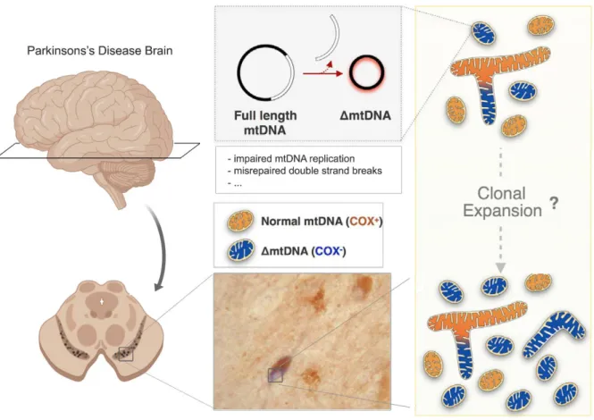
2006) and even more abundant in PD patients (Bender et al. 2006). These studies further showed that respiratory chain deficiency of SNc DaNs correlated with increased mtDNA deletions, providing a direct link between accumulating deletions and mitochondrial dysfunction (see Fig. 1-2).
MtDNA deletions in SNc DaNs vary widely in size (~2000 – 9500bp) and are thought to be somatically acquired via clonal expansion (Reeve et al. 2008). Their accumulation is driven by the catecholamine metabolism (Neuhaus et al. 2014; Neuhaus et al. 2017), explaining why especially DaNs are hotspots for such alterations in the mtDNA. A possible mechanistic explanation for the differential outcome during normal ageing and PD was provided by the groups of Tzoulis and Bindoff: Whereas in SNc DaNs of healthy individuals, the population of wild-type mtDNA is maintained by increased total mtDNA copy number upon deletion accumulation, this compensatory upregulation fails and results in depletion of the wild-type mtDNA population in PD patients (Dolle et al. 2016). These findings could be confirmed by two established mouse models with impaired mtDNA maintenance and mtDNA replication,
Fig. 1-2 Accumulation of mtDNA deletions in SNc DaNs of PD patients. With increasing age, SNc DaNs accumulate deletions of mtDNA, a process which is accelerated in PD. Deletion accumulation is associated with mitochondrial dysfunction, presented by COX deficiency (blue stained neuron). Defective replication as well as misrepaired double strand breaks of mtDNA are proposed to result in deletion formation. Deleted molecules are
Introduction
respectively. Depletion of mtDNA in DaNs via cell type-specific KO of the mitochondrial transcription factor A (TFAM) has been shown to mimic the key features of PD pathology in mice. These so called MitoPark mice revealed selective neurodegeneration and progressive motor impairment (Ekstrand et al. 2007). Furthermore, DaNs were able to compensate multiple mtDNA deletions upon mutation of mitochondrial DNA polymerase γ (POLγ) by the upregulation of mtDNA copy number (Trifunovic et al. 2004; Perier et al. 2013). However, crossing these Mutator mice with Parkin KO mice, which do not show any impairment of DaNs (Perez and Palmiter 2005), caused selective loss of DaNs and an accompanied motor dysfunction (Pickrell et al. 2015). This highlights that the success of the compensatory increase in mtDNA copy number upon deletion accumulation further depends on functional mitochondrial quality control. SNc DaNs of idiopathic PD patients have been further shown to reveal low TFAM levels, accompanied with reduced mtDNA copy number and complex I deficiency (Grunewald et al. 2016). It is thereby likely that in PD, the significant pathological event is the depletion of wild-type mtDNA as a result of accumulating deletions, rather than solely the presence of mtDNA deletions. If balancing the amount of wild-type mtDNA and mtDNA deletions, by increasing mtDNA copy number and holding the amount of deleted mtDNA molecules at tolerable levels, fails in SNc DaNs, this will inevitably cause mitochondrial dysfunction and subsequently provoke neuron death.
The mechanism for the formation of mtDNA deletions is still not fully understood. For a long period of time, incorrect mtDNA replication in the form of strand displacement has been thought to be primarily responsible (Clayton 1982). The contribution of the replication machinery on deletion formation in general is evidenced by a variety of studies reporting multiple mtDNA deletions in patients with mutations in genes that are responsible for mtDNA replication, such as Twinkle (Suomalainen et al. 1992; Hudson et al. 2005; Hanisch et al. 2015) and POLγ (Di Fonzo et al. 2003; Ferreira et al. 2011; Nissanka et al. 2018). However, this scenario was challenged to also occur in SNc DaNs (Reeve et al. 2008). Simultaneously, mtDNA deletions were suggested to rather arise as a consequence of misrepaired double strand breaks upon mtDNA damage (Krishnan et al. 2008). The connection between deletion formation and mtDNA double strand breaks has been subsequently confirmed in neurons in vitro (Fukui and Moraes 2009) as well as in vivo (Fukui and Moraes 2009), including DaNs (Pickrell et al. 2011).
The fact that mtDNA double strand breaks are promoted by ROS (Pinto et al. 2017), could further explain the accumulation of mtDNA deletions in healthy individuals, with regard to increasing ROS-induced mtDNA damage with normal ageing. Thereby, mtDNA deletions underscore the significance of ageing in the development of PD (Liu 2014; Rizzo et al. 2016;
Collier, Kanaan, and Kordower 2017).
The potential of measuring mtDNA levels as a putative biomarker for PD has been intensively
studied by various groups in recent years. After the identification of reduced levels of circulating
Introduction
cell-free mtDNA in the cerebrospinal fluid (CSF) in Alzheimer’s disease patients (Podlesniy et al. 2013), those results could be shown in PD patients as well (Pyle et al. 2015) and seem to be further influenced upon treatment (Lowes et al. 2020). This indicates that alterations in the amount of CSF mtDNA could rather be used for the diagnosis of ongoing neurodegeneration in general than specifically for PD. Reduced mtDNA copy number has been further detected in peripheral blood cells and SNc DaNs of PD patients, in contrast to healthy individuals (Pyle, Anugrha, et al. 2016), however, these findings could not be repeated in PD patients with depression (Pyle, Lowes, et al. 2016). A recent study confirmed that blood mtDNA copy number is unchanged in PD patients and controls, indicating that it is not an adequate biomarker for PD (Davis et al. 2020). Correspondingly, fibroblasts from idiopathic PD patients as well as patients with a LRRK2 mutation background did not show any changes in mtDNA copy number (Podlesniy et al. 2019). In face of the unsolved mechanistic relation between neurodegeneration and reduced circulating cell-free mtDNA as well as the majority of data reporting no alterations of the mtDNA copy number in diverse biopsies of PD patients, mtDNA levels are currently no promising candidate for reliable PD diagnosis.
1.1.5 Oxidative Stress
Oxidative stress is a widespread term characterizing a pathophysiological cellular state with increased presence of ROS not balanced by the cellular anti-oxidative systems. The three main ROS are superoxide (O
2-), hydrogen peroxide (H
2O
2) and hydroxyl radical (OH
-), all being produced from molecular oxygen (O
2) (Collin 2019). Mitochondria are the primary intracellular source of ROS through the interaction of unpaired electrons with molecular O
2at the respiratory chain (Cadenas and Davies 2000; Andreyev, Kushnareva, and Starkov 2005). In particular, complex I and III have been shown to be the major ROS producers in isolated mitochondria (Kudin et al. 2004; Kudin, Debska-Vielhaber, and Kunz 2005; Kussmaul and Hirst 2006). Increased O
2-production at complex I is observed upon high matrix NADH concentrations and high mitochondrial membrane potential, respectively, whereas at complex III, high O
2-levels are induced by selective inhibition. Thus, ROS contribution by complex III is rather thought to be of secondary importance (Murphy 2009).
Mitochondria possess their own antioxidant system in order to counteract increased ROS
levels. Reduced glutathione (GSH) can scavenge ROS and thereby protects biomolecules
from oxidative damage (Ruszkiewicz and Albrecht 2015). Moreover, mitochondrial matrix
enzymes with antioxidant activity, such as manganese-dependent superoxide dismutase
(MnSOD), are able to convert O
2-into H
2O
2(Miriyala, Holley, and St Clair 2011). Glutathione
peroxidase (GPx) subsequently transforms H
2O
2into H
2O while oxidizing GSH. The oxidized
form, GSSG, is in turn reduced back to GSH by glutathione reductase (GR) and the usage of
Introduction
mitochondria-located isoform peroxiredoxin 5 has been reported to protect cells from neurodegeneration upon treatment with MPTP (De Simoni et al. 2013) and rotenone (Wang, Huang, et al. 2019), respectively.
Constantly keeping mitochondrially-derived ROS at low levels, DaNs additionally have to face another source, since the metabolism of DA is associated with the production of a variety of ROS and hence impede antioxidant processes in this cell type (Delcambre, Nonnenmacher, and Hiller 2016). Consequently, signs of oxidative damage (Dexter et al. 1989; Alam et al.
1997; Floor and Wetzel 1998; Bosco et al. 2006) as well as low GSH concentrations (Sofic et al. 1992; Sian et al. 1994; Jenner and Olanow 1996) have been detected early in the SNc of PD patients. In addition, the deficiency of complex I, the primary ROS producer (Murphy 2009), in SNc DaNs leads to elevated ROS levels, thereby causing a vicious circle that might greatly contribute to cell death in PD (Dias, Junn, and Mouradian 2013). The importance of oxidative stress in PD pathology is further confirmed by the fact that mutations in CHCHD2 or DJ-1 encoded by PARK7 as well as treatment with MPTP, Rotenone and 6-OHDA (see above) are associated with oxidative damage and degeneration of DaNs.
1.2 Dopaminergic Neurons in Parkinson’s disease
Cardinal symptoms in PD only occur with the loss of DaNs in the SNc to a certain degree. The
‘shaking palsy’, as it was initially termed by James Parkinson in 1817 (Parkinson 2002), considerably improved our understanding of voluntary movement control. Besides the SNc, other DA-enriched brain regions are affected in PD and linked to prodromal non-motor symptoms, such as hyposmia and depression (Postuma, Aarsland, et al. 2012). However, though multiple and promising insights have been gained up to now, the especially severe vulnerability of nigral DaNs is not fully understood and treatments to slow down neurodegeneration are still lacking.
1.2.1 Selective Vulnerability
PD is a complex neurological disorder that occurs in a heterogeneous manner and includes
the loss of differential neuronal subtypes in a variety of areas in the brain. In addition to the
SNc mentioned above, among others, neurons in the locus coeruleus, nucleus basalis of
Meynert, pedunculopontine nucleus, raphe nucleus, dorsal motor nucleus of the vagus,
olfactory bulb (OB), amygdala and hypothalamus are also affected (Dickson 2012). Thus,
neurodegeneration in PD is not only restricted to DaNs but also involves serotonergic,
noradrenergic, cholinergic, GABAergic and glutamatergic neurons (Brichta, Greengard, and
Flajolet 2013). This heterogeneous damage of diverse neurological systems is primarily linked
to non-motor symptoms which can precede motor impairment by up to ten years (Martinez-
Martin et al. 2011; Postuma, Aarsland, et al. 2012; Khoo et al. 2013; Duncan et al. 2014).
Introduction
However, motor symptoms remain the key pathology and the pivotal event for diagnosis of PD.
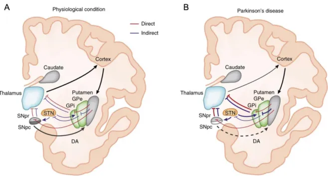
Motor dysfunction is primarily caused by the loss of DaNs in the SNc and the accompanying depletion of DA in the dorsal striatum (Michel, Hirsch, and Hunot 2016; Obeso et al. 2017). It is the DaNs that are therefore in the focus of potential cell replacement therapies (Sonntag et al. 2018). Interestingly, a neighboring DaN-enriched midbrain region, the ventral tegmental area (VTA), is much less affected (Alberico, Cassell, and Narayanan 2015) although both SNc and VTA DaNs are closely related with only <1% (Grimm et al. 2004) and <3% (Greene, Dingledine, and Greenamyre 2005), respectively, of genes being differentially expressed. This selectivity in the death of DaNs is occupying scientists since decades. Whereas the favored loss of SNc DaNs upon treatment with MPTP and 6-OHDA, which rely on cell penetrance via DAT, could be explained by the lower expression of the transporter in VTA DaNs (Lammel et al. 2008), the explanation for rotenone, which should be able to access any cell but still causes selective DaN death via diverse routes of administration, is more complex. Monogenic mutations causing familial forms of PD are widespread in the brain and complex I deficiency has been shown to occur throughout the whole brain of idiopathic PD patients (Flones et al.
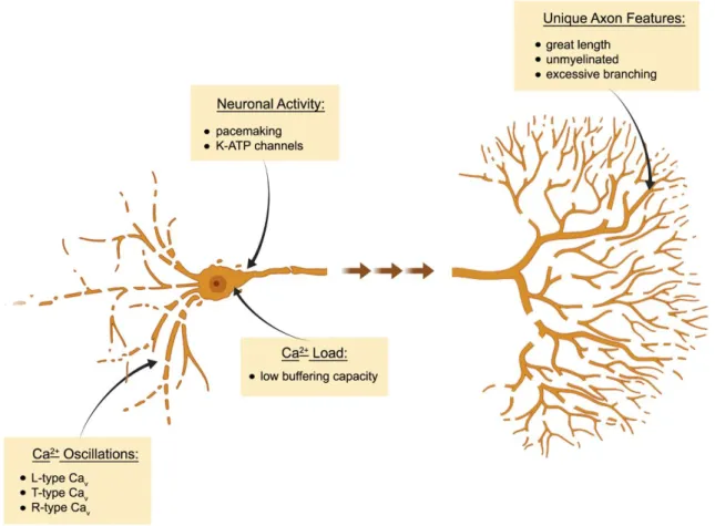
2018). Furthermore, both SNc and VTA DaNs of wildtype mice have been shown to equally accumulate mtDNA deletions with increasing age (Neuhaus et al. 2014). Consequently, there must be further cell-type specific factors rendering SNc DaNs highly vulnerable to mitochondrial dysfunction (see Fig. 1-3).
Investigation of electrophysiological patterns of both midbrain DaN populations has greatly contributed to a better understanding of the selective vulnerability. SNc as well as VTA DaNs demonstrate autonomous pacemaking (Grace and Bunney 1984; Puopolo, Raviola, and Bean 2007; Khaliq and Bean 2010). In juvenile SNc DaNs and generally in VTA DaNs, voltage- dependent Na
+channels are mainly responsible for pacemaker activity, while in adult SNc DaNs, L-type voltage-gated Ca
2+channels [Ca
v1 (Catterall et al. 2005)] contribute to the autonomous firing (Chan et al. 2007). Whereas Ca
v1 activity is not essential for pacemaking (Guzman et al. 2009; Poetschke et al. 2015), it is associated with somatodendritic Ca
2+oscillations (Chan et al. 2007) and on the one hand thought to assist pacemaker activity and directly boost oxidative phosphorylation (Surmeier, Guzman, and Sanchez-Padilla 2010). On the other hand, however, Ca
2+oscillations are a disservice to SNc DaNs, as they cause oxidative stress by unknown mechanisms (Guzman et al. 2010). Ca
2+entry via Ca
v1 channels might be further promoted in PD via the inhibition of the hyperpolarization-activated current (I
h). MPTP treatment blocked I
hin SNc DaNs (Masi et al. 2013) and thereby increased neuronal activity (Masi et al. 2015) as well as elevated intracellular Ca
2+levels (Carbone et al. 2017).
The metabolic challenge upon increased Ca
2+concentrations in SNc DaNs, in contrast to VTA
DaNs (Philippart et al. 2016), is thought to render these neurons more vulnerable to
Introduction
epidemiological studies revealed a 20-30% lower risk for the development of PD in patients treated for high blood pressure with Ca
v1 inhibitors (Lang, Gong, and Fan 2015), so called dihydropyridines (DHP) which block both Ca
v1.2 and Ca
v1.3 channels (Koschak et al. 2001;
Xu and Lipscombe 2001). Despite controversial in vivo results regarding dose efficiency as well as specificity, the DHP isradipine, has been tested in a phase III clinical trial on PD patients (Biglan et al. 2017). This clinical trial failed ('Isradipine Versus Placebo in Early Parkinson Disease: A Randomized Trial' 2020), supporting recent concerns about the dosage’s efficiency (Ortner et al. 2017).
Other voltage-gated Ca
2+channels have been linked to increased cytosolic Ca
2+levels and the accompanying vulnerability of SNc DaNs. T-type voltage-gated Ca
2+channels have been shown to contribute to Ca
2+oscillations (Guzman et al. 2018) and to drive degeneration of SNc DaNs in a familial form of PD (Tabata et al. 2018). In addition, a recent study identified the R- type channel Ca
v2.3 to be most strongly expressed among all voltage-gated Ca
2+channels in
Fig. 1-3 Cell-autonomous factors contributing to the vulnerability of SNc DaNs to mitochondrial dysfunction. Schematic summary of selected cellular key features that contribute to the selective vulnerability of SNc DaNs in PD upon mitochondrial dysfunction. Autonomous pacemaking is associated with Ca2+ oscillations mediated by distinct voltage-gated Ca2+ channels (Cav). Low Ca2+ buffering capacity further contributes to enhanced intracellular Ca2+ load, which in turn causes oxidative stress by unknown mechanisms. SNc DaNs have a long, unmyelinated axon which is extremely branched, leading to high energetic demands and a strong dependence on functional mitochondria. ATP-sensitive potassium (K-ATP) channels open in metabolic demand situations, and thereby connect neuronal activity with the energetic state of the cell. When mitochondrial function is impaired, however, hyperactivity of K-ATP channels can result in the complete loss of neuronal excitability and contributes to neuron death (Figure was generated by using BioRender, according to Surmeier et al. 2017).