dibenzodiazepinone-type muscarinic M
2-receptor antagonists conjugated to fluorescent dyes or
small peptides
DISSERTATION
ZUR ERLANGUNG DES DOKTORGRADES DER NATURWISSENSCHAFTEN (DR. RER. NAT.) AN DER FAKULTÄT FÜR CHEMIE UND PHARMAZIE
DER UNIVERSITÄT REGENSBURG
vorgelegt von Corinna G. Weinhart
(geboren: Gruber) aus Fensterbach, 2020
Fakultät IV – Chemie und Pharmazie – der Universität Regensburg.
Promotionsgesuch wurde eingereicht am: Oktober 2020 Tag der mündlichen Prüfung:
Vorsitzender des Prüfungsausschusses: Prof. Dr. Sigurd Elz
Erstgutachter: PD Dr. Max Keller
Zweitgutachter: Prof. Dr. Pierre Koch
Drittprüfer: Prof. Dr. Frank-Michael Matysik
ACKNOWLEDGMENTS
First and foremost, I would like to thank Prof. Dr. Armin Buschauer, who died far too early, for giving me the opportunity to work in his research group on this versatile and interesting project.
Another very important person for the realization of this thesis was Dr. Max Keller, who supervised and accompanied this whole project. I would like to express my deepest gratitude to him for his continuous, excellent scientific support and input.
Especially, I am very grateful that he continuously offered an open door, to discuss difficulties and ideas occurring during the work with his patient and constructive manner.
I want to express my sincere gratitude to Susanne Bollwein, Brigitte Wenzl and Maria Beer-Krön, for excellent technical assistance, extremely patient performances of assays and for cultivation of the needed cells. Furthermore, I want to thank Peter Richthammer for helping me to solve several technical problems.
Moreover, I want to thank Dr. David Wifling for the induced-fit docking studies in Chapter 2 and for the valuable ideas. I also want to thank Maximilian Schmidt and Eduard Neu as well as their supervisors Timothy Clark and Peter Gmeiner for the performances of the latest induced-fit docking studies for Chapter 2.
For the association to the research training group 1910 “Medicinal Chemistry of Selective GPCR Ligands” and for their financial support I want to thank the Deutsche Forschungsgemeinschaft (DFG). I am very grateful for the organisation of numerous interesting events and critical helpful scientific discussions during our retreats.
Furthermore, I want to thank all employees of the analytical department of the University of Regensburg for the measurement of numerous NMR and mass spectra. Especially, I want to express my gratitude to Fritz Kastner and Josef Kiermaier for many helpful discussions and excellent assistance, as well as several amusing moments.
Beyond that, I thank all colleagues of the Buschauer- and Elz- group for their help, support and ideas and the excellent and amusing working atmosphere. Especially,
synthesis, and for many amusing, as well as constructive and helpful conversations.
Moreover I want to express my gratitude to Carina Höring for the development of the mini-Gi recruitment functional assay for the M2R and for her patient assistance doing my first functional assays, as well as for the generation and cultivation of HEK- cells expressing NlucN-mini-Gαi and M2R-NlucC constructs.
I want to thank Silvia Heinrich for offering friendly assistance for organizational, administrative and bureaucratic matters.
Zu guter Letzt möchte ich meiner Familie für ihre uneingeschränkte Unterstützung danken. Der größte Dank gebührt jedoch meinem Michael. Für seine Liebe, aufmunternden Worte, immerwährende Unterstützung und für jeden einzelnen Moment, den wir miteinander verbringen.
Regensburg, 22.10.2020
PUBLICATIONS, PRESENTATIONS AND PROFESSIONAL TRAINING
Peer- reviewed journal articles
(published or submitted prior the submission of this thesis)
A. Pegoli*, D. Wifling*, C.G. Gruber*, X. She, H. Hubner, G. Bernhardt, P. Gmeiner, M.
Keller, Conjugation of short peptides to dibenzodiazepinone-type muscarinic acetylcholine receptor ligands determines M2R selectivity, J. Med. Chem., 62 (2019) 5358-5369.
*equally contributed
X. She, A. Pegoli, C.G. Gruber, D. Wifling, J. Carpenter, H. Hübner, J. Wan, G.
Bernhardt, P. Gmeiner, N.D. Holliday, M. Keller, Red-emitting dibenzodiazepinone derivatives as fluorescent dualsteric probes for the muscarinic acetylcholine M2 receptor, J. Med. Chem., 63 (2020) 4133-4154.
C.G. Gruber, A. Pegoli, C. Müller, L. Grätz, X. She, M. Keller, Differently fluorescence- labelled dibenzodiazepinone-type muscarinic acetylcholine receptor ligands with high M2R affinity, RSC Med. Chem., 11 (2020) 823-832.
L. Grätz, T. Laasfeldb, A. Allikaltb, C. G. Gruber, A. Pegoli, M.-J. Tahk, M.-L. Tsernant, M.
Keller, A. Rinken, BRET- and fluorescence anisotropy-based assays for on-line monitoring of ligand binding to muscarinic M2 receptors, Biochim. Biophys. Acta, Mol Cell Res., 2020, submitted
C. G. Weinhart, D. Wifling, M. F. Schmidt, E. Neu, C. Höring, T. Clark, P. Gmeiner and M.
Keller., Dibenzodiazepinone-type muscarinic receptor antagonists conjugated to basic peptides: impact of the linker moiety and unnatural amino acids on M2R selectivity, Eur. J.
Med. Chem., 2020, submitted
Poster presentations
C. Gruber, A. Pegoli, D. Wifling, G. Bernhardt, M. Keller, Conjugation of
dibenzodiazepinone-type muscarinic acetylcholine receptor ligands to short peptides as a new strategy for the development of highly selective M2R ligands, 8th Austrian Peptide Symposium, December 2018, Salzburg, Austria
C. Gruber, A. Pegoli, D. Wifling, G. Bernhardt, M. Keller, Conjugation of
dibenzodiazepinone-type muscarinic acetylcholine receptor ligands to short peptides as a new strategy for the development of highly selective M2R ligands, 9th International
Summer School "Medicinal Chemistry", September 2018, Regensburg, Germany C. Gruber, A. Pegoli, D. Wifling, M. Keller, Conjugation of dibenzodiazepinone-type muscarinic acetylcholine receptor ligands to short peptides as a new strategy for the development of highly selective M2R ligands, Frontiers in Medicinal Chemisty, March 2020, Freiburg, Germany (cancelled due to Corona pandemic)
Professional training
Since January 2017 member of the research training group 1910 “Medicinal Chemistry of Selective GPCR Ligands” funded by the Deutsche Forschungsgemeinschaft
Content
CHAPTER 1 General introduction ... 1
1.1. G-Protein coupled receptors ... 2
1.1.1. GPCRs: Overview and classification ... 2
1.1.2. G-protein-dependent signaling ... 3
1.2. Muscarinic acetylcholine receptors ... 4
1.2.1. General aspects, expression and physiological functions ... 4
1.2.2. MR crystal structures and the dualsteric/bitopic ligand approach ... 6
1.3. Subtype preferring or selective MR ligands ... 8
1.3.1. General aspects ... 8
1.3.2. M1 receptor preferring ligands ... 9
1.3.3. M2 receptor preferring ligands ... 10
1.3.4. M3 receptor preferring ligands ... 12
1.3.5. M4 receptor preferring ligands ... 13
1.3.6. M5 receptor preferring ligands ... 14
1.4. Scope and objectives ... 15
1.5. References ... 17
CHAPTER 2 Dibenzodiazepinone-type muscarinic receptor antagonists conjugated to basic peptides: impact of the linker moiety and unnatural amino acids on M2R selectivity ... 27
2.1. Introduction ... 29
2.2. Results and Discussion ... 31
2.2.1. Chemistry ... 31
2.2.2. Radioligand competition binding studies with [3H]NMS ... 36
2.2.3. Mini-Gsi protein recruitment assay ... 43
2.2.4. Induced-fit docking ... 44
2.3. Conclusion ... 46
2.4. Experimental section ... 47
2.4.1. General experimental conditions ... 47
2.4.3. Solid-phase peptide synthesis (SPPS)...50
2.4.4. General Procedure for the Synthesis of Compounds 53-60, 63-65, 73-82, 85- 99, 101-126, 130, 131, 134, 135, 137-139 ...50
2.4.5. Experimental synthetic protocols and analytical data of compounds 9, 10, 13, 17-19, 23-25, 28, 31, 33, 35, 37, 38, 40, 43, 53-60, 63-65, 73-82, 85-99, 101-126, 130, 131, 134, 135, 137-139 ...51
2.4.6. Cell culture ... 113
2.4.7. Radioligand competition binding assay ... 113
2.4.8. Mini-Gsi protein recruitment assay ... 114
2.4.9. Induced-fit Docking ... 116
2.5. Supplementary Information ... 117
2.6. References ... 130
CHAPTER 3 Further DIBA-peptide conjugates and stability studies in human plasma ... 135
3.1. Results and Discussion ... 137
3.1.1. Chemistry ... 137
3.1.2. Radioligand competition binding studies ... 137
3.1.3. Stability studies in human plasma ... 139
3.2. Experimental section ... 140
3.2.1. General experimental conditions... 140
3.2.2. Compound characterization ... 141
3.2.3. Screening for pan-assay interference compounds (PAINS). ... 141
3.2.4. Solid-phase peptide synthesis (SPPS)... 142
3.2.5. General procedure for the synthesis of compounds 144-147 ... 142
3.2.6. Experimental synthetic protocols and analytical data of compounds 144-149 ... 142
3.2.7. Cell culture ... 146
3.2.8. Radioligand competition binding ... 146
3.2.9. Investigation of the stability of 3, 4 and 146 in human plasma. ... 146
3.2.10. Data processing ... 148
Content
CHAPTER 4 Fluorescently labeled dibenzodiazepionone-type muscarinic M2R
ligands ... 151
4.1. Introduction ... 153
4.2. Results and Discussion ... 154
4.2.1. Chemistry ... 154
4.2.2. Radioligand competition binding studies with [3H]NMS ... 156
4.2.3. Fluorescence properties ... 158
4.2.4. Flow cytometric M2R saturation binding studies ... 158
4.3. Conclusion ... 160
4.4. Experimental section ... 160
4.4.1. General experimental conditions ... 160
4.4.2. Compounds characterization ... 161
4.4.3. Experimental synthetic protocols and analytical data ... 162
4.4.4. Investigation of chemical stability ... 165
4.4.5. Cell culture ... 166
4.4.6. Determination of excitation and emission spectra ... 166
4.4.7. Radioligand competition binding assay ... 166
4.4.8. Flow cytometric saturation binding experiments ... 167
4.4.9. Data processing ... 167
4.5. Supplementary Information ... 169
4.6. References ... 172
Summary ... 177
Appendix ... 181
1H and 13C-NMR spectra of compounds 6, 23-25, 28, 31, 35, 40, 43, 53-60, 63-65, 73-82, 85-99, 101-126, 134, 135 and 1H-NMR spectra of compounds 137-139 (Chapter 2) . 182 RP-HPLC chromatograms of compounds 6, 13, 23-25, 31, 35, 40, 43 53-60,63-65, 73- 82, 85-99, 101-126, 134 and 135 (Chapter 2) ... 295
1H and 13C-NMR spectra of compounds 144-149 (Chapter 3) ... 320
RP-HPLC chromatograms of compounds 144-149 (Chapter 3) ... 329
RP-HPLC chromatograms of compounds 164-169 (Chapter 4) ... 336
Abbreviations ... 339
Overview of bold compound numerals and lab codes ... 342
Eidesstattliche Erklärung ... 344
CHAPTER 1
General introduction
Chapter 1
1.1. G-Protein coupled receptors
1.1.1. GPCRs: Overview and classification
G-protein coupled receptors (GPCRs), also referred to as seven transmembrane (7TM) receptors, are versatile proteins constituting the largest and most intensively studied family of membrane receptors. As they are responsible for transducing signals across plasma membranes, GPCRs are essential nodes of communication between the intra- and extracellular environment.1, 2 Hence, GPCRs are substantially involved in the regulation of physiological processes, and represent attractive and important drug targets.3-7 GPCRs are encoded by approximately 800 genes in humans and consist of a single polypeptide chain, which contains seven hydrophobic alpha helices spanning the plasma membrane.8, 9 The N-terminus is located extracellularly and the C-terminus intracellularly. Consequently, GPCRs exhibit three extracellular and three intracellular loops, located between the extracellular ends and the intracellular ends, respectively, of adjacent transmembrane domains.10, 11 In humans, about 450 GPCRs have sensory function (olfaction, taste, light perception, pheromone signaling) and ca. 350 GPCRs are non-sensory receptors, transmitting an extracellular stimulus into the cell by binding of an (endogenous) ligand, acting as an agonist.12 GPCRs bind a tremendous variety of ligands, covering small molecules such as biogenic amines (e.g. acetylcholine), peptides (e.g. neuropeptide Y), nucleotides and Ca2+ ions, as well as proteins (e.g. the 28 kDA CXCR6 chemokine receptor agonist CXCL16), pheromones and exogenous ligands such as fragrances or flavours (Figure 1.1).12, 13
Based on sequence homologies and functional roles, the vertebrate GPCRs can be classified into five families: rhodopsin-like receptors (class A), secretin receptor-like GPCRs (class B), class C GPCRs (comprising metabotropic glutamate receptors, γ-aminobutyric acid receptors and Ca2+-sensing receptors), adhesion GPCRs and the family of frizzled proteins.12 With ca.
430 sensory and 300 non-sensory receptors, class A comprises most GPCRs.12 Approximately 80 non-
Figure 1.1. Schematic illustration of a generic GPCR and a heterotrimeric G-protein. GPCRs exhibit seven transmembrane helices (7TM) with an extracellular N-terminal and an intracellular C-terminal domain. Reception of an extracellular stimulus (e.g. photons, ions, odorants, pheromones, hormones, neurotransmitters) induces conformational changes in the receptor that mediates the activation of heterotrimeric G- proteins. G-proteins, in turn, interact with a various effectors, controlling intracellular messengers. (modified from literature11)
identified.12, 14
As GPCRs are involved in the regulation of numerous physiological functions (e.g. smooth muscle contraction, pain modulation, cognition, cell proliferation, heart function, etc.), dysfunctions of GPCRs are associated with a multitude of different diseases, such as asthma, diabetes type 2, schizophrenia (SZ), Alzheimer’s disease (AD) or various autoimmune diseases.15 This, and the fact that GPCRs are easily druggable, account for their predominating role among biological targets of approved drugs, hence, to date, drugs that target GPCRs account for approx. 27% of the global market share of therapeutic drugs.15
1.1.2. G-protein-dependent signaling
The guanine nucleotide-binding proteins (G-Proteins) are heterotrimeric proteins, consisting of an α-subunit, exhibiting GTPase activity, a β-subunit and a γ-subunit.16 In its inactive, GDP-bound form, the α-subunit is tightly connected to the β/γ-heterodimer. Typically, the active conformational state of a GPCR is induced by binding of an agonist to the orthosteric binging site of the receptor. Consequently, the G-protein complex associates with the intracellular site of the receptor via the GDP bound α-subunit, resulting in the release of GDP and the rapid binding of GTP instead. This, in turn, results in the dissociation of the GPCR/Gαβγ complex into the GPCR, the α-subunit and the βγ-complex.11, 17 Both components of the G-protein are then capable of activating different downstream effectors.
In its GTP bound state, the α subunit is “switched” into its active form, which can associate with its respective downstream effector proteins, including adenylyl cyclases (ACs), phospholipase C-β or ion channels.11, 16, 18 Effector proteins, addressed by the β/γ-subunit, are, e.g., ACs, GPCR kinases (GRKs) or ion channels.19 Upon hydrolysis of GTP to GDP by the intrinsic GTPase activity of the α-subunit, the latter is “switched off”, i.e. turns back to the inactive state resulting in the re-association of the α- and β/γ-subunits, which can then undergo a new activation cycle.20 As various isoforms of α-, β- and γ-subunits exist, multiple heterotrimeric G-proteins can be constituted, exhibiting distinct effector interaction profiles.11, 21, 22 Based on their sequence homology and function, Gα-subunits were subdivided into four groups (αs, αi/o, αq/11 and α12/13).13, 17, 20, 22 Gαs is responsible for the stimulation of ACs, leading to an increase of intracellular cAMP (3’-5’-cylclic adenosine monopohsopate) levels and consequently to an activation of proteinkinase A (PKA),16, 22 whereas its counterpart, Gαi/o, leads to an inhibition of the enzymatic activity of ACs and thus in decreased intracellular cAMP formation (Figure 1.2).22, 23 A third major Gα subtype
Chapter 1
represents Gαq/11, which activates effector proteins from the phospholipase C-β (PLC-β) class (Figure 1.2). PLC-β catalyses the formation of inositol-1,4,5- trisphosphat (IP3) and 1,2- diacylclygerol (DAG) by hydrolysis of phosphatidylinositol 4,5- bisphpsophate (PIP2).24 IP3 is a second messenger that mediates the efflux of Ca2+ from intracellular stores, in particular from the endoplasmatic reticulum (ER).25 DAG, however, activates protein kinase C (PKC), which is responsible for the phosphorylation of various proteins.25 The β- and γ-subunits, forming a tightly associated βγ-
complex, were also identified as signal transducers. They transmit signals to various downstream effector proteins, such as ACs26, PLCs27 and different types of ion channels28-
30. Furthermore, the GPCR signaling is essentially influenced by receptor expression as well as by desensitization and internalization of activated receptors. Upon agonist binding, the activated receptor is prone to phosphorylation at specific sites at the intracellular loops and carboxyl-terminal tail trough, e.g., GRKs.31 This results in a decreased G-protein binding to the receptor, but in an increased affinity of β-arrestin, which also binds to the intracellular side of the receptor mediating its internalization.31, 32
1.2. Muscarinic acetylcholine receptors
1.2.1. General aspects, expression and physiological functions
In humans, the family of muscarinic acetylcholine receptors (MRs) comprises five subtypes, designated M1R, M2R, M3R, M4R and M5R.33 Whereas the M1R, M3R and M5R preferably couple to Gq-type G-proteins, resulting in phospholipase C activation, hydrolysis of PIP2 and an increase in intracellular Ca2+, the M2 and M4 receptor mainly activate Gi/o-type G-proteins, leading to a reduction of cAMP formation by the inhibition of ACs.33-35 Physiologically, MRs and nicotinic acetylcholine receptors (nAChRs) are activated by acetylcholine (ACh) (cf.
Figure 1.3), which binds to the five MRs with rather low affinity: pK values: 4.9 (M R),36 4.3-
Figure 1.2. Schematic overview of major GPCR signalling pathways. Agonist (“A”) binding results in an exchange of GDP by GTP, leading to a dissociation of the αβγ-complex in a α−subunit and βγ-dimer. (A) Stimulation of the Gαs-subfamily activates ACs leading to increased cAMP formation from ATP.
(B) Activation of Gαi-coupled GPCRs leads to a decrease in AC activity. (C) Activation of Gαq-subunits results in a stimulation of PLC-β, which then promotes the formation of IP3 and DAG.
The second messenger IP3 mediates the release of Ca2+ from the ER (modified from literature22)
is formed by choline acetyl transferase from acetyl-Coenzyme A and choline, and degraded by acetylcholinesterase (Figure 1.3).
Figure 1.3. Schematic illustration of the enzymatic formation and degradation of acetylcholine (ACh). Ach acts as agonist at muscarinic receptors (GPCRs) and nicotinic receptors (ligand-gated cation channels).
Initially, several naturally occurring ligands addressing the muscarinic acetylcholine receptors were described, including the agonists muscarine (a toxin from the mushroom Aminita muscaria; the family of MRs was named after this toxin) and pilocarpine (therapeutic agent for glaucoma from the Pilocarpus genus belonging to the family Rutacea) as well as antagonists such as atropine (from Atropa belladonna).
Muscarinic receptors are widely distributed in peripheral organs and in the central nervous system (CNS). In the CNS, MRs exert neuromodulatory functions. In the periphery, they are a key component of the parasympathetic nervous system, regulating the function of various organs and glands (cf. Table 1.1).Moreover, MRs were reported to be involved in the regulation of cell proliferation and to be expressed on human immune cells, suggesting a modulatory role of MRs in inflammatory and immune response.35, 40
Consequently, MRs have emerged as attractive and important drug targets for the treatments of numerous diseases such as chronic obstructive pulmonary disease (COPD), asthma, abdominal spasms, overactive bladder, glaucoma and neurological disorders, such as AD, Parkinson’s disease (PD), drug addiction, depression or schizophrenia (SZ).35, 41-47 It should be mentioned, that the neuromodulatory effects mediated by muscarinic receptors have not been unequivocally assigned to the individual subtypes due to a lack of highly subtype selective MR agonists and antagonists, which are needed as pharmacological tools, as well as co-expression of several subtypes in the same brain regions. For this reason, the list of MR modulated physiological functions, presented in Table 1.1, is incomplete.
acetyl-CoA + HO N
choline O-acetyl- transferase (EC 2.3.1.6)
O N
O
acetylcholine (ACh)
acetylcholin- esterase (EC 3.1.1.7)
HO N
OH
O +
Chapter 1
1.2.2. MR crystal structures and the dualsteric/bitopic ligand approach
Among the five muscarinic acetylcholine receptor subtypes, the orthosteric (acetylcholine) binding pocket is highly conserved. Thus, the development of highly subtype-selective MR ligands has been very challenging and potent and selective therapeutics without side effects (attributed to actions at undesired MR subtypes) are still an unfulfilled need. Over the past
Table 1.1. Expression and physiological functions of muscarinic acetylcholine receptors.a Muscarinic
receptor Tissue expression (human)b Physiological and therapeutic relevance (the latter indicated by ‘’) M1 o CNS: e.g. cerebral cortex, striatum,
basal ganglia, hypothalamus, limbic system (e.g. amygdala and
hippocampus), putamen o periphery: e.g. eye (iris), lung,
prostate, urinary bladder, vasculature
o modulation of attention, cognition and learning processes AD, dementia, o mediation of bronchoconstriction SZ o mediation of gastric acid secretion
gastric ulcers (M1R antagonists) M2 o CNS: widely distributed, e.g.
brainstem, cerebral cortex, hypothalamus, hippocampus and thalamus
o periphery: e.g. heart, lung, GI tract, eye (ciliary body), prostate,
submandibular gland, ureter, urinary bladder
o reduction of heart rate and cardiac contractility
o modulation of smooth muscle contraction glaucoma
o autoreceptor function in the central and peripheral nervous system (modulation of ACh release) AD, SZ (M2R antagonists)
M3 o CNS: e.g. hippocampus, hypothalamus
o periphery: e.g. blood vessels, eye (ciliary body, iris), GI tract, lung, pancreatic gland, prostate, retina, salivary gland, testis, ureter, urinary bladder
o stimulation of saliva, gastric acid and insulin secretion dry mouth syndrome
o mediation of smooth muscle
contraction (trachea, bronchi, ileum, iris sphincter, urinary bladder, vasculature) COPD, overactive bladder, irritable bowel syndrome M4 o CNS: e.g. striatum, amygdala,
neocortex, hippocampus,
hypothalamus, putamen, spinal cord o periphery: e.g. autonomic ganglia,
eye (ciliary body, iris), urinary bladder
o modulation of ACh release
o modulation of dopamine release SZ, PD
M5 o CNS: e.g. substantia nigra pars compacta hippocampus,
hypothalamus, ventral tegmental area o periphery: e.g. eye (ciliary body),
ureter, urinary bladder, vasculature
o modulation of striatal dopamine release drug addiction o mediation of vasorelaxation
aInformation were taken from the following sources: Cortes et al.,48 Levey et al.,49 Levey et al.,50 Langmead et al.,51 Zhang et al.,52 Nietgen et al.,53 Tyagi et al.,54 Wess,55 Wess et al.,47 Andersson,34 Bolbecker et al.,41 Bubser et al.,56 Buels et al.,57 Eglen,35 Ehlert et al.,58 Harvey,59 Mitchelson,60 McDonald et al.,61 Sellers et al.,62 Nishtala et al.,63 Clader et al.,64 Lebois et al.,65 Raffa..
bExpression analysis was based on the identification of mRNA, on autoradiography using radiolabeled receptor ligands or on immunocytochemistry using antibodies.
cGiven are well understood physiological functions. Note: MRs are involved in the regulation of further physiological processes, such as thermoregulation, nociception and circadian rhythm, but an unambiguous subtype-specific elucidation of these mechanisms has not been achieved to date.
acetylcholine receptors were reported (Table 1.2).
These crystal structures confirmed the high structural similarity of MRs within the 7-TM bundle harbouring the orthosteric binding site, a phenomenon that had earlier been concluded from the high sequence homology among MRs within the transmembrane domains and from poorly pronounced selectivity profiles of orthosteric MR ligands. On top of the orthosteric binding site, i.e. near the receptor surface, MRs exhibit, as many other GPCRs, another pocket, referred to as the receptor vestibule. In the case of MRs, this well- shaped vestibule is often called the common allosteric site, which is less conserved compared to the orthosteric binding pocket.73, 76 Therefore, the development of allosteric MR ligands or modulators has been seen as a promising approach to overcome the lack of highly selective MR ligands. Indeed, this gave rise to use MRs as model receptors to study allosterism at GPCRs.46, 77-81 Binding of allosteric modulators induces a change in receptor conformation, resulting in a modulation of the binding affinity (and eventually also potency and efficacy) of orthosteric ligands.82
In 1976 gallamine was identified as the first negative allosteric modulator of muscarinic acetylcholine receptors, inhibiting the action of acetylcholine and carbachol.83 Since then, a wide variety of allosteric MR modulators with moderate to high MR subtype selectivity has been discovered, most of them interacting with the receptor within the common allosteric site.76 The major challenge regarding the development of allosteric MR ligands seems to be high receptor affinity as most described allosteric MR ligands exhibit rather low receptor affinity, with dissociation constants > 0.1 µM.12 This issue can be addressed by designing
Table 1.2. Crystal or cryo-EM structures of muscarinic acetylcholine receptors.
MR subtype Activation state Bound ligand Ref.
M1R inactive tiotropium (antagonist) a
active iperoxo (agonist) (cryo-EM structure) b
M2R inactive QNB (antagonist) c
inactive NMS (antagonist) d
inactive AF-DX 384 (antagonist) d
active iperoxo (agonist) e
active iperoxo (agonist) and allosteric modulator
LY2119620 e
active iperoxo (agonist) (cryo-EM structure) b
M3R inactive BS46 (antagonist) f
inactive tiotropium (antagonist) g,h
inactive NMS (antagonist) h
M4R inactive tiotropium (antagonist) a
M5R inactive tiotropium (antagonist, inverse agonist) i References are as follows: (a) Thal et al.;67 (b) Maeda et al.;68 (c) Haga et al.;69 (d) Suno et al.;70 (e) Kruse et al.;71 (f) Liu et al.;72 (g) Kruse et al.;73 (h) Thorsen et al.;74 (i) Vuckovic et al.75
Chapter 1
dualsteric (bitopic) ligands, which occupy the orthosteric site and (in part) also the less conserved common allosteric site, a strategy called the dualsteric ligand approach.46, 78, 84- 88 Interactions within the orthosteric pocket should contribute to high receptor affinity and interactions within the allosteric site are anticipated to mediate subtype selectivity.
1.3. Subtype preferring or selective MR ligands
1.3.1. General aspects
A multitude of therapeutic drugs addressing MRs have been identified over the past decades, but many of these ligands show no or only low subtype selectivity, causing adverse effects.56, 57, 62 Interestingly, the lack of subtype selective MR ligands seems to give justification for calling ligands “selective”, which would have been declared as “subtype preferring” or “non-selective” outside the muscarinic receptor field. For illustration, two examples are given in the following:
(1) In many scientific reports pirenzepine is called an M1R selective antagonist and has been used as such in pharmacological studies. However, with pKi values of 8.0 (M1), 6.3 (M2), 6.8 (M3), 7.0 (M4), 6.9 (M5), corresponding to only 10-fold higher M1R affinity compared to the M4R and M5R, one should question if M1Rs can be selectively blocked by this compound (cf. Figure 1.4).62
(2) The so-called M2R selective antagonist AF-DX 384 exhibits the following selectivity profile (pKi values): 7.51 (M1), 8.22 (M2), 7.18 (M3), 8.0 (M4), 6.27 (M5).89 It is obvious that AF-DX 384 does not discriminate between the M2 and M4 muscarinic receptor subtypes.
Furthermore, the M2R affinity is only 5-fold and 11-fold higher compared to the M1R and M3R affinity, respectively.
Several MR ligands, mainly representing peripherally acting MR antagonists (antimuscarinics), are in clinical use, e.g. ipratropium (Berodual®) and tiotropium (Spiriva®) to treat asthma and COPD, or butylscopolamine (Buscopan®) to treat abdominal spasms.
The lack of highly selective MR orthosteric ligands demands alternative approaches to enhance subtype selectivity in order to reduce side effects. Therefore, there is a need for highly subtype selective MR agonists and antagonists in order to improve established clinical applications as well as to pave the way to new therapeutic approaches.
In the following, for each MR subtype, the most selective reported MR ligands are briefly discussed.
1.3.2. M receptor preferring ligands
In 2019, spiropiperidine 1 (SPP1) was developed and presented as a potent partial M1R orthosteric agonist with selectivity according to results from functional assays.12, 90 The selectivity of the brain penetrant M1R ligand SPP1 was assessed in vitro in functional assays in relevant tissues (rat atrium (M2R), rat ileum (M3R)), where SPP1 was devoid of agonistic effects, but showed antagonistic effects by blocking the antagonist carbachol in these tissues.90 However, in terms of MR binding, SPP1 only slightly prefers the M1R over the M2, M4 and M5 receptor (cf. Figure 1.4):90
Figure 1.4. Chemical structures of M1R preferring ligands (A = agonist; Ant = antagonist; AM = allosteric modulator) reported in literature. References are as follows: (a) Broad et al.;90 (b) Bolden et al.;91 (c) Sellers et al.;62 (d) Dörje et al.,89 note: only binding data (pKi) of the (R)-enantiomere are shown; (e) Sheffler et al.;92 (f) Lebois et al.;93 *reported Ki or Kd values were converted to pKi or pKd values; note: in the case of AMs, the determination of binding data by competition binding with labeled orthosteric ligands is not feasible.
Prior to this, the development of selective orthosteric M1R ligands had been unsuccessful.
Therefore, the design of M1R selective ligands has focused on targeting a less conserved allosteric binding site or aiming at a bitopic binding mechanism at the M1R. This yielded ligands with selectivity according to results of functional assays, however, binding data were not provided in these studies.93-95 The most promising compound of this series was VU0364572 (cf. Figure 1.4), a bitopic M1R agonist, with almost no functional activity at the other MR subtypes (EC50 (M1R) = 110 nM, EC50 (M2-M5R) > 30 µM), however, radioligand competition binding data were again not provided.93
Several non-selective MR agonists, including cevimeline, milameline, sabcomeline, talsaclidine and xanomeline were developed for the treatment of AD, in which the
Chapter 1
therapeutic effects were considered to be mediated by M1R activation in the CNS. However, due to the promiscuity of these orthosteric MR agonists with respect to MR binding, the roles of the individual MR subtypes in observed in vivo effects remain unclear.56 All of these compounds reached various stages of clinical trials for the treatment of AD, however, due to their poor subtype selectivity and associated cholinergic side effects, e.g. nausea and diarrhoea, the clinical trials exhibited a high drop-out rate, limiting the administrable doses of the drug candidates.51, 96 Cevimeline (Evoxac®) (Ki M1R/M2R/M3R/M4R = 1:0.18:0.53:0.21),97 which was currently approved by the FDA for the treatment of xerostomia (dry mouth) associated with Sjögren's syndrome, still exhibits several cholinergic side effects due to the lacking subtype selectivity.98
Likewise, there is also a lack of highly selective M1R antagonists. In 2009, VU0255035 was introduced as a selective orthosteric M1 receptor antagonist, reducing pilocarpine-induced seizures in mice.92 This ligand exhibited a rather good M1R selectivity (Figure 1.4),92 and was therefore further investigated in several studies with respect to its potential clinical use.99-101
As mentioned above, the so called M1R selective antagonist pirenzepine (Gastrozepin®), which is used in the treatment of peptic, gastric and duodenal ulcers, shows only a 10 to 16-fold higher M1R affinity compared to the M3R, M4R and M5R (Figure 1.4). Likewise, the clinically approved MR ligands biperiden (Akineton®) and trihexyphenidyl (Artane®) act as antagonists at M1R and M4R, showing a slight selectivity for M1R, and are used to relieve smooth muscle tone, sweating, salvation and to reduce rigor and tremor in patients with Parkinsonism.102 Biperiden only shows up to 13-fold higher affinity to the M1R, which causes several cholinergic side effects such as nausea, constipation, gastric irritation, urinary retention or dry mouth due to antagonism at other MR subtypes.
To date, no highly selective M1R agonists and antagonist are available as licensed drugs.
1.3.3. M2 receptor preferring ligands
Whereas the search for selective M2R agonists has been neglected (compound (−)-5 (in Scapecchi et al.) is the only reported agonist showing a preference for M2R over the other four subtypes),103 several attempts have been made to design highly selective M2R antagonists (Figure 1.5). Early discoveries were based on natural products, dibenzodiazepinones, piperazines, and piperidines. The dibenzodiazepinone DIBA and the pyridobenzodiazepinone BIBN 99, both acting as M2R antagonists, showed high M2R affinity and moderate M2R selectivity over the other muscarinic receptors (Figure 1.5), representing privileged scaffolds to develop selective M2R antagonists.104, 105 Thereupon,
only few ligands showed a slightly improved M2R selectivity, for instance BIBN 140.106-111 Himbacine, an alkaloid isolated from the bark of Australian magnolias, exhibits high M2R affinity (pKi = 8.00) and modest M2R selectivity (cf. Figure 1.5), rendering himbacine a promising starting point in AD research (mode of action: selective antagonism at M2
Figure 1.5. Chemical structures of M2R preferring ligands (A = agonist; Ant = antagonist) reported in literature.
References are as follows: (a) Scapecchi et al.,103 (b) Clader et al.,112 note: Ki values for MR subtypes M1, M3, M4 and M5 were not provided; (c) Wang et al.,113 note: Ki values for MR subtypes M1, M3, M4 and M5 were not provided; (d) Doods et al.,104 (e) Doods et al.,114 (f) Gitler et al.,105 (g) Dörje et al.,89 (h) Lachowicz et al.,115 (i) Lachowicz et al.,116 (j) Pegoli et al.;117 n.a. not analyzed, *reported Ki or Kd values were converted to pKi or pKd
values.
autoreceptors in the CNS).89 However, the development of M2R selective antagonists, derived from himbacine, failed.
SCH 57790 was the first compound of a new class of piperazine-based M2R preferring compounds, presented in 2001.115 The incorporation of a terminal piperidine moiety resulted in the discovery of SCH 72788, which exhibited an improved M2R selectivity over M1R, M3R and MR (cf. Figure 1.5).116 Another structural modulation of SCH 57790 led to the
Chapter 1
piperidinylpiperidine analogue SCH 211803, showing an enhanced M2R selectivity, which had entered phase 1 clinical trials.113, 118 Based on SCH 211803, several compounds were developed and characterized, i.e. compound 30 (in Wang et al.)113 and 21 (in Clader et al.)112, showing the best M2R selectivity profiles in this series.
Using the dualsteric ligand approach, a series of dibenzodiazepinone derivatives, containing a short peptide, was developed, where the peptide moiety, supposed to interact with the receptor via allosteric sites, was shown to modulate MR subtype selectivity.117 UR- AP148 (3) exhibited the highest M2R selectivity in this series (Figure 1.5) and is further discussed in chapter 2.
As antagonism at presynaptic M2Rs represents an alternative approach to increase cholinergic transmission by increased ACh levels in patients with, e.g. AD or SZ, but selective M2R ligands are not yet available as licensed drugs, there is still a need for highly selective M2R antagonists.
1.3.4. M3 receptor preferring ligands
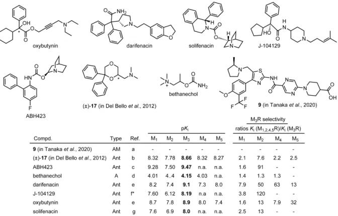
Although potent anticholinergics for the treatment of, e.g. COPD or overactive bladder (OAB), are being therapeutically used, most have little subtype selectivity. Oxybutynin (Dridase®), darifenacin (Emselex®) and solifenacin (Vesikur®), used for the treatment of OAB, are the only licensed drugs, exhibiting a certain M3R selectivity.
The M3R antagonist J-104129 shows a high degree of selectivity for the M3R over M2R (Figure 1.6).119 However, the bronchodilating activity of J-104129, when applied orally in COPD patients, was inferior to the dilating effect achieved by inhalation of the rather unselective compound ipratropium bromide, and additional side effects like dry mouth were more pronounced in the J-104129 treatment group of a 6-week phase II study comprising 412 COPD patients.120 This study demonstrated, that high M3R over M2R selectivity of an oral antimuscarinic agent is not enough to improve the therapeutic effect in patients with COPD compared with inhaled non-selective antimuscarinics.121 The experimental ligand ABH423 (Figure 1.6) derived from the non-selective antagonist 3-quinuclidinylbenzilate (QNB) showed 91-fold selectivity over the M2R and was further modified in various positions, yielding ligands exhibiting 46- to 68-fold M3R over M2R selectivity (data not shown).122
The tertiary amine (±)-17 (in Del Bello et al.), derived from 1,4-dioxane, preferred M3R by approx. 2-fold over M1,4,5R and by approx. 8-fold over M2R and proved to be effective in reducing the contraction of rat urinary bladder (Figure 1.6)123 Moreover, highly selective M3R agonists have not been described as well. Bethanechol (Myocholine-Glenwood®), which stimulates contraction of the bladder and expulsion of urine, is the only M3R agonist showing
Figure 1.6. Chemical structures of M3R preferring ligands (A = agonist; Ant = antagonist; AM = allosteric modulator) reported in literature. References are as follows: (a) Tanaka et al.,124 (b) Scapecchi et al.,103 (c) Fischer et al.,122 note: Ki values for MR subtypes M4 and M5 were not provided; (d) Jakubik et al.,38 note: Ki
value for M5R was not provided; (e) Abrams et al.,125 (f) Mitsuya et al.,119 (g) Ikeda et al.;126 n.a. not analyzed;
*reported Ki values were converted to pKi values. note: in the case of AMs, the determination of binding data by competition binding with labeled orthosteric ligands is not feasible.
In 2020, the positive allosteric modulator (PAM) 9 (in Tanaka et al.) was discovered (for structure see Figure 1.6), showing moderate to high subtype selectivity over the other MR subtypes, which was assessed by the Carbachol (CCh) dependent Ca2+ increase in functional assays.124 When used at a concentration of 10 µM, compound 9 (in Tanaka et al.) caused a 269-fold decrease in the EC50 of CCh (M3R activation), whereas the decrease in the EC50 of CCh was lower in the case of other MR subtypes (M1R: 2.3-fold, M2R: 1.3- fold, M4R: 1.0-fold, M5R: 94-fold).124
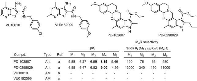
1.3.5. M4 receptor preferring ligands
To date, no orthosteric selective M4R agonists, which might be useful for treating positive symptoms (e.g. hallucinations, delusions) of SZ associated with hyperdopaminergia,127-129 have been discovered. However, activation of the muscarinic M4 receptor can also be mediated by allosteric ligands. The positive allosteric modulator (PAM) VU10010 represented a breakthrough when described in 2008 (Figure 1.7).130 When used at a concentration of 10 µM, VU10010 induced a 47-fold potentiation of M4R ACh potency, whereas the response to ACh in cell lines expressing each of the other MR subtypes was unaffected.130
Chapter 1
Figure 1.7. Chemical structures of M4R preferring ligands (Ant = antagonist; AM = allosteric modulator) reported in literature. References are as follows: (a) Böhme et al.131 (b) Shirey et al.,130 (c) Brady et al.;132 note: in the case of AMs, the determination of binding data by competition binding with labeled orthosteric ligands is not feasible.
Subsequent optimization approaches led to the discovery of VU0152100, which induced a dose-dependent shift in ACh potency (maximal increase over control: 70-fold).132
On the other hand, also the development of selective M4R antagonists, which were suggested to restore the dopamine acetylcholine balance in patients with PD, has been challenging.133 To date, only PD-102807, PD-0298029 and a few other benzoxazine analogues were described (Figure 1.7).131 The former is still used in scientific research for studying the effects of the different MR subtypes in the brain and in the periphery.134-136 PD- 0298029 exhibited poor bioavailability and rapid metabolism in animal studies, which limits its use to in vitro studies.137
In summary, selective M4R ligands, representing drug candidates, e.g. for the treatment of AD, SZ or PD, are not available to date.
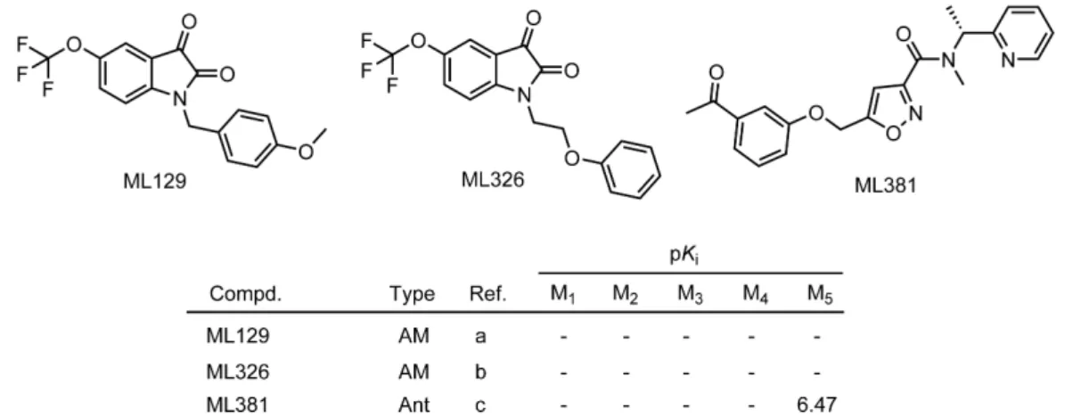
1.3.6. M5 receptor preferring ligands
To date, no selective M5R agonists have been discovered, however, a series of M5R positive allosteric modulators was described (Figure 1.8). The first PAM for the M5R, ML129, was reported in 2009, provoking higher potentiation of ACh induced intracellular Ca2+- mobilization at the M5R compared to the M1R-M4R (EC50 values of ACh in the presence of ML129: M1R-M4R: >30 µM, M5R: 1.16 µM).138 Only a few years later, ML326 was discovered as the first sub-micromolar, selective M5R PAM (EC50 values of ACh in the presence of ML326: M1R-M4R: >30 µM, M5R: 410 nM) (Figure 1.8).139
Figure 1.8 Chemical structures of the positive allosteric M5R modulators ML129 and ML326 and the M5R antagonist ML381 (Ant = antagonist; AM = allosteric modulator). References are as follows: (a) Bridges et al.,138 (b) Gentry et al.,139 (c) Gentry et al.;140 note: in the case of AMs, the determination of binding data by competition binding with labeled orthosteric ligands is not feasible.
Shortly thereafter, in 2014, the orthosteric M5R antagonist ML381 was reported (Figure 1.8).
According to the results from functional studies, i.e. the inhibition of ACh induced Ca2+
response, this compound displayed M5R selective antagonism (IC50 (M1R) = >10 µM;
IC50 (M2R-M4R) = >30 µM; IC50 (M5R) = 0.45 µM). However, MR binding data of ML381 were only reported for the M5R (Figure 1.8).140
1.4. Scope and objectives
Muscarinic acetylcholine receptors (MRs) are widely distributed in the central and peripheral nervous system, being (potential) targets for the treatment of various diseases, such as Alzheimer’s disease (AD), Parkinson’s disease (PD), schizophrenia (SZ), overactive bladder (OAB) or chronic obstructive pulmonary disease (COPD). Due to the high conservation of the orthosteric binding pocket within the family of MRs, the development of highly selective MR ligands has been extremely challenging, and to date, no MR ligands, which are selective for one of the five subtypes, are available as licensed drugs. Highly subtype-selective MR ligands are needed as new therapeutics with lower side effects, but also as molecular tools required, e.g. for pharmacological studies. As the vestibule (common allosteric binding site) of MRs is less conserved than the orthosteric binding pocket, the dualsteric ligand approach, i.e. the design of ligands interacting with both, the orthosteric and the allosteric binding site, has been considered useful for the development of highly subtype selective MR ligands,84-88 This approach benefits from recently reported MR crystal structures, which are available for all MR subtypes.67-75
Lately, conjugation of small peptides to the M2R preferring dibenzodiazepinone-type MR antagonist DIBA (referred to as DIBA-peptide conjugates) was shown to considerably influence MR selectivity.117 The introduction of basic amino acids into the peptide moiety,

![Figure 2.3. Radioligand displacement curves obtained from competition binding experiments with [ 3 H]NMS (0.2 nM (M 1 R, M 2 R, M 3 R), 0.1 nM (M 4 R) or 0.3 nM (M 5 R) and compounds 89, 103-105, 109 and 95 at intact CHO hM x R cells (x = 1-5)](https://thumb-eu.123doks.com/thumbv2/1library_info/3726883.1508307/54.892.121.751.131.808/figure-radioligand-displacement-obtained-competition-binding-experiments-compounds.webp)