Contents lists available atScienceDirect
Algal Research
journal homepage:www.elsevier.com/locate/algal
Production of extracts with anaesthetic activity from the culture of Heterosigma akashiwo in pilot-scale photobioreactors
J.J. Gallardo-Rodríguez
a,b,⁎, A. Astuya-Villalón
c, V. Avello
c, A. Llanos-Rivera
c, B. Krock
d, C. Agurto-Muñoz
e,f, A. Sánchez-Mirón
a, F. García-Camacho
aaDepartment of Chemical Engineering, University of Almería, Spain
bDepartment of Chemical Engineering, Faculty of Engineering, University of Concepción, Concepción, Chile
cLaboratory of Cell Culture and Marine Genomics, Department of Oceanography, Faculty of Natural and Oceanographic Sciences, COPAS Sur-Austral Program, University of Concepción, Concepción, Chile
dAlfred Wegener Institut-Helmholtz Zentrum für Polar- und Meeresforschung, Chemische Ökologie, Am Handelshafen 12, 27570 Bremerhaven, Germany
eDepartment of Food Science and Technology, Faculty of Pharmacy, University of Concepcion, Concepcion, Chile
fGIBMAR, Biotechnology Centre, Concepción University, Concepción, Chile
A R T I C L E I N F O
Keywords:
Raphidophytes Anaesthetic Bioprocess Photobioreactor
A B S T R A C T
The shear-sensitive microalgaHeterosigma akashiwois known to produce brevetoxin-like compounds that are active in voltage-dependent sodium channels. In this work, we present a study on the production of anaesthetic extracts fromH. akashiwobiomass obtained in low-shear bioreactors at different growth phases. The photo- bioreactors (PBRs) used had specific configurations and were operated in such a way as to avoid cellular damage by hydrodynamic stress. Cultures were developed in small static-controlflasks and PBRs with volumes ranging from 1.5 L to 200 L. The bioactivity of the produced extracts was assessed in vitro (Neuro-2a cell-based assay) and in vivo (Zebrafish model). Bioactivity depended slightly on the growth phase and culture system, with greater toxicity with the Neuro-2a model when stationary-phase extracts were used. Interestingly, extracts were not cytotoxic against other human cell lines. Steady production of anaesthetic bioactives was observed. In vivo anaesthetic efficacy, assessed with the Zebrafish model, was similar to that of commercial products, and without any observed mortality at 24-h post exposure.
1. Introduction
Dinoflagellate and raphidophyte microalgae have gained bio- technological importance in recent years due to their ability to produce bioactive molecules, mostly polyketides. The wide inter- and intra- species diversity implies the potential of discovering new molecules [1].
However, because it is difficult to obtain stable and repeatable cultures of these microalgae species, there have been only a few marketable applications to date [2]. Polyketides from fungi and microalgae are among the chemical groups screened for antitumor drugs. Other in- teresting bioactivities of this family of structures that are being studied as potential therapeutic drugs include immunosuppressors and drugs for infectious diseases [3]. Most dinoflagellate and raphidophyte mi- croalgae produce multiple bioactive compounds, some of which are chemical species that typically share structural features (e.g. polyke- tides) but have different bioactivities. For instance,Karenia brevissyn- thesises brevetoxins and its antagonist, brevenal [4]. While this is
interesting from an ecological or chemical point of view, it is challen- ging for bioprocessing. In these cases, culture conditions that trigger the synthesis of bioactives must be determined. The bioreactor culture continues to be the only alternative for obtaining microalgal bioactives.
Although technologically possible and an option for derivatives, che- mical synthesis is not feasible from an economic point of view.
Lack of knowledge about metabolism and optimal growth condi- tions and conditions to enhance bioactive production are among the difficulties in culturing dinoflagellate and raphidophyte microalgae [2].
WithProtoceratium reticulatum,it has been observed that certain oper- ating conditions increase toxin cell content, such as the level of agita- tion [5]. However, stable long-term agitated cultures significantly de- crease yessotoxin production [6]. The major culture challenge is microalgal sensitivity to hydrodynamic stress and bioreactor design.
Concentrations of dinoflagellate microalgae in photosynthetic cultures of dinoflagellate have generally been low compared to those of non- dinoflagellate microalgae, although there have been advances in recent
https://doi.org/10.1016/j.algal.2019.101760
Received 9 August 2019; Received in revised form 7 December 2019; Accepted 9 December 2019
⁎Corresponding author at: Department of Chemical Engineering, University of Almería, Carretera Sacramento s/n, E04120 Almería, Spain.
E-mail address:jgr285@ual.es(J.J. Gallardo-Rodríguez).
2211-9264/ © 2019 Elsevier B.V. All rights reserved.
T
years. Recent works withAmphidiniumandKarlodiniumhave demon- strated that bioprocesses are feasible with some shear-sensitive species [7,8]. In highly controlled systems, biomass concentrations can reach nearly 1 g·L−1[7]. Nevertheless, metabolite production is still a bot- tleneck because of the low quantities (pg per cell) and the high degree of dependence on culture conditions [6,9].
Raphidophytes like Heterosigma akashiwo, Fibrocapsa japonica, Chattonella spp. andPseudochattonellaare known to cause mass mor- talities of cultivated and wildfish [10]. In particular,H. akashiwohas had an enormous impact on the coasts of Japan [11] and in other parts of the world [12,13]. Attempts have been made to determine the ich- thyotoxic mechanisms of most HAB-forming species [14]. Proposed mechanisms include the production of reactive oxygen species (ROS) as the main source of damage, although the release of high free fatty acids has a synergistic effect. There is also an undeniable ichthyotoxic effect of toxins produced by several species, includingH. akashiwo[15–17].
Some of these molecules can cause respiratory and/or cardiac paralysis and abnormal behavior [17,18]. The raphidophyceae class (e.g. H.
akashiwo) has been suggested as a brevetoxin or brevetoxin-like com- pound producer [19], although some authors have questioned this [20].
The amount of brevetoxin and brevetoxin-like compounds produced is highly variable, and can even be at undetectable levels [19–21]. The crude extract ofH. akashiwoCCMP302 showed an inhibitory effect on the voltage-dependent sodium channel in the neuronal model neuro-2a [16]. Interestingly, this suggests the presence of compounds with an- aesthetic activity, with potential commercial value because their effects are similar to those of saxitoxin and tetrodotoxin [16].
This work reports on the feasibility of the microalgaH. akashiwo CCMP302 to produce valuable bioactives when it is cultured photo- autotrophically at different scales. Biomass extracts harvested at dif- ferent growth phases were evaluated for anaesthetic activity (models based on Zebrafish and Neuro-2a CBA) and cytotoxicity.
2. Materials and methods 2.1. The microalga
The marine microalgaHeterosigma akashiwo(strain CCMP302) was used. The alga was obtained from the Provasoli-Guillard National Center for the Culture of Marine Phytoplankton (Maine, USA). Inocula of H. akashiwo were grown in static flasks (without hydrodynamic stress) at 20 ± 1 °C under continuous illumination. 58 W fluorescent lamps were used for illumination and the irradiance at the surface of the cultureflasks was 100μmol photon m−2s−1. Flasks were not aer- ated, but gently agitated manually once a day. The culture medium was supplemented with L1 and prepared in autoclaved natural seawater [22].
2.2. Cultivation in photobioreactors
Two types of PBRs were used for microalgal biomass production: (i) a 1.5-L conventional stirred glass bioreactor (New Brunswick, BioFlo model No. 030–032) with a six-bladed Rushton turbine as an impeller (SPBR1.5); (ii) three bubble columns with culture capacities of 2 L (PBR2), 12 L (PBR12) and 200 L (PBR200). The SPBR1.5 was baffled and had the following relevant dimensions: 0.11 m culture depth;
0.11 m vessel diameter; 1.5 cm clearance of the impeller from the bottom of the vessel; 0.05 m impeller diameter. The impeller blades were 0.75 cm long and 0.4 cm wide.
The three bubble columns, PBR2, PBR12 and PBR200, were tubes made of transparent polyvinyl chloride (PVC; Type I, Grade I compound with a cell classification of 12,454 per ASTM D1784). The tubes had internal diameters of 0.11 m, 0.14 m and 0.45 cm, respectively. Cell suspensions were agitated by air sparging, with filtered air from a compressor and injected through a nozzle sparger placed at the bottom of the column. The gas-free liquid heights were approximately 0.78 m,
1.25 m and 1.25 m, respectively.
Photoautotrophic growth of H. akashiwowas investigated in the PBRs. The culture temperature was controlled at 20 ± 1 °C. The cul- ture pH was not controlled. In all cases the initial pH was set at 8.1. All PBRs were continuously illuminated with spirally wound cool-white- light LEDs. The irradiance at the center of the photobioreactor (filled with seawater) was measured using a QSL-100 quantum scalar irra- diance sensor (Biospherical Instruments, San Diego, USA). Incident ir- radiance (photosynthetically active) was 100μmol photon m−2s−1. The photobioreactors and associated pipework were cleaned before the experiments to remove salt deposits and sterilized as described pre- viously [6]. The culture medium was prepared as described above. The fresh medium was inoculated with 10%v/v of an inoculum containing algal cells in the late exponential growth phase. The cell concentration in the freshly inoculated photobioreactors was around 20,000 cells mL−1.
Two experimental sets were carried out. Firstly, as a prior step to PBR mass culture, tolerance of H. akashiwo to averaged energy dis- sipation rates (EDRs) developed in mechanical (SPBR1.5) and pneu- matic photobioreactors (PBR2) was characterized with short-term cul- tures (250 min). The agitation rate (N) was varied from 50 rpm to 200 rpm in SPBR1.5. The specific airflow rate (Q) was varied from 0.2 to 0.6 v v−1min−1in PBR2. The assays are summarized in detail in Table 1. Once EDR-damage thresholds were determined from the re- sults of thefirst experimental set. A conservativeQvalue was selected to mass cultureH. akashiwoin the aerated photobioreactors PBR12 and PBR200. The effect of scaling-up was also studied in the second set. A culture in a 1-Lflask was used as a control. The cultures were produced batchwise in all PBRs.
2.3. Hydrodynamic characterization of photobioreactors
The acid-tracer method as described previously was used to measure the mixing time (θm) [6]. Briefly, the pH level was altered by adding hydrochloric acid (35% wt/vol). The mixing time was calculated as the time elapsed to reach 95% of thefinal pH value. All experiments were carried out in triplicate. TheEDRvalue in the stirred bioreactor was calculated using the broadly applicable method of Ruszkowski [23], as follows:
=⎛
⎝⎜ ⎞
⎠⎟ EDR A T
θ D
·
m·
3
13 (1)
whereAis a constant with a value of 5.9,Dis the impeller diameter (m),Tis the tank diameter (m) andθmis the measured mixing time. Eq.
(1)indicates that the relationship betweenEDRandθmis independent of the type of stirrer.EDRin the bubble columns is related to surface gas velocity (ug), according to Eq.(2)[24],
= ∙
EDR g ug (2)
where g is gravitational acceleration constant. Theugvalue can easily Table 1
Summary of the experimental design to investigate the tolerance ofH. akashiwo to the hydrodynamic stress in mechanically agitated (SPBR1.5) and aerated (PBR2) cultures. (N: impeller rotational speed;EDR: averaged energy dissipa- tion rate;θm: mixing time;λ: Kolmogorov's microscale of turbulence).
PBR N, rpm Q, v v−1min−1 θm, s EDR, mW kg−1 λ,μm
SPBR1.5 50 – 14.0 1.8 51.6
100 – 9.3 6.9 36.9
150 – 6.5 19.9 28.4
200 – 6.0 25.3 26.7
250 – 5.5 32.9 25.0
PBR2 – 0.2 16.0 3.4 44.0
– 0.4 10.5 6.9 37.0
– 0.6 6.3 10.3 33.4
be obtained by dividing the gasflow rate by the column cross-section area. Kolmogorov's microscale of turbulence was obtained from the following equation:
⎜ ⎟
= ⎛
⎝
⎞
⎠
∙ −
λ μ
ρL EDR
L 3/4
1/4
(3) whereμLandρLare broth viscosity and density, respectively.Table 1 shows the values ofθm,EDRandλfor thefirst experimental set.
2.4. Kinetic parameters
Growth was determined by cell counts. Broth samples (1 mL) were collected daily,fixed with Lugol's solution, and then cells were counted on a Sedgewick-Rafter counting slide. The maximum cell-specific growth rate (h−1) in the exponential growth phase was calculated using the following equation:
= −
μ N N
t t ln( / )
max
0
0 (4)
whereNandN0are cell concentrations (cells mL−1) at culture timest andto(days), respectively, in the interval when the specific growth rate is at a maximum. The doubling time (Td) was calculated as:
= T Ln
μ (2)
d
max (5)
The maximum cell-specific death ratekd(h−1) in short-term culture experiments to determine EDR thresholds was calculated using the following equation:
= − ∙
(
NN)
k tln d
0 (6)
whereNandN0are the cell concentrations (cells mL−1) at timestand initial time, respectively, andkdis the maximum death rate.
2.5. Preparation of H. akashiwo extracts
Broth samples of 20 mL were taken from theH. akashiwocultures in exponential and stationary phase conducted inflasks and PBRs. The cell pellets obtained by centrifuging the samples at 3000 g were extracted with 4 mL of methanol. The extracts were centrifuged at 10000gfor 5 min, and the supernatants were stored in darkness at 20 °C prior to use. The activity of the extracts was evaluated by the cell bioassay Neuro-2a (see M&M Section 2.7). The extracts were subjected to gravity-flow purification. 500-mg C-18 columns were used, previously conditioned with HPLC grade methanol and deionized water. Subse- quently, 1 to 1.5 mL of the methanolic extract was loaded. The sample was eluted with 100% MeOH, dried in a stream of nitrogen at 45 °C and then resuspended in DMSO.
2.6. Tandem mass spectrometric analysis of brevetoxins and brevenal The presence of brevetoxins (PbTx and BTX) and brevenal in the extracts was analyzed in a Waters XEVO TQ-XS tandem quadrupole atmospheric pressure (API) mass spectrometer (Waters, Eschborn, Germany) equipped with a high-performance ZSpray dual-orthogonal multi-mode (ESI/SPCI/ESCi) source, coupled to a Waters Acquity UPLC system consisting of a Waters Acquity UPLC I-Class Solvent Manager, a Waters Acquity UPLC I-Class flow-through-needle (FTN) Sample Manager, and a Acquity UPLC I-Class column manager (CM-A).
Potential lipophilic toxins in the extracts were separated by injection 0.5μL of sample by reversed-phase chromatography on a C18 phase.
The analytical column (50 × 2.1 mm) was packed with 130 Å and 1.7μm of C18, and maintained at 40 °C. The flow rate was 0.6 mL·min−1and gradient elution was performed with two eluants.
Eluant A was water containing 0.01% formic acid and 0.05%
ammonium hydroxide, and eluent B was acetonitrile containing 0.01%
formic acid. Initial conditions were 0.5 min column equilibration with 30% eluant B, followed by a linear gradient to 90% B in 3 min and isocratic elution until 4 min with 90% B. The system was then returned in 0.1 min to initial conditions (total run time: 5 min).
Full scan experiments were performed in the mass range ofm/z600 tom/z1000 in the positive ion mode. Mass spectrometric parameters were as follows: capillary voltage 3.5 kV, cone voltage 40 V, desolvation temperature 600 °C, desolvation gas flow 1000 L·h−1, cone gasflow 150 L·h−1, nebuliser pressure 7.0 bar, collision gasflow 0.15 mL·min−1, collision 20 eV, and a source temperature of 150 °C.
Collision-induced dissociation (CID) produced ion spectra were re- corded for the pseudomolecular ion of brevenalm/z657. CID spectra were recorded with the same instrument in the ScanWave Daughter scan (ScanWave DS) modus in a mass range ofm/z50 to 660, and in a positive ionization and unit resolution mode. All mass spectrometric parameters were as described above for the full scan experiment. The mass transitions of brevetoxins and brevenal were set as detailed in Table 2.
2.7. Neuro-2a cell-based assay (CBA)
The neuro-2a cytotoxicity bioassay, as reported by Astuya et al. [16]
was used to detect brevetoxin-like neurotoxins acting on voltage-gated sodium channels (VGSCs). Briefly, neuro-2a mouse neuroblastoma cells (ATCC, CCL-131) were treated with the Na+/K+-ATPase inhibitor ouabain (0.3 mM), the sodium channel-activator veratridine (0.03 mM) andH. akashiwo extract (12.5–100μg·mL−1). The reduction of cyto- toxicity in the presence of ouabain/veratridine (0.3/0.003 mM), or“cell rescue”, is indicative of sodium channel blocking or paralyzing toxins.
The enhancement of cytotoxicity in the presence of ouabain/veratridine (0.1/0.01 mM) is indicative of potent sodium channel activators like brevetoxin-like toxins [25–27]. The cytotoxicity of the H. akashiwo extract unrelated to the VGSC effect was ruled out by exposing neuro-2a cells to the extract in the absence of ouabain/veratridine. Cell viability was measured after 24 h-incubation using MTT (3-(4,5-dimethylthiazol- 2-yl)-2,5-diphenyltetrazolium bromide, Life Technologies, Carlsbad, CA, USA) [25]. Absorbance was read at 570 nm using an automated multi-well scanning spectrophotometer (Synergy H1, Biotek, Vermont, USA). Cell viability was expressed as a percentage to control cells without the extracts (100% viability). Thus, the relative viability effect was represented as cell rescue. The cell rescue results of the neuro-2a CBA (presence of O/V) were expressed relative to the viability of cul- tures where saxitoxin standard (50 nM) was used as a control. These control tests showed viabilities > 90%. The standard was obtained from the Institute for Marine Biosciences, National Research Council of Canada. Concentrations of extracts of 100 mg·L−1were excluded since toxicity would mask cell rescue. Maximum cell rescue was obtained in the range of 25–50 mg·L−1.
Table 2
Mass transitions (m/zQ1 > Q3) used to detect brevetoxins and brevenal.
Toxin Q1 mass (m/z) Q3 mass (m/z)
PbTx-1 867.5 849.5
PbTx-2 895.5 877.5
PbTx-3 897.5 879.5
PbTx-6 911.5 893.5
PbTx-7 869.5 851.5
PbTx-9 899.5 863.4
PbTx-10 871.5 853.5
BTX-B1 985.5 967.5
BTX-B2 1034.6 1016.6
Brevenal 657.4 639.4
2.8. Determination of the anaesthetic efficiency of the H. akashiwo methanolic extracts
Rearing, handling and experimental procedures were approved by the Bioethics Committee of the University of Concepción. A breeding stock of zebrafish (Danio rerio) wild type strain was maintained in ac- cordance with international guidelines for zebrafish housing and hus- bandry; at 27 ± 1 °C, with a 12:12 h light:dark photoperiod and con- stant waterfiltration. Thefish were fed withArtemiasp. nauplii and TetraMin tropical flakes (Tetra, Hannover, Germany). To obtain the larvae needed for the experiments, spawning was induced daily and the healthy embryos were incubated at 27 °C in embryo medium until hatching occurred. Concentrations of H. akashiwomethanolic extract ranging from 50 to 200 mg·L−1were previously assayed to define an adequate dose for anesthesia induction times below 5 min. Zebrafish larvae at 7–8 days post fertilization (dpf) were exposed toH. akashiwo extract in wells (volume of 200μL). The induction and recovery times of light anesthesia were then evaluated as described earlier [28]. Briefly, light anaesthetic stage is considered when there is partial loss of zeb- rafish larvae equilibrium and no reaction to external stimuli is ob- served. The results indicated that a concentration ofH. akashiwome- thanolic extract of around 200 mg·L−1 induced anesthesia. This concentration was selected to evaluate anaesthetic activity of extracts prepared from PBR cultures. Induction and recovery times were mea- sured for all larvae (N= 5 per experiment). Post-exposure survival was recorded 24 h after the experiments.
2.9. Determination of in vitro cytotoxicity
Antiproliferative assays were conducted as described elsewhere [29,30] with crude methanolic extracts obtained from H. akashiwo biomass as were performed. Four human tumor cell lines obtained from the American Type Culture Collection (ATCC) were used, namely A549 (ATCC CCL-185) (lung carcinoma, NSCLC), HT-29 (ATCC HTB-38) (colon adenocarcinoma), MDA-MB-231 (ATCC HTB-26) (breast ade- nocarcinoma) and PSN-1 (ATCC CRL-3211) (pancreas adenocarci- noma). Cells were maintained in Dulbecco's modified Eagle's medium (DMEM), supplemented with 10% fetal bovine serum (FBS), 2 mML- glutamine, 100 U/mL penicillin and 100 U/mL streptomycin at 37 °C, 5% CO2and 98% humidity. Cells were harvested for the experiments from subconfluent cultures by trypsinization and resuspended in fresh medium before counting and plating. Human tumor cells were seeded in 96-well microtiter plates, at 5000 cells per well in aliquots of 150μL and allowed to attach to the plate surface for 18 h (overnight) in drug- free medium. One control (untreated) plate for each cell line was then fixed and used for the time zero reference value. Cells on plates were then exposed to different methanolic extract concentrations in DMSO/
DMEM (10, 30 and 100μg mL−1). Cell survival was measured after treatment for 72 h. Results are provided as survival percentages: 100%
means there are as many cells as the control (extracts do not inhibit growth); 0% means that there is the same number of cells as at the start of the trial (no growth, extracts inhibit mitosis);−100% means that all the cells were lysed (strong cytotoxic effect). Measurements were taken for triplicate samples. The standard deviations were in most cases <
10%.
2.10. Statistics and data treatments
All experimental data are presented as means ± SD of three in- dependent experiments performed with duplicate determinations. Data were treated and plotted using Prism 4.0 software (GraphPad Inc.).
3. Results and discussion
3.1. Damage energy dissipation rate threshold in agitated cultures of H.
akashiwo
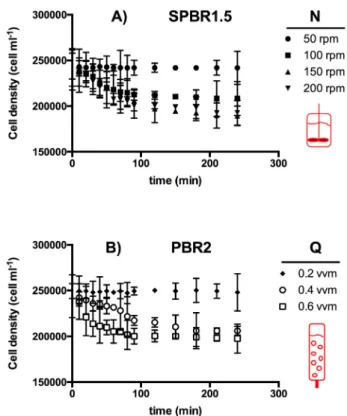
Fig. 1shows the growth kinetics of various short-term agitatedH.
akashiwobatch cultures. The disappearance of cells due to lysis was evident above an impeller speed (N) of 50 rpm with the mechanically agitated photobioreactor (SPBR1.5) (seeFig. 1A) and above an aeration rate of 0.2 v v−1min−1 with the pneumatically agitated photo- bioreactor (PBR2) (seeFig. 1B). Based on the results shown inFig. 1, the maximum specific death rates (kd) were calculated with Eq.(6).
Fig. 2shows thekdvalues and correspondingEDRvalues (seeTable 2).
No EDR-associated damage was observed with either SPBR1.5 and PBR2 at similar EDR values, 1.8 mW kg−1 and 3.4 mW kg−1, Fig. 1.Short-term effect of (A) the mechanical agitation rate (SPBR1.5), and (B) the aeration rate (PBR2) onH. akashiwocell density. Each value is average of triplicate assays. Error bars represent standard deviation.
Fig. 2.Kinetic death rate constant (kd) ofH. akashiwoversus energy dissipation rate (EDR) for mechanical agitation (PBR1.5) and bubbling (PBR2) in bench- scale photobioreactors. Each value is average of triplicate assays. Error bars represent standard deviation.
respectively. As can be observed,kdincreased withEDRin both PBRs, the increase in PBR2 being significantly higher than in PBR1.5. This difference is consistent withfindings in similar shear sensitivity studies reported in the literature. In typical bioreactors, such as stirred tank and bubble column PBRs, cells are exposed to an inhomogeneous EDR field [31]. Local EDR values can be as much as two orders of magnitude higher in the impeller regions or on the liquid surface where bubbles break. Such high EDR values have been estimated to contribute around 70% of the total power in a typical bioreactor. The EDRfields and the proportion of both extensionalflow and shearflow in stirred tanks and bubble columns are also entirely different at similar average EDRs. This means that the history and type of EDR experienced by cells in SPBR1.5 and PBR2 were probably different. The frequency of cell exposure to hydrodynamic extensional stress during the bubble bursting process in PBR12 is the probable cause of the higher kd values than those in PBR1.5, in which cells were grown without bubbling. This extreme sensitivity of some microalgae to bubbling has been reported previously [5,32].
3.2. Scale-up of Heterosigma akashiwo cell yield
Fig. 2showsEDR-associated damage thresholds for the two PBRs:
between 1.8 and 6.9 mW kg−1 for PBR1.5, and between 3.4 and 6.9 mW kg−1for PBR2. It is more difficult to maintain an average EDR in those ranges during PBR scale-up of PBRs with stirred reactors than with bubble columns because the mixing times in stirred reactors in- crease more rapidly with the scale size [31]. The percentage of the mixing power that is non-homogeneously dissipated near the stirrer increases with increases in scale size [33]. The EDR in the bubble col- umns can be kept at low levels by reducing the ratio of gas flow to culture volume. Consequently, the operational EDR values for the PBR12L and PBR200L cultures were established at 5.3 and 5.1 mW kg−1by setting gasflow rates at 0.042 and 0.025 v v−1min−1, respectively, both equivalent to a surface gas velocity of 5.4 10−4m s−1. Theseflow rates allowed the cells to be homogeneously distributed in the PBR. However, degassing and the average time re- quired for a single cell to travel from dark to illuminated areas were clearly lower in the larger PBR.
The scale-up conditions described above were compared in terms of biomass yield for the photoautotrophic growth of H. akashiwo. Two types of culture devices of three sizes were used: (i) static culture (without agitation) in a 1-L Erlenmeyer flask in which CO2was pro- vided by diffusion through a culture-air interface; and (ii) pneumati- cally-agitated cultures in the PBR12 and PBR200, in which mixing and CO2was provided via air bubbling in the culture medium. CO2was not controlled in the PBRs in order to observe mainly the effect of EDR during the scale-up process. The PBR200 also allowed us to determine if growth limitation caused by CO2and/or light had an impact on the bioactive production.Table 3shows the corresponding surface–to–vo- lume ratios in decreasing order in the culture devices.Fig. 3shows the growth kinetics ofH. akashiwo. Maximum specific growth rates in the exponential phase were different among the three culture systems (see Table 3). Kinetics converged at a stationaryfinal concentration around 300,000 cell mL−1in theflask and PBR12 cultures, while a lower cell
concentration of approximately 150,000 cell mL−1 was reached in PBR200.
The slowest doubling time (μmax) was with the PBR200, while the most rapid was with the PBR12 (Table 3). Doubling required nearly two days with the PBR12. These results were expected in photosynthetic cultures due to the advantages of bubbling columns with a high height- to-diameter ratio compared to those with lower ratios [34]. The light availability was maximum for the PBR12 because of a shorter light path. A shorter light path with the same incident irradiance supposes less auto-shading. PBR200 may have been limited by available light since exponential growth was slower. However, the low airflow rate fixed in the PBR200 suggests that CO2 limitation was also an issue during most the culture period. Evidence for this is the linear growth phase of the PBR200 culture from the thirteenth day to the end of the culture in Fig. 3. Only one or both of the two nutrients that were continuously supplied to the culture (CO2 at 0.03% vol/vol in the aeration gas and light) could have caused it and therefore limited growth. That is, the linear growth phase was established when CO2
and/or light energy consumptions balanced the CO2 and photons transfer. To establish if the limiting process was the CO2transfer, an approach based on characteristic time analysis was used as reported elsewhere [35]. Thus, two different characteristic times, such as mass transfer time (tMT) and CO2 consumption time (tC), were taken into account. The characteristic mass transfer time for CO2was evaluated as:
=
t K a
1
MT
L (7)
whereKLais the volumetric mass transfer coefficient for CO2. For augof 5.4 10−4m s−1in the PBR2000, a KLa value of 3.32 10−4s−1 was obtained from the following equation reported for seawater in pilot bubble column PBRs [36]:
= − −
K a u 2.222
L 1
g1.171
(8) Thus, from Eq.(7) atMT value of 3020 s was calculated. During growth,tCwas expressed as
=
∗
t C
C COμC
Y b
2 1
CO2 (9)
whereCCO2∗is the equilibrium concentration of CO2with a value es- timated of 0.45 g m−3 at 20 °C, Cb is the biomass concentration ex- pressed in terms of dry weight (g m−3) and YCO2 is the CO2 yield coefficient (a value of 0.55 was assumed) [35]. The conversion of the cell concentration data (N) for PBR200 inFig. 3toCbwas performed from the existing correlation betweenCband cell biovolume (Vcell) [7]:
=
Cb ξ N V· · cell (10)
whereVcellwas calculated from the average equivalent mean cell dia- meter (14 10−6m) measured byflow cytometry. A conservativeξvalue Table 3
Kinetic parameters determined from results of Heterosigma akashiwo batch cultures grown in PBRs of different scales: maximum specific growth rate,μmax; maximum doubling time,Td; S/V is the surface–to–volume ratio. Each value is average of triplicate assays.
PBR S/V ratio Mixing μmax(day−1) Td(day)
Flask 4.75 Static 0.264 2.446
PBR12 1.28 Bubbling 0.36 1.882
PBR200 0.79 Bubbling 0.12 5.385
Fig. 3.Effect of scale-up on the cell yield ofHeterosigma akashiwogrown in different photobioreactors. Each value is average of triplicate assays. Error bars represent standard deviation.
has been recently suggested to be around 0.180 g d.w. mL−1in basis to a fresh microalgal cells density of 1.2 g mL−1and a dry mass fraction of fresh cells of 25% [7]. In linear phase, the productμ·Cbin Eq.(10)is constant with a value of 4.7 g m−3day−1. Thus, the characteristic consumption time for CO2,tC, estimated from Eq.(9)was of 190 s. AstC
was clearly lower thantMT, CO2-consumption was controlled by CO2- transfer from the gas phase. Regarding the light, the daily mean volu- metric photon flux absorbed by the culture (Γ) was evaluated to de- termine if the linear phase in the PBR200 culture was co-limited by light. Because the photons received by the walls of the PBR200 were virtually absorbed by the culture in the linear phase (i.e. the light path in the PBR200 and cell concentrations achieved in the linear phase were such that no photonicflow escaped from the PBR200 wall),Γhave to be constant, independent of Cb and close to the maximum photon flux potentially absorbable by the culture (i.e.Γmax). Assuming a two-di- mensional totally diffuse incident light,Γmaxcan be estimated from the followingexpression [8]:
=
Γ I
Rπ 2
max o
(11) whereIo(100μE m−2s−1) is the irradiance measured in the center of the PBR200filled with seawater and R(22.5 10−2m) is the PBR200 radius. The value ofΓmaxcalculated was 283μE m−3s−1, a more than three-fold lesser relative to PBR12. The value ofCbat which Γmax is virtually reached in PBR200 is close to 0.031 kg m−3on day 13, which is the biomass concentration where linear growth phase starts. There- fore, the irradiance supplied to PBR200 seemed to be too low and po- tentially co-responsible for the appearance of the linear growth phase.
This is also supported by previous results obtained with other micro- algae. For example, the linear growth phase of an airlift PBR culture of Phaedactylum tricornutumcommenced with aCbnear 1.2 kg m−3when a Γmaxof 16,500μE m−3s−1was absorbed by the culture [35]. In the case ofAmphidinium carterae, aΓmaxvalue of 2510μE m−3s−1caused a linear growth phase at close toCb0.2 kg m−3[7]. For aΓmaxof 283μE m−3s−1as that measured in the PBR200, both aforementioned mi- croalgae would have hypothetically commenced their linear growth phase at around 0.02 kg m−3, in line with that above estimated forH.
akashiwoin the PBR200.
There have been few reports on pilot-plant scale culturing of the ichthyotoxic microalgaeH. akashiwo, although there are a number of works cataloguing this species as a producer of interesting bioactives [16,19,37]. Fuentes-Grünewald et al. [38] provided an example of in- tensive culture of an H. akashiwostrain. However, research has gen- erally been performed with uncontrolled cultures at laboratory scales with volumes not exceeding a liter. At this scale,H. akashiwogrows optimally [15,39]. As in the present work, volumetric cell productivity was higher, and the maximum specific growth rate was greater in the PBR12.
The scale-up to PBR200 based on maintaining the optimalQvalue constant for PBR12 (i.e. 0.042 v v−1min−1) implied setting EDR at approximately 8.85 mW kg−1in PBR200. Nevertheless, this EDR pro- duced cell lysis in PBR2 (seeFig. 2). There were also noticeable dif- ferences in the aspect ratio (i.e. length-to-diameter ratio) among the bioreactors: 5.6 in PBR12 and 2.8 in PBR200. The interaction of PBR hydrodynamics and geometry (S/V ratio and aspect ratio) provided a more efficient illumination and CO2-transfer regime than that in PBR200. Optimal growth of highly shear-susceptible microalgae in bubble column PBRs is complex because varied design and operational parameters have to be carefully selected to minimize turbulence-asso- ciated damage to cells [40]. Airflow rate alone should not be used to improve cell growth and mitigate damage in sparged photobioreactors because other parameters such as CO2molar fraction in the injected air, column volume, sparger geometry details, and mass transfer, should also be taken into consideration. A model reported for shear-sensitive dinoflagellate microalgae provides guidance on how several of these types of parameters can be manipulated to reach adequate mass transfer
and to avoid cell damage [40,41]. In principle, pneumatically-agitated culture systems like bubble columns are candidate PBRs for the pro- duction of bioactives fromH. akashiwo. Any further scale up of pro- duction should rely on increasing the number of PBRs. Future research should be aimed at optimizing the PBR unit to be used.
3.3. Functional biological characterization of H. akashiwo extracts Neurotoxins acting on VGSCs were assessed according to the pro- tocol described by [16]. First, we evaluated the cytotoxic effect of the extract ofH. akashiwoon the neuro-2a model and then determined the
“rescue activity”caused by a blockage of the Na+channel. The ability to block the Na+channel is similar to that of TTX or brevenal, which have been shown to produce anesthesia [16,42]. Thefirst assessment was carried out to determine cytotoxicity unrelated to any effect on the Na+channel.Fig. 4A shows the percentages of viable neuro-2a cells exposed to H. akashiwo methanolic extracts ranging from 12.5 to 100 mg L−1. Extracts were prepared from biomass harvested in the exponential and stationary phases from the cultures in PBR12 and the Erlenmeyerflask. There were evident cytotoxic effects in extracts from cultures in PBR12 and the flasks at concentrations of 50 and 100 mg L−1, respectively. The mortality rate was 100% in the PBR12 culture extracts of the exponential and stationary growth phases. The viability offlask-grown neuro-2a cells from the exponential and sta- tionary phases decreased respectively by 30 and 50%. Although the cytotoxic effects on the cultures grown inflasks and PBR12 were si- milar, the effects on extracts from the PBR12 culture were much greater, with little difference among growth phases. For the flask Fig. 4.A) % Viability of neuro-2a CBA withHeterosigma akashiwomethanolic extracts prepared from biomass harvested in exponential and stationary growth phases fromflask and PBR12 cultures; B) Normalized cell rescue (increase in viability) of neuro-2a cells exposed to ouabaine/veratridine andH. akashiwo extracts. A value of 1 represents recovery of total viability using saxitoxin standard (50 nM). Each value is average of triplicate assays. Error bars re- present standard deviation.
cultures, the extract obtained during the exponential growth phase had a greater impact on viability than the stationary one.
Interestingly, the 100 mg·L−1 extract from PBR12 did not show cytotoxic effects against four human tumor cell lines typically used in standard antiproliferative assays. This indicates that the cytotoxic effect on neuro-2a cells cannot be extrapolated to other cell lines. This is confirmed byFig. 4B, which shows the experimental data of cellular rescue produced by the Heterosigma akashiwoextracts when the O/V opens the Na+channel. The results indicate a biological effect of the extracts similar to those of other paralyzing marine biotoxins [16].
However,Fig. 4B shows a deviation in this pattern for the 50 mg·L−1- extract from the PBR12-exponential biomass because there was less rescue activity.
A brevetoxin-like bioactive produces cell death rather than cell rescue in the presence of O/V (Fig. 4B). In that case, negative values of normalized cell rescue are obtained. Overall, the results shown in Fig. 4B indicate that the compounds in the extracts from the PBR12 and flask cultures with bioactivity related to the Na+channel are similar.
Saxitoxin-type bioactives seem the most likely candidates. Assuming that the viability values in Fig. 4A are a proxy for concentration of saxitoxin-type compounds in the extracts, the effect of growth phase and PBR type on the viability of neuro-2a cells confirms that the bio- synthesis of this kind of bioactives is a complex process, as reported for other microalgae [44].
3.4. Chemical analysis of the H. akashiwo extract
Although functional assays have clearly shown that extracts ofH.
akashiwo have brevenal-like activity, nothing is known about the identity of the chemical compounds eliciting these effects. However, several studies have reported the presence of brevetoxins in raphido- phytes [19,44,45]. Astuya et al. [16] reported the presence of a pa- ralyzing toxin (HaTx) in H. akashiwo extracts that block or inhibit voltage-dependent sodium-channels (VDSCs), a behavior similar to those of saxitoxin and tetrodotoxin, but also to that of brevenal, which acts as an antagonist of brevetoxins [4].
In the light of these reports, the extracts of the raphidophyte H.
akashiwowere analyzed for the presence of brevetoxins and brevenal.
None of the brevetoxins listed inTable 1were detected in any of the analyzed CCMP302 H. akashiwo extracts. The detection limit (S/
N > 3) was calculated as 0.1 fg cell−1for PbTx-2 and was in the same order of magnitude for the other brevetoxins, of which standard solu- tions were available (PbTx-3,−6 and−9). The absence of brevetoxins has also been reported for the raphidophyteFibrocapsa japonica[20], and raises questions about previous reports of the production of bre- vetoxins by raphidophytes. In contrast to the absence of any known brevetoxins, all extracts showed a brevenal peak at mass transition (m/z 657.4 > 639.4) at the retention time of 3.5 min. As there was no brevenal standard solution available for compound identification, a collision induced dissociation (CID) spectrum was recorded to obtain more detailed information about this compound. The pseudo-molecular ion was the base peak of the CID spectrum of them/z657H. akashiwo compound, with very few fragments. The second most abundant frag- ment was water loss from the pseudo-molecular ion and smaller frag- ments in low abundance atm/z109, 121, 149 and 215. In contrast, the spectrum of brevenal was characterized by six consecutive water loss events from the pseudo-molecular ion, which is very characteristic of polyketides like brevenal, and a few of smaller fragments, of which the most abundant werem/z159 and 255. The discrepancies among the fragments of brevenal and the isobaric compound inH. akashiwoare clear evidence thatH. akashiwodoes not produce brevenal and that the isobaric compound found inH. akashiwois not chemically related to polyketides of the brevetoxin group. The absence of brevetoxins and brevenal in theH. akashiwoextracts shows thatH. akashiwoproduces chemical compound(s) that are structurally unrelated to brevetoxins, but that elicit similar biological functions. The lack of knowledge about
secondary bioactive metabolites of raphidophytes and other ichthyo- toxic species clearly demonstrates the need for further research in this field.
3.5. Anaesthetic efficiency
The in vivo anaesthesic effect ofH. akashiwoextracts was evaluated with the zebrafish larval model. Zebrafish have been widely used in the evaluation of anaesthetics, both from the perspective of animal care [46,47] and the search for optimal anaesthetics for vertebrates [48].
Recently, zebrafish larvae have been proposed for testing analgesic efficacy and nociception [49,50], given that larvae have a similar re- sponse to a noxious challenge to that of adult zebrafish and other vertebrates. Induction and recovery times in our assays were below 6 min when extracts of 200 mg·L−1 were used (Fig. 5). We did not observe mortality for 24 h after exposure. There were differences in anaesthetic induction times based on culture methods and phases, with the shortest times in extracts fromflask cultures harvested in the sta- tionary phase (Fig. 5). Recovery times were lower in extracts from the PBR12 culture (ANOVA p < 0.001) in stationary phase. Nevertheless, differences remained low (Fig. 5). Although there were significant differences in anaesthetic efficiency between culture methods and phase of cultivation, the ranges of induction and recovery times showed that culture in the PBR12 did not change the ability of the extracts to induce anesthesia. The induction and recovery times were close to those desired for an anaesthetic agent (induction 3 min, recovery 5 min [28]).
As well, the same quantified order of magnitude was observed for zebrafish [51] and species of commercial importance [52] as with an- aesthetics currently in use.
4. Conclusions
Cultures ofH. akashiwowere grown in PBRs of up to 200 L at an EDR of 5.1 mW kg−1with a gasflow of 0.025 v v−1min−1. Under these conditions, we observed a homogeneous distribution of microalgal cells in the PBRs and the absence of hydrodynamic damage. However, growth was optimal in the PBR12 culture due to the narrower light path and better gassing/degassing. Chemical analysis of the biomass re- vealed that H. akashiwo produces chemical compounds unrelated structurally to brevetoxins or brevenal. The methanolic extract of har- vested biomass did not show cytotoxic (up to 50 mg·L−1) or saxitoxin- like effects. PBR cultures produced biomasses with similar character- istics in the different growth phases, although the stationary phase showed higher toxicity in the neuro-2a cellular model. However, cy- totoxicity evaluated on four human tumor cell lines showed that the crude methanolic extract did not present cytotoxicity. The anaesthetic effect of the methanolic extract, assessed according to the Zebrafish model, was similar to those of commercial products, and without Fig. 5.Light anesthesia induction and recovery times in Zebra fish larvae produced byHeterosigma akashiwomethanolic extracts (200 mg·L−1) prepared from biomass harvested fromflask and PBR12 cultures in the exponential and stationary growth phases. Each value is average of triplicate assays. Error bars represent standard deviation.
observed mortality at 24-h post-exposure.
“No conflicts, informed consent, or human rights are applicable to this study. Animal rights: Rearing, handling and experimental work with zebrafish larvae were carried out under protocols approved by Bioethics Committee of University of Concepción and in accordance with internationally established procedures.”
Declaration of competing interest
The authors declare that:
o All authors have participated in (a) conception and design, or ana- lysis and interpretation of the data; (b) drafting the article or re- vising it critically for important intellectual content; and (c) ap- proval of thefinal version.
o This manuscript has not been submitted to, nor is under review at, another journal or other publishing venue.
o The authors have no affiliation with any organization with a direct or indirectfinancial interest in the subject matter discussed in the manuscript.
o The authors have no other conflict of interests regarding the sub- mitted manuscript.
Acknowledgements
This research was mainly funded by the projects Fondecyt 1170515 (Conicyt, Chile) and Fondef IDEA 2017 ID17I10100 (Conicyt, Chile).
This research has been also partially funded by the State Research Agency (grants RTC-2017-6405-1 and CTQ2014-55888-C3-02 (MARBIOM)) of the Ministry of Science, Innovation and Universities and the European Regional Development Fund Program and by the Helmholtz-Gemeinschaft Deutscher Forschungszentren through the re- search programme PACES II of the Alfred Wegener Institut-Helmholtz Zentrum für Polar- und Meeresforschung. We would also like to thank Dr. Fernando de la Calle Verdú (PharmaMar S.A., Spain) by performing the antiproliferative assays.
References
[1] J. Assunção, A. Catarina Guedes, F. Xavier Malcata, Biotechnological and phar- macological applications of biotoxins and other bioactive molecules from dino- flagellates, Mar. Drugs 15 (2017),https://doi.org/10.3390/md15120393.
[2] J.J. Gallardo-Rodríguez, A. Sánchez-Mirón, F. García-Camacho, L. López-Rosales, Y. Chisti, E. Molina-Grima, Bioactives from microalgal dinoflagellates, Biotechnol.
Adv. 30 (2012) 1673–1684,https://doi.org/10.1016/j.biotechadv.2012.07.005.
[3] R. Zhang, C. Li, J. Wang, Y. Yang, Y. Yan, Microbial production of small medicinal molecules and biologics: from nature to synthetic pathways, Biotechnol. Adv. 36 (2018) 2219–2231,https://doi.org/10.1016/j.biotechadv.2018.10.009.
[4] A.J. Bourdelais, S. Campbell, H. Jacocks, J. Naar, J.L.C. Wright, J. Carsi, D.G. Baden, Brevenal is a natural inhibitor of brevetoxin action in sodium channel receptor binding assays, Cell. Mol. Neurobiol. 24 (2004) 553–563,https://doi.org/
10.1023/B:CEMN.0000023629.81595.09.
[5] F. García Camacho, J.J. Gallardo Rodríguez, A. Sánchez Mirón, M.C. Cerón García, E.H. Belarbi, E. Molina Grima, Determination of shear stress thresholds in toxic dinoflagellates cultured in shakenflasks, Process Biochem. 42 (2007) 1506–1515, https://doi.org/10.1016/j.procbio.2007.08.001.
[6] F.G. Camacho, J.J.G. Rodríguez, A.S. Mirón, E.H. Belarbi, Y. Chisti, E.M. Grima, Photobioreactor scale-up for a shear-sensitive dinoflagellate microalga, Process Biochem. 46 (2011) 936–944,https://doi.org/10.1016/j.procbio.2011.01.005.
[7] A. Molina-Miras, A. Morales-Amador, C.R. de Vera, L. López-Rosales, A. Sánchez- Mirón, M.L. Souto, J.J. Fernández, M. Norte, F. García-Camacho, E. Molina-Grima, A pilot-scale bioprocess to produce amphidinols from the marine microalga Amphidinium carterae: isolation of a novel analogue, Algal Res. 31 (2018) 87–98, https://doi.org/10.1016/j.algal.2018.01.010.
[8] L. López-Rosales, F. García-Camacho, A. Sánchez-Mirón, E. Martín Beato, Y. Chisti, E. Molina Grima, Pilot-scale bubble column photobioreactor culture of a marine dinoflagellate microalga illuminated with light emission diodes, Bioresour. Technol.
216 (2016) 845–855,https://doi.org/10.1016/j.biortech.2016.06.027.
[9] J.J. Gallardo Rodríguez, A. Sánchez Mirón, F. García Camacho, M.C. Cerón García, E.H. Belarbi, E. Molina Grima, Culture of dinoflagellates in a fed-batch and con- tinuous stirred-tank photobioreactors: growth, oxidative stress and toxin produc- tion, Process Biochem. 45 (2010) 660–666,https://doi.org/10.1016/j.procbio.
2009.12.018.
[10] J.J. Gallardo-Rodríguez, A. Astuya-Villalón, A. Llanos-Rivera, V. Avello-Fontalba, V. Ulloa-Jofré, A critical review on control methods for harmful algal blooms, Rev.
Aquac. (2018) 1–24,https://doi.org/10.1111/raq.12251.
[11] Y. Fukuyo, I. Imai, M. Kodama, K. Tamai, Red tides and other harmful algal blooms in Japan, in: F.J.R. Taylor, V.L. Trainer (Eds.), Harmful Algal Bloom, PICES Reg.
North Pacific, North Pacific Marine Science Organization (PICES), Sidney, 2002, pp.
7–20.
[12] A.L. Clement, Phytoplankton monitoring program in thefish farming region of south Chile, in: T.J.S. Smayda (Ed.), Toxic Phytoplankt. Bloom. Sea, Elsevier Science Publishers B.V., Amsterdam, 1993, pp. 223–258.
[13] J.-S. Ki, M.-S. Han, Nuclear rDNA and chloroplast rbcL, rbcS and IGS sequence data, and their implications from the Japanese, Korean, and North American harmful algae, Heterosigma akashiwo (Raphidophyceae), Environ. Res. 103 (2007) 299–304,https://doi.org/10.1016/j.envres.2006.08.014.
[14] J.J. Dorantes-Aranda, A. Seger, J.I. Mardones, P.D. Nichols, G.M. Hallegraeff, Progress in understanding algal bloom-mediatedfish kills: the role of superoxide radicals, phycotoxins and fatty acids, PLoS One 10 (2015),https://doi.org/10.
1371/journal.pone.0133549.
[15] A. Astuya, A. Rivera, K. Vega-Drake, C. Aburto, F. Cruzat, V. Ulloa, T. Caprile, J.J. Gallardo-Rodríguez, Study of the ichthyotoxic microalga Heterosigma akashiwo by transcriptional activation of sublethal marker Hsp70b in Transwell co-culture assays, PLoS One 13 (2018),https://doi.org/10.1371/journal.pone.0201438.
[16] A. Astuya, A.E. Ramírez, A. Aballay, J. Araya, J. Silva, V. Ulloa, J. Fuentealba, Neurotoxin-like compounds from the ichthyotoxic red tide alga Heterosigma aka- shiwo induce a TTX-like synaptic silencing in mammalian neurons, Harmful Algae 47 (2015) 1–8,https://doi.org/10.1016/j.hal.2015.04.006.
[17] M. Endo, Y. Onoue, A. Kuroki, Neurotoxin-induced cardiac disorder and its role in the death offish exposed to Chattonella marina, Mar. Biol. 112 (1992) 371–376, https://doi.org/10.1007/BF00356281.
[18] J.H. Landsberg, The effects of harmful algal blooms on aquatic organisms, Rev. Fish.
Sci. 10 (2002) 113–390,https://doi.org/10.1080/20026491051695.
[19] S. Khan, O. Arakawa, Y. Onoue, Neurotoxins in a toxic red tide of Heterosigma akashiwo (Raphidophyceae) in Kagoshima Bay, Japan, Aquac. Res. 28 (1997) 9–14, https://doi.org/10.1046/j.1365-2109.1997.t01-1-00823.x.
[20] M.K. de Boer, C. Boerée, S.B. Sjollema, T. de Vries, A.D. Rijnsdorp, A.G.J. Buma, The toxic effect of the marine raphidophyte Fibrocapsa japonica on larvae of the commonflatfish sole (Solea solea), Harmful Algae 17 (2012) 92–101,https://doi.
org/10.1016/j.hal.2012.03.005.
[21] C.J. Keppler, A.J. Lewitus, A.H. Ringwood, J. Hoguet, T. Staton, Sublethal cellular effects of short-term raphidophyte and brevetoxin exposures on the eastern oyster Crassostrea virginica, Mar. Ecol. Prog. Ser. 312 (2006) 141–147,https://doi.org/
10.3354/meps312141.
[22] R.R.L. Guillard, P.E. Hargraves, Stichochrysis immobilis is a diatom, not a chyrso- phyte, Phycologia 32 (1993) 234–236.
[23] S. Ruszkowski, A rational method for measuring blending performance, and com- parison of different impeller types, Proc. 8th Eur. Conf. Mix, 1994, pp. 283–291.
[24] Y. Chisti, Shear sensitivity, Encycl. Bioprocess Technol. Ferment. Biocatal. Biosep, Wiley, New York, 1999, pp. 2379–2406http://www.scopus.com/inward/record.
url?eid=2-s2.0-0000037343&partnerID=tZOtx3y1.
[25] R.L. Manger, L.S. Leja, S.Y. Lee, J.M. Hungerford, M.M. Wekell, Tetrazolium-based cell bioassay for neurotoxins active on voltage-sensitive sodium channels: semi- automated assay for saxitoxins, brevetoxins, and ciguatoxins, Anal. Biochem. 214 (1993) 190–194,https://doi.org/10.1006/abio.1993.1476.
[26] E. Cañete, J. Diogène, Comparative study of the use of neuroblastoma cells (Neuro- 2a) and neuroblastoma × glioma hybrid cells (NG108-15) for the toxic effect quantification of marine toxins, Toxicon 52 (2008) 541–550,https://doi.org/10.
1016/j.toxicon.2008.06.028.
[27] A. Caillaud, P. de la Iglesia, H.T. Darius, S. Pauillac, K. Aligizaki, S. Fraga, M. Chinain, J. Diogène, Update on methodologies available for ciguatoxin de- termination: perspectives to confront the onset of ciguaterafish poisoning in Europe, Mar. Drugs 8 (2010) 1838–1907,https://doi.org/10.3390/md8061838.
[28] L.G. Ross, B. Ross (Eds.), Anaesthetic and Sedative Techniques for Aquatic Animals, Third, Wiley, New York, 2008, ,https://doi.org/10.1002/9781444302264.
[29] C. Schleissner, L.M. Cañedo, P. Rodríguez, C. Crespo, P. Zúñiga, A. Peñalver, F. de la Calle, C. Cuevas, Bacterial production of a pederin analogue by a free-living marine alphaproteobacterium, J. Nat. Prod. 80 (2017) 2170–2173.
[30] R.H. Shoemaker, The NCI60 human tumour cell line anticancer drug screen, Nat.
Rev. Cancer 6 (2006) 813.
[31] K. van't Riet, R.G.J.M. van der Lans, Mixing in bioreactor vessels, in: M. Moo-Young (Ed.), Compr. Biotechnol, second ed., Elsevier B.V., 2011, pp. 63–80, ,https://doi.
org/10.1016/B978-0-08-088504-9.00083-0Second Edi.
[32] L. López-Rosales, F. García-Camacho, A. Sánchez-Mirón, A. Contreras-Gómez, E. Molina-Grima, An optimisation approach for culturing shear-sensitive dino- flagellate microalgae in bench-scale bubble column photobioreactors, Bioresour.
Technol. 197 (2015) 375–382,https://doi.org/10.1016/j.biortech.2015.08.087.
[33] J. Varley, J. Birch, Reactor design for large scale suspension animal cell culture, Cytotechnology 29 (1999) 177–205,https://doi.org/10.1023/A:1008008021481.
[34] A. Sánchez Mirón, A.C. Gómez, F.G. Camacho, E.M. Grima, Y. Chisti, Comparative evaluation of compact photobioreactors for large-scale monoculture of microalgae, Prog. Ind. Microbiol. 35 (1999) 249–270,https://doi.org/10.1016/S0079- 6352(99)80119-2.
[35] A. Contreras, F. García, E. Molina, J.C. Merchuk, Interaction between CO2-mass transfer, light availability, and hydrodynamic stress in the growth of Phaeodactylum tricornutum in a concentric tube airlift photobioreactor, Biotechnol. Bioeng. 60 (1998) 317–325.
[36] A.S. Mirón, F.G. Camacho, A.C. Gómez, E.M. Grima, Y. Chisti, Bubble-column and
airlift photobioreactors for algal culture, AICHE J. 46 (2000) 1872–1886https://
www.scopus.com/inward/record.uri?eid=2-s2.0-0034281532&partnerID=40&
md5=2d24466d5600ea4411e5e847f5be71e5.
[37] M.J. Twiner, S.J. Dixon, C.G. Trick, Extracellular organics from specific cultures of Heterosigma akashiwo (Raphidophyceae) irreversibly alter respiratory activity in mammalian cells, Harmful Algae 3 (2004) 173–182,https://doi.org/10.1016/j.hal.
2003.10.003.
[38] C. Fuentes-Grünewald, E. Garcés, E. Alacid, S. Rossi, J. Camp, Biomass and lipid production of dinoflagellates and raphidophytes in indoor and outdoor photo- bioreactors, Mar. Biotechnol. 15 (2013) 37–47http://www.scopus.com/inward/
record.url?eid=2-s2.0-84871668893&partnerID=40&md5=
a5cb16bf5e416822ef830c1b5c718871.
[39] C.M. Bianco, J.J. Stewart, K.R. Miller, C. Fitzgerald, K.J. Coyne, Light intensity impacts the production of biofuel intermediates in Heterosigma akashiwo growing on simulatedflue gas containing carbon dioxide and nitric oxide, Bioresour.
Technol. 219 (2016) 246–251,https://doi.org/10.1016/j.biortech.2016.07.119.
[40] L. López-Rosales, A. Sánchez-Mirón, A. Contreras-Gómez, F. García-Camacho, F. Battaglia, L. Zhao, E. Molina-Grima, Characterization of bubble column photo- bioreactors for shear-sensitive microalgae culture, Bioresour. Technol. 275 (2019) 1–9,https://doi.org/10.1016/j.biortech.2018.12.009.
[41] L. López-Rosales, F. García-Camacho, A. Sánchez-Mirón, A. Contreras-Gómez, E. Molina-Grima, Modeling shear-sensitive dinoflagellate microalgae growth in bubble column photobioreactors, Bioresour. Technol. 245 (2017) 250–257,https://
doi.org/10.1016/j.biortech.2017.08.161.
[42] F.R. Nieto, E.J. Cobos, M.Á. Tejada, C. Sánchez-Fernández, R. González-Cano, C.M. Cendán, Tetrodotoxin (TTX) as a therapeutic agent for pain, Mar. Drugs 10 (2012),https://doi.org/10.3390/md10020281.
[43] D.M. Anderson, D.M. Kulis, J.J. Sullivan, S. Hall, C. Lee, Dynamics and physiology of saxitoxin production by the dinoflagellatesAlexandrium spp, Mar. Biol. 104 (1990) 511–524,https://doi.org/10.1007/BF01314358.
[44] A.J. Bourdelais, C.R. Tomas, J. Naar, J. Kubanek, D.G. Baden, Newfish-killing alga in coastal Delaware produces neurotoxins, Environ. Health Perspect. 110 (2002) 465–470,https://doi.org/10.1289/ehp.02110465.
[45] C.J. Band-Schmidt, A. Martínez-López, J.J. Bustillos-Guzmán, L. Carreón-Palau, L. Morquecho, N.O. Olguín-Monroy, T. Zenteno-Savín, A. Mendoza-Flores, B. González-Acosta, F.H. Hernández-Sandoval, C. Tomas, Morphology, biochem- istry, and growth of raphidophyte strains from the Gulf of California, Hydrobiologia 693 (2012) 81–97,https://doi.org/10.1007/s10750-012-1088-y.
[46] C. Collymore, A. Tolwani, C. Lieggi, S. Rasmussen, Efficacy and safety of 5 anes- thetics in adult zebrafish (Danio rerio), J. Am. Assoc. Lab. Anim. Sci. 53 (2014) 198–203https://www.ncbi.nlm.nih.gov/pubmed/24602548.
[47] T. Martins, A.M. Valentim, N. Pereira, L.M. Antunes, Anaesthesia and analgesia in laboratory adult zebrafish: a question of refinement, Lab. Anim. 50 (2016) 476–488,https://doi.org/10.1177/0023677216670686.
[48] V. Bedell, E. Buglo, D. Marcato, C. Pylatiuk, R. Mikut, J. Stegmaier, W. Scudder, M. Wray, S. Züchner, U. Strähle, R. Peravali, J.E. Dallman, Chapter ten - zebrafish: a pharmacogenetic model for anesthesia, in: R.G. Eckenhoff, I.J.B.T.-M., E. Dmochowski (Eds.), Chem. Biochem. Approaches Study Anesth. Funct. Part a, Academic Press, 2018, pp. 189–209, ,https://doi.org/10.1016/bs.mie.2018.02.
004.
[49] J. Lopez-Luna, Q. Al-Jubouri, W. Al-Nuaimy, L.U. Sneddon, Impact of analgesic drugs on the behavioural responses of larval zebrafish to potentially noxious tem- peratures, Appl. Anim. Behav. Sci. 188 (2017) 97–105,https://doi.org/10.1016/j.
applanim.2017.01.002.
[50] J. Lopez-Luna, Q. Al-Jubouri, W. Al-Nuaimy, L.U. Sneddon, Reduction in activity by noxious chemical stimulation is ameliorated by immersion in analgesic drugs in zebrafish, J. Exp. Biol. 220 (2017) 1451 LP–1458,https://doi.org/10.1242/jeb.
146969.
[51] F.J. Sánchez-Vázquez, M.I. Terry, V.O. Felizardo, L.M. Vera, Daily rhythms of toxicity and effectiveness of anesthetics (MS222 and eugenol) in zebrafish (Danio rerio), Chronobiol. Int. 28 (2011) 109–117,https://doi.org/10.3109/07420528.
2010.538105.
[52] M.A. Husen, S. Sharma, Anaesthetic efficacy of MS-222 and AQUI-S®in advanced size fry of rohu, Labeo rohita, (Hamilton-Buchanan), Aquac. Res. 47 (2016) 2496–2505,https://doi.org/10.1111/are.12698.