The FASEB Journal • Research Communication
N-cadherin promoter polymorphisms and risk of osteoarthritis
Anke Ruedel,*
,1Klaus Stark,
†,‡,1Simone Kaufmann,* Richard Bauer,
储Joerg Reinders,
§Jozef Rovensky,
¶Stanislava Blažiková,
¶Peter J. Oefner,
§and Anja K. Bosserhoff*
,2*Institute of Pathology,
†Department of Internal Medicine II,
‡Department of Genetic Epidemiology, and
§Institute of Functional Genomics, University of Regensburg, Regensburg, Germany;
储
Department of Oral and Maxillofacial Surgery, University Hospital Regensburg, Regensburg, Germany; and
¶National Institute of Rheumatic Diseases, Piestany, Slovakia
ABSTRACT
Osteoarthritis (OA) is the most common form of arthritis. It is characterized by cartilage de- struction and bone remodeling, mediated in part by synovial fibroblasts (SFs). Given the functional signifi- cance of cadherins in these cells, we aimed at determin- ing the role of genetic variants of N-cadherin (CDH2) in OA of the knee and hip. Six single-nucleotide polymor- phisms in the genomic region of the
CDH2gene were genotyped in 312 patients with OA and 259 healthy control subjects. Gene expression of
CDH2was ana- lyzed by qRT-PCR. Liquid chromatography-mass spec- trometry was used to identify a transcription factor isolated by DNA pulldown. Its potential for binding to gene variants was examined by electrophoretic mobility shift assay, enzyme-linked immunosorbent assay, and chromatin immunoprecipitation. Genetic analysis iden- tified a polymorphism located in the
CDH2promoter region to be associated with risk of OA. The minor allele of rs11564299 had a protective effect against OA.
Compared to carriers of the major allele, carriers of the minor allele of rs11564299 displayed increased N- cadherin levels in SFs. Based on
in silicoanalysis, the minor allele was predicted to generate a novel tran- scription factor binding site, Direct-binding assays and mass spectrometric analysis identified hnRNP K as binding selectively to the minor allele. In summary, a
CDH2promoter polymorphism influences the risk of OA, and hnRNP K was found to be involved in the regulation of elevated N-cadherin expression in pa- tients with OA carrying the minor allele of rs11564299.—
Ruedel, A., Stark, K., Kaufmann, S., Bauer, R., Reinders, J., Rovensky, J., Blaži ková, S., Oefner, P. J., Bosserhoff, A. K. N-cadherin promoter polymorphisms
and risk of osteoarthritis.
FASEB J.28, 683– 691 (2014).
www.fasebj.org
Key Words: genetic association study
䡠
CDH2䡠
hnRNP K䡠
rs11564299Osteoarthritis (OA) is the
most common form of arthritis. Cartilage destruction and bone remodeling characterize this painful and disabling disease. OA is a complex disease, and both environmental and genetic factors play a role in disease susceptibility. Several genetic loci with genome-wide significance have been identified for hip and knee OA (1–3). Three important points came up in these genetic studies: first, the effects of the common variants are low to moderate; second, differences between European and Asian study samples have been reported; and third, joint-specific genetic effects are involved (4, 5). Despite the replication of genetic risk variants, the molecular pathogenesis is still poorly understood. However, there is growing evidence that fibroblasts of the synovial membrane from OA patients [OA synovial fibroblasts (OASFs)] play an important role. It has been shown that activated OASFs are capable of migrating into and destroying the carti- lage (6), thereby contributing to disease progression and severity.
Cadherins are a family of structurally related, calcium- dependent cell– cell adhesion proteins that play impor- tant roles in cell adhesion and migration in processes ranging from embryogenesis to cancer metastasis (7).
Human
in vitroand murine
in vivostudies have estab- lished a critical role of cadherin 11 in the development and architecture of the synovium and in modulating the capacity of synovial fibroblasts (SFs) to produce cytokines, chemokines, and other inflammatory factors (8, 9). Less is known about the role of N-cadherin, which is encoded by the
CDH2gene (10), has been found to be coexpressed with cadherin 11 on murine
1These authors contributed equally to this work.
2Correspondence: Institute of Pathology, University of Regensburg, Franz-Josef-Strauss-Allee 11, D-93053 Regens- burg, Germany. E-mail: anja.bosserhoff@klinik.uni-regensburg.de
doi: 10.1096/fj.13-238295 Abbreviations: ChIP, chromatin immunoprecipitation; ds-
oligonucleotides, double-stranded oligonucleotides; ELISA, enzyme-linked immunoabsorbent assay; EMSA, electropho- retic mobility shift assay; hnRNP K, heterogeneous nuclear ribonucleoprotein K; HWE, Hardy-Weinberg equilibrium;
LD, linkage disequilibrium; OA, osteoarthritis; OASF, osteo- arthritis synovial fibroblast; PCR, polymerase chain reaction;
qRT-PCR, quantitative reverse transcription polymerase chain reaction; SF, synovial fibroblast; SNP, single-nucleotide poly- morphism
SFs (11), and has been reported to suppress or pro- mote invasion in different kinds of cancer. For exam- ple, the loss of N-cadherin has been reported to lead to reduced cell– cell adhesion in osteosarcoma. There- fore, the absence of N-cadherin promotes metastasis in these cells (12). In contrast, it has been shown that N-cadherin expression in, for example, melanoma cells or pancreatic cancer cells leads to increased metastasis;
therefore, overexpression promotes migration (13–15).
Here, we describe changes of N-cadherin expression at the mRNA level in OASFs in association with a single- nucleotide polymorphism (SNP) in the N-cadherin promoter. Bringing together both genetic association studies and functional data could lead to the identifi- cation of novel pathways of OA onset and progression.
MATERIALS AND METHODS
Study populationA total of 571 Slovak individuals (127 males, 444 females) were included in the study: 312 patients with OA (60 males, 252 females) and 259 healthy control subjects (67 males, 192 females). OA disease status of the knee, hand, and hip were clinically and radiographically ascertained. The control sub- jects were selected by clinical exclusion of arthritic or degen- erative symptoms. Further phenotype details are shown in Table 1. Our study case patients and control subjects did not differ in gender, but the latter were significantly younger (P⬍0.001).
Subjects’ written consent was obtained according to the current Declaration of Helsinki. The study was approved by the ethics committees of the Slovakia National Institute of Rheumatic Diseases (Piestany, Slovakia) and the University of Regensburg (Regensburg, Germany).
Marker selection and genotyping
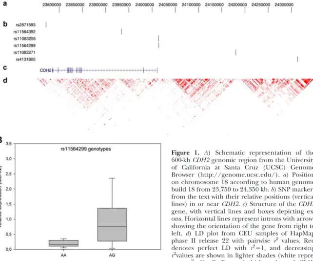
SNPs in theCDH2genomic region (NM_001792.3) on chro- mosome 18 were selected to cover the gene without redun- dancy with respect to linkage disequilibrium (LD) structure:
1 SNP at the distal promoter (rs11083271), 1 SNP at the proximal promoter (rs11564299), 1 SNP in intron 2 (rs11564392), and 1 SNP at the 3=end of the gene (rs2871593). In addition, rs4131805, located 302 kb upstream of theCDH25=end, was
included because of a reported association with bone density and geometry (16). Therefore, 542 kb of the genomic region was analyzed withⱖ1 SNP in each distinct LD block (Fig. 1A).
After a first round of association testing, SNP rs11083255 was analyzed as an additional marker in the CDH2 promoter region. For information on all SNP markers analyzed, see Table 2.
Genomic DNA was isolated from whole-blood specimens with the PureGene DNA Blood Kit (Qiagen, Hilden, Ger- many). DNA samples were genotyped with 5= exonuclease TaqMan technology (Life Technologies; Applied Biosystems, Foster City, CA, USA), according to the manufacturer’s instructions. In brief, for each genotyping experiment, 10 ng DNA was used in a total volume of 5 l containing 1⫻
TaqMan Genotyping Master Mix. Polymerase chain reaction (PCR) and post-PCR end point plate reading was performed on an Applied Biosystems 7900HT Real-Time PCR System.
Sequence Detection System 2.3 software (Life Technologies) was used to assign genotypes applying the allelic discrimina- tion test. Case and control DNA were genotyped together on the same plates with duplicates of samples (10%) to assess intraplate and interplate genotype quality. No genotyping discrepancies were detected. Assignment of genotypes was performed by a person without knowledge of the proband’s affection status.
Cell culture, RNA extraction, and real-time quantitative reverse transcription PCR (qRT-PCR) analysis
Synovial tissue specimens were obtained from patients with OA immediately after the knee joint capsule was opened (17).
Synovial cells from patients with OA were cultured in Dulbecco’s modified Eagle’s medium (DMEM; Sigma-Aldrich, Munich, Germany), 10% fetal calf serum (FCS; PAN Biotech, Aiden- bach, Germany), and penicillin and streptomycin (P/S) (PAA, Piscataway, NJ, USA). The same medium was used to grow the cell lines HSE (immortalized rheumatoid SFs) and K4Im (immortalized normal SFs), which were kindly pro- vided by Prof. W. K. Aicher (University of Tübingen, Tübin- gen, Germany). Total cellular RNA was isolated from cultured cells with the RNeasy kit (Qiagen), and cDNAs were gener- ated by reverse transcription. In parallel, genomic DNA from cultured cells for genotyping was isolated with the QIAamp DNA Mini Kit (Qiagen). qRT-PCR was performed with SYBR Green master mix (Qiagen), with the primer pair 5=-TGGAT- GAAGATGGCATGG-3= and 5=-AGGTGGCCACTGTGCT- TAC-3=, targeting exon A to exon B ofCDH2. Relative gene expression was normalized to -actin mRNA levels by using
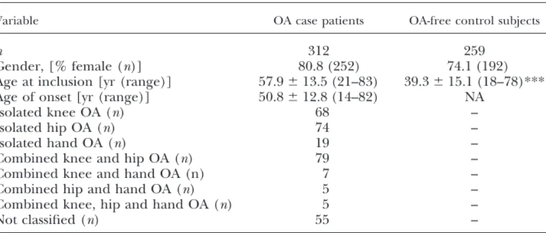
TABLE 1.Characteristics of study population for association analyses
Variable OA case patients OA-free control subjects
n 312 259
Gender, [% female (n)] 80.8 (252) 74.1 (192)
Age at inclusion [yr (range)] 57.9⫾13.5 (21–83) 39.3⫾15.1 (18–78)***
Age of onset [yr (range)] 50.8⫾12.8 (14–82) NA
Isolated knee OA (n) 68 –
Isolated hip OA (n) 74 –
Isolated hand OA (n) 19 –
Combined knee and hip OA (n) 79 –
Combined knee and hand OA (n) 7 –
Combined hip and hand OA (n) 5 –
Combined knee, hip and hand OA (n) 5 –
Not classified (n) 55 –
Values are means⫾sd. NA, not applicable. ***P⬍0.001.
the comparative cycle threshold (Ct) method. The Mel-Im cell line was used as a calibrator sample.
Preparation of nuclear extracts
Nuclear extracts from OASFs were prepared by using the method of Dignamet al.(18).
Enzyme-linked immunoabsorbent assay (ELISA)
For quantitative measurement of the binding capacity of transcription factors, a sandwich ELISA was established based
on a previously published protocol (19). 5=-Biotinylated (Btn) forward oligonucleotides that contained either variant A [5=-(Btn)- TATTCCTTCTTGTAA-3=] or variant G of rs11564299 [5=- (Btn)TATTCCTCCTTGTAA-3=] were annealed overnight to reverse complementary, nonbiotinylated oligonucleotides (5=-TTACAAGAAGGAATA-3=and 5=-TTACAAGGAGGAATA- 3=, respectively) and then were bound onto streptavidin- coated plates. After 3 washes of the plates with PBS, nonspe- cific binding was blocked with incubation buffer (MIA ELISA;
Roche Applied Sciences, Mannheim, Germany) for 1 h.
Nuclear proteins from HSE cells, 6g in 150l of incubation buffer, were allowed to bind to the oligonucleotides for 1 h.
Figure 1. A) Schematic representation of the 600-kbCDH2genomic region from the University of California at Santa Cruz (UCSC) Genome Browser (http://genome.ucsc.edu/). a) Position on chromosome 18 according to human genome build 18 from 23,750 to 24,350 kb.b) SNP markers from the text with their relative positions (vertical lines) in or nearCDH2.c) Structure of theCDH2 gene, with vertical lines and boxes depicting ex- ons. Horizontal lines represent introns with arrows showing the orientation of the gene from right to left. d) LD plot from CEU samples of HapMap phase II release 22 with pairwise r2 values. Red denotes perfect LD with r2⫽1, and decreasing r2values are shown in lighter shades (white repre- sents r2⫽0). B) Box-and-whisker plots of CDH2 mRNA expression for rs11564299 genotypes. Boxes represent the 25th to 75th percentiles; horizontal lines within boxes represent median values. Whiskers show the 5th and 95th percentiles.
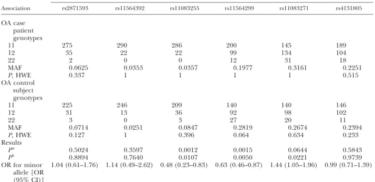
TABLE 2. SNP markers
SNP TaqMan assay Position on chromosome 18a Localization
Alleles
Call rateb
Major Minor
rs2871593 C_15841206_20 23,771,322 3=Intergenic A G 0.95
rs11564392 C_30810483_10 23,933,730 Intron 2 T C 0.95
rs11083255 C_27837839_10 24,013,162 Promoter T C 0.97
rs11564299 C_30810437_20 24,014,026 Promoter A G 0.95
rs11083271 C_417876_10 24,180,057 5=Intergenic C T 0.95
rs4131805 C_26772835_20 24,313,110 5=Intergenic T C 0.95
aNational Center for Biotechnology Information (NCBI; Bethesda, MD, USA) build 36.3 (human genome build 18).bBased onn⫽571 samples.
For displacement analysis, double-stranded oligonucleotides (ds-oligonucleotides) without the biotin label were incubated with the nuclear extracts for 10 min before incubation on the plate. The plates were then washed 3 times with incubation buffer, followed by incubation with a specific antibody against ETS-1 (1:500; Novocastra; Leica Biosystems, Wetzlar, Ger- many) or heterogeneous nuclear ribonucleoprotein K (hn- RNP K; 1:500; Acris Antibodies, San Diego, CA, USA) for 1 h.
After 3 washes with incubation buffer, a secondary antibody, anti-mouse or anti-rabbit (1:1000; Cell Signaling Technology, Danvers, MA, USA) conjugated with horseradish peroxidase, was added for 30 min. The binding was measured by using ABTS solution (Roche Applied Sciences).
Pulldown assay
5=-Biotinylated forward oligonucleotides containing either vari- ant A or G of rs11564299 were annealed overnight to their respective complementary, nonbiotinylated strands [5=- (Btn)TATTCCTTCTTGTAA-3= and 5=-TTACAAGAAGGAATA- 3=; and 5=-(Btn)TATTCCTCCTTGTAA-3= and 5=- TTACAAGGAGGAATA-3=]. Streptavidin beads (GE Healthcare Europe GmbH, Freiburg, Germany) were washed with 1 ml of ice-cold PBS. Then, the beads were incubated with the annealed 5=-biotinylated oligonucleotides overnight. Nuclear extracts from the HSE cells were precleared by incubation with strepta- vidin beads overnight. On the next day, the precleared lysate was incubated with the oligonucleotides bound to streptavidin beads for 4 h. The samples were analyzed by SDS-PAGE, followed by silver staining. The bands selectively occurring in the samples of the minor variant of rs11564299 were cut out and analyzed by mass spectrometry (MS).
MS
Respective bands were excised and washed according to Shevchenkoet al. (20) with slight modifications. Briefly, gel pieces were washed 3 times alternately with 50l of 50 and 25 mM NH4HCO3 in 50% acetonitrile. Subsequently, the gel slices were dried in a vacuum centrifuge. Trypsin solution (5
l; 12.5 ng/l in 50 mM NH4HCO3) was added to each gel piece and incubated at 37°C overnight for in-gel digestion.
The obtained peptides were eluted with 20l of 5% formic acid and subjected to nano-LC-MS/MS-analysis on an Ulti- mate 3000 nano-HPLC system (Dionex GmbH, Idstein, Ger- many). The samples were preconcentrated on a 300m i.d., 5 mm C18-PepMap precolumn (5m, 300 Å; Thermo Fisher Scientific, Idstein, Germany), with 0.1% formic acid and a flow rate of 40l/min. The peptides were then separated on a 75 m i.d., 15 cm, C18-PepMap-column (flow rate 300 nl/min; Thermo Fisher Scientific), with a 1 h binary gradient from 4 to 40% of solvent B (solvent A: 0.1% formic acid;
solvent B: 0.1% formic acid in acetonitrile). The nano-HPLC was directly coupled to a quadrupole time-of-flight (QTOF) mass spectrometer (QStar XL; AB Sciex, Darmstadt, Ger- many) acquiring 1 full MS and 2 MS/MS spectra of the most intense ions in the respective full-MS scan, repeatedly. The MS/MS spectra were searched against the Uniprot database with the Mascot Daemon and the Mascot algorithm (version 2.2; Matrix Science Ltd., London, UK), with trypsin used as the protease with a maximum of 1 missed cleavage site, oxidation of methionine as a variable modification, and 0.2 Da tolerance for MS and MS/MS signals. Only proteins with ⱖ2 significantly scored peptide spectra that passed manual inspection were considered to be positive identifications.
Electrophoretic mobility shift assay (EMSA)
Two double-stranded oligomeric binding sites with either variant A (5=-TACAAGAAGGAA-3=) or variant G (5=- TA- CAAGGAGGAA-3=) of rs11564299 were generated. The ds- oligonucleotides were end labeled with T4 polynucleotide kinase (Roche Applied Sciences) and [32P]ATP[␥P] (Am- ersham-GE Healthcare, Munich, Germany). Band shifts were performed by incubating 5g recombinanthnRNP K (Cusabio Biotech, Wuhan, China) in 5⫻ mobility shift buffer [1g of poly(dI-dC)(dI-dC), 40% glycerol, 25 mM MgCl2, 1 mM EDTA, 25 mM dithiothreitol, 250 mM KCl, and 25 mM HEPES/KOH, pH 7.9) with the DNA probe for 10 min before separation on a 6% nondenaturing poly- acrylamide gel. DNA-protein complexes were resolved on a nondenaturing polyacrylamide gel at 250 V, 50 mA, and 100 W for 1.5 h (21).
Chromatin immunoprecipitation (ChIP)
ChIP was performed as described by the manufacturer (ChIP-IT Enzymatic; Active Motif, La Hulpe, Belgium). Spe- cific antibodies against hnRNP K (Sigma-Aldrich), ETS-1 (Novocastra), polymerase II (Active Motif), and IgG (Active Motif) were used. For qRT-PCR, primers spanning the region around rs11564299 were designed (hCDH2 rs11564299 for- ward, 5=-CTTCTTGTAATCAGAGGCC-3=, and hCDH2 rs11564299 reverse, 5=- ATTGTTTTAGCATCTTGCC-3=). For quantifica- tion of the positive control, commercially available GAPDH primers (Active Motif) were used.
Statistical analyses and bioinformatics tools
Differences between dichotomous traits were calculated by2 test. Differences in continuous variables between groups were calculated with a 2-tailedttest for normally distributed values or with the nonparametric Wilcoxon rank sum test for variables that failed normal distribution, as determined by the Shapiro-Wilk test. Expression data between the 3 genotype groups were compared by Kruskal-Wallis test. Association analysis was carried out in a logistic regression model with gender as a covariate. When age of onset was included as a covariate, for the controls age at inclusion was used. Preva- lence ORs with their 95% CIs were reported. Values ofP⬍ 0.05 were considered significant. To determine whether the genotypes of case patients and control subjects of all SNPs deviated from Hardy-Weinberg equilibrium (HWE), actual and predicted genotype counts of both groups were com- pared by an exact test (22). Association analyses were per- formed with PLINK 1.07 (23), and the statistical software package JMP 7.0.2 (SAS Institute, Inc., Cary, NC, USA) was used for other analyses. For LD testing, HaploView 4.2 was used (24) with HapMap phase II data from releases 22, 24, and 27 (25). MatInspector 8.04, and SNPInspector 2.2 (Geno- matix Software GmbH, Munich, Germany) were used to analyze the potential functional effects of SNPs on transcrip- tion factor binding sites.
RESULTS
Testing for genetic association
All genotyped markers fulfilled our criteria of a
ⱖ95%
call rate and showed no deviation from HWE (Table 3).
The 5 SNPs genotyped initially were not in tight LD (maximum
r2⫽0.03 in the entire study sample of 571 persons). There was a significant association (P
⫽1.5
⫻10
⫺3) between rs11564299 and risk of OA in logistic regression analysis after adjustment for gender. Signif- icance decreased to
P ⫽5.0
⫻10
⫺3after additional adjustment for age of recruitment (Table 3). In the fully adjusted model, SNP rs11083271 was nominally associated with OA risk (P⫽ 0.02; Table 3). Because rs11564299 is localized in the promoter region 2583 bp upstream of the
CDH25= end, we searched the HapMap data for polymorphisms closer to the
CDH2transcrip- tion start site. The SNP marker rs11083255 is localized 1719 bp upstream of
CDH2and was included in a second round of genotyping. Both rs11564299 and rs11083255 were in weak LD in our study sample (r
2⫽0.215). A significant association between rs11083255 and OA risk was detected (P
⫽1.2
⫻10
⫺3for logistic regression ad- justed for gender;
P⫽1.1
⫻10
⫺2for logistic regression adjusted for gender and age at recruitment; Table 3).
The effect direction for both SNPs was the same, with a protective effect of the minor allele (Table 3). Taking multiple testing with a total of 6 markers into account, only rs11564299 showed significant association with OA risk in our study (P
⫽5.0
⫻10
⫺3⫻6 tests
⫽0.03).
Conditional logistic regression analysis with each nominally associated SNP showed that rs11083255 and rs11564299 were not fully independently associated with OA risk (P
⫽0.040, OR
⫽0.69 for rs11564299, after adjustment for rs11083255;
P⫽0.166, OR
⫽0.61 for rs11083255, after adjustment for rs11564299)—that
is, loss of significance of
ⱖ1 order of magnitude was observed. As assumed by LD, rs11083271 did not fully explain the association results for rs11564299 (P⫽ 0.018, OR
⫽0.67) and rs11083255 (P
⫽0.020, OR
⫽0.46).
Because our study sample included different pheno- types of OA (Table 3), we performed separate analyses on samples of knee OA and hip OA. A comparison of 68 patients with isolated knee OA with 259 control subjects showed that 3 markers from the combined analysis (rs11083255, rs11564299, and rs11083271) re- mained significantly associated (logistic regression us- ing gender and age at inclusion as covariates;
P⫽0.016,
P⫽0.036, and
P⫽0.042, respectively). For hip OA (n
⫽74 cases), no association was detected (P
⫽0.639,
P⫽0.071, and
P⫽0.887, respectively).
Expression analysis
A total of 29 synovial cell specimens were available from patients with knee OA. Comparison of
CDH2mRNA expression levels between heterozygotes (n
⫽3) and homozygotes (n⫽26) for the major allele of rs11083255 did not reach the significance level (P
⫽0.43). The 3 genotypes of rs11564299 were strongly associated with
CDH2mRNA expression (P
⫽0.0086). Comparison be- tween rs11564299 heterozygotes (n⫽ 9) and homozy- gotes (n
⫽19) for the major allele yielded
P ⫽0.0073 (Fig. 1B). For both SNPs, the mean expression of
CDH2was higher in carriers of the minor alleles (e.g., rs11564299:
heterozygotes, mean
⫽1.04
⫾0.94; homozygotes, mean
⫽0.26
⫾0.33).
TABLE 3.Results of an SNP association analysis in an OA case– control sample
Association
SNP
rs2871593 rs11564392 rs11083255 rs11564299 rs11083271 rs4131805
OA case patient genotypes
11 275 290 286 200 145 189
12 35 22 22 99 134 104
22 2 0 0 12 31 18
MAF 0.0625 0.0353 0.0357 0.1977 0.3161 0.2251
P, HWE 0.337 1 1 1 1 0.515
OA control subject genotypes
11 225 246 209 140 140 146
12 31 13 36 92 98 102
22 3 0 3 27 20 11
MAF 0.0714 0.0251 0.0847 0.2819 0.2674 0.2394
P, HWE 0.127 1 0.396 0.064 0.634 0.233
Results
Pa 0.5024 0.3597 0.0012 0.0015 0.0644 0.5843
Pb 0.8894 0.7640 0.0107 0.0050 0.0221 0.9739
OR for minor allele [OR (95% CI)]
1.04 (0.61–1.76) 1.14 (0.49–2.62) 0.48 (0.23–0.83) 0.63 (0.46–0.87) 1.44 (1.05–1.96) 0.99 (0.71–1.39)
Numbers of the genotypes (i.e., 11, 12, 22) are according to the alleles in Table 1. MAF, minor allele frequency.aLogistic regression adjusted for sex.bLogistic regression adjusted for age at inclusion and sex.
In silico
data
That the G allele of rs11564299 was associated with elevated
CDH2mRNA levels in OASF led to the hypoth- esis that this allele may generate a novel transcription factor binding site.
In silicoanalysis with Genomatix (Genomatix Software GmbH) predicted the loss of a TEF (26) binding site and a gain in sites for ETS-like transcription factors (27) and ZNF35 (28).
Analysis of a potential transcription factor binding site
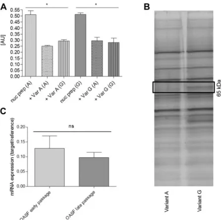
A quantitative ELISA with ds-oligonucleotides contain- ing either variant A or G of rs11564299 as bait was established to verify the predicted ETS-like binding site.
After incubation with nuclear proteins and detection with a specific ETS-1 antibody and peroxidase-labeled secondary antibodies, no differences in binding effi- ciency to one of the variants were observed (Fig. 2A).
Therefore, ETS-1 was excluded as a transcription factor binding selectively to the minor allele of rs11564299.
Subsequently, a pulldown assay was performed for identification of the transcription factor responsible for the higher
CDH2mRNA levels observed with the minor rs11564299 allele. The pulldown was performed with nuclear extracts of the cell line HSE, which had tested heterozygous for rs11564299, by using the specific oligonucleotides for alleles A and G of rs11564299.
Pulled down proteins were separated on an acrylamide gel and stained silver, followed by excision, tryptic digestion, and MS analysis of those protein bands that had been pulled down selectively with ds-oligonucleo-
tides containing variant G (Fig. 2B). This method revealed hnRNP K as a potential binding protein.
Expression of hnRNP K in early (P3) and late (P8) passages of OASFs was determined by qRT-PCR, and no significant differences were observed between the dif- ferent groups (Fig. 2C). For analysis of differential binding of hnRNP K to the minor allele (G) compared to the major allele (A) of rs11564299, a quantitative ELISA with a specific hnRNP K antibody was estab- lished. The ELISA showed that hnRNP K bound only to ds-oligonucleotides containing variant G, but not to those containing variant A (Fig. 3A). The binding was inhibited by preincubation with ds-oligonucleotides containing variant G but not variant A (Fig. 3A). As shown before, performing the ELISA with an ETS-1 antibody revealed no differential binding of ETS-1 to ds-oligonucleotides containing either variant of the SNP rs11564299 (Fig. 2A). As a negative control, a setting with the forward biotinylated ssDNA oligonucle- otides was performed, but did not show any binding of hnRNP K to the ssDNA (data not shown). EMSAs of similar oligonucleotides yielded the same results. A shift was observed only for probes carrying variant G after incubation with recombinant hnRNP K (Fig. 3B).
This result showed that hnRNP K bound exclusively to the minor allele of rs11564299, indicating that hnRNP K may be responsible for elevated
CDH2mRNA expres- sion. To further elucidate the binding of hnRNP K to the minor variant of rs11564299, a ChIP assay was performed. Antibodies against ETS-1 (positive control 1), pol II (positive control 2), and IgG (negative control) were used in addition to the anti-hnRNP K
Figure 2. A) ELISA, performed with an ETS-1 antibody, did not show preferential binding to one of the 2 alleles of rs11564299. Here, com- petition was possible with variants A and G.B) Silver-stained acrylamide gel of a pulldown assay using oligonucleotides containing either allele A or G of rs11564299. A band, showing only the pulldown with variant G, was analyzed by MS and revealed hnRNP K as a potential transcrip- tion factor binding to theCDH2promoter with the minor allele G. C) mRNA expression of hnRNP K was not altered during cell culture cultivation of OASFs.
antibody. The ChIP assay was performed with HSE cells, which are heterozygous for rs11564299, and K4Im, a cell line of immortalized SFs that are homozy- gous for the major allele of rs11564299. hnRNP K bound only to chromatin in HSE cells, whereas no binding was observed when the homozygous wild-type K4Im cells were tested (Fig. 3C). These findings con- firmed that hnRNP K bound exclusively to the N- cadherin promoter in the presence of the minor allele of rs11564299, therefore making hnRNP K a candidate for inducing the elevated levels of N-cadherin.
DISCUSSION
Genetic association studies have shown that SNPs con- tribute significantly to the susceptibility and severity of many diseases (29). Also, associations of genetic vari- ants with OA onset and progression have been pub- lished (2, 30). As shown before, SNPs in the promoter region of a gene can influence disease susceptibility (31, 32). Our results indicate that
CDH2SNP rs11564299 influences susceptibility to OA prevalence, as the minor G allele is more common among the control population than the OA population. We found that the allelic state of rs11564299, which is located 2583 bp upstream of the
CDH2transcription start site, significantly affected mRNA expression levels of
CDH2in SFs, with the AG heterozygotes (HSE cells) express- ing significantly more
CDH2than the AA homozygotes (K4Im cells). This result indicates stronger activity of the
CDH2promoter in patients carrying the minor
allele, probably caused by an allele-specific transcrip- tion factor binding site. However, none of the transcrip- tion factors predicted
in silicoto bind to the variant promoter sequence showed differential binding to the minor allele of rs11564299. Rather, MS analysis identi- fied hnRNP K as a factor that bound exclusively to the G allele of rs11564299 and not to the A allele. hnRNP K is one of the major pre-mRNA binding proteins. It has also been described to bind dsDNA and to act as a transcription factor (33). Binding of hnRNP K was confirmed by a specific ELISA and ChIP. Thus, hnRNP K appears to contribute to the increased N-cadherin expression levels in SFs carrying the G allele of rs11564299. As N-cadherin is a core component of adherens junctions, the increased levels of it may affect cell– cell contacts, tissue architecture, and cell motility (34, 35). It has been described before that N-cadherin levels are critical for cell invasion and that N-cadherin re-expression in glioma cells and in human arterial smooth muscle cells leads to less migration (36, 37).
Therefore, higher N-cadherin levels in SFs may lead to a decreased migratory potential of the cells. As shown by Schubert
et al.,(6) migration of SF into cartilage leads to the degradation of the extracellular matrix and cartilage loss. Thus, inhibition of migration by en- hanced cell– cell contact may prevent or reduce carti- lage destruction. All evidence combined, our study implicates for the first time
CDH2encoding N-cadherin as a susceptibility gene for OA, especially of the knee.
Some limitations of our study should be discussed. First, we had no replication study for the genetic association data, and therefore our finding of rs11564299 as a risk
Figure 3.A) For quantitative measurement of the binding capacity of hnRNP K, an ELISA was established with specific oligonucleotides containing variant A or G. A clear binding to variant G was observed, whereas no binding K to variant A was measured. Binding was repressed only in competition experiments with variant G oligonucleotides, not with variant A.B) EMSAs with the variant A or G probes showed binding only to variant G.C) Specific binding of hnRNP K to variant G was confirmed by ChIP. In heterozygous HSE cells, binding was observed, whereas in K4Im cells, which are homozygous for variant A, only binding to ETS-1 was detected. Top panel: HSE. Bottom panel: K4Im. Lane 1, input; lane 2, IgG antibody; lane 3, Pol II antibody; lane 4, hnRNP K antibody;lane 5, ETS-1 antibody; lane 6, H2O control. (Differential binding of ETS-1 in the control setting is due to base pair changes in theGAPDHpromoter region of K4Im, which resulted in an ETS-1 binding site; data not shown.)
factor for OA susceptibility could be a false positive.
However, the apparent link between rs11564299 allelic state and
CDH2mRNA expression lends functional support to this hypothesis. Second, the effect size of the rs11564299 minor allele on OA risk is small (OR
⫽0.63,
i.e.,⬃1.6 times increased risk in carriers of the major allele). However, it has become obvious that common variants are often associated with OR
⬍1.5 (38). Third, because the control subjects were selected by clinical exclusion of arthritic or degenerative diseases, appar- ent OA was excluded. However, the preclinical state of joint disorders within the controls could have affected the association results. Fourth, because of our marker selection, we did not fully cover the whole
CDH2gene when tagging SNPs. This deficiency is one that could be solved by screening existing genome-wide association data on OA (2). Since SFs are only 1 candidate cell type in the joint that is potentially responsible for OA onset and progression, future studies should expand N-cad- herin analysis to other cells and their interaction within the joint.
The exact role of N-cadherin for OA susceptibility has yet to be defined. Further analyses should be aimed at the interaction of genetic variants in
CDH2with known risk factors for OA, such as age, increased body-mass index, mechanical stress, and microinjuries of the joint.
The authors thank Rainer Straub (Internal Medicine I, University Hospital of Regensburg) for supplying the re- search material and for helpful discussions. This study was supported by Deutsche Forschungsgemeinschaft (DFG) grant FOR 696 to A.K.B. and K.S.
REFERENCES
1. Evangelou, E., Valdes, A. M., Kerkhof, H. J. M., Styrkarsdottir, U., Zhu, Y., Meulenbelt, I., Lories, R. J., Karassa, F. B., Tylza- nowski, P., Bos, S. D., Akune, T., Arden, N. K., Carr, A., Chapman, K., Cupples, L. A., Dai, J., Deloukas, P., Doherty, M., Doherty, S., Engstrom, G., Gonzalez, A., Halldorsson, B. V., Hammond, C. L., Hart, D. J., Helgadottir, H., Hofman, A., Ikegawa, S., Ingvarsson, T., Jiang, Q., Jonsson, H., Kaprio, J., Kawaguchi, H., Kisand, K., Kloppenburg, M., Kujala, U. M., Lohmander, L. S., Loughlin, J., Luyten, F. P., Mabuchi, A., McCaskie, A., Nakajima, M., Nilsson, P. M., Nishida, N., Ollier, W. E. R., Panoutsopoulou, K., van de Putte, T., Ralston, S. H., Rivadeneira, F., Saarela, J., Schulte-Merker, S., Shi, D., Slag- boom, P. E., Sudo, A., Tamm, A., Thorleifsson, G., Thorsteins- dottir, U., Tsezou, A., Wallis, G. A., Wilkinson, J. M., Yoshimura, N., Zeggini, E., Zhai, G., Zhang, F., Jonsdottir, I., Uitterlinden, A. G., Felson, D. T., van Meurs, J. B., Stefansson, K., Ioannidis, J. P. A., and Spector, T. D. (2011) Meta-analysis of genome-wide association studies confirms a susceptibility locus for knee osteoarthritis on chromosome 7q22.Ann. Rheum. Dis.70,349 – 355
2. arcOGEN Consortium and arcOGEN Collaborators (2012) Identification of new susceptibility loci for osteoarthritis (arcOGEN): a genome-wide association study.Lancet380,815–
823
3. Evangelou, E., Kerkhof, H. J., Styrkarsdottir, U., Ntzani, E. E., Bos, S. D., Esko, T., Evans, D. S., Metrustry, S., Panoutsopoulou, K., Ramos, Y. F. M., Thorleifsson, G., Tsilidis, K. K., Arden, N., Aslam, N., Bellamy, N., Birrell, F., Blanco, F. J., Carr, A., Chapman, K., Day-Williams, A. G., Deloukas, P., Doherty, M., Engstrom, G., Helgadottir, H. T., Hofman, A., Ingvarsson, T.,
Jonsson, H., Keis, A., Keurentjes, J. C., Kloppenburg, M., Lind, P. A., McCaskie, A., Martin, N. G., Milani, L., Montgomery, G. W., Nelissen, R. G. H. H., Nevitt, M. C., Nilsson, P. M., Ollier, W. E., Parimi, N., Rai, A., Ralston, S. H., Reed, M. R., Riancho, J. A., Rivadeneira, F., Rodriguez-Fontenla, C., Southam, L., Thorsteinsdottir, U., Tsezou, A., Wallis, G. A., Wilkinson, J. M., Gonzalez, A., Lane, N. E., Lohmander, L. S., Loughlin, J., Metspalu, A., Uitterlinden, A. G., Jonsdottir, I., Stefansson, K., Slagboom, P. E., Zeggini, E., Meulenbelt, I., Ioannidis, J. P., Spector, T. D., van Meurs, J. B. J., and Valdes, A. M. (2013) A meta-analysis of genome-wide association studies identifies novel variants associated with osteoarthritis of the hip. [E-pub ahead of print] Ann. Rheum. Dis.doi: 10.1136/annrheumdis- 2012–203114
4. Gonzalez, A. (2013) Osteoarthritis year 2013 in review: genetics and genomics.Osteoarthritis Cartilage21,1443–1451
5. Panoutsopoulou, K., and Zeggini, E. (2013) Advances in osteo- arthritis genetics. [E-pub ahead of print] J. Med. Genet. doi:
10.1136/jmedgenet-2013-101754
6. Schubert, T., Denk, A., Mägdefrau, U., Kaufmann, S., Bastone, P., Lowin, T., Schedel, J., and Bosserhoff, A. K. (2009) Role of the netrin system of repellent factors on synovial fibroblasts in rheumatoid arthritis and osteoarthritis. Int. J. Immunopathol.
Pharmacol.22,715–722
7. Becker, S. F., Langhe, R., Huang, C., Wedlich, D., and Kashef, J.
(2012) Giving the right tug for migration: cadherins in tis- sue movements.Arch. Biochem. Biophys.524,30 –42
8. Chang, S. K., Noss, E. H., Chen, M., Gu, Z., Townsend, K., Grenha, R., Leon, L., Lee, S. Y., Lee, D. M., and Brenner, M. B.
(2011) Cadherin-11 regulates fibroblast inflammation. Proc.
Natl. Acad. Sci. U. S. A.108,8402–8407
9. Lee, D. M., Kiener, H. P., Agarwal, S. K., Noss, E. H., Watts, G. F. M., Chisaka, O., Takeichi, M., and Brenner, M. B. (2007) Cadherin-11 in synovial lining formation and pathology in ar- thritis.Science315,1006 –1010
10. Grunwald, G. B., Pratt, R. S., and Lilien, J. (1982) Enzymic dissection of embryonic cell adhesive mechanisms: III, immu- nological identification of a component of the calcium-depen- dent adhesive system of embryonic chick neural retina cells.J.
Cell Sci.55,69 –83
11. Agarwal, S. K., Lee, D. M., Kiener, H. P., and Brenner, M. B.
(2008) Coexpression of two mesenchymal cadherins, cadherin 11 and N-cadherin, on murine fibroblast-like synoviocytes.Ar- thritis Rheum.58,1044 –1054
12. Kashima, T., Nakamura, K., Kawaguchi, J., Takanashi, M., Ishida, T., Aburatani, H., Kudo, A., Fukayama, M., and Grigo- riadis, A. E. (2003) Overexpression of cadherins suppresses pulmonary metastasis of osteosarcoma in vivo.Int. J. Cancer104, 147–154
13. Nakajima, S., Doi, R., Toyoda, E., Tsuji, S., Wada, M., Koizumi, M., Tulachan, S. S., Ito, D., Kami, K., Mori, T., Kawaguchi, Y., Fujimoto, K., Hosotani, R., and Imamura, M. (2004) N-cadherin expression and epithelial-mesenchymal transition in pancre- atic carcinoma.Clin. Cancer Res.10,4125–4133
14. Li, G., Satyamoorthy, K., and Herlyn, M. (2001) N-cadherin- mediated intercellular interactions promote survival and migra- tion of melanoma cells.Cancer Res.61,3819 –3825
15. Kuphal, S., and Bosserhoff, A. K. (2006) Influence of the cytoplasmic domain of E-cadherin on endogenous N-cadherin expression in malignant melanoma.Oncogene25,248 –259 16. Kiel, D. P., Demissie, S., Dupuis, J., Lunetta, K. L., Murabito,
J. M., and Karasik, D. (2007) Genome-wide association with bone mass and geometry in the Framingham Heart Study.BMC Med. Genet.8,S14
17. Miller, L. E., Jüsten, H. P., Schölmerich, J., and Straub, R. H.
(2000) The loss of sympathetic nerve fibers in the synovial tissue of patients with rheumatoid arthritis is accompanied by in- creased norepinephrine release from synovial macrophages.
FASEB J.14,2097–2107
18. Dignam, J. D., Martin, P. L., Shastry, B. S., and Roeder, R. G.
(1983) Eukaryotic gene transcription with purified components.
Methods Enzymol.101,582–598
19. Benotmane, A. M., Hoylaerts, M. F., Collen, D., and Belayew, A.
(1997) Nonisotopic quantitative analysis of protein-DNA inter- actions at equilibrium.Anal. Biochem.250,181–185
20. Shevchenko, A., Wilm, M., Vorm, O., and Mann, M. (1996) Mass spectrometric sequencing of proteins silver-stained polyacryl- amide gels.Anal. Chem.68,850 –858
21. Wenke, A.-K., Grässel, S., Moser, M., and Bosserhoff, A. K.
(2009) The cartilage-specific transcription factor Sox9 regulates AP-2epsilon expression in chondrocytes.FEBS J.276,2494 –2504 22. Wigginton, J. E., Cutler, D. J., and Abecasis, G. R. (2005) A note on exact tests of Hardy-Weinberg equilibrium. Am. J. Hum.
Genet.76,887–893
23. Purcell, S., Neale, B., Todd-Brown, K., Thomas, L., Ferreira, M. A. R., Bender, D., Maller, J., Sklar, P., de Bakker, P. I. W., Daly, M. J., and Sham, P. C. (2007) PLINK: a tool set for whole-genome association and population-based linkage analy- ses.Am. J. Hum. Genet.81,559 –575
24. Barrett, J. C., Fry, B., Maller, J., and Daly, M. J. (2005) Haplo- view: analysis and visualization of LD and haplotype maps.
Bioinformatics21,263–265
25. Frazer, K. A., Ballinger, D. G., Cox, D. R., Hinds, D. A., Stuve, L. L., Gibbs, R. A., Belmont, J. W., Boudreau, A., Hardenbol, P., Leal, S. M., Pasternak, S., Wheeler, D. A., Willis, T. D., Yu, F., Yang, H., Zeng, C., Gao, Y., Hu, H., Hu, W., Li, C., Lin, W., Liu, S., Pan, H., Tang, X., Wang, J., Wang, W., Yu, J., Zhang, B., Zhang, Q., Zhao, H., Zhao, H., Zhou, J., Gabriel, S. B., Barry, R., Blumenstiel, B., Camargo, A., Defelice, M., Faggart, M., Goyette, M., Gupta, S., Moore, J., Nguyen, H., Onofrio, R. C., Parkin, M., Roy, J., Stahl, E., Winchester, E., Ziaugra, L., Altshuler, D., Shen, Y., Yao, Z., Huang, W., Chu, X., He, Y., Jin, L., Liu, Y., Shen, Y., Sun, W., Wang, H., Wang, Y., Wang, Y., Xiong, X., Xu, L., Waye, M. M. Y., Tsui, S. K. W., Xue, H., Wong, J. T.-F., Galver, L. M., Fan, J.-B., Gunderson, K., Murray, S. S., Oliphant, A. R., Chee, M. S., Montpetit, A., Chagnon, F., Ferretti, V., Leboeuf, M., Olivier, J.-F., Phillips, M. S., Roumy, S., Sallée, C., Verner, A., Hudson, T. J., Kwok, P.-Y., Cai, D., Koboldt, D. C., Miller, R. D., Pawlikowska, L., Taillon-Miller, P., Xiao, M., Tsui, L.-C., Mak, W., Song, Y. Q., Tam, P. K. H., Nakamura, Y., Kawaguchi, T., Kitamoto, T., Morizono, T., Nagashima, A., Ohnishi, Y., Sekine, A., Tanaka, T., Tsunoda, T., Deloukas, P., Bird, C. P., Delgado, M., Dermitzakis, E. T., Gwilliam, R., Hunt, S., Morrison, J., Powell, D., Stranger, B. E., Whittaker, P., Bentley, D. R., Daly, M. J., de Bakker, P. I. W., Barrett, J., Chretien, Y. R., Maller, J., McCarroll, S., Patterson, N., Pe’er, I., Price, A., Purcell, S., Richter, D. J., Sabeti, P., Saxena, R., Schaffner, S. F., Sham, P. C., Varilly, P., Stein, L. D., Krishnan, L., Smith, A. V., Tello-Ruiz, M. K., Thorisson, G. A., Chakravarti, A., Chen, P. E., Cutler, D. J., Kashuk, C. S., Lin, S., Abecasis, G. R., Guan, W., Li, Y., Munro, H. M., Qin, Z. S., Thomas, D. J., McVean, G., Auton, A., Bottolo, L., Cardin, N., Eyheramendy, S., Freeman, C., Marchini, J., Myers, S., Spencer, C., Stephens, M., Donnelly, P., Cardon, L. R., Clarke, G., Evans, D. M., Morris, A. P., Weir, B. S., Mullikin, J. C., Sherry, S. T., Feolo, M., Skol, A., Zhang, H., Matsuda, I., Fukushima, Y., Macer, D. R., Suda, E., Rotimi, C. N., Adebamowo, C. A., Ajayi, I., Aniagwu, T., Marshall, P. A., Nkwodimmah, C., Royal, C. D. M., Leppert, M. F., Dixon, M., Peiffer, A., Qiu, R., Kent, A., Kato, K., Niikawa, N., Adewole, I. F., Knoppers, B. M., Foster, M. W., Clayton, E. W., Watkin, J., Muzny, D., Nazareth, L., Sodergren, E., Weinstock, G. M., Yakub, I., Birren, B. W., Wilson, R. K., Fulton, L. L., Rogers, J., Burton, J., Carter, N. P., Clee, C. M., Griffiths, M., Jones, M. C.,
McLay, K., Plumb, R. W., Ross, M. T., Sims, S. K., Willey, D. L., Chen, Z., Han, H., Le Kang, Godbout, M., Wallenburg, J. C., L’Archevêque, P., Bellemare, G., Saeki, K., Wang, H., An, D., Fu, H., Li, Q., Wang, Z., Wang, R., Holden, A. L., Brooks, L. D., McEwen, J. E., Guyer, M. S., Wang, V. O., Peterson, J. L., Shi, M., Spiegel, J., Sung, L. M., Zacharia, L. F., Collins, F. S., Kennedy, K., Jamieson, R., and Stewart, J. (2007) A second generation human haplotype map of over 3.1 million SNPs.Nature449, 851–861
26. Fonjallaz, P., Ossipow, V., Wanner, G., and Schibler, U. (1996) The two PAR leucine zipper proteins, TEF and DBP, display similar circadian and tissue-specific expression, but have differ- ent target promoter preferences.EMBO J.15,351–362 27. Virbasius, J. V., and Scarpulla, R. C. (1991) Transcriptional
activation through ETS domain binding sites in the cytochrome c oxidase subunit IV gene.Mol. Cell. Biol11,5631–5638 28. Pengue, G., Cannada-Bartoli, P., and Lania, L. (1993) The
ZNF35 human zinc finger gene encodes a sequence-specific DNA-binding protein.FEBS Lett.321,233–236
29. Hindorff, L. A., Sethupathy, P., Junkins, H. A., Ramos, E. M., Mehta, J. P., Collins, F. S., and Manolio, T. A. (2009) Potential etiologic and functional implications of genome-wide associa- tion loci for human diseases and traits. Proc. Natl. Acad. Sci.
U. S. A.106,9362–9367
30. Bijsterbosch, J., Kloppenburg, M., Reijnierse, M., Rosendaal, F. R., Huizinga, T. W. J., Slagboom, P. E., and Meulenbelt, I.
(2013) Association study of candidate genes for the progression of hand osteoarthritis.Osteoarthritis Cartilage21,565–569 31. Shirasawa, S. (2004) SNPs in the promoter of a B cell-specific
antisense transcript, SAS-ZFAT, determine susceptibility to au- toimmune thyroid disease.Hum. Mol. Genet.13,2221–2231 32. Waldron-Lynch, F., Adams, C., Amos, C., Zhu, D. K.,
McDermott, M. F., Shanahan, F., Molloy, M. G., and O’Gara, F.
(2001) Tumour necrosis factor 5= promoter single nucleotide polymorphisms influence susceptibility to rheumatoid arthritis (RA) in immunogenetically defined multiplex RA families.Genes Immun.2,82–87
33. Michelotti, E. F., Michelotti, G. A., Aronsohn, A. I., and Levens, D. (1996) Heterogeneous nuclear ribonucleoprotein K is a transcription factor.Mol. Cell. Biol.16,2350 –2360
34. Collinet, C., and Lecuit, T. (2013) Stability and dynamics of cell-cell junctions.Prog. Mol. Biol. Transl. Sci.116,25–47 35. Ivanov, A. I., and Naydenov, N. G. (2013) Dynamics and
regulation of epithelial adherens junctions: recent discoveries and controversies.Int. Rev. Cell. Mol. Biol.303,27–99
36. Blindt, R. (2004) Downregulation of N-cadherin in the neoin- tima stimulates migration of smooth muscle cells by RhoA de- activation.Cardiovasc. Res.62,212–222
37. Péglion, F., and Etienne-Manneville, S. (2012) N-cadherin ex- pression level as a critical indicator of invasion in non-epithe- lial tumors.Cell. Adh. Migr.6,327–332
38. Ku, C. S., Loy, E. Y., Pawitan, Y., and Chia, K. S. (2010) The pursuit of genome-wide association studies: where are we now?
J. Hum. Genet.55,195–206
Received for publication July 17, 2013.
Accepted for publication October 15, 2013.