FROM THE FACULTY OF MEDICINE OF THE UNIVERSITY OF REGENSBURG PROF. DR. MARKUS J. RIEMENSCHNEIDER
DEPARTMENT OF NEUROPATHOLOGY
ANALYSIS OF THE SIGNIFICANCE OF miRNAs OF THE miRNA-200 FAMILY IN THE MALIGNANT PHENOTYPE OF GLIOBLASTOMA
Inaugural-Dissertation to obtain the title of Doctor of medicine (Dr. med.)
from the Faculty of Medicine of the University of Regensburg
presented by
Natalia Vélez Char, M.D., M.Sc.
2017
Dean: Prof. Dr. Dr. Torsten E. Reichert
1. Rapporteur: Prof. Dr. Markus J. Riemenschneider 2. Rapporteur: Prof. Dr. Peter Hau
Oral examination date: Wednesday, August 2nd 2017
Declaration
This doctoral thesis was designed, encouraged, supported and supervised by Prof. Dr. Markus J.
Riemenschneider.
I hereby declare that I have done the present work independently and without any other tools or resources, except where otherwise stated, than the ones described in this thesis.
That where I have used data, concepts or resources taken directly or indirectly from other sources, this has always been clearly attributed and the corresponding source cited.
I have not directly nor indirectly received any remuneration of intermediary or advisory services (promotion consultants or other persons); and that no one has received directly or indirectly monetary benefits from this institution or me for work related to this content.
Furthermore, I certify that this research thesis or any part of it has not been previously submitted for a degree or any other qualification at the University of Regensburg or any other institution in Germany or abroad.
Regensburg, ………... ....…………..………
Natalia Vélez Char
i
Zusammenfassung
Der miR200-Familie werden Tumorsuppressor-Eigenschaften in verschiedenen malignen epithelialen Tumoren zugeschrieben. Ihre Herunterregulation ist mit Tumoraggressivität, metastatischer Erkrankung, Tumorprogression, Chemoresistenz und schlechterer Prognose assoziiert. Im Glioblastom wurde gezeigt, dass einige Mitglieder der miR200-Familie auf sehr niedrigem Niveau exprimiert werden, und dieses Phänomen ist ebenfalls mit einer schlechten Prognose assoziiert.
Auch wenn somit eine Rolle der miRNA 200-Familie bei Glioblastomen naheliegt, gibt es noch keine detaillierten Daten über die funktionalen Effekte einer Dysregulation ihrer beiden Strängen (3p vs.
5p) bzw. Daten darüber, welcher der beiden Stränge funktionell aktiv ist und ob eine Dysregulation einzelner miRNAs relevante funktionelle Effekte besitzt.
Zu diesem Zweck wurden U-87 MG- und U-251 MG-Zellen mit den jeweiligen 3p- und 5p-miRNA- Mimics der gesamten miR200-Familie transfiziert und funktionelle Assays sowie Next generation sequencing (NGS)-Analysen von RNA-Proben nach Transfektion durchgeführt, um die Effekte der miR200-Familie in Glioblastomzellen genauer zu charakterisieren. Die Ergebnisse dieser Arbeit zeigten, dass Glioblastom-Zelllinien ein niedrigeres Werte Expressionsniveau der 3p-Stränge der miR200-Familienmitglieder im Vergleich zu resezierten Tumoren und nicht-neoplastischem Hirngewebe aufzeigten und dass die induzierte Überexpression dieser Stränge zu einer Abnahme der Proliferation und einer Zunahme der Apoptose in U-87 MG- und U-251 MG-Zellen sowie zu einer Abnahme der Expression von ZEB1 und ZEB2 führte. Darüber hinaus zeigten sich in den NGS- Analysen eine funktionelle Gruppierung der Zellen mit einer Überexpression der 3p-Stränge nach ihren Seed-Sequenzen und eine klare Trennung von den Zellen mit Hochregulation der 5p Stränge.
Die 3p-Überexpression induzierte eine Hochregulation von Targets des G2-M-DNA-Damage- Checkpoints, von E2F-Targets sowie von Targets der c-myc- und NFkß-Signalwege. Weiterhin induzierte die 3p-Überexpression eine Herrunterregulation von Targets des CDH1-Gens und der Hypoxie-Antwort (einschließlich HIF1α).
In Zusammenschau, all dieser Befunde deuten die Ergebnisse dieser Arbeit darauf hin, dass die 3p- Stränge der miR200-Familie in Gliomen tumorsuppressive Eigenschaften vermitteln, indem sie pathogenetisch relevante Signalwege beeinflussen und Mechanismen der DNA-Schädigung/- Reparatur, des Zellzyklus und der Zellvitalität, der Zellmotilität und Adhäsion sowie der Hypoxie regulieren.
ii
Abstract
The miR200 family has been described to exert tumor suppressing functions in different malignant epithelial tumors, and their down regulation has been associated with aggressiveness, metastatic disease, tumor progression, chemoresistance und worse prognosis. In glioblastoma, some members of the miR200 family have been shown to be expressed at very low levels, and this phenomenon has been associated to poor prognosis. Even though a role of the miRNA 200 family in glioblastomas has been suggested, no detailed data has been provided regarding differences in functional effects among their two strands. In other terms, it has not yet been established which of the two strands is functional active, and if dysregulation of isolated miRNAs of this family has relevant functional effects.
For this purpose, U-87 MG and U-251 MG cells were transfected with 3p and 5p miRNA mimics of the miR200 family, and functional assays as well as next generation sequencing of RNA samples after transfection were performed. Results showed that glioblastoma cell lines express lower levels of the 3p strands of the miR200 family members compared to glioma biopsy samples and non-neoplastic brain tissue, and that inducing overexpression of these strands leads to a decrease in proliferation and increase in apoptosis in U-87MG and U-251 MG cells, as well as a decrease in the expression of ZEB1 and ZEB2. Furthermore, NGS analyses showed a functional clustering of cells overexpressing the 3p strands in two groups according to their seed sequences, with clear separation of cells overexpressing the 5p strands. 3p overexpression caused upregulation of G2/ M DNA damage checkpoint and E2F target genes. Also, targets of the c-myc and NFkß signaling pathways where up- and targets of the CDH1 gene and of hypoxia response (including HIF1α) were downregulated.
In conclusion, the findings of this work suggest that the 3p strands of the miR200 family carry tumor suppressive functions in gliomas by interacting with major pathogenic pathways implicated in DNA damage/ repair, cell cycle, cell viability, cell motility/ adhesion and hypoxia response.
iii
Table of Contents
Zusammenfassung ... i
Abstract ... ii
1. Introduction ... 1
1.1. The miRNA 200 Family ... 1
1.1.1. Micro RNAs ... 1
1.1.2. The miRNA 200 family ... 2
1.2. Epithelial to mesenchymal transition (EMT) in cancer ... 3
1.2.1. Epithelial to mesenchymal transition ... 3
1.2.2. The role of the miRNA 200 Family in EMT ... 4
1.3. Glioblastoma ... 5
1.3.1. Glioblastoma ... 5
1.3.2. EMT and Glioblastoma ... 5
1.3.3. The role of the miRNA 200 Family in Glioblastoma ... 5
2. Aim of the study ... 7
3. Materials and Methods ... 8
3.1. Materials ... 8
3.1.1. Cell lines ... 8
3.1.2. Chemical substances ... 8
3.1.3. Expendable Supplies and Materials ... 9
3.1.4. Electrical Equipment ... 10
3.1.5. Kits ... 11
3.1.6. Medium and Supplements for Cell Culture and in vitro Assays ... 12
3.1.7. miRNA Primer Assays for qRT-PCR ... 12
3.1.8. miRNA Sequences for transient transfection ... 13
3.1.9. Non-expendable Supplies and Materials ... 14
3.1.10. Primer pairs for qRT-PCR... 15
3.1.11. RNA samples ... 15
3.1.12. Solutions and other biochemical compounds/ materials ... 17
iv
3.1.13. Software ... 17
3.2. Methods ... 18
3.2.1. Cell Biology Methods ... 18
3.2.2. Molecular Biology Procedures ... 21
3.2.3. Biochemical Methods ... 27
3.2.4. Bioinformatics ... 28
4. Results ... 32
4.1. Expression of the miR200 family... 32
4.1.1. Expression of the miR200 family in glioblastoma samples extracted from The Cancer Genome Atlas (TCGA) ... 39
4.1.2. Expression of ZEB1 and ZEB2 in glioblastoma samples extracted from The Cancer Genome Atlas (TCGA) ... 42
4.2. Transfection ... 43
4.2.1. Expression of the other miR200 family members after transfection with one mimic ... 45
4.2.2. Effects of upregulation of the miR200 family members in glioma cell proliferation and apoptosis ... 47
4.2.3. Expression of ZEB1 and ZEB2 in cells transfected with mimics of the miR200 family ... 50
4.3. NGS ... 51
4.3.1. Multivariate Analysis ... 51
5. Discussion ... 58
6. Conclusion ... 63
Bibliography ... 64
1
1. Introduction
1.1. The miRNA 200 Family
1.1.1. Micro RNAs
Micro RNAs (miRNAs) are small (21-25 nt long), non-coding RNAs that regulate gene expression post- transcriptionally by binding to 3´untranslated regions or open reading frames of target mRNAs, leading to the their degradation or repression of mRNA translation1, 2. They are transcribed either mono- or polycistronically in the cell nucleus by an RNA polymerase type II, resulting in hundred to thousand nucleotides long, polyadenylated and capped primary transcripts (primary miRNA, pri- miRNA)1, 2, which will later on be cleaved by Drosha, a type III RNase, to ~70 nucleotides long precursor miRNAs (pre-miRNA). These pre-miRs bind to exportin-5 to be transported to the cytosol, where they are cleaved again by a type III RNase called Dicer, to a final ~21–22 nucleotide long miRNA duplex. One of both strands (non-functional) will end up being degraded, while the remaining, functionally active strand will bind to Argonaute and enter the RNA induced silencing complex (RISC)1, 2. One of the two miRNA strands, usually miRNA 5p, was originally believed to have no functional effect and be the one to get degraded, but recent evidence suggests that any of the two strands (3p or 5p) could be functionally active and therefore of biological significance (Yang et al., Nucleic Acids Res. 2013). Once bound to RISC, miRNAs bind, according to their seed sequences, to complementary sequences in the 3′UTRs of target mRNAs, leading to mRNA degradation or translation inhibition3.
Figure 1. miRNA synthesis and function. Pri-miRNAs are transcribed and cleaved in the nucleus (pre-miRNAs). Pre-miRNAs are transported to the cytosol where they are cleaved again into a final 22 nucleotide long mature miRNA. After binding to RISC, miRNAs bind according to their seed sequence to mRNA to either silence or degrade it (Source: Takahashi et al. Frontiers in Genetics 2014).4
2
1.1.2. The miRNA 200 family
The miRNA 200 family is composed of 5 members coded in two different genomic clusters: miRNA- 200a, -200b and -429 in chromosome 12, and miRNA-200c and -141 clustered in chromosome 1.
They possess a seed sequence located at position 2 to 5 from the 5´ end of their functional, mature miRNA strand, which is responsible for the specificity of the miRNA-mRNA targeting. The seed sequences among all 5 members differ in only one nucleotide in position 3, clustering the miRNAs members further, in two functional groups, which each share the same seed sequence: miRNA-200a together with -141, and miRNAs-200b, -200c and -429.1, 2
Figure 2. Genomic and functional clustering of the miRNA 200 family members. While miRNAs 200a, 200b and 429 cluster in chromosome 1 and miRNAs 200c and 141 in chromosome 12, they also cluster functionally according to their seed sequenece in two different groups, where miRNAs 200b, 200c and 429 and miRNA 200a and 141 cluster together in (Source: Humphries et al. Oncotarget 2015).2
3
1.2. Epithelial to mesenchymal transition (EMT) in cancer
1.2.1. Epithelial to mesenchymal transition
In several types of cancer, the embryonic process known as epithelial mesenchymal transition (EMT) has been described and associated with increased migration, invasive capacities and metastasis5-7, as well as with the acquisition of molecular and functional properties of cancer cells8, 9. EMT is an embryonic process that triggers the transformation of epithelial cells into a mesenchymal, motile phenotype that allows the migration of cells required during embryogenesis6, 8. In adults, this process comes active during wound repair and tissue regeneration10. It is characterized by a gain of spindle-shaped morphology in cells, the loss of E-cadherin and the de novo expression of mesenchymal associated genes like N-cadherin, fibronectin, α-smooth muscle actin and vimentin10.
Figure 3. Epithelial to mesenchymal transition (EMT) in cancer cells. Cells in primary tumor undergo EMT, acquiring motility and the capacity to invade new organs, where a reverse mesenchymal to epithelial transformation (MET) occurs leading to tumor cell colonization, proliferation and metastasis (Source: Thiery et al. Cell 2009).5
Various transcription factors and extracellular stimuli have been described as de novo inducers of EMT.
4
1.2.2. The role of the miRNA 200 Family in EMT
In different types of epithelial tumors, the miRNA 200 family has been shown to be deregulated, and this phenomenon was associated with either oncogenic or tumor suppressive functions2, 10-12. Most studies have shown a decreased expression of the miRNA 200 family in different types of cancers, which correlates to the activity of the key master regulators of the epithelial to mesenchymal transformation (EMT), ZEB1 and ZEB210, 11, and consequently to the inhibition of EMT signature proteins like E-cadherin and Vimentin12, 13. Target sites for the miR200 Family in the 3´UTR of both ZEB1 and ZEB2 have been identified14, 15. Furthermore, silencing of ZEB1 has been shown to induce miR200 expression in a negative feedback loop by direct binding to Ebox sites present upstream of both miR200 clusters16, 17. Via these same mechanisms, expression of the miR200 family has been shown to play a regulatory role in tumor aggressiveness and metastasis, in cancer stem cell self- renewal and differentiation as well as in chemoresistance, and has even shown to have an impact in overall prognosis of patients with high grade gliomas2, 18.
Figure 4. Role of the miRNA200 family in EMT. The miRNA200 family have been associated with the activity of ZEB1/ ZEB2 and the expression of e-cadherin, as well as with the preservation of an epithelial phenotype in tumor cells. (Source: Thiery et al. Cell 2009).5
5
1.3. Glioblastoma
1.3.1. Glioblastoma
WHO Grade IV astrocytoma, also known as glioblastoma, is a malignant tumor characterized by hypercelullarity, nuclear atypia, mitotic figures and evidence of angiogenesis and/or necrosis upon histological examination. Their typical diffuse tissue-distribution pattern, with extensive dissemination of cancer cells in the brain’s parenchyma makes a microscopically total surgical resection an almost impossible task19. They can arise de novo (primary GBM) or from the malignant progression of a low-grade astrocytoma (secondary GBM)20. These tumors harbor major genetic alterations. Primary GBM frequently bear amplification and/or mutations of the gene encoding the epidermal growth factor receptor (EGFR), occurring in up to 60% of all tumors21-23. The most common mutation is a gain of function of EGFR, which might be associated to proliferation and invasion; other major genetic alterations include deletion of the lipid phosphatase gene, PTEN, which results in increased AKT/mTOR activity, and may be responsible of promoting cancer cell survival, proliferation and invasion22, 24-26. Hypermethylation of the promoter gene encoding the DNA-repair enzyme, MGMT, is also a frequent occurrence, being present in 36% of primary GBM and in 75% of secondary GBM, and is associated with a better response to chemotherapy27-29. The prognosis of glioblastoma is poor, and the median survival for these patients is in average 12-18 months29, 30, with a median survival when radiotherapy and chemotherapy are combined of 14.6 months31.
1.3.2. EMT and Glioblastoma
In 2010, Carro et al. published a reverse-engineering and unbiased interrogation study of a glioma- specific regulatory network, which revealed that similar processes to EMT in epithelial cancers also play a role in gliomas, and that a complex transcriptional regulatory network produces a more aggressive mesenchymal glioma cell phenotype32. Moreover, glioblastomas can be subclassified according to different patterns of gene expression into various categories (classical, mesenchymal, neural and proneural) in which the mesenchymal subtype has been associated with a higher grade of aggressiveness (Veshaak et al., Cancer Cell 2010).
1.3.3. The role of the miRNA 200 Family in Glioblastoma
The microRNA 200 family has been shown to have an effect on cell proliferation, cell cycle, and tumor growth in gliomas and in brain tumor initiating cells33. One of its members, miR-200a, has
6
been proven to downregulate single-minded homolog 2-short form (SIM2-s), a protein which has been found to be overexpressed in many human cancers including gliomas35. Another of its members, miR200b, has been shown to be downregulated in high grade gliomas34, 35, and to suppress tumor cell growth when it is overexpressed33. Furthermore, Men et al. could confirm that lower miR-200b expression correlates with worse progression-free survival and overall survival in patients with WHO grade III and IV gliomas36. In U-87 MG glioma cells an epigenetic silencing of the miRNA 200a/200b/429 clustercould be shown, a phenomenon mainly mediated in a synergistic co- work between DNA methyltransferase 1 (DNMT1) and the PcG protein Enhancer of Zeste homolog 2 (EZH2), a histone methyltransferase37. In an experimental study with the chemotherapeutical molecule NPV-LDE-225 an inhibition of EMT by upregulating E-cadherin and inhibiting N-cadherin, Snail, Slug, and Zeb1 through modulating the miR-200 family was observed in brain tumor initiating cells38. Furthermore, the ZEB1-miR-200 feedback loop has been shown to play a key role in tumorigenesis, invasion and chemoresistance in glioblastoma through activation of downstream effectors like ROBO1 and c- MYB, as well of MGMT39. Siebzehnrübl et al. concomitantly showed that ZEB1 expression in glioblastoma patients is predictive of shorter survival and poor Temozolomide response39.
7
2. Aim of the study
In light of the findings listed in 1.3.3. there seems to be a relevance of miRNA 200 family signaling in glioblastomas, however, the functionalities have not yet been systematically assessed.
Based on the findings from Carro et al.32, which revealed that similar processes to EMT in epithelial cancers also play a role in gliomas, and that a complex transcriptional regulatory network produces a more aggressive mesenchymal glioma cell phenotype, the aim of this study was to more specifically define the role of the miRNA 200 family in the formation of the malignant phenotype of glioblastoma.
8
3. Materials and Methods
3.1. Materials
3.1.1. Cell lines
For all experiments carried out in this work, human adherent glioblastoma cell lines were used.
These were obtained from CLS Cell Lines Service GmbH (Eppelheim)40.
Table 1. List of cell lines used. N/A = not available.
Cell line Tumor of Origin Age Sex
U-87 MG Glioblastoma (WHO-Grade IV) 44 years Male
U-118 MG Glioblastoma (WHO-Grade IV) 50 years Male
U-251 MG Glioblastoma (WHO-Grade IV) N/A Male
RNA from seven human glioblastoma stem cell lines cultured and expanded in our laboratory (NCH421k, NCH465, NCH601, NCH636, NCH644, NCH660h, NCH1425)41 was also used in a series of experiments (gene expression analysis by qRT-PCR). These cell lines were kindly provided by Professor Christel Herold-Mende, Department of Neurosurgery at the University Clinic Heidelberg.
STR (Short Tandem Repeat)-Analyses were performed in all glioblastoma cell lines (CLS Cell Lines Service GmbH (Eppelheim)) in order to confirm their origin42.
3.1.2. Chemical substances
Table 2. List of all chemical substances used in the experiments described in this work, including their manufacturing company.
Substance Manufacturer
Acetic acid Carl Roth GmbH & Co. KG, Karlsruhe, Germany
Agarose (universal) peqGOLD Universal Agarose, pequLab, VWR
International, Ismaning, Germany
9
beta-Mercaptoethanol Carl Roth GmbH & Co. KG, Karlsruhe, Germany
Chloroform (Trichlormethane) Carl Roth GmbH & Co. KG, Karlsruhe, Germany
DMSO Carl Roth GmbH & Co. KG, Karlsruhe, Germany
EDTA Carl Roth GmbH & Co. KG, Karlsruhe, Germany
Ethanol (70%, denaturated) Otto Fischar & Co. KG, Saarbrücken, Germany
Ethanol (ROTIPURAN® ≥ 99,8%) Carl Roth GmbH & Co. KG, Karlsruhe, Germany
Orange G (C.I.16230) Carl Roth GmbH & Co. KG, Karlsruhe, Germany
RedSafeTM nucleic acid staining solution iNtRON Biotechnology, Seongnam, Korea
Trizma base Sigma Aldrich, St. Louis (MO), USA
3.1.3. Expendable Supplies and Materials
Table 3. List of all expendable supplies and materials used in experiments carried out in this work. The manufacturing company is listed on the right column.
Supply/ Material Manufacturer
BD Discardit™ II syringes (2 ml) BD GmbH, Heidelberg, Germany
BD Microbalance™ 3 Needles 20G 20 G x 11/2" Nr.1, 0,9 mm x 40 mm
BD GmbH, Heidelberg, Germany
Combitips (1, 2 and 5 ml)
Eppendorf, Hamburg, Germany
Cellstar® Cell Culture Flasks Greiner Bio-One GmbH, Frickenhausen, Germany
10
(T-25cm2, T-75cm2)
Disposable gloves Peha-soft, nitrile (Fino), HARTMANN Gruppe, Heidenheim an der Brenz, Germany; NeoTouch, Neoprene (powder free), Ansell, Iselin (NJ), USA
Disposable glass Pasteur pipettes VWR International GmbH, Ismaning, Germany
Filter tips
(10, 20, 200 and 1.000 µl)
Biopsphere® filtered, Sarstedt, Nümbrecht, Germany
96-Well PCR Plates 4titude, Dorking, UK
qPCR seal sheets 4titude, Dorking, UK
Sterile serological Pipettes (2, 5, 10 and 25 ml)
Sarstedt, Nümbrecht, Germany
Test plates
(6-well, 24-well, 96-well)
Sarstedt, Nümbrecht, Germany (Clear 6-well plates);
Greiner Bio-One, Frickenhausen, Germany (96-wells plates with black walls, clear floor); Thermo Fisher Scientific, Waltham (MA), USA (96-well plates with black walls and floor)
Test tubes
(8-Lid Chain PCR tubes (flat), SafeSeal 1,5 and 2 μl, 15 and 30 ml Tubes)
Sarstedt, Nümbrecht, Germany
3.1.4. Electrical Equipment
Table 4. List of all electrical equipment used, including the name of the manufacturing/ developing company.
Equipment Model Producer
CO2-Incubator HERAcell240i Thermo Fisher Scientific, Waltham (MA) USA
Safety cabinet (cell culture laminar flow Hood)
HERAsafe KS/KSP Thermo Fisher Scientific, Waltham (MA) USA
Centrifuge Rotina 420R HettichLab, Tuttlingen, Germany
Centrifuge Mini Star VWR International GmbH, Ismaning,
Germany
Centrifuge Mikro 200R HettichLab, Tuttlingen, Germany
11
Elektrophoresis chamber Wide Mini-Sub Cell GT Bio-Rad, Hercules (CA), USA Elektrophoresis Power Supply POWER PAC 3000 Bio-Rad, Hercules (CA), USA
Lab Scale PBS/PBJ Kern & Sohn GmbH, Balingen, Germany
Lab tubes rotator Revolver™ Adjustable Lab Rotator H5600
VWR International GmbH, Ismaning, Germany
Microplate Reader FLUOStar Omega BMG Labtech, Ortenberg, Germany
Microscope Leitz DM IL Leica Microsystems, Wetzlar, Germany
Multipipette plus Eppendorf, Hamburg, Germany
PCR-System (real-time) StepOnePlusTM Applied Biosystems, Foster City (CA), USA
pH-Meter FiveEasyTM Mettler Toledo, Columbus (OH), USA
Pipette controller accu-jet® pro Brand GmbH & Co. KG, Wertheim, Germany
Test tube shaker 100-2500 1/min VWR International GmbH, Ismaning, Germany
Sequencer (NGS) HiSeq 1000 Illumina, San Diego (CA), USA
Spectrophotometer NanoDrop 2000 Thermo Fisher Scientific, Waltham (MA), USA
Thermocycler T3000 Biometra, Göttingen, Germany
Thermo-Mixer compact Eppendorf, Hamburg, Germany
Water bath AL 25 Lauda, Lauda-Königshofen, Germany
3.1.5. Kits
Table 5. Overview of the Kits used in this work, including the name of the company they were acquired from and the assays they were used for. N/A = not available.
Kit Producer Use
Apo-ONE® Homogeneous Caspase- 3/7 Assay
Promega, Madison (WI), USA (Cat. No. G7790)
Apoptosis
Cell proliferation ELISA, BrdU (chemiluminescent)
Roche/ Sigma Aldrich, St. Louis (MI), USA
(Cat. No. 11669915001)
Proliferation
miRNeasy Mini Kit Qiagen, Hilden, Germany miRNA isolation
12
(Cat. No. 217004) miScript II RT Kit (50) Qiagen, Hilden, Germany
(Cat. No. 218161)
cDNA transcription
miScript SYBR® Green PCR Kit Qiagen, Hilden, Germany (Cat. No. 218075)
qRT-PCR
SensiFASTTM SYBR® Hi-ROX Mix Bioline, London, UK (Cat. No. BIO-92005)
qRT-PCR
TruSeq® RNA Sample Preparation Kit v2
Illumina, San Diego (CA), USA (Cat. No. RS-122-2002)
miRNASeq
3.1.6. Medium and Supplements for Cell Culture and in vitro Assays
Table 6. List of all media and reagents used for cell culture, including their producing company.
Solution Producer
Dulbecco's modified Eagle´s Medium (DMEM) With high Glucose (4500mg/dl, L-Glutamine and sodium bicarbonate, without sodium pyruvate, liquid, sterile-filtered, suitable for cell culture.
Sigma Aldrich, St. Louis, MO, USA
Fetal Bovine Serum Premium (10% v/v) PAN Biotech GmbH, Aidenbach, Germany
Opti-MEM® I Reduced Serum Medium GIBCO™ (Life Technologies), Thermo Fisher Scientific, Waltham (MA), USA
Dulbecco's Phosphate Buffered Saline (PBS) Sigma Aldrich, St. Louis (MO), USA
Penicillin (100U/ml)/Streptomycin (100 µg/ml) Life Technologies, Thermo Fisher Scientific, Waltham (MA), USA
Trypsin-EDTA Solution Sigma-Aldrich, St. Louis (MO), USA
3.1.7. miRNA Primer Assays for qRT-PCR
Table 7. List of micro RNAs PCR primer assays used to assess expression of the miRNA200 family through qRT-PCR.
miRNA Mature miRNA Sequence Source
Hs_miR-200a_1 miScript Primer 5'-UAACACUGUCUGGUAACGAUGU-3' Cat. No. MS00003738
13
Assay (miR200a 3p) Qiagen, Hilden, Germany
Hs_miR-200a*_2 miScript Primer Assay (miR200a 5p)
5'-CAUCUUACCGGACAGUGCUGGA-3' Cat. No. MS00009009 Qiagen
Hs_miR-200b_3 miScript Primer Assay (miR200b 3p)
5'-UAAUACUGCCUGGUAAUGAUGA-3' Cat. No. MS00009016 Qiagen
Hs_miR-200b*_1 miScript Primer Assay (miR200b 5p)
5'-CAUCUUACUGGGCAGCAUUGGA-3' Cat. No. MS00009023 Qiagen
Hs_miR-200c_1 miScript Primer Assay (miR200c 3p)
5'-UAAUACUGCCGGGUAAUGAUGGA-3' Cat. No. MS00003752 Qiagen
Hs_miR-200c*_1 miScript Primer Assay (miR200c 5p)
5'-CGUCUUACCCAGCAGUGUUUGG-3' Cat. No. MS00009030 Qiagen
Hs_miR-141_1 miScript Primer Assay (miR141 3p)
5'-UAACACUGUCUGGUAAAGAUGG-3' Cat. No. MS00003507 Qiagen
Hs_miR-141*_1 miScript Primer Assay (miR141 5p)
5'-CAUCUUCCAGUACAGUGUUGGA-3' Cat. No. MS00008680 Qiagen
Hs_miR-429_1 miScript Primer Assay (miR429)
5'-UAAUACUGUCUGGUAAAACCGU-3' Cat. No. MS00004193 Qiagen
3.1.8. miRNA Sequences for transient transfection
Table 8. List of micro-RNA mimics, including their sequence and manufacturing company, used in transient transfection assays.
miRNA Mature miRNA Sequence Source
hsa-miR-200a-3p mirVana®
mimics
5'-UAACACUGUCUGGUAACGAUGU-3' Assay Id. MC10991 Thermo Fischer Scientific, USA
hsa-miR-200a-5p mirVana®
mimics
5'-CAUCUUACCGGACAGUGCUGGA-3' Assay Id. MC10250 Thermo Fischer Scientific hsa-miR-200b-3p mirVana®
mimics
5'-UAAUACUGCCUGGUAAUGAUGA-3' Assay Id. MC10492 Thermo Fischer Scientific hsa-miR-200b-5p mirVana® 5'-CAUCUUACUGGGCAGCAUUGGA-3' Assay Id. MC12857
14
mimics Thermo Fischer Scientific
hsa-miR-200c-3p mirVana®
mimics
5'-UAAUACUGCCGGGUAAUGAUGGA-3' Assay Id. MC11714 Thermo Fischer Scientific hsa-miR-200c-5p mirVana®
mimics
5'-CGUCUUACCCAGCAGUGUUUGG-3' Assay Id. MC12741 Thermo Fischer Scientific hsa-miR-141-3p mirVana®
mimics
5'-UAACACUGUCUGGUAAAGAUGG-3' Assay Id. MC10860 Thermo Fischer Scientific hsa-miR-141-5p mirVana®
mimics
5'-CAUCUUCCAGUACAGUGUUGGA-3' Assay Id. MC13054 Thermo Fischer Scientific Has-miR-429 mirVana® mimics 5'-UAAUACUGUCUGGUAAAACCGU-3' Assay Id. MC10221
Thermo Fischer Scientific
3.1.9. Non-expendable Supplies and Materials
Table 9. List of all non-expendable supplies and materials used in this work, including the name of the company they were purchased from.
Supply/ Material Manufacturer
Precision cover slips 18 x 18 mm Carl Roth GmbH & Co. KG, Karlsruhe, Germany
Magnetic Stand-96 Life Technologies, Thermo Fisher Scientific, Waltham (MA), USA
Neubauer cell counting chamber (0,1 mm) Assistent, Glaswarenfabrik Karl Hecht GmbH & Co KG, Sondheim/Rhön, Germany; Marienfeld-Superior, Lauda-Königshofen, Germany
Pipettes, Research plus
(0,5-10 µl; 2-20 µl; 20 200 µl; 100-1.000 µl)
Eppendorf, Hamburg, Germany
15
3.1.10. Primer pairs for qRT-PCR
Table 10. List of PCR primers including their sequences, used in qRT-PCR assays.
Gene Forward 5’ 3’ Reverse 3’ 5’ Size Source
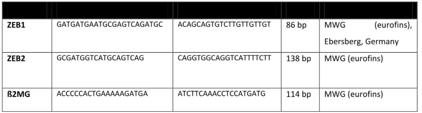
ZEB1 GATGATGAATGCGAGTCAGATGC ACAGCAGTGTCTTGTTGTTGT 86 bp MWG (eurofins), Ebersberg, Germany ZEB2 GCGATGGTCATGCAGTCAG CAGGTGGCAGGTCATTTTCTT 138 bp MWG (eurofins)
ß2MG ACCCCCACTGAAAAAGATGA ATCTTCAAACCTCCATGATG 114 bp MWG (eurofins)
3.1.11. RNA samples
Table 11. Purchased RNA samples from human, non-neoplastic brain tissue. Sample concentration, patient data and manufacturing company are listed on the right columns.
RNA Sample Conc. [µg/µl] Age Sex Company
Temporal lobe 2,25 26 M BioChain®, Newark (CA), USA
Frontal lobe 2,53 27 M BioChain®, Newark (CA), USA
Occipital lobe 1,90 41 M BioChain®, Newark (CA), USA
Corpus callosum 1,69 26 M BioChain®, Newark (CA), USA
Total human brain 1 18 M BioChain®, Newark (CA), USA
Total adult brain 1 66 F Agilent Technologies, Santa Clara (CA), USA
Human Universal Reference 1 N/A N/A Agilent Technologies, Santa Clara (CA), USA
In Table 12 is a list of RNA samples from glioma tumors obtained from patients that underwent surgery at the Department of Neurosurgery (Prof. Dr. A. Brawanski and Prof. Dr. M. Proescholdt) at Regensburg University Hospital, after informed and written consent had been given. Tissue samples were initially snap frozen and stored at -80°C. RNA was extracted only from tissue samples that had a tumor content of more than 80%. Experiments were approved by the ethics committee of the University of Regensburg (#13-101-0005).
Table 12. List of RNA extracted from glioma tumors obtained from the department of neurosurgery of the University of Regensburg Hospital. Patient information as well as RNA concentration are also listed above.
RNA Sample Conc. [µg/ml] Tumor Age Sex
A2 303 Diffuse Astrozytoma (WHO grade II) 40 M
AA7 367 Anaplastic Astrozytoma (WHO grade III) 37 M
16
GB42 1213 Glioblastoma (WHO grade IV) 68 F
GB44 1434 Glioblastoma (WHO grade IV) 45 M
GB45 323 Glioblastoma (WHO grade IV) 69 M
GB46 130 Glioblastoma (WHO grade IV) 61 F
GB47 222 Glioblastoma (WHO grade IV) 51 F
GB49 283 Glioblastoma (WHO grade IV) 65 M
GB50 230 Glioblastoma (WHO grade IV) 71 F
GB52 61 Glioblastoma (WHO grade IV) 43 M
GB53 118 Glioblastoma (WHO grade IV) 48 M
Moreover, RNA from other commercially available immortalized glioblastoma cell lines was retrieved and included in gene expression assays. RNA from MelJuSo cells, corresponding to a melanoma cell line with epithelial morphology43, 44, was kindly provided by the research group of Prof. Anja Bosserhoff (Chair of biochemistry and molecular medicine at the University of Erlangen).
Table 13. List of cell lines with their corresponding tumor of origin, patient data.
RNA Sample Tumor of origin Age Sex
A-172 Glioblastoma (WHO-Grade IV) 53 M
T98G Glioblastoma (WHO-Grade IV) 61 M
TP365 Glioblastoma (WHO-Grade IV) N/A F
MelJuSo Melanoma 58 F
Also, RNA from seven human glioblastoma stem cell lines was included in this work.
Table 14. RNA concentration extracted from glioblastoma initiating stem cell lines.
RNA Sample NCH644 NCH421K
NCH601 NCH465 NCH660K
NCH711 NCH636 NCH14225
17
3.1.12. Solutions and other biochemical compounds/ materials
Table 15. List of solutions and other biochemical compunds/ materials used in this work.
Solution/ Coumpund/ Material Manufacturer
100 bp-DNA-Ladder Fermentas, Waltham (MA), USA
DEPC-Water (DNase/RNase-free) Bioline, London, UK
RNase-free Water Qiagen, Hilden, Germany
Tris-EDTA buffer (JETquick Plasmid Miniprep Spin Kit)
Genomed GmbH, Löhne, Germany
3.1.13. Software
In the following table all software used throughout the course of this work are listed.
Table 16. List of softwares and their developing companies which were used during this work.
Software Developer
CASAVA1.8.2 Illumina, San Diego (CA), USA
cutadapt v1.11 Department of Computer Science, TU Dortmund, Germany
DAVID (Database for Annotation, Visualization and Integrated Discovery)
Leidos Biomedical Research, Inc., National Cancer Institute at Frederick, Frederick (MD), USA
EndNote (Version X7.5) Thomson Reuters, New York City (NY), USA
FASTQC v 0.11.5 Babraham Bioinformatics, Cambridge, UK.
Genome Reference Consortium Human Build 38 (GRCh38.p7)
ENSEMBLE gene annotation system, European Bioinformatics Institute
18
GraphPad Prism (Version 6.1) GraphPad Software, La Jolla (CA), USA
GenePattern GSEA module v16 Broad Institute, Cambridge (MA), USA
hisat2-2.0.4 Center for Computational Biology, Johns Hopkins University, Baltimore (MD), USA
MARS Data Analysis Software (Version 2.41)
BMG Labtech, Ortenberg, Germany
Microsoft Office (Version 2007) Microsoft, Redmond (WA), USA
NanoDrop 2000-Software (Version 1.4.2)
Thermo Fisher Scientific, Waltham (MA), USA
Omega Control-Software (Version 3.00)
BMG Labtech, Ortenberg, Germany
R (Version 3.1.2) https://www.r-project.org/
StepOne™ Software (Version 2.1) Applied Biosystems, Foster City (CA), USA
Venny 2.1.0. BioinfoGP, Spanish National Biotechnology Center, Madrid, Spain
3.2. Methods
3.2.1. Cell Biology Methods
3.2.1.1. Cell thawing and cultureAll cells were purchased from CLS cell lines service (see section 3.1.). These were frozen in FBS containing 10 % DMSO, and stored in liquid Nitrogen. For subculturing purposes, frozen-stored cells were quickly thawed in a water bath adjusted to 37°C. The cell-containing solution was then
19
pipetted under the laminar flow hood, into a culture Flask containing 10 ml DMEM cell culture medium with 10% FBS (v/v), 100 U/ml Penicillin and 100 ug/ml Streptomycin(cDMEM)45. A medium change was done 24 hours after.
3.2.1.2. Culture of glioblastoma cell lines
For the experiments carried out in this work, cells were cultured under normoxic conditions (21% O2
and 5% CO2, with 95% atmospheric humidity, at 37°C), in cDMEM medium, as explained before.
When cultured cells achieved a confluency between 70 to 80%, they were splitted with the use of Trypsin 1X (Sigma-Aldrich). Trypsin is a catalytic enzyme, which cleaves proteins on cell surfaces allowing cells to detach from one another as well as from the culture flask46. For this purpose, old medium was first removed by aspiration, and cells were washed with prewarmed 10 ml DPBS. After complete removal of DPBS by aspiration, 1.5 ml (for T75 m2 Flasks) of trypsin was given directly to the cells. Incubation was performed at 37°C in the cell culture incubator under normoxic conditions, for a maximum of 2 minutes. In order to stop the lytic actions of trypsin in the cells, 8.5 ml fresh cDMEM medium was added, and remaining attached cells were mechanically detached from the flasks bottom (by pipetting). The complete solution (medium containing detached cells) was recollected in a 15 ml Tube and centrifuged for 4 minutes at room temperature at a maximum of 1,000 x g. After removing the supernatant, 10 ml fresh Medium (containing FCS and antibiotics) was added to dissolve the cell pellet. For culture, 1 ml of the cell suspension was added to a new flask containing 9 ml fresh Medium (1:10 splitting).
3.2.1.3. Cell Harvesting
Single cell suspensions were obtained either for gene expression analyses (qRT-PCR, transfection) or functional assays (transfection, proliferation). For harvesting single cell suspensions, medium was aspirated and the cells were washed with DPBS. Afterwards, 1.5 ml trypsin-EDTA were added and incubated for a maximum of 2 minutes at 37°C. After all cells were detached and passed on to a 15 ml tube, they were centrifuged at 1,000 x g for 4 minutes at room temperature. Supernatant was discarded; 10 ml medium was added to dissolve the cell pellet. Once the cell pellet was dissolved in cDMEM, approximately 1 ml was transferred onto a 1.5 ml tube and counted with a Neubauer Chamber. The cell counting chamber should be prepared by fixing a glass cover on its central area.
Afterwards, around 10 μl of cell suspension were carefully pipetted onto the chamber. Under the microscope, cells lying inside the four external counting grids were counted (see Figure 1), using the following formula: concentration of cells/ml = No. of cells/ Volumen (ml)) x 10,000, where Volume is
20
the total amount of grids counted (4) and 10,000 accounts for the total volume of Medium in which cells were diluted in. In this way a total amount of cells per ml was obtained.47
Figure 1. Neubauer cell counting chamber. The four external counting grids are used during this process. Viable cells inside the squares black dots) are counted and used in the formula described above to determine the number of cells per ml. (Images adapted from Celeromics.com).48
3.2.1.4. Transfection of GBM cell lines with miRNA mimics
Transfection is the process of introducing foreign nucleic acids into cells48. For this procedure, Lipofectamine™ 2000 transfection reagent was used. Lipofectamine™ 2000 is a cationic liposome reagent that allows the passage of nucleic acids across the cell membrane by wrapping them in liposomes, a process known as lipofection49.
RNAi. RNA interference (RNAi) is the process of regulating the expression of protein-coding genes, using double-stranded RNA20, by loading on to Argonaute 2 (Ago2), the core catalytic component of the RNA-induced-silencing-complex (RISC)51, 52. In the cytoplasm, small interfering RNAs are cleaved, the so called “passenger” strand is degraded, and the remaining strand, called “guide” strand, will bind to RISC, and guide it into recognizing and binding to the target mRNA53.
miRNA mimics. For transient miRNA transfections, miRNA mirVana mimics (Thermo Fischer Scientific, Waltham (MA), USA) were purchased. Micro-RNA (miRNA) mimics are chemically synthesized, 20-23 nucleotide-long, double-stranded, RNA molecules that mimic endogenous miRNAs and enable functional analysis of miRNAs54. The negative control miRNA mirVana mimic Negative Control #1 was used. All mimics came lyophilized and were dissolved in sterile conditions
21
under a laminar flow hood using RNAse-free water to obtain a concentration of 5 µM. Aliquots were prepared and stored at -20°C.
Transfection. For transfection, depending on the plate´s size, a determined amount of cells (see table 18) were seeded in DMEM medium with 10% FBS without antibiotics. After 24 hours, when a confluency between 70-80% was reached, cells were transfected with miRNA mimics. Two separate tubes were prepared, containing 50 to 250 μl of prewarmed OPTI-MEM® I medium. In the Lipofectamine™ 2000 mix 0.75 - 5 µl/well Lipofectamine™ 2000 was added, mixed by gentle tapping, and incubated at room temperature for 5 minutes. In the miRNA-containing mix, 30nM/well miRNA mimic was added and mixed by gentle tapping. Afterwards, Lipofectamine™ 2000- and miRNA- containing solutions were mixed into a same tube and incubated at room temperature for 20 minutes. During this time, medium was exchanged (DMEM with 10% FBS without antibiotics).
Afterwards, Lipofectamine™ 2000 plus miRNA solution was added in a dropwise fashion into each well. Cells were then incubated at 37 °C and 5% CO2 conditions. After 48 hours post-transfection cells were either harvested and lysed for RNA isolation, or used in functional assays.55, 56
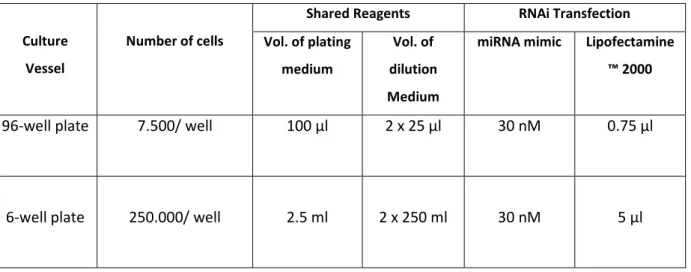
Table 17. Components for miRNA Transfection with Lipofectamine™ 2000. Table adapted from Invitrogen.com.
Culture Vessel
Number of cells
Shared Reagents RNAi Transfection Vol. of plating
medium
Vol. of dilution Medium
miRNA mimic Lipofectamine
™ 2000
96-well plate 7.500/ well 100 µl 2 x 25 µl 30 nM 0.75 µl
6-well plate 250.000/ well 2.5 ml 2 x 250 ml 30 nM 5 µl
3.2.2. Molecular Biology Procedures
3.2.2.1. miRNA ExtractionTotal RNA containing miRNAs was extracted from transfected and untransfected cells using the Qiagen miRNeasy mini kit (Qiagen, Hilden, Germany). Its principle relays on a silica membrane with selective binding properties, and a specialized high-salt buffer system that allows the binding of miRNA molecules to the silica membrane57.
22
Cells in culture plates or cell pellets containing ~500,000 cells were used for this procedure. For RNA isolation, cells and/or pellets were washed with DPBS and lysed using 750 µl of QIAzol lysis reagent (included in the miRNeasy Mini Kit). This solution is a highly denaturing monophasic solution of phenol and guanidine thiocyanate, which inactivates RNases to ensure RNA integrity. The solution was homogenized by pipetting the lysate at least 5 times up and down. In order to separate nucleic acids from other organic cellular components, chloroform (140 µl) was added and mixed with the lysate, which was afterwards centrifuged 15 minutes at 4°C at a speed of 12,000 x g. The aqueous upper phase was then transferred into a new collection tube, and the lower phase was discarded. To allow appropriate RNA binding conditions, one volume (~550µl) of RNase-free 70% EtOH was added, and the solution was well mixed by pipetting. The whole solution was then transferred to an RNeasy spin column placed in a 2ml collection tube. It was centrifuged for 15 seconds at 8,000 x g at RT and the flow through was discarded. The membrane was washed with 700 µl of buffer RWT. Afterwards, samples were washed adding 500µl RPE buffer to the RNeasy spin column and centrifuging it for 15 seconds at 8,000 x g. The flow-through was discarded, and this step was repeated prolonging the centrifuging time to 2 minutes. The spin column was then placed in a new 2ml collection tube, and centrifuged once more at 12,000 x g for 1 minute. After this step, the miRNeasy spin column was placed in a new 1.5 ml tube, and 30 µl RNase-free water was added directly to the spin column membrane. RNA was eluted by centrifugation at 8,000 x g. Samples were placed on ice immediately.
To determine the final RNA concentration, absorbance at 260 nm was measured.
3.2.2.2. Reverse Transcription of cDNA
Reverse transcription is the process by which a reverse transcriptase (RNA-dependent polymerase) transcribes from a single strand RNA a complementary DNA strand (cDNA). For this procedure, the miScript RT II Kit from Qiagen (Qiagen, Hilen, Germany) was used. 1 µg total RNA was used for reverse transcription. Afterwards a 2 µl reverse transcriptase-containing Master Mix was added, to achieve a final volume of 20 µl (see tables 20 and 21). For the RT-containing master mix miScript HiFlex Buffer was used. This kit allows to specifically detect miRNAs, by polyadenilation of mature miRNAs. During cDNA reverse transcription, oligo-dT primers which possess a 3' degenerate anchor are used, and a universal tag sequence on the 5' end is added, which allows amplification of mature miRNA during qRT-PCR58. The reverse transcription reaction was at last placed on the Thermocycler using the program described in table 19. cDNA samples were stored at -20°C.
23
Table 18. RT Master Mix Components (1x). *The use of HiFlex Buffer allows real-time PCR quantification of mature miRNA, precursor miRNA, ncRNA, and/or mRNA using individual miRNA primer assays.
Reagent Volume of 20 µl/ sample
HiFlex Buffer* 4 µl
10x nucleic mix 2 µl
RNase-free Water Variable
miScript reverse transcriptase mix 2 µl
Template RNA variable
Table 19. cDNA Transcription Program in Thermo Cycler.
Time Temperature
60 minutes 37°C
5 minutes 95°C
Indefinitely 4°C
3.2.2.3. Quantitative Real Time PCR
Real-time quantitative reverse-transcription PCR, also known as qRT-PCR, is the measurement of the increase in double stranded DNA (dsDNA) in the course of the reaction (in real time)59 and is therefore used for quantification of gene expression. It follows usually 3 steps: denaturation, annealing and elongation. During denaturation, the cDNA samples are exposed to high temperatures in order to break apart their hydrogen bonds to separate the DNA strands. In the annealing phase primers will attach to the cDNA strand. During elongation the DNA-polymerase will add complementary nucleotides on to the single-stranded cDNA forming new double-stranded DNA fragments.
SensiFAST™ SYBR® Hi-ROX Mix (Bioline, Luckenwalde, Germany) and miScript SYBR Green PCR Kit (Qiagen, Hilden, Germany) were used for this purpose. SYBR Green is an asymmetrical cyanine dye that binds to the DNA double strand. The resulting DNA-dye complex absorbs blue light (λmax = 488 nm) and emits green light (λmax = 522 nm). During the exponential phase of the PCR reaction, the fluorescence is proportional to the amount of PCR products obtained.
24
SensiFAST™ SYBR® Hi-ROX Mix was used to evaluate the expression of ZEB1 and ZEB2 in control and transfected cells, using ß2-MG as reference (housekeeping) gene. miScript SBYR Green PCR Kit was used to confirm the overexpression of miRNAs 200a/b/c, 141 and 429 in transfected cells compared to cells transfected with a negative control as well as to untransfected cells. Samples were prepared in triplicates, including a negative, water only-containing control (RNase-free water from Qiagen).
Samples were all previously diluted 1:10 with RNase-free water. The qRT-PCR reaction was performed as described below.60
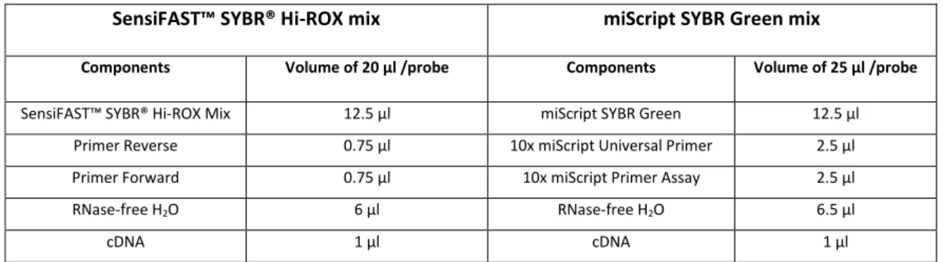
Table 20. Components of the master mixes prepared for qRT-PCR reactions using both Hi-ROX and miScript SYBR Green mixes.
SensiFAST™ SYBR® Hi-ROX mix miScript SYBR Green mix
Components Volume of 20 µl /probe Components Volume of 25 µl /probe
SensiFAST™ SYBR® Hi-ROX Mix 12.5 µl miScript SYBR Green 12.5 µl
Primer Reverse 0.75 µl 10x miScript Universal Primer 2.5 µl
Primer Forward 0.75 µl 10x miScript Primer Assay 2.5 µl
RNase-free H2O 6 µl RNase-free H2O 6.5 µl
cDNA 1 µl cDNA 1 µl
Primer assays to evaluate the expression of the miRNA 200 Family (200a, 200b, 200c, 141 and 429) in glioblastoma cell lines, glioblastoma tumors (RNA), glioblastoma stem cells (RNA) and normal brain tissue (RNA) were purchased from Qiagen (miScript primer assays (Qiagen, Hilden, Germany)).
These are synthesized mature miRNA forward primer sequences (see Table 8, section 3.1.8) that, together with the use of a universal reverse primer (supplied in the miScript SYBR Green Kit (Qiagen, Hilden, Germany)), specifically target each one of the miRNAs mentioned above. As housekeeping RNA Hs_SNORD68_11, an extra miScript primer assay coding small nucleolar RNA, C/D box 6861, was used. The primers came lyophilized and were dissolved in sterile conditions under a laminar flow hood, using 550 µL TE Buffer (TE contained in the JETKit from Genomed GmbH, Löhne, Germany) as suggested by the manufacturer. They were aliquoted and stored at -20°C.
All other primers
for qRT-PCR
were custom-synthesized (Eurofins, Jena, DE). The ZEB1/ ZEB2 primer pair sequences were obtained from the Harvard primer bank62. ß2-Microtubulin (ß2MG) was used as housekeeping gene. Primers came lyophilized, and were later dissolved in sterile conditions under a cell culture hood, using RNAse-free water (from Qiagen, Hilden, Germany) to obtain a concentration of 100 µM. They were stored at -20°C, and thawed shortly before being used in a reaction.
PCR-plates were used for this procedure. All reagents were kept on ice at all times.
25
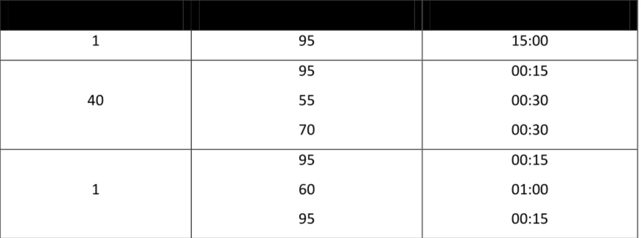
Table 21. Parameter settings for the qRT-PCR using Eurofins primers.
Number of Cycles Temperature in °C Duration in minutes
1 95 02:00
40
95 60
00:50 00:15
1
95 60 95
00:15 01:00 00:15
Table 22. Parameter Setting for the qRT-PCR using miScript primer assays primers.
Number of Cycles Temperature in °C Duration in minutes
1 95 15:00
40
95 55 70
00:15 00:30 00:30
1
95 60 95
00:15 01:00 00:15
Analyses were performed using the comparative Ct(2-ΔΔCt) method module within the StepOne™
Software (Version 2.1) (Applied Biosystems (Foster city (CA), USA)).
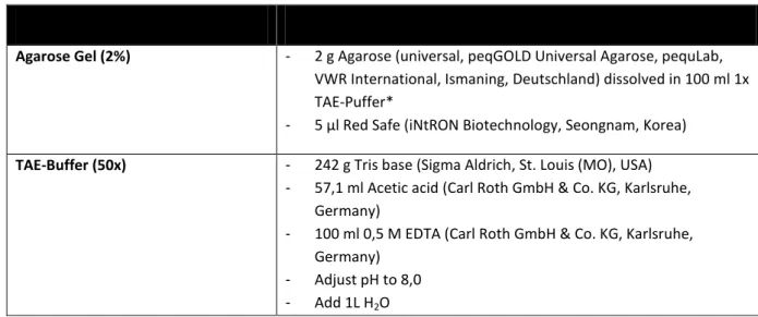
3.2.2.4. Agarose-Gel electrophoresis
PCR products can be analyzed by agarose-gel electrophoresis, in order to confirm specificity of the replicated DNA fragment by assessing its length (nucleotides). For this purpose, PCR samples were mixed with loading buffer (Orange G (Carl Roth GmbH & Co. KG, Karlsruhe, Germany)) and loaded onto a 2% agarose gel. Electrophoresis was done at 120 V. Smaller molecules are able to advance further on the gel compared to bigger ones. The size of the molecules is assessed with the help of a DNA-ladder loaded parallel with the PCR products, which displays bands along the gel with a determined size based on amount of base pairs.
26
Table 23. Composition of agarose gel and TAE-Buffers used for Agarose-Gel Electrophoresis of PCR products. *(1x TAE buffer is made out of 200 ml 50x TAE and 800 ml H2O).
Buffer/ Solution Composition
Agarose Gel (2%) - 2 g Agarose (universal, peqGOLD Universal Agarose, pequLab, VWR International, Ismaning, Deutschland) dissolved in 100 ml 1x TAE-Puffer*
- 5 µl Red Safe (iNtRON Biotechnology, Seongnam, Korea) TAE-Buffer (50x) - 242 g Tris base (Sigma Aldrich, St. Louis (MO), USA)
- 57,1 ml Acetic acid (Carl Roth GmbH & Co. KG, Karlsruhe, Germany)
- 100 ml 0,5 M EDTA (Carl Roth GmbH & Co. KG, Karlsruhe, Germany)
- Adjust pH to 8,0 - Add 1L H2O
The DNA was visualized using UV light (Gel iX manager, Intas, Göttingen, DE).
3.2.2.5. Next Generation Sequencing (NGS)
mRNA libraries. Libraries for Next Generation Sequencing (NGS) were prepared using 500 ng mRNA previously isolated from transfected and untransfected cells (miRNease mini Kit (Qiagen, Hilden, Germany)) and the TruSeq® RNA Sample Preparation Kit v2 (Illumina, San Diego, CA, USA).
Instructions from the manufacturer were adapted to a 60% protocol.
The first step was to purify and denaturate the RNA molecules. For this purpose, RNA purification beads (RPB) were added to the samples, incubated (Thermocycler) at 65°C for five minutes - to allow RNA to bind to the beads - and later on washed on a 96-well plate (Bead Washing Buffer, BWB), eluted (Elution Buffer, EB) and allowed to rebind (Bead Binding Buffer). The manufacturer provided all buffers and reagents. After a second wash, RPBs were re-suspended in Elution, Prime, Fragment mix (EPF) and incubated (Thermocycler) at 94°C for eight minutes.
The next step was to transcribe RNA into cDNA. This starts with the synthesis of a first strand cDNA using a SuperScript II reverse-transcriptase-containing first strand master mix (supplied by Illumina).
The incubation (Thermocycler) times were as follows: 10 minutes at 25°C, 50 minutes at 42°C and 15 minutes at 70°C. Afterwards, a second strand cDNA was synthesized incubating the RBDs with a second strand mix at 16°C for one hour. The resulting double stranded cDNA was purified using AMPure XP beads (Beckman Coulter, Brea, CA, USA).
27
After creating a double stranded cDNA an end repair had to be performed. For this step, the RPBs were incubated (Thermocycler) in End Repair Mix (ERM) for 30 minutes at 30°C, and afterwards cleaned up again with AMPure XP beads. Up to this point, the following step was to add A- nucleotides to the 3´end of the cDNA by adding an A-tailing mix (incubation details: 37°C for 30 minutes followed by 70°C for five minutes). Indexed adapters were ligated to the end-repaired A- tailed cDNA at 30°C for ten minutes, a Ligation Control mix was added and the libraries were cleaned up again with AMPure XP beads.
Finally an enrichment of DNA fragments was done through PCR amplification. This was performed with PCR reagents provided in the kit. The following cycling conditions were applied: denaturation at 98°C for 30 seconds, 15 cycles of 10 seconds at 98°C, 30 seconds at 60°C, 30 seconds at 72°C and a final extension at 72°C for five minutes. The amplified libraries were one last time purified with AMPure XP beads. Quality control was assessed with the use of a Bioanalyzer® (Agilent, Santa Clara (CA), USA) and Next Generation Sequencing (NGS) was finally performed using a HiSeq 1000 sequencer (Illumina, San Diego (CA), USA).
Next Generation Sequencing. Sequencing of the miRNA libraries was performed in a Core Facility on Campus of the University of Regensburg, in the competence Center for fluorescence bioanalytics (Kompetenzzentrum für Bioanalytik (KFB)) using a HiSeq 1000 sequencer from Illumina (Illumina, San Diego (CA), USA). Libraries were quantified using the KAPA SYBR FAST ABI Prism Library Quantification Kit (Kapa Biosystems, Woburn (MA), USA). Equimolar amounts of each library were used for cluster generation on the cBot with the TruSeq SR Cluster Kit v3 (Illumina, San Diego (CA), USA). The sequencing run was performed using the indexed, 50 cycles single read (SR) protocol and the TruSeq SBS v3 Kit (Illumina, San Diego, CA, USA).
3.2.3. Biochemical Methods
3.2.3.1. Proliferation AssayBromodeoxyuridine (5-bromo-2'-deoxyuridine, BrdU) is a synthetic nucleoside, analogous to thymidine, which is commonly used in the detection of proliferating cells in living tissues. BrdU substitutes thymidine during DNA replication, being able to be incorporated itself into the DNA of the cell. The amount of incorporated BrdU can be measured by detection with a specific antibody.63
For this assay, cells were seeded in black 96-well plates with clear bottom (Greiner Bio-One GmbH, Frickenhausen, Deutschland) and transfected as previously described (see transfection section