Case report:
THERAPEUTIC POTENTIAL OF PERIPHERAL BLOOD STEM CELL TRANSPLANTATION IN ONE CIRRHOTIC PATIENT CAUSED
BY HBV COMBINED WITH HCV
Li Yan1, 2, Ying Han1, 2, Jianhong Wang1, Huohong Han1, Xinmin Zhou1, Jingmei Liu1, Yuan- long He1, and Daiming Fan1*
1 State Key Laboratory of Cancer Biology, Institute of Digestive Diseases, Xijing Hospital, The Fourth Military Medical University, Xi'an, Shanxi Province, China.
2 Li Yan and Ying Han contribute equally to this paper.
* corresponding author: Daiming Fan, E-mail: fandaim@fmmu.edu.cn; Phone:
86-29-84773974; Fax: 86-29-82539041
ABSTRACT
Stem cell based therapy was very attractive in decompensated liver cirrhosis currently.
The possible mechanism might be due to its potential to help tissue regeneration with mini- mally invasive procedures. Here we report the case of a 44-year-old man, infected by hepatitis B virus (HBV) combined with hepatitis C virus (HCV) for longer than 10 years, who eventu- ally developed decompensated liver cirrhosis. After being infused with mobilized peripheral blood stem cells, the patient showed significantly elevated serum albumin level, cholesterol (CHO), cholinesterase (CHE) and decreased PT (prothrombin time) during the 26 months of follow-up. To our knowledge, this is the first case of transplanting mobilized PBSCs to treat the HBV combined with HCV related decompensated liver cirrhosis.
Keywords:HBV, HCV, decompensated liver cirrhosis, Peripheral blood stem cell transplan- tation
INTRODUCTION
Decompensated liver cirrhosis was one of the most common end-stage liver dis- eases in China (Liu and Fan, 2007). Cur- rently, liver transplant provides the only definite cure, but it is limited by donor or- gans, operative damage, risk of rejection, and high costs. Therefore, an alternative therapy to improve liver function of the cirrhotic patients was more urgent.
There were several reports that bone-marrow (BM) derived stem cells might accelerate the recovery and im- provement of the liver function in the ani-
mal model of liver injury (Wang et al., 2003;
Korbling et al., 2002; Lagasse et al., 2000).
Based on the results of animal experiments, a few clinical trials showed that bone mar- row-derived stem cells or granulocyte- colony stimulating factor (G-CSF)- mobilized bone marrow-derived hemato- poietic stem cells could contribute to the liver function of the cirrhotic (Terai et al., 2006; Yannaki et al.,2006; am Esch et al.,2005; Gordon et al., 2006; Lorenzini and Andreone, 2007). In our previous study, we have demonstrated that peripheral blood
monocytes from patients with HBV related decompensated liver cirrhosis could differ- entiate into functional hepatocytes and con- tribute to liver function (Yan et al., 2007), although this matter was still discussed controversially by the previous reports (Nussler et al., 2006; Hengstler et al., 2005).
Although all the preliminary results seemed to be attractive, the follow-up time of the treated patients was too short to fully evaluate the safety and efficacy of stem cell therapy in liver cirrhosis. Here, we describe the case of one patient with decompensated liver cirrhosis causing by HBV combined with HCV, as a result of lasting ameliora- tion of the clinical course with infusion of mobilized peripheral blood stem cells.
CASE REPORT
A 44-year-old man was admitted to Xijing Hospital in August 2005 for a detailed ex- amination of liver dysfunction. Laboratory data disclosed the following abnormal val- ues: Total Protein (TP) 53 g/L (normal 60–87), Albumin (ALB) 31 g/L (37–55), To- tal Bilirubin (TBIL) 19.4 µmol/L (6–19.2), Cholesterol (CHO) 2.23 mmol/L (3.50-6.50), Prothrombin Time (PT) 19.2 S (10.5-12.8), Activated Partial Thromboplastin Time (APTT) 49.9 S (26.8-37.5), Fibrinogen (FIB) 1.92 g/L (2.0-4.0), Thrombin Time (TT) 21.2 S (14.2-19.6), Prothrombin Activity (AT ) 58.9 % (83-96%), International Normalized Ratio (INR) 1.60 (0.92-1.11), cholinesterase (CHE) 3021 IU/L (5300–12900). Peripheral blood studies disclosed white blood cell count at 1.53×10⁹/L (3.5-10×109/L) (neutrophils 49.6 %, lymphocytes 33.3 %, monocytes 9.2 %, eosinophils 7.2 %, basophils 0.7 %), HGB 93 g/L (115-180) and platelets 27×109/L (80-300×109/L). Virus markers were positive for Hepatitis B surface anti- gen(HBsAg), Hepatitis B core antibody (HBcAb), and Hepatitis C virus antibody
(HCVAb), negative for Hepatitis B surface antibody (HBs-Ab), Hepatitis B e antigen (HBe Ag), Hepatitis B e antibody (HBe-Ab) and human immunodeficiency virus (HIV);
HBV DNA was 9.5×10⁶ copy/ml (< 1000).
A questionnaire revealed that the patient was infected by HBV combined with HCV 11 years before, and had undergone an ex- amination for liver function and was diag- nosed with chronic hepatitis B combined with hepatitis C. He had no history of al- cohol abuse or blood transfusion. His spouse was negative for HBsAg and HCVAb. A physical examination showed that he had mild abdominal distention, splenomegaly, gingival bleeding, jaundice, and edema of lower limbs. No lymphade- nopathy or skin rash was observed. Ultra- sound disclosed a cirrhotic liver, splenome- galy, and mild ascites. Gastroendoscopy revealed an esophageal varix, and a gastric scattered congestion. Histological findings of liver biopsy were not done for the pa- tient’s refusal.
Liver transplant was rejected by the pa- tients and their family members. Autolo- gous PBSCT was carried out in this patient after he assigned a formal written informed consent. The patient was mobilized with recombinant human granulocyte colony stimulating factor (rhG-CSF, Qi Lu Phar- maceutical Co, LTD, China) at 5-10 µg/kg/d administered subcutaneously daily for 4 days to induce the bone mar- row-derived stem cells into the peripheral blood, then PBSC was collected by means of Apheresis, using the COBE(R) Spectra TM Apheresis System (Gambro BCT Inc, Stockholm, Sweden). The duration of the procedure was 3 hours until the number of PBSC reached 108/ml. Then, 50 ml of the PBSC was returned to the patients via he- patic artery in the Imaging Department. Pa- tients were discharged after 5 days’ bed rest.
The therapy project above was approved by
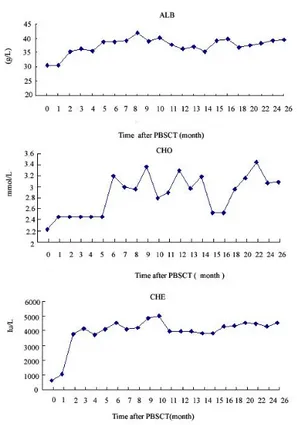
the ethics committee of Xijing hospital of the Fourth Military Medical University. The patient was followed-up to 26 months to evaluate the clinical effect of PBSCT. Dur- ing the follow-up period, medication was unchanged and the patient did not receive antiviral therapies. Liver function related serum markers including albumin, alanine aminotransferase (ALT), aspartate ami- notransferase (AST), TBIL, CHO, CHE, and prothrombin time were assayed to evaluate the liver function. Liver of the pa- tient was also examined by abdominal ul- trasonography. The results of the follow-up study indicated that liver synthetic function related markers including serum albumin, CHO, and CHE were significantly in- creased after PBSCT (Figure 1), and prothrombin time was decreased after PBSCT (Figure 2).
Figure 1: After PBSCT, serum ALB, CHO and CHE were significantly increased dur- ing the follow-up.
Figure 2: After PBSCT, prothrombin time was decreased during the follow-up.
However, data for ALT, AST, and TBIL did not show significant changes after PBSCT, and serum HBV-DNA copy also didn’t show significant changes at all (data not shown). Abdominal ultrasonography showed that ascites was disappeared at 2 weeks after PBSCT. Taken together, the cirrhotic patient showed a lasting ameliora- tion of clinical course after PBSCT.
DISCUSSION
Bone marrow-derived stem cells were known to contribute to the cirrhotic livers currently(Mohamadnejad et al., 2007; Ab- del Aziz et al., 2007). HBV-related or
HCV-related decompensated liver cir- rhosis was very common worldwide, espe- cially in China, because there were about more than 10 % HBV carriers. Once the decompensated liver cirrhosis occurred, liver transplant provides the only definite cure; however, postoperative recurred virus related hepatitis or tumor limited is widely used. Stem cell based therapy provides one inspiring therapy for hepatitis virus related decompensated liver cirrhosis.
This is the first report about evaluating liver function of HBV combined with HCV related cirrhotic patient who underwent PBSCT. In addition, the time of follow-up was the longest compared with other re- ports about PBSCT contributing to liver function of decompensated liver cirrhosis.
After PBSCT, the patient acquired lasting
amelioration of the liver function, which included liver synthetic function related markers including serum albumin; CHO, CHE, and prothrombin time were signifi- cantly improved after PBSCT. However, serum ALT, AST, and TBIL did not show significant changes after PBSCT, indicating that G-CSF mobilization combined with PBSCT therapy could not change the con- dition of virus copy. What’s more, serum HBV-DNA copy was still unchanged during the follow-up. There were two factors in- volved in the lasting amelioration of the clinical course, including G-CSF mobiliza- tion and PBSCT. G-CSF is a pleiotropic cytokine that plays a major role in regulat- ing hematopoiesis and innate immune re- sponses (Franzke, 2006). A recent study reported that G-CSF could mobilize BM stem cells into infarcted cardiac tissue and accelerates their differentiation into vascu- lar cells and cardiac myocytes (Takano et al., 2006), and the effect of G-CSF has also been demonstrated in Crohn’s disease and ulcerative colitis (Baert and Rutgeerts, 2000). Moreover, a current report demon- strated that hematopoietic mobilization could increase the presence of bone mar- row–derived hepatocytes via in vivo cell fusion (Quintana-Bustamante et al., 2006).
Thus, our favorable result might be ex- plained both by G-CSF mobilization and PBSCT. Firstly, G-CSF could mobilize a large number of PBSCs into the circulation and secret some cytokines or growth factors to promote hepatocytes functions by paracrine mechanisms. Secondly, trans- planted PBSC via hepatic artery might in- duce the higher concentration of PBSC homing to the injured liver and contribute to the liver function by cell differentiation or fusion. To fully demonstrate the thera- peutic value of this protocol, results of long-term follow-up in more patients with hepatitis related cirrhosis are needed.
REFERENCES
Abdel Aziz MT, Atta HM, Mahfouz S, Fouad HH, Roshdy NK, Ahmed HH, Rashed LA, Sabry D, Hassouna AA, Hasan NM. Therapeutic potential of bone mar- row-derived mesenchymal stem cells on experimental liver fibrosis. Clin Biochem 2007;40:893-9.
am Esch JS2nd, Knoefel WT, Klein M, Ghodsizad A, Fuerst G, Poll LW, Piechaczek C, Burchardt ER, Feifel N, Stoldt V, Stockschläder M, Stoecklein N, Tustas RY, Eisenberger CF, Peiper M, Häussinger D, Hosch SB. Portal application of autologous CD133+ bone marrow cells to the liver: a novel concept to support he- patic regeneration. Stem Cells 2005;23:
463-70.
Baert FJ, Rutgeerts PJ. Medical therapies for ulcerative colitis and Crohn’s disease.
Curr Gastroenterol Rep 2000;2:446-50.
Franzke A. The role of G-CSF in adaptive immunity. Cytokine Growth Factor Rev 2006;17:235-44.
Gordon MY, Levicar N, Pai M, Bachellier P, Dimarakis I, Al-Allaf F, M'Hamdi H, Thalji T, Welsh JP, Marley SB, Davies J, Dazzi F, Marelli-Berg F, Tait P, Playford R, Jiao L, Jensen S, Nicholls JP, Ayav A, Nohandani M, Farzaneh F, Gaken J, Dodge R, Alison M, Apperley JF, Lechler R, Habib NA.
Characterization and clinical application of human CD34+ stem/progenitor cell popula- tions mobilized into the blood by granulo- cyte colony-stimulating factor. Stem Cells 2006;24:1822-30.
Hengstler JG, Brulport M, Schormann W, Bauer A, Hermes M, Nussler AK, Fandrich F, Ruhnke M, Ungefroren H, Griffin L, Bockamp E, Oesch F, von Mach MA. Gen- eration of human hepatocytes by stem cell technology: definition of the hepatocyte.
Expert Opin Drug Metab Toxicol 2005;1:
61-74.
Körbling M, Katz RL, Khanna A, Ruifrok AC, Rondon G, Albitar M, Champlin RE, Estrov Z. Hepatocytes and epithelial cells of donor origin in recipients of periph- eral-blood stem cells. N Engl J Med 2002;346:738-46.
Lagasse E, Connors H, Al-Dhalimy M, Reitsma M, Dohse M, Osborne L, Wang X, Finegold M, Weissman IL, Grompe M. Pu- rified hematopoietic stem cells can differ- entiate into hepatocytes in vivo. Nat Med 2000;6:1229-34.
Liu J, Fan D. Hepatitis B in China. Lancet 2007;369:1582-3.
Lorenzini S, Andreone P. Stem cell therapy for human liver cirrhosis: a cautious analy- sis of the results. Stem Cells 2007;
25:2383-4.
Mohamadnejad M, Alimoghaddam K, Mohyeddin-Bonab M, Bagheri M, Bashtar M, Ghanaati H, Baharvand H, Ghavam- zadeh A, Malekzadeh R. Phase 1 trial of autologous bone marrow mesenchymal stem cell transplantation in patients with decompensated liver cirrhosis. Arch Iran Med 2007;10:459-66.
Nussler A, Konig S, Ott M, Sokal E, Christ B, Thasler W, Brulport M, Gabelein G, Schormann W, Schulze M, Ellis E, Kraemer M, Nocken F, Fleig W, Manns M, Strom SC, Hengstler JG. Present status and perspec- tives of cell-based therapies for liver dis- eases. J Hepatol 2006;45:144-59.
Quintana-Bustamante O, Alvarez-Barrien- tos A, Kofman AV, Fabregat I, Bueren JA, Theise ND, Segovia JC. Hematopoietic mo- bilization in mice increases the presence of bone marrow-derived hepatocytes via in vivo cell fusion. Hepatology 2006;43: 108-16.
Takano H, Qin Y, Hasegawa H, Ueda K, Niitsuma Y, Ohtsuka M, Komuro I. Effects of G-CSF on left ventricular remodeling and heart failure after acute myocardial in- farction. J Mol Med 2006;84:185-93.
Terai S, Ishikawa T, Omori K, Aoyama K, Marumoto Y, Urata Y, Yokoyama Y, Uchida K, Yamasaki T, Fujii Y, Okita K, Sakaida I.
Improved liver function in patients with liver cirrhosis after autologous bone mar- row cell infusion therapy. Stem Cells 2006;24:2292-8.
Wang X, Ge S, McNamara G, Hao QL, Crooks GM, Nolta JA. Albumin-expressing hepatocyte-like cells develop in the livers of immune-deficient mice that received transplants of highly purified human hema- topoietic stem cells. Blood 2003;101:
4201-8.
Yan L, Han Y, Wang J, Liu J, Hong L, Fan D. Peripheral blood monocytes from pa- tients with HBV related decompensated liver cirrhosis can differentiate into func- tional hepatocytes. Am J Hematol 2007;
82:949-54.
Yannaki E, Anagnostopoulos A, Kapetanos D, Xagorari A, Iordanidis F, Batsis I, Kaloyannidis P, Athanasiou E, Dourvas G, Kitis G, Fassas A. Lasting amelioration in the clinical course of decompensated alco- holic cirrhosis with boost infusions of mo- bilized peripheral blood stem cells. Exp Hematol 2006;34:1583-7.