Qualitive and quantitive mass spectrometric analysis of neuroactive substances from single
insect neurons
Inaugural-Dissertation zur
Erlangung des Doktorgrades
der Mathematisch-Naturwissenschaftlichen Fakultät der Universität zu Köln
vorgelegt von
Max Raoul Diesner
aus Frankfurt am Main
Köln 2018
Berichterstatter:
Dr. Susanne Neupert Prof. Dr. Marcus Krüger Tag der mündlichen Prüfung:
18.1.2019
I Abstract
Organisms need to constantly adapt their behavior to the changing environment as well as react towards changes in their internal state. The nervous system perceives and processes such stimuli and coordinates the corresponding reactions of the body. This system is based on regulated cell- cell communication, utilizing a wide range of different chemical signaling molecules and receptors. If one wants to fully grasp how neural circuits process, modulate and relay incoming information, then the involved neuroactive substances, their cellular distribution, temporal and quantitative dynamics have to be analyzed on single cell resolution. Single cell mass spectrometry (SCMS) allows the interrogation of chemical profiles from individual cells, including neuroactive substances such as neuropeptides and biogenic amines. Matrix assisted laser desorption/ionization – time-of-flight mass spectrometry (MALDI-TOF MS) has established itself as a fast and reliable tool for the analysis of neuropeptides from single neurons of invertebrates and vertebrates alike. However, the detection of small signaling molecules, such as biogenic monoamines, by MALDI-TOF SCMS has been challenging. Biogenic monoamines play key roles in orchestrating and modulating neural circuits, therefore a MALDI-TOF SCMS based method for their detection and quantification is highly desirable.
Additionally, biogenic monoamines can be co-localized with neuropeptides. Therefore the development of a MALDI-TOF SCMS based method capable of detecting both neuroactive substances would help to reveal such overlapping expression profiles.
In the current thesis, I focused on the development of a MALDI-TOF SCMS based method that allows the detection and quantification of biogenic monoamines from single somata of insect neurons. The study focused on the insect octopaminergic/tyraminergic system, with an emphasis on octopamine (OA), which is considered to be homologous to the vertebrate noradrenalin/adrenalin system. By using chemical derivatization of amine moieties of OA and tyramine (TA) and an optimized sample preparation, I was able to lower the respective detection limits to single cell concentrations. Additionally, I could show that the chemical derivatization does not interfere with the detection of neuropeptides from the same sample, hence allowing the simultaneous detection of both substance classes. Further, I could show that absolute quantification of OA and TA is possible from single cell sample volumes using isotopically labeled synthetic standards. I used the developed protocol for the qualitative and quantitative analysis of OA/TA from genetically labeled and manually microdissected somata of interneurons from the fruit fly Drosophila melanogaster. Using the newly developed approach,
II I analyzed intracellular OA/TA ratios, compared somatic OA titers between sexes and two different OAergic cell clusters and revealed that prolonged cooling of animals has an increasing effect on detectable OA titers in the analyzed neurons.
Furthermore, I used the developed protocol to analyze changes in somatic OA titers of aggression modulating OAergic neurons from the gnathal ganglion in socially naive and experienced adult male D. melanogaster. I could show that the somatic OA titer increases in these neurons when flies had social contact with the same sex compared to naive flies, which is possibly mediated by an input from pheromone detecting gustatory receptor neurons. To my knowledge, this is the first study to report a quantified increase of a somatic biogenic monoamine titer detected directly from individual isolated neurons of intact insect brains between two behavioral states by mass spectrometric analysis.
In a collaborative study, I employed the developed protocol to intracellular recorded descending dorsal unpaired median neurons from the Indian stick insect Carausius morosus and was able to confirm that these neurons contain OA and TA and thus could be OAergic.
Finally, as a starting point in an effort to create a map of neuropeptidergic neurons and their repertoire of neuroactive substances in adult D. melanogaster, I was involved in the analysis of single genetically labeled neuropeptidergic neuron somata using MALDI-TOF SCMS. In summary, we could describe a total of 10 different cell types characterized by their expressed neuropeptides and their location in the CNS. Future studies will focus on analyzing these cell types towards potential co-localized aminergic transmitters using the developed protocol.
III Zusammenfassung
Organismen müssen ihr Verhalten ständig an die sich verändernde Umwelt anpassen sowie auf Veränderungen in ihrem physiologischen Zustand reagieren. Das Nervensystem nimmt solche Reize wahr, verarbeitet sie und koordiniert die entsprechenden Reaktionen des Körpers. Dieses System basiert auf einer geregelten Zell-Zell-Kommunikation, bei der eine Vielzahl verschiedener chemischer Signalmoleküle und Rezeptoren zum Einsatz kommen. Um zu verstehen, wie neuronale Schaltkreise eingehende Informationen verarbeiten, modulieren und weiterleiten, müssen die beteiligten neuroaktiven Substanzen auf zellulärer Ebene charakterisiert und ihre zeitliche und quantitative Dynamik auf Einzelzellnivaeu analysiert werden. Die Einzelzell-Massenspektrometrie (SCMS) ermöglicht die Analyse von chemischen Profilen einzelner Zellen, einschließlich neuroaktiver Substanzen wie z.B. Neuropeptide und biogene Monoamine. Die matrixgestützte Laser Desorption/Ionisation – Flugzeit Massenspektrometrie (matrix assisted laser desorption/ionization mass spectrometry; MALDI- TOF MS) hat sich als schnelle und zuverlässige Methode zur Analyse von Neuropeptiden aus einzelnen Neuronen von Invertebraten und Vertebraten etabliert. Die Detektion von kleinen Signalmolekülen, wie z.B. biogenen Monoaminen, durch MALDI-TOF SCMS stellt jedoch bis heute eine analytische Herausforderung dar. Da biogene Monoamine eine Schlüsselrolle bei der Orchestrierung und Modulation neuronaler Schaltkreise haben, ist eine MALDI-TOF SCMS gestützte Methode für deren Nachweis und Quantifizierung äußerst erstrebenswert. Biogene Monoamine können zu dem mit Neuropeptiden kolokalisiert seien. Somit würde eine neu entwickelte durch MALDI-TOF SCMS gestützte Methode, mit der ein Nachweis beider neuroaktiven Substanzklassen aus einer Zellprobe möglich ist, es deutlich erleichtern entsprechend überlappende Expressionsmuster nachzuweisen.
In der vorliegenden Arbeit konzentrierte ich mich auf die Entwicklung einer MALDI-TOF SCMS basierten Methode, die den Nachweis und die Quantifizierung von biogenen Monoaminen aus einzelnen Insektenneuronen ermöglicht. Die Studie konzentrierte sich auf das octopaminerge/tyraminerge System von Insekten, mit dem Schwerpunkt auf Octopamin (OA), das als homolog zum Noradrenalin/Adrenalin-System der Wirbeltiere gilt. Durch die chemische Derivatisierung der Aminogruppe von OA und Tyramin (TA) und eine optimierte Probenvorbereitung konnte ich die jeweiligen Nachweisgrenzen auf Einzelzellkonzentrationen senken. Zudem konnte ich zeigen, dass die chemische Derivatisierung den Nachweis von Neuropeptiden aus derselben Probe nicht stört und somit den gleichzeitigen Nachweis beider
IV Substanzklassen ermöglicht. Außerdem konnte ich zeigen, dass die Quantifizierung von OA und TA aus Einzelzellprobenvolumina mit isotopenmarkierten synthetischen Standards möglich ist. Ich entwickelte das Protokoll zur qualitativen und quantitativen Analyse von OA/TA mittels genetisch markierten und manuell mikrodissektierten Somata von Interneuronen der Taufliege Drosophila melanogaster. Mit dem neu entwickelten Ansatz untersuchte ich intrazelluläre OA/TA Verhältnisse, verglich somatische OA-Titer zwischen den Geschlechtern sowie zwei verschiedenen OAergen Zellclustern und zeigte, dass eine Kühlung der Tiere vor der Präparation zu einem Anstieg der nachweisbaren OA-Titer in den analysierten Neuronen führt.
Darüber hinaus habe ich das entwickelte Protokoll verwendet, um Veränderungen in somatischen OA-Titern von aggressionsmodulierenden OAergen Neuronen aus dem gnathalen Ganglion in sozial naiven und erfahrenen erwachsenen männlichen D. melanogaster zu analysieren. Ich konnte zeigen, dass der somatische OA-Titer in diesen Neuronen zunimmt, wenn Fliegen sozialen Kontakt mit dem gleichen Geschlecht hatten, verglichen mit naiven Fliegen. Dies ist möglicherweise auf einen Input von gustatorische Rezeptorneuronen zurückführen welche Pheromone detektieren. Nach meiner Kenntnis ist dies die erste Studie, die einen quantifizierten Anstieg eines somatischen biogenen Monoamin-Titers zeigt, der direkt aus einzelnen isolierten Zellen eines intakten Insekten Gehirns zwischen zwei Verhaltenszuständen mittels massenspektrometrischer Analyse nachgewiesen wurde.
In einem Kooperationsprojekt untersuchte ich mit Hilfe des entwickelten Protokolls intrazellulär abgeleitete dorsal ungepaarte mediane Neurone der indischen Stabheuschrecke Carausius morosus in Bezug auf ihre aminerge Zusammensetzung. Ich konnte nachweisen, dass diese Neurone OA und TA enthalten und somit OAerg seien können.
Als Ausgangspunkt für die Erstellung einer Karte neuropeptiderger Neurone und ihrer Repertoires an neuroaktiven Substanzen in adulten D. melanogaster, war ich an der Analyse einzelner genetisch markierte neuropeptiderge Neuronenzellkörper mittels MALDI-TOF SCMS beteiligt. Insgesamt konnten wir 10 verschiedene Zelltypen beschreiben, die sich durch ihre Neuropeptidzusammensetzung und ihre Lage im Gehirn auszeichnen. Zukünftige Studien werden sich darauf konzentrieren, diese Zelltypen mit Hilfe des entwickelten Protokolls zur biogenen Monoamincharakterisierung auf mögliche Kolokalisierungen aminerger Transmitter zu analysieren.
V Table of Contents
Abstract ... I Zusammenfassung ... III Table of Contents ... V Abbreviations ... VII
1. General Introduction ... 1
2. Development of a MALDI-TOF MS based workflow for the identification and quantification of octopamine and tyramine from single identified D. melanogaster neurons . 28 2.1 Introduction ... 29
2.2 Materials and Methods ... 31
2.3 Results and Discussion ... 35
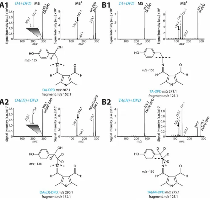
2.3.1 Analysis of non-derivatized and derivatized synthetic OA and TA ... 35
2.3.2 Detection of OA and TA from individual Drosophila neurons ... 41
2.3.3 Quantification of synthetic OA and TA ... 43
2.3.4 Stability of stored samples ... 47
2.3.5 Isomeric ion species and neuropeptide profiling ... 49
2.3.6 Discrimination of OA and DA ... 50
2.3.7 Quantification of OA and TA in VMlb somata ... 52
2.3.8 Temperature-dependent variability of detectable OA titers in VMlb neurons ... 53
2.3.9 Sexual dimorphism of VMlb OA concentrations ... 54
2.3.10 Quantification of OA from two OA/TA cell populations... 55
2.4 Conclusion ... 57
3. Quantification of Drosophila VMlb octopamine titers in social driven male aggression by MALDI-TOF SCMS ... 58
3.1 Introduction ... 59
3.2 Materials and Methods ... 63
3.3 Results and Discussion ... 66
3.3.1 Influence of age, sex and genetic construct on OA VMlb titers... 66
3.3.2 Influence of social experience on VMlb OA titers ... 67
3.4 Conclusion ... 72
4. Mass spectrometric detection of octopamine and tyramine from intracellular recorded desDUM neurons of Carausius morosus ... 73
4.1 Introduction ... 74
4.2 Materials and Methods ... 76
4.3 Results and Discussion ... 79
4.4 Conclusion ... 83
5. Mass spectrometric survey and mapping of neuropeptides in DIMMED neurons from the adult Drosophila brain ... 84
VI
5.1 Introduction ... 85
5.2 Materials and Methods ... 87
5.3 Results and Discussion ... 90
5.3.1 Cell type #1: Allatostatin-C (Ast-C) neurons ... 92
5.3.2 Cell type #2: Myosuppressin (DMS) neurons ... 94
5.3.3 Cell type #3: Allatostatin-A (Ast-A) + myoinhibiting peptide (MIP) + Natalisin (Nat) neurons ... 94
5.3.4 Cell type #4: CAPA neurons ... 97
5.3.5 Cell type #5: Hugin/Pyrokinin (huginPK) neurons ... 99
5.3.6 Cell type #6: Sulfakinin (SK) neurons ... 100
5.3.7 Cell type #7: Corazonin (Cor)/short Neuropeptide F (sNPF) neurons ... 101
5.3.8 Cell type #8: Pigment dispersing factor (PDF) neurons ... 103
5.3.9 Cell type #9: Neuropeptide-like precursor 1 (NPLP1) neurons ... 103
5.3.10 Cell type #10: Neuropeptide F (NPF) neurons ... 106
5.4 Conclusion ... 108
6. General Discussion ... 110
6.1 Methodological aspects of the developed MALDI-TOF MS based approach ... 110
6.1.1 Range of detectable analytes ... 111
6.1.2 Limit of detection and quantification ... 113
6.1.3 Sample isolation ... 114
6.2 Applications and implications for further research ... 117
7. Conclusion ... 123
8. List of Figures ... 124
9. List of Tables ... 125
10. Bibliography ... 126
11. Part Publications ... 152
12. Acknowledgments ... 154
13. Erklärung ... 155
14. Curriculum vitae ... 156
VII Abbreviations
1,5-DAN 1,5-diaminonaphtalene
5-HT 5-Hydroxytryptamine/serotonin
7-T (z)-7-tricosense
ACh acetylcholine
ACN acetonitrile
CHCA α–cyano-4-hydroxycinnamic acid
AD adrenaline
Akh adipokinetic hormone
AP action potential
Ast-A allatostatin-A
Ast-C allatostatin-C
CA 4-hydroxy-3-methoxycinnamaldehyde
cAMP 3’,5’-cyclic adenosine monophosphate
CE capillary electrophoresis
CFME carbon-fiber mircoelectrodes
Cha choline acetyltransferase
CID collision-induced dissociation
CNiFERs cell-based neurotransmitter fluorescent engineered reporters
CNS central nervous system
cVA 11-cis-vaccenyl acetate
CPG central pattern generator
DA dopamine
DAG diacylglycerol
DESI desorption electrospray ionization
DPD 2,5-dimethyl-1H-pyrrole-3,4-dicarbaldehyde desDUM descending dorsal unpaired median
DHB 2,5-dihydroxybenzoic acid
DIM DIMMED (transcription factor)
DMS drosomyosuppressin
DTT dithiothreitol
DUM dorsal unpaired median
ECD electrochemical detection
VIII
ER endoplasmatic reticulum
ESI electrospray ionization
FDA US Food and Drug Administration
FRET fluorescence resonance energy transfer
FSCV fast-scan cyclic voltammetry
GABA γ-aminobutyric acid
GFP green fluorescent protein
GNG gnathal ganglion
GPCR G-protein-coupled receptors
GRASP GFP reconstitution across synaptic partners
GRN gustatory receptor neuron
HA histamine
HPLC high performance liquid chromatography ICLI inferior contralateral interneuron
IP3 1,4,5-triphosphate
IS internal standard
ISD in-source decay
LDCV large dense core vesicle
LEAP large, episodically-releasing, amidating peptide producing cells, that contain the transcription factor DIMMED
LIF laser-induced fluorescence detection LINF laser-induced native fluorescence detection
LOD limit of detection
LOQ limit of quantification
LLOQ lower limit of quantification
MALDI matrix-assisted laser desorption/ionization
MBON mushroom body output neurons
MEKC micellar elektrokinetic capillary chromatography
MIP myoinhibitory peptide
MN motor neuron
MP bilaterial medial protocerebral neurons
MS mass spectrometry
MS² tandem mass spectrometry
MSI mass spectrometry imaging
IX
m/z mass-to-charge ratio
NAD noradrenaline
nanoLC nano liquid chromatography
Nat natalisin
NPF neuropeptide F
NPLP1 neuropeptide-like precursor 1
OA octopamine
OA(d3) (±)-p-octopamine-α,β,β-d3
OAR octopamine receptors
ORN olfactory receptors neurons
PAM protocerebral anterior medial
PBS phosphate buffer solution
PDF pigment dispersing factor
PK pyrokinin
PM paired median
PP precursor peptides
PPL1 protocerebral posterior lateral 1
PSD post-source decay
PTM posttranslational modification
PVK periviscerokinin
rpm revolutions per minute
RSD relative standard deviation
SCMS single cell mass spectrometry SIMS secondary ion mass spectrometry
SK sulfakinin
sNPF short neuropeptide F
TA tyramine
TA(d4) 2-(4-hydroxyphenyl)ethyl-1,1,2,2-d4-amine
Tβh tyrosine-β-hydroxylase
TDC tyrosine decarboxylase
TFA trifluoroacetic acid
TK tachykinin
TNT tetanus neurotoxin light chain
TOF time-of-flight
X TrpA1 transient receptor potential cation channel A1
Tyr tyrosine
UM unpaired median neuron
UV ultraviolet
VL ventrolateral
VMlb ventral midline labial
VMmd ventral midline mandibular
VMmx ventral midline maxillary
VNC ventral nerve cord
VPM ventral paired median
VUM ventral unpaired median
1 1. General Introduction
Organisms need to constantly adapt their behavior to the changing environment as well as react towards changes in their internal state. The nervous system perceives internal and external stimuli, integrates this information in the context to the current state of the organism and generates an appropriate reaction of the body. Regulated cell-cell communication is the basis for such targeted transduction, integration and modulation of information in the nervous system.
For cell-cell communication in the nervous system, neurons can convert their electrical activity into the release of neuroactive substances targeting neurons and/or other cell types. Target cells express receptors which bind neuroactive substances with high specificity and selectivity.
When a receptor binds to its specific ligand, intracellular signal cascades are activated altering the physiology and “behavior” of the corresponding cell, for example by changing gene expression or activation of membranous ion channels. A plethora of different neuroactive substances have been identified from nervous systems of invertebrates and vertebrates, and it has been shown that the cellular expression patterns, receptors and corresponding functions of a single neuroactive substance can be conserved throughout different evolutionary lineages (Bräunig & Pflüger 2001; Roeder 2005; Nässel & Wegener 2011).
In order to fully comprehend how the nervous system and underlying neural circuits shape and control the reactions of an organism, it is essential to analyze involved neuroactive substances, their cellular distribution, temporal and quantitative dynamics on a single cell resolution.
However, neuroactive substances often show overlapping expression patterns in nervous system, e.g. biogenic monoamines and neuropeptides (Merighi 2011; Fricker 2012; Nässel 2018), which makes it challenging to reveal the full signaling capabilities of a given neuron.
Thus, methodological approaches that enable the simultaneous detection of multiple classes of substances in a single experiment are highly desirable. Ideally, such methods would be quantitative to allow the analysis of changing neuroactive substance concentration in a given cell in relation to other signaling molecules, drugs or different behavioral states of the analyzed organism. Mass spectrometry (MS) offers the possibility to simultaneously detect and quantify different substance classes from biological samples such as single cells. For example, matrix assisted laser desorption/ ionization – time-of-flight single cell mass spectrometry (MALDI- TOF SCMS) is a widely used tool for the detection of neuropeptides from neuron somata of invertebrates and vertebrates alike (e.g. Li, Garden, et al. 2000; Neupert & Predel 2005;
Rubakhin & Sweedler 2007; Neupert et al. 2007). However, the detection of potentially co-
2 localized small neuroactive substances, like biogenic monoamines, has been challenging using this approach.
The main focus of the current thesis was the development of a MALDI-TOF SCMS based strategy for the detection and quantification of biogenic monoamines from single insect neuron somata. The focus laid on the insect octopamine (OA)/tyramine (TA) system, with an emphasis on OA, which is considered to be homologous to the vertebrate noradrenalin/adrenalin system.
Moreover, since biogenic monoamines can be co-localized with neuropeptides in neurons, the simultaneous detection of these substance classes was investigated. To set the context of my work, I want to introduce insect nervous systems, OA and TA, neuropeptides and mass spectrometry as an analytical method in more detail in the following sections.
Insect nervous systems and their role in neuroscience
Insect nervous systems are arranged in single units called ganglia organized in a metameric fashion, which is based on their segmental body plan. These ganglia can represent fusion products of so called neuromeres or represent a single neuromere. Neuromeres represent a segment specific part of the central nervous system (CNS) that contains the neural circuitry which processes sensory information and controls movement of segmental appendages and segmental muscles (Niven et al. 2008). Single neuromeres are linked by a pair of connectives, which are still present in fused ganglia as axonal tracts. The insect CNS is comprised of a brain (supraesophageal ganglion), the gnathal ganglion (GNG; old name subesophagial ganglion; Ito et al. 2014), and the ventral nerve cord (VNC). The brain is a fusion product of four pregnathal neuromeres, while the GNG represents a fusion product of the three gnathal neuromeres (Urbach & Technau 2003; Niven et al. 2008). The VNC consists of three thoracic ganglia, the pro-, meso- and metathoracic ganglion, connected to a varying number of abdominal ganglia.
Ganglia in the VNC can be fused or remain free depending on the examined insect lineage, but in general represent a total of eleven neuromeres (Niven et al. 2008). Further, comparable to the vertebrate autonomic nervous system, a system of peripheral ganglia exists in insects, the stomatogastric nervous system. It is comprised of four main parts: the frontal ganglion, the hypocerebral ganglion and the paired/unpaired ingluvial and proventricular ganglia (Bilingsley
& LeHane 1996; Hartenstein 1997).
3 Even though insect nervous systems are only comprised of a small number of neurons compared to mammal nervous systems, they produce a wide variety of complex behavior patterns such as mating, collision avoidance and spatial navigation as well as many properties of insect neurons show striking resemblance in their mammalian counterparts (Namiki et al. 2009). Moreover, extensive similarities of neuronal circuits underlying sensory systems, synaptic plasticity and neuromodulation between invertebrates and vertebrates suggest a deep homology between central parts of their distinct CNS (Strausfeld & Hirth 2013; Haberkern & Jayaraman 2016).
The investigation of these “simple” nervous systems, therefore, facilitates our understanding of nervous systems in general on a broader scale (Namiki et al. 2009). Aside from these benefits, a multitude of practical advantages exist when working with insects and their nervous systems.
Especially, their low cost mass rearing and short generation times facilitate fast repetition rates of experiments. Furthermore, the limited size and accessibility of insect nervous systems is highly beneficial for modern microscopy based methods, like immunohistochemistry or in vivo imaging, as well as electrophysiological applications. It is therefore not surprising that insects have established themselves as model organisms for the functional analysis of behavior and neuronal networks throughout the last century.
Another more recent driving force has been the development of non-invasive genetic tools for the targeted expression of reporter and effector genes like the GAL4/UAS system (Jarman et al. 1993) or the LaxA-LexAop system (Lai & Lee 2006) and their ongoing extension in the small fruit fly D. melanogaster (Yoshihara & Ito 2012). The combination of these tools with the fully sequenced D. melanogaster genome (Adams et al. 2000), enables the targeted analysis of gene expression by genetic labeling, as well as the targeted interference of gene expression rates via methods such as RNA interference (Roignant et al. 2003). Moreover, the system allows the activation or silencing of single neurons by light (Lima & Miesenböck 2005), temperature (Kitamoto 2001; McGuire et al. 2003) or the expression of ion channels (Luan et al. 2006) for the analysis of neuronal network functions. Furthermore, the GAL4/UAS system has been transferred to another insect model the red flour beetle Tribolium castaneum (Schinko et al.
2010), which will allow to use the depicted genetic tools in this insect species.
Finally, large scale projects which aim to decipher a high number of various insect transcriptomes and genomes, like the 1kite project (www.1kite.org) or the i5k project (i5k.github.io), in combination with the newly discovered CRISPR/Cas gene editing method (Jinek et al., 2012; Gasiunas et al., 2012) will allow the transfer of the already developed genetic
4 tools to other insect species. In the current thesis, I used the GAL4/UAS system to label different subsets of neurons in adult D. melanogaster to allow their repeatable dissection and identification between different individual flies.
Biogenic monoamines
Biogenic monoamines are small biogenic neuroactive substances which have a single functional amine moiety. The group of biogenic monoamine transmitter includes: the imidazole amines (histamine [HA]), catecholamines (adrenaline [AD], noradrenaline [NAD], dopamine [DA]), indolamines (serotonin [5-HT]), tryptamines (tryptamine), phenethylamines (e.g.: TA, OA, synephrine) and thyronamines (3-Iodothyronamine). Biogenic monoamines are versatile in their mode of action and can function as neurotransmitters, neuromodulators and/or neurohormones. They are involved in the regulation of nearly all vital functions in vertebrates and invertebrates alike, such as cardiovascular control, movement, endocrine regulation, circadian rhythms, learning and memory (e.g. Nässel & Winther, 2010).
All biogenic monoamines are synthesized from proteinogenic amino acids and their derivates, like tyrosine (Tyr), phenylalanine, tryptophan or histidine, via amino acid decarboxylases (Brady et al. 2012). They are synthesized in the cytosol of the neuron cell body as well as the nerve terminals, where they are stored and released from synaptic vesicles or large dense core vesicles (LDCV) by stimulus induced Ca2+ dependent exocytosis. Cytosolic biogenic monoamines are actively transported into small synaptic vesicles or LDCVs by vesicular H+ dependent monoamine transporters and can be co-localized with other neuroactive substances, like neuropeptides (Brady et al. 2012; Gallo et al. 2016). Nearly all biogenic monoamines are recognized by GPCR receptors, with the exception of 5-HT which binds to an ionotropic receptor (Wu et al. 2015). Secreted biogenic monoamines are inactivated either by degradation or reuptake. The primary mechanism for inactivation is cytosolic reuptake by specialized Na+/Cl- dependent plasma membrane transporters (e.g.: DA transporter; Giros & Caron 1993;
Ueno & Kume 2014). Cytosolic catabolism of monoamine transmitters after reuptake is mainly mediated by monoamine oxidases (Westlund et al. 1985; Eisenhofer 2004; Yamamoto &
Vernier 2011) whereas extra-neuronal inactivation is mediated by catechol-o- methyltransferases (Eisenhofer 2004; Yamamoto & Vernier 2011). However, in invertebrates biogenic monoamine inactivation is enzymatically diverse and can also be mediated by N-
5 acetylation, N-methylation, sulfation and other mechanisms (Sloley & Juorio 1995; Sloley 2004; Sotnikova & Gainetdinov 2010).
The analysis of the wide range involvement of biogenic monoamine signaling in complex behaviors and emotions led to the understanding that impairment of these systems can lead to severe disease and disorders such as depression, obsessive-compulsive disorder, neuropathic pain, and others (Pitman et al. 2011; Yousuf & Kerr 2016). Therefore, methods which allow us to foster our understanding of their cellular distribution, intracellular dynamics and effects on target cells are highly desirable. In this thesis I focused on two biogenic monoamines OA and TA in insect nervous systems, with an emphasis on OA.
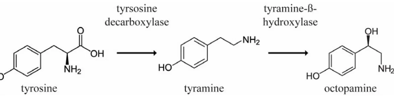
Figure 1.1Synthesis pathway of OA. Tyrsoine is decarboxylated into TA, and TA is subsequently hydroxylated into OA.
Octopamine
Octopamine, or p-hydroxyethanolamine, was first discovered in the salivary glands of Octopus vulgaris (Erspamer & Boretti 1951). It is synthesized by hydroxylation of its precursor TA, via the tyramine-β-hydroxlase (Tβh) in invertebrates (Fig. 1.1). OA is the most abundant biogenic monoamine transmitter in invertebrates (Verlinden et al. 2010; Fang et al. 2011), however, in vertebrates it is considered a trace amine and only found in very small amounts (Berry 2004).
While OA function in the CNS of invertebrates is a subject of numerous studies and serves as a perfect model to investigate general features of neuromodulation, only recently interest in trace amine function in vertebrate nervous systems has resparked due to the discovery of mammalian trace amine receptors (Borowsky et al. 2001; Lindemann et al. 2005; khan & nawaz 2016). A growing body of evidence suggests an involvement of OAergic signaling in neurosynaptic transmission and seems to be connected to vertebrate neurological disorders like depression, migraine or Parkinson´s disease (khan & nawaz 2016). The chemical structure of OA is analogous to the vertebrate NAD and the invertebrate OA/TA signaling system is often
6 seen as homologous to the vertebrate NAD/AD signaling system, due to similar functions in physiology and behavior as well as structural features (Roeder 2005; Verlinden et al. 2010).
Furthermore, a recent phylogenetic analysis of biogenic monoamine receptors suggested that OAergic and adrenergic systems coexisted in the last common ancestor of bilaterians (Roeder 2005; Bauknecht & Jékely 2017). Adrenergic signaling seems to be unique to the deuterostome lineage and has no physiological relevance in invertebrates to date, while OA signaling in vertebrates was believed to be physiologically irrelevant as well (see above). Due to this lineage specificity and the fact that OA signaling has regulating effects in virtually all tested organs and tissues of insects it has been the subject of a plethora of studies as a potential target for insecticides (Roeder 2005; Verlinden et al. 2010; Farooqui 2012)
In insects, only a small number of neurons express OA as neuroactive substance, which has also been reported for other biogenic monoamines, projecting to most neuropil regions of the brain, the thoracic nervous system and peripheral organs. In the larval D. melanogaster CNS, for example, only around 80 OAergic neurons have been identified (Monastirioti et al. 1995;
Selcho et al. 2012; Selcho et al. 2014), whereas the imago yields about 100 OAergic neurons (Monastirioti et al. 1995; Sinakevitch & Strausfeld 2006; Busch et al. 2009). Furthermore, the distribution and number of OAergic neurons has been studied in various insects including honey bees (Sinakevitch & Strausfeld 2006; Schröter et al. 2007), blowflies (Sinakevitch & Strausfeld 2006), cockroaches (Eckert et al. 1992; Sinakevitch et al. 2005), hawkmoths (Dacks et al. 2005), crickets (Sporhase-Eichmann et al. 1992), and locust (Konings et al. 1988). The projection patterns and terminal areas of these OAergic neurons seem to be conserved and analogous counterparts can be identified between different insect species (Braunig 1991; Bräunig &
Pflüger 2001; Schröter et al. 2007; Busch et al. 2009; Busch & Tanimoto 2010). Mainly two populations of OAergic neurons have been described in insect nervsous systems; paired median (PM) and non-median neurons that can be located in the brain, the GNG and some segmental ganglia, as well as unpaired median neurons (UM) located solely in segmental ganglia and the GNG of insects (Bräunig & Pflüger 2001; Busch et al. 2009; Selcho et al. 2014). UM neurons can be subdivided into dorsal UM (DUM) and ventral UM (VUM) neurons depending on the cell body position at the midline of the respective ganglion (Bräunig & Pflüger 2001; Busch et al. 2009; Selcho et al. 2014). In D. melanogaster three clusters comprised of VUM and ventral PM (VPM) neurons located in the GNG have been described, with VUM neurons of the mandibular (VMmd) and maxillary (VMmx) cluster (8-9 VUM neurons in each cluster; Busch et al. 2009; Schneider et al. 2012) showing only innervation of the brain in various higher brain
7 regions as well as the GNG. Individual VUMs of different clusters show very similar ramifications and projection patterns in the brain and it is suggested that they developed through a duplication of an evolutionary older set of OAergic VUMs (Busch et al. 2009; Busch &
Tanimoto 2010). Because of their similarities in their projection patterns these neurons are characterized into morphological groups (Busch et al. 2009). Most VUMs of the labial cluster (VMlb; ~4-5 neurons) in adult D. melanogaster, however, send their projections towards the VNC with unknown targets (VUMd1-3; Busch et al. 2009; Schneider et al. 2012). Aside from these descending VUM neurons of the VMlb cluster, one pair of VPM neurons of the VMmd cluster, VPM1, project asymmetrical to the ipsilateral side of the thoracic neuromeres and the abdominal neuromeres (Busch et al. 2009). In the adult locust, homologous OAergic cells, so called descending DUM (desDUM) neurons, project to neuropil regions processing information of leg sensory organs (Bräunig & Burrows 2004). Finally, five sets of VPM neurons exist in D.
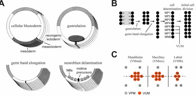
melanogaster, with VPM1 and 2 neurons being located in the close vicinity of the VMmd cluster, VPM3 neurons at the VMmx cluster and VPM4 and 5 neurons at the VMlb cluster (Busch et al. 2009; Busch & Tanimoto 2010; see Fig. 1.2). Each VUM cluster develops from a single median precursor cell of the embryonic midline, while it has been suggested that VPM neurons develop from paired lateral neuroblasts (see Fig. 1.2; Klämbt et al. 1991; Busch &
Tanimoto 2010).
OA solely mediates its regulatory and modulatory functions through G protein coupled receptors (GPCR) in insects. In the genetic model organism D. melanogaster, for example, five OA GPCRs have been identified with different binding affinities, second messenger cascades and distributions (Farooqui 2012; El-Kholy et al. 2015; Qi et al. 2017). These five OA receptors (OAR) are grouped into three classes based on their structural and signaling similarities to vertebrate adrenergic receptors: α-adrenergic-like receptors (OAMB [OctαR; two isoforms, OAMB-K3; OAMB-AS]; Octα2R [two isoforms, Octα2R-L; Octα2R-S]), β-adrenergic-like receptors (Octß1R; Octß2R; Octß3R) and octopamine/tyramine receptors (Farooqui 2012; El- Kholy et al. 2015; Qi et al. 2017). OAR elicit their function though G-proteins activating various second messenger cascades including cAMP, inositol 1,4,5-triphosphate (IP3), and diacylglycerol (DAG). The latter second messenger cascades affect intracellular Ca2+
concentrations, via IP3 for example, or regulate phosphorylation via the protein kinase A, by cAMP, or protein kinase C by DAG (Farooqui 2012).
8 Octopamine in insect behavior and physiology
DUM/VUMs are considered the main source of OA in the brain and the thoracic nervous system of insects. Subpopulations of gnathal DUM/VUMs innervate most neuropil regions of the insect brain and it has been shown that these are involved in controlling and regulating various complex behaviors, whereas thoracic and segmental DUM/VUM populations project to the periphery and are hypothesized to be the main source of peripheral OA signaling (Bräunig &
Pflüger 2001; Roeder 2005; Farooqui 2007; Farooqui 2012).
Figure 1.2 Development of VUM/VPM neurons in D. melanogaster. (A) Development of the midline during four embryonic stages in D. melanogaster. Four midline precursor cells are located at both ends of the mesodermal anlage, forming the mesectoderm. During gastrulation, the mesoderm invaginates so that the midline precursor cells become juxtaposed at the ventral midline. During germ band elongation these precursor cells become a single line with eight cells per segment. Before these stem cells divide, they delaminate from the neuronal ectoderm into the neuroblast layer. (B) Schematic drawing of the development of the midline as described in (A). The midline consists of 7 medial precursor cells and the medial neuroblast. The medial precursor cells 5-7 develop into VUM neurons in D. melanogaster. (C) Outline of gnathal OAergic cell clusters and their VUM/VPM neurons in adult D. melanogaster. Numbers indicate a distinct morphological cell type with similar projections. Modified from (Klämbt et al. 1991; Busch & Tanimoto 2010).
OA signaling in the CNS involves modulating a plethora of different behaviors and underlying neuronal circuits such as reward and appetitive reinforcement (D. melanogaster: Schwaerzel et al. 2003; Schroll et al. 2006; Burke et al. 2012; Apis mellifera: Hammer 1993; Hammer &
Menzel 1998; Gryllus bimaculatus: Unoki et al. 2005; Unoki et al. 2006), taste (D.
melanogaster: LeDue et al. 2016), aggression (D. melanogaster: Hoyer et al. 2008; Zhou et al.
2008; Hoopfer 2016; Watanabe et al. 2017; G. bimaculatus: Stevenson et al. 2000; Stevenson
9 2005) and vision (A. mellifera: Erber & Kloppenburg 1995; Locusta migratoria: Stern et al.
1995; Stern 2009). Other processes modulated by OA in the CNS include the initiation, maintenance and modulation of motor patterns and motor circuits as described for walking and flying in locusts (Schistocerca americana: Sombati & Hoyle 1984; Schistocerca gregaria:
Rillich et al. 2013), flight in the hawk moth (Manduca sexta: Claassen & Kammer 1986), larval locomotion and adult flight in the fruit fly (D. melanogaster. larvae: Saraswati et al. 2004; Fox et al. 2006; Selcho et al. 2012; Imago: Brembs et al. 2007; van Breugel et al. 2014), and walking in the stick insect (Carausius morosus, Büschges et al. 1993). Furthermore, the role of OA in sensitization and dishabituation has been studied in various insects such as in the visual system of the locust (L. migratoria: Stern 2009) or the honey bee proboscis extension reflex (PER;
Mercer & Menzel 1982; Braun & Bicker 1992).
OA has also modulating effects in the periphery on skeletal and visceral muscles, organs and glands. In homology to the vertebrate adrenergic system, OA is involved in the physiological body adaptation during energy demanding tasks such as the fight or flight response (Stevenson et al. 2000). As response to “stressful” stimuli and subsequent hyperglycemia, OA can be released into the hemolymph, were it acts as a neurohormone, to increase hemolymphatic lipid and sugar concentrations by either affecting energy storages in the fat body directly (Fields &
Woodring 1991) or by regulating the release of the neuropeptide adipokinetic hormone (Akh) from the corpora cardiaca (Pannabecker & Orchard 1986; Lorenz & Gäde 2009). Other peripheral effects of OA signaling involve the insect immune system, where OA mediates haemocyte activity, such as phagocytosis and nodule formation (Baines et al. 1992), and bounds to the surface of bacterial pathogens for better recognition (Dunphy & Downer 1994).
Furthermore, OA can modulate sensory organs and neurons by increasing or decreasing receptor sensitivity, receptor density or affecting neurotransmitter release (Farooqui 2007). For example, it has been shown that OA sensitizes sensory neurons of the forewing stretch receptor in locust (Ramirez et al. 1993). Finally, OA acts on a wide range of different muscles e.g. on oviduct contraction (D. melanogaster.: Rodríguez-Valentín et al. 2006; L. mirgatoria: Orchard
& Lange 1986), extensior-tibiae muscles in S. gregaria (Evans & O’Shea 1977) and body wall muscle in D. melanogaster larvae (Monastirioti et al. 1995; Selcho et al. 2012).
10 Tyramine
Tyramine, or 4-Hydroxy-phenylethylamin, is not only the chemical precursor of OA but can rather act as neuroactive substance on its own. TA modulation is often described as antagonistic to OA mediated effects, homologous to adrenergic system in vertebrates. TA is synthesized by decarboxylation of Tyr via the tyramine-decarboxylase (Tdc, Fig. 1.1; Cole et al. 2005; Roeder 2005; Farooqui 2012). In Drosophila, two Tdc genes were described and subsequent gene products show Tdc activity in vivo, however Tdc1 is expressed non-neurally, e.g. in malpighian tubules, rectal papillae and digestive tract musculature, whereas Tdc2 is expressed solely in neurons (Cole et al. 2005).
Studies focusing on the effects of OA signaling often reduced TA to an intermediate product in the synthesis pathway of OA (Lange 2009; Farooqui 2012). Nowadays, a growing body of evidence is starting to overturn this picture and more studies focus on potential roles of TA signaling in physiology and behavior. However, unraveling the functions of TA signaling is difficult due to dependency of OA synthesis on TA and only limited access to selective and strong TA receptor antagonists (Lange 2009). Nevertheless, the identification of three Drosophila TA GPCRs (Oct/TyrR [TyrR], TyrRII, III; Saudou et al. 1990; Cazzamali et al.
2005; El-Kholy et al. 2015) and characterization of some of their signaling properties (Cazzamali et al. 2005; Bayliss et al. 2013; Farooqui 2012), as well as the analysis of their expression pattern in the CNS by creation of specific transgenetic driver lines in D.
melanogaster (El-Kholy et al. 2015) has led to first insights in TA signaling in the fly and to the understanding that TA acts as independent neuroactive substances in the CNS.
Tyramine in insect behavior and physiology
Tyramine is co-localized with OA in DUM/VUM neurons and it has been speculated that both substances can be co-released from OAergic neurons (Lange & da Silva 2007; Lange 2009;
Pyakurel et al. 2016). Aside from co-releasing, neurons expressing exclusively TA have been described in the D. melanogaster larval brain and VNC by immunostainings against TA, OA and synthesizing enzymes (Monastirioti et al. 1995; Nagaya et al. 2002; Selcho et al. 2014).
Furthermore, studies on the TA/OAergic innervation of the locust oviduct from abdominal DUM/VUMs and paired dorsal neurons revealed also potentially exclusive TAergic neurons (Stevenson et al. 1994; Donini & Lange 2004; Lange & da Silva 2007; da Silva & Lange 2008).
11 TA signaling has often been described as antagonistic to OA signaling, homologous to the adrenergic system in vertebrates. TA signaling is involved in regulating motor patterns and circuits, as it has been shown that increased neuronal TA titers decrease larval crawling distance in D. melanogaster (Nagaya et al. 2002; Saraswati et al. 2004; Fox et al. 2006) as well as reduce flight duration in adults (Brembs et al. 2007; Ryglewski et al. 2017). In the locust S. gregaria it has been shown that TA is modulating central pattern generators for walking and flight (Buhl et al. 2008; Rillich et al. 2013). Sensory systems can also be the target of TA modulation, which has been shown by a D. melanogaster tyramine receptor mutant honoka, which showed reduced behavioral responses to odorant repellents compared to wild type flies (Kutsukake et al. 2000).
Furthermore, TA signaling modulates appetitive regulation as shown in the blowfly Phormia regina, where injections of TA led to a decreased PER with regard to experiences of appetitive and nonappetitive food flavors (Nisimura 2005). In A. mellifera, however, thoracic injection of TA led to an increased sugar responsiveness and a better performance in appetitive learning (Scheiner et al. 2002; Scheiner, Reim, et al. 2017), which is suggested to be mediated by caste specific differential expression of TA GPCRs in the honey bees fat body (Scheiner, Entler, et al. 2017).
Neuropeptides
Neuropeptides are the most diverse neuroactive substances found in metazoan organisms and are involved in controlling and modulating a plethora of physiological processes and behavioral patterns such as food intake, reproduction or muscle control (Nässel 2002; Nässel & Winther 2010; Wegener & Veenstra 2015; Schoofs et al. 2017). The term neuropeptide was coined by David de Wied in the 70s who studied the activity of pituitary peptide hormones in rats on motivation, learning and memory (de Wied 1971; de Wied 2000). The proposed definition was that neuropeptides are produced and secreted by neurons and affect functions of the CNS (de Wied et al. 1974; de Wied 2000). A more recent and better fitting definition states that a neuropeptide is defined as a bioactive peptide, secreted by neurons or non-neuronal cells, with similarities in their expressed genetic information, synthesis, processing as well as their binding to similar receptor families affecting neuronal tissue (Kastin 2000; Fricker 2012).
Neuropeptides usually contain 2-100 amino acids and are produced by neurosecretory cells and interneurons in the nervous system but can also be expressed by various non-neuronal cells of the endocrine system (Zitnan 1996; Kingan & Adams 2000; Fricker 2012; Wegener & Veenstra
12 2015). A single cell is not limited to the expression of a distinct neuropeptide gene but can express multiple neuropeptide genes at the same time, sometimes even co-localized with classical neurotransmitter or biogenic monoamines (Fricker 2012; Nässel 2018). Neuropeptides are versatile in their mode of action and can act as neurotransmitter, neuromodulator and/or neurohormone depending on the secreting synapse or neuroheamal site (Nässel 2009; Nässel &
Winther 2010).
Neuropeptides are produced in the cell body and are matured throughout the regulated secretory pathway, in contrast to classical neurotransmitters which are in general enzymatically synthesized at the synapse (Merighi 2011; Fricker 2012). Neuropeptide synthesis starts with the translation of a peptide-precursor, the prepropeptide or preprohormone, containing a signal peptide which guides the preproprotein to the rough endoplasmic reticulum (ER; Perone et al.
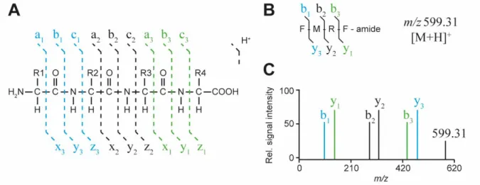
1997). As a first enzymatic step a signal peptidase removes the N-terminal signal sequence, which is rapidly degraded (Tuteja 2005). The resulting propeptide or prohormone can undergo further modification (e.g.: glycosylation) and is transported to the Golgi network, passing the network from the cis- to the trans-face, and subsequently packaged in LDCVs for further maturation, storage and ultimately secretion (Merighi 2011; Fricker 2012). During Golgi transit and maturation in LDCVs the propeptide is cleaved by endoproteases (furins, prohormone convertases) at designated internal dibasic and/or monobasic cleavage sites, such as Lys-Arg (KR), Lys-Lys (KK), Arg-Arg (RR), Arg (R) and Lys (K) (Veenstra 2000; Rholam & Fahy 2009; Fricker 2012). C-terminal basic amino acids, remaining from the endoprotease enzymatic cleavage, are subsequently removed by exoproteases (carboxyproteases) leading to mature and/or immature peptides (Merighi 2011; Fricker 2012). Often, a precursor is cleaved into multiple neuropeptides (isoforms) with structurally related C-terminal motifs. The C-terminal motif plays a crucial role during interaction of the neuropeptide and its designated receptor, in general a GPCR (Nässel & Winther 2010; Merighi 2011; Fricker 2012). Neuropeptides with a conserved structural motif are classified into neuropeptide families (Coast & Schooley 2011).
Subsequently, resulting peptides can be subjected to further modification during maturation.
Such post-translational modifications (PTM) affect the activity and degradation of mature neuropeptides. Amidation of the C-terminus is a widespread PTM of neuropeptides and is necessary for proper receptor recognition (Kolhekar et al. 1997; Prigge et al. 2000). Cyclization of N-terminal Gln into pyroglutamate is another frequently observed PTM in neuropeptides (e.g.: corazonin; Veenstra 1989). Other PTMs involve the addition of functional groups to
13 amino acid side chains such as sulfation of Tyr (Holman et al. 1986), phosphorylation (Browne et al. 1981; Gäde et al. 2006) and glycosylation (Lu et al. 2002).
This multifarious synthesis, processing and packaging of neuropeptides leads to a vast repertoire of different potential bioactive neuroactive substances. To unravel such neuropeptidergic systems it is vital to gain knowledge of their expressed and processed mature products as well as other co-expressed neuroactive substances on a single cell level in nervous systems.
An introduction to mass spectrometry
The identification, structural characterization and quantification of organic and inorganic chemical compounds from complex sample mixtures, such as organs/tissues/cells and their extracts, is today mainly achieved by MS and adjacent technologies such as chromatography.
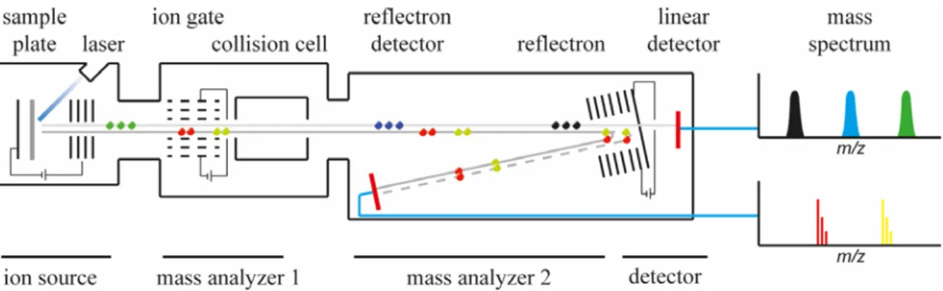
All mass spectrometers are based on the same principle, which is the separation of ionized molecules by means of their mass-to-charge ratio (m/z) in the gas-phase. In order to achieve this separation, mass spectrometers are working under high vacuum to circumvent potential collisions or diversions of produced ions with air molecules such as nitrogen. All mass spectrometers have three main components; (1) an ion source, which produces negatively or positively charged analyte ions, (2) the mass-analyzer, which separates the produced ions by their m/z ratio, and (3) a detector, detecting the separated ions (Fig. 1.3). Finally, a mass spectrum is generated by plotting the relative abundance of detected ions in a defined scanned mass range against their m/z ratio (Figure 1.3 A). This basic outline, also termed MS-mode or MS1, illuminates the chemical complexity of a given sample, with information regarding relative abundance and masses of ionized analyte molecules. Ultimately, masses of detected ions can be compared to databanks, such as METLIN (metlin.scripps.edu) or UNIPROT (www.uniprot.org), in which already identified substances are deposited and identified via
“mass match”. However, this identification solely rests on the comparison of recorded masses without any information on the chemical structure of a recorded ion and is often seen as a non- conclusive verification.
To obtain information on the chemical structure of an analyte of interest, mass spectrometers utilize a second mode called MS² or tandem mass spectrometry, which involves fragmentation of the ion of interest, the so called precursor ion, into specific and reproducible product ions.
14 This process involves multiple sequential steps of MS analysis according to their m/z ratio (Fig.
1.3). After the generation of analyte ions in the source, they are separated by their m/z ratio in a first mass analyzer (MS1). Distinct precursor ion species can now be filtered from the remaining ion background and subjected to fragmentation. A second mass analyzer (MS²) than separates the produced ion fragments or product ions and ultimately guides them to the detector (Fig. 1.3). To date various fragmentation methods have been observed and developed, which can be exclusive to a given instrumentation, such as in-source decay (ISD; Brown & Lennon 1995; Sellami et al. 2012), post-source decay (PSD; Spengler et al. 1992; Kaufmann et al.
1994), collision-induced dissociation (CID; Levsen & Schwarz 1976; Mitchell Wells &
McLuckey 2005), photodissociation methods (Brodbelt 2015), electron capture and transfer methods (Qi & Volmer 2016; Lermyte et al. 2018) and others.
The detection of specific analytes can be hampered from biological samples due to isometric and/or isobaric chemical compounds, substances with concentrations lower than the limit of detection (LOD) or ion suppression effects caused by salts, ion-pairing agents, endogenous compounds, and additives such as drugs or metabolites. In order to tackle such sample-derived problems and enable a successful detection by MS, mass spectrometers are often combined with chromatographic or electro kinetic separation techniques, enzymatic digestions, chemical derivatizations or a combination of these.
While MS or even MS² can give information about relative concentrations of sample analytes an absolute quantification of these compounds can be achieved by the addition of a known internal standard (IS) or reference with a defined concentration to the sample. While synthetic isotopically marked standards of a known substance are the gold standard, these can be very expensive or unavailable for purchase. To fill this gap, chemically related substances can be used instead, however, to guarantee a precise quantification their ionization properties and linear correlation compared to the ion of interest have to be assessed carefully. Another possibility for absolute quantification is to analyze synthetic standards of the analyte of interest before the main experiment, in a corresponding sample matrix with the identical instrumentation, and compare the obtained signal intensities from both experiments.
A variety of different ion sources, analyzers and detectors have been developed and combined in the last decade, leading to numerous analytical platforms with unique features and drawbacks. In this study, I used MALDI-TOF MS as the main analytical tool.