Cancer Medicine. 2019;8:7065–7073. wileyonlinelibrary.com/journal/cam4 | 7065
O R I G I N A L R E S E A R C H
Increased cytoplasmatic expression of cancer immune
surveillance receptor CD1d in anaplastic thyroid carcinomas
Florian Weber1
|
Henrik Junger2|
Jens M. Werner2|
Natalia Velez Char1|
Carolina Rejas2
|
Hans J. Schlitt2|
Matthias Hornung2This is an open access article under the terms of the Creative Commons Attribution License, which permits use, distribution and reproduction in any medium, provided the original work is properly cited.
© 2019 The Authors. Cancer Medicine published by John Wiley & Sons Ltd.
Florian Weber and Henrik Junger equally contributed to this work.
1Department of Pathology, University Hospital Regensburg, Regensburg, Germany
2Department of Surgery, University Hospital Regensburg, Regensburg, Germany
Correspondence
Matthias Hornung, Department of Surgery, University Hospital Regensburg, 93042 Regensburg, Germany.
Email: matthias.hornung@ukr.de Funding information
This research did not receive any specific grant from any funding agency in the public, commercial, or not‐for‐profit sector.
Abstract
Background: Anaplastic thyroid carcinomas are associated with rapid tumor growth, short survival time and without any promising therapy to improve the poor progno- sis. In this study, expression of immunoregulative receptor CD1d and lymphocyte infiltration in different thyroid tumors as well as in healthy tissue were analyzed in order to find new targets for an immunotherapeutic approach.
Methods: CD1d immunohistochemistry was performed in samples of 18 anaplastic, 17 follicular, 27 papillary, and 4 medullary thyroid carcinomas as well as in 19 speci- mens from normal thyroid tissue and additionally in 10 samples of sarcoma, seven malignant melanoma and three spindle‐cell lung carcinoma. Furthermore, thyroid samples were stained with antibodies against CD3, CD20, CD56, CD68, and LCA in order to analyze lymphocyte infiltration.
Results: For the first time CD1d receptor expression on normal thyroid tissue could be demonstrated. Moreover, anaplastic thyroid carcinomas showed significantly higher expression levels compared to other thyroid samples. Most astonishingly, CD1d expression disappeared from the cellular surface and was detected rather in the cytoplasm of anaplastic thyroid carcinoma cells. In addition, histologically simi- lar tumors to anaplastic carcinoma like sarcoma and malignant melanoma revealed distinct CD1d staining patterns. Furthermore, infiltration of T cells, B cells, and mac- rophages in anaplastic thyroid carcinomas was different when compared to normal thyroid tissue and all other thyroid carcinomas.
Conclusions: Anaplastic thyroid carcinomas show significantly higher expression of CD1d, a receptor for NKT cells, which are subject of several anticancer therapy stud- ies. These results may offer a novel approach to explore immunotherapeutic treat- ment options.
K E Y W O R D S
CD1d, immunotherapy, lymphocyte infiltration, NKT cell, thyroid carcinoma
1
|
INTRODUCTIONThe incidence of malignant thyroid tumors has increased over the last few years.1 These tumors represent a very heterogeneous group of malignancies, with distinct differ- ences in histology and likewise in clinical outcome.1,2 Most thyroid malignancies are described as follicular or papil- lary thyroid carcinomas (FTC/PTC) but about 1.7% of the patients are diagnosed with anaplastic thyroid carcinoma (ATC).3 ATC is a rare, yet rapidly growing neoplasm of the thyroid gland with a poor prognosis.4 In contrast to FTC or PTC, which can be treated postoperatively by radioiodine therapy, ATCs are not sensitive to any further treatment and therefore have the worst prognosis with a median survival of only 5 months.3,5 However, immunotherapy as new ther- apeutic approach has been introduced against a wide range of solid tumors recently. Oncologic trials have focused on developing immunomodulating therapies to restore the functional ability of different immune cells against neo- plastic cells and showed promising results in several tumor types, such as lung cancer, melanoma, and colon cancer.6,7 Regarding malignant thyroid tumors, recent advances ap- proached several strategies for immunomodulation, such as inhibiting recruitment of tumor‐associated macrophages (TAM), blocking and inhibiting of several immune check- points, as well as identification of tumor‐specific antigens.8 In this context, natural killer T (NKT) cells, which are con- sidered to be regulators of an inflammatory immune response, also represent a promising target for antitumor therapy.9-11 They are a subset of lymphocytes that co‐express NK cell markers and the T‐cell receptor.12 In humans, NKT cell popu- lation expresses the Vα24 and Jα18 gene segments, which are preferentially associated with Vβ11.13,14 NKT cells are acti- vated through a specific receptor called CD1d, which belongs to a group of CD1 molecules associated with β2‐microglob- ulin, and is known to present lipids, including glucosylcer- amides and glycosylphosphatidylinositol to NKT cells.15 It is already known that activation of CD1d leads to the production of interleukin (IL)‐4 and interferon (IFN)‐g and an increase in the cytolytic activity of NKT cells.16 However, expression of CD1d, a MHC class I‐related molecule, could just be shown on hematopoietic cells and on epithelial cells of the intestine.9,17,18
In this study, the expression of CD1d and lymphocyte in- filtration in healthy thyroid tissue as well as in several thyroid carcinomas was analyzed in order to find new targets for an immunotherapy approach.
2
|
MATERIALS AND METHODS 2.1|
Tissue samplesFFPE tissue samples were collected from the archives of the Institute for Pathology, Regensburg. Tumor entity was
confirmed before anonymization. Two tissue micro arrays (TMAs), containing a total of 18 samples of ATC, 17 sam- ples of FTC (12 grossly invasive, 5 minimally invasive), 27 samples of PTC, and 4 samples of medullary thyroid carcinoma (MTC), were created with subsequent histo- morphological control of the tumor samples by a trained pathologist (FW) using conventional H&E staining. The TMAs also contained 19 samples of normal thyroid tissue, as well as 10 samples of sarcoma (SRC; two myofibroblas- tic sarcomas, two dedifferentiated liposarcomas, and one fibrosarcoma, leimyosarcoma, biphasic synovial sarcoma, clear cell sarcoma, carcinosarcoma and sarcomatoid renal cell carcinoma, respectively), seven samples of malignant melanoma (MM), and three samples of spindle‐cell lung carcinoma (SLC).
2.2
|
ImmunohistochemistryFor standard immunohistochemistry, 5 µm thick paraffin sections were processed using routine diagnostic antibodies and immunostaining protocols (Ventana immunostainer) of the Institute for Pathology, Regensburg for staining of CD3, CD20, CD56, CD68 (KP1), and LCA (CD45). The number of positively stained cells (lymphocytes or monocytes) per high‐power field (HPF, 400x magnification) for each tis- sue sample was recorded and used for further analysis. For CD1d immunohistochemistry, a mouse monoclonal anti- body against human CD1d (LifeSpan BioSciences, code:
LS‐B6766, clone: ms R3) was used in a dilution of 1:50.
The dewaxed paraffin sections were subjected to heat‐in- duced epitope retrieval for 5 minutes at 120°C in Tris‐EDTA buffer at pH 8.5. The antigen localization was carried out using polymer‐based detection systems and an automated system setting with diaminobenzidine as the final substrate.
Immunohistochemistry was evaluated by an expert patholo- gist (FW); for CD3, CD20, CD56, CD68, and LCA, the num- ber of specifically positive cells per high‐power field (HPF, 400×) were counted. For CD1d, in addition to the number of specifically positive cells per HPF, the immunostaining of tumor/normal thyroid cells was assessed by an expert pathologist (FW) and a trained pathologist (NVC) accord- ing to relative intensity of cytoplasmic (c) and membranous (m) staining, that is c > m, c = m, or m > c; some samples showed a completely negative staining reaction, and only one sample had nuclear positivity (Figure 1). In case of differing results between the two observers, a mutual consensus was found and recorded.
2.3
|
MicroscopyStained sections were viewed with a Leica light micro- scope (Leitz DMRBE; Leitz). Images were acquired using a MIRAX digital slide scanner (Zeiss).
2.4
|
In situ hybridizationmRNA detection of CD1d (Hs‐CD1D #496771, ACDBio) was performed using the RNAscope 2.5 HD RED Reagent Kit (#322350, ACDBio) on 3‐μm FFPE sections of tissue micro arrays (TMAs) according to the manufacturer's instructions. RNA quality was evalu- ated for each sample with a probe specific to the house- keeping gene ubiquitin (Hs‐UBC, #310041, ACDBio).
Negative control background staining was evaluated using a probe specific to the bacterial DapB gene (DapB,
#310043, ACDBio). Only samples with an average of >4 dots per cell with the housekeeping gene probe staining and an average of <1 dot per 10 cells with the nega- tive control staining were assayed with target probes.
Analysis was performed by two independent research- ers on a Leica light microscope (Leitz DMRBE; Leitz) using a 40× objective. The RNAscope signal is scored on the basis of number of dots per cell as recommended
by the manufacture (ACDscore) and was applied as fol- lows 0 = 0 dot/cell, 1 = 1‐3 dots/cell, 2 = 4‐9 dots/cell, 3 = 10‐15 dots/cell, and 4 = >15 dots/cell with > 10% of dots in clusters).
2.5
|
Statistical analysisD'Agostino & Pearson normality test did not confirm a nor- mal distribution of the data set. Therefore, results are shown as median ± interquartile range (IQR). Statistical analyses were performed using a nonparametric Kruskal‐Wallis test followed by Dunn's multiple comparison. Differences were considered significant at P < .05. GraphPad Prism 7.0b (GraphPad Software, Inc) was used for significance tests and generating plots.
2.6
|
EthicsThe study had been approved by the institutional review board of the University Hospital of Regensburg (# 14‐160‐0029).
3
|
RESULTS3.1
|
Immune cell populations in normal thyroid tissueInitially, normal thyroid tissue was stained for several im- mune cell populations by immunohistochemistry. As shown in Figure 1, using specific antibodies for different immune cell markers normal thyroid tissue showed a low number of T cells (CD3 pos., 3 ± 4.1 per HPF), macrophages (CD68 pos., 2.06 ± 1.8 per HPF), and B cells (CD20 pos., 0.33 ± 0.8 per HPF).
3.2
|
Lymphocyte infiltration and thyroid cancerThen, different thyroid carcinomas were stained for cells of the immune system (Figure 1). As summarized in Figure 2, ATC specimens showed a significantly higher number of CD3 (36, IQR 19.5‐67.25) and CD68 (37, 18.5‐54.5)‐posi- tive cells in comparison to FTCs (CD3: 4; 2‐9.5, P = .0009;
CD68: 1, 0‐3, P = .005), PTCs (CD3: 19, 3‐52.5, n.s.;
CD68: 20.5, 11.75‐27.25, n.s.) and MTCs (CD3: 0, 0‐2.25, P = .0009; CD68: 6, 2.25‐10.5, n.s.), as well as normal thyroid tissue (CD3: P < .0001; CD68: P < .0001). In ad- dition, cells expressing CD20, a B cell marker, could be de- tected significantly more frequently in ATCs (2.5, 0‐6) than in normal thyroid tissue (P = .005) but the differences to FTCs, PTCs, and MTCs were not significant. Interestingly, FTCs revealed similar numbers of CD3‐positive cells when compared to normal thyroid tissue, but less than PTCs (Figure 2).
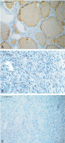
FIGURE 1 Examples of CD3 immunohistochemistry are shown for normal thyroid tissue (A), ATC with numerous positive lymphocytes (B) and FTC with only one positive lymphocyte (C)
A
B
C
3.3
|
CD1d expression on thyroid tissueNKT cells could not be detected in thyroid tissue, most likely due to the fact that to our knowledge it has not yet been possible to stain these specific cells by immunohistochem- istry. Nevertheless, the expression of the NKT cell activat- ing receptor CD1d.19,20 was analyzed and as expected, there were more immune cells expressing CD1d in ATCs (Figure 3). Furthermore, for the first time CD1d could be detected in thyroid tissue. Staining of normal thyroid tissue revealed
membranous expression of CD1d (Figure 4) as it has been already shown for hematopoiesis‐derived and intestinal epi- thelial cells.17,18,21
3.4
|
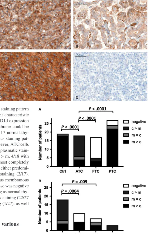
CD1d expression and thyroid cancerAdditionally, different thyroid carcinomas were stained for CD1d expression (Figure 4). Interestingly, ATC was asso- ciated with a significantly higher number of CD1d‐positive cells compared to other tumor entities and normal thyroid FIGURE 2 Mean number of positive cells per HPF for CD3 (A), CD20 (B) and CD68 (C) in ATC, FTC, MTC, PTC, and normal thyroid tissue
FIGURE 3 A, Mean number of positive immune cells per HPF for CD1d and (B) CD1d mRNA (Score 0‐3) in ATC, FTC, MTC, PTC, and normal thyroid tissue
tissue. Additionally, looking at the respective staining pattern for CD1d in individual tumor cells, different characteristic features with regard to the localization of CD1d expression either in the cytoplasm or on the cell membrane could be found (Figure 5A). In the control group of 17 normal thy- roid tissue samples, all showed a membranous staining pat- tern for CD1d expression (m > c 17/17). However, ATC cells showed a very strong and predominantly cytoplasmatic stain- ing of CD1d expression (12/18 cases with c > m, 4/18 with m > c, 2 with c = m). FTC cells stained almost completely negative (12/17 neg.), with only five cases of either predomi- nantly membranous (3/17) or cytoplasmic staining (2/17).
MTC cells were mostly stained homogeneous membranous and cytoplasmic (3/4 with c = m), only one case was negative for CD1d. PTC cells showed a similar staining as normal thy- roid tissue with a predominantly membranous staining (22/27 with m = c) and even partial nuclear staining (1/27), as well as two completely negative stainings (2/27).
3.5
|
CD1d mRNA expression in various thyroid cancer typesCD1d transcripts were detected by chromogenic alka- line phosphates‐based in situ hybridization in FFPE tissue (RNAscope). All study section showed very good mRNA quality, indicated by very high abundant signal (mRNA score >3) for the positive control ubiquitin (Figure 6A), and further high assay quality was revealed by no unspecific sig- nal (mRNA score = 0) for the negative control DapB (Figure 6B). Figure 6 shows representative areas of CD1d mRNA expression in ATC, FTC, MTC, PTC, and normal thyroid tissue. CD1d was almost exclusively expressed, at relatively high levels, in ATC; the lowest CD1d mRNA expression was seen in normal thyroid tissue (Figure 3B), consistent with our IHC data (Figure 4). CD1d mRNA expression in
ATC was significantly higher than that in FTC (P = .02) and normal thyroid tissue (P < .0001; Figure 3B). FTC and MTC showed only sporadic CD1d mRNA expression levels FIGURE 4 Different staining
patterns for CD1d immunohistochemistry with predominantly cytoplasmic staining in ATC (A), predominantly membranous staining in PTC (B), equally membranous and cytoplasmic staining in SRC (C) or no staining in FTC (D)
A B
C D
FIGURE 5 Staining patterns of normal thyroid tissue and different thyroid cancer types (ATC: anaplastic thyroid carcinoma;
FTC: follicular thyroid carcinoma; MTC: medullary thyroid carcinoma; PTC: papillary thyroid carcinoma) (A) as well as other malignant tumors (SRC: sarcoma; MM: malignant melanoma; SLC:
spindle‐cell lung cancer) (B). Staining patterns are indicated as predominantly membranous (m > c), predominantly cytoplasmic (c > m), equally membranous and cytoplasmic (c = m), negative (neg.) and nuclear (n)
(Figure 3B). In contrast to FTC and MTC, PTC showed a moderately increased CD1d mRNA expression, signifi- cantly higher than that in normal thyroid tissue (P = .0025;
Figure 3B).
3.6
|
CD1d expression as a diagnostic toolSince CD1d expression was very strong and localized predominantly cytoplasmic in ATC cells, we next wanted to assess if this could be used as histopathological tool to
help in the differential diagnosis of ATC and other medi- astinal tumors. Therefore, specimens of sarcoma (SRC), MM, and SLC were examined for their respective stain- ing profiles (Figure 7). SRC cells were either negative (6/11) or showed a predominantly membranous staining pattern (5/11), while MM cells showed a heterogeneous profile with all patterns present (3/7 with m > c, 2/7 with c > m, 2/7 neg.). All three SLC cases showed a predomi- nantly cytoplasmic staining pattern (3/3) (Figures 5B and 6).
FIGURE 6 Expression of UBC (positive control) and DapB (negative control) with red chromogenic ISH (RNAscope) in a representative ATC (A and B) and normal thyroid tissue (C and D). Different levels of CD1d mRNA detected with red chromogenic ISH (RNAscope) in PTC (ACD mRNA score 1) (E) and several ATC (ACD mRNA score 1, 2 and 3) (F, G and H). All images at 4× magnification
A
B
C
D
E
F
G
H
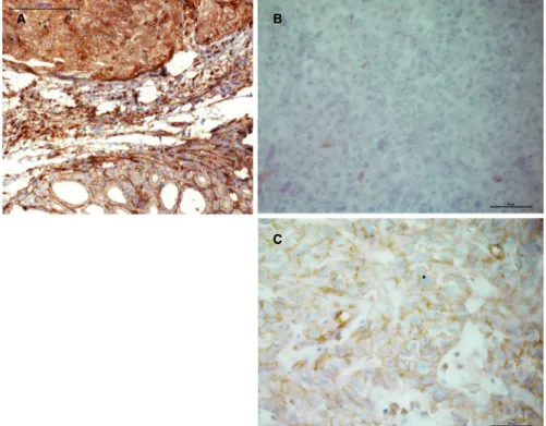
FIGURE 7 CD1d‐positive staining in ATC (A) compared to negative staining in MM (B) and weak membranous staining in SRC (C)
A B
C
4
|
DISCUSSIONIn this study, for the first time CD1d receptor expres- sion could be detected on thyroid tissue. So far, expres- sion of CD1d was described only on hematopoietic and epithelial cells of the intestine.9,17,18 Furthermore, analy- sis of different thyroid tumor entities revealed significant higher expression of CD1d and CD1d mRNA in ATCs compared to normal thyroid tissue and all other thyroid tumor types. On average, patients with ATCs survive only a few months after primary diagnosis.22 Although ra- diotherapy and different chemotherapies result in slightly improved survival rates,23,24 outcome is still very poor.22 Therefore, several treatment strategies are currently under research.22 Regarding cancer therapy, in the last few years a lot of effort was put into the research of immunotherapy with NKT cells, for which CD1d serves as target recep- tor.11,25-27 Activation pathways of NKT cells in cancer, CD1d mechanism, and even CD1d escape strategies in tu- mors are topics of current studies.28 In this regard, a CD1d‐
binding glycolipid for specific stimulation of iNKT cells has been developed for the treatment of breast cancer.29 Interestingly, CD1d expression in ATCs was not located on the cell surface but rather in the cytoplasm of the tumor cell. It has already been shown that CD1d expression is decreased in several diseases. Virus infection inhibits NKT cell‐CD1d function among others through downregulation of CD1d.30,31 In this context, herpes simplex virus 1 phos- phorylates the type II kinesin motor protein KIF3A which lead to the suppression of CD1d.32 Otherwise, in systemic lupus erythematosus TLR9‐induced microRNA‐155 and Ets‐1 decrease membrane CD1d levels.33 But also in ma- lignancies CD1d degradation could be observed. In chronic myeloid leukemia, activation of the Rho‐associated protein kinase (ROCK) mediates the reduction of membrane ex- pression of CD1d but not the reduction of intracytoplas- matic levels or mRNA transcripts34 and a certain kind of invasive breast cancer is also associated with downregu- lated CD1d.35 Regarding the CD1d receptor molecule, a Thr/Ser residue motif in the cytoplasmatic tail seems to be pivotal for the surface expression.36 Nevertheless, future research has to focus on the mechanism of CD1d down- regulation, especially in ATCs. Although CD1d downregu- lation seems to represent dedifferentiation, a correlation between cytoplasmatic, respectively membranous expres- sion and survival time could not be found; this is may be due to the limited number of patients.
Infiltrating lymphocytes and their function related to dif- ferent pathologies are subject to ongoing research. Concerning the thyroid gland, lymphocytes are mainly analyzed in Grave's disease and Hashimoto's thyroiditis due to the autoimmune background.37 These data were compared to lymphocyte in- filtration into thyroidal malignant tumors, mainly focused on
the immunoregulatory potential.37-39 Although previously published data revealed no increase of lymphocytes in ATCs compared to other thyroid carcinomas,40,41 Lindhorst et al42 showed clear evidence that lymphocyte infiltration is more frequent in ATCs, which can be confirmed by results of the present study. Interestingly, in ATC patients, myeloid‐derived suppressor cells are increased in the peripheral blood and cell‐mediated immune responses seem to be suppressed.43 Furthermore, in ATCs, increased secretion of CXCL10 has been reported.44 In turn, CXCL10 binds to CXCR3 subse- quently leading to T and NKT cell activation.45,46 In addition to its function in coordinating inflammation and immune re- sponse, it has been already shown that CXCR3 and its ligand CXCL10 are involved in the progression of several different cancer types.47-52 However, further research will be required to evaluate if immunotherapy can be an option for ATCs in the future and to prove if CD1d represents an appropriate tar- get for such treatment.
An interesting finding in our study is the possibility to use CD1d immunohistochemistry in an extended panel for the differential diagnosis of mediastinal lesions in a difficult clin- icopathologic setting. Due to its location near the upper me- diastinum and often advanced stage with infiltration of other organs and structures in the vicinity, it can be difficult to dis- tinguish ATC from other malignant neoplasms in the mediasti- nal area or metastases, such as poorly differentiated squamous carcinoma (SCC) of the head and neck,53 sarcoma (SRC),54,55 malignant melanoma (MM),56 sarcomatoid mesothelioma or lung cancer.57 In some cases, diagnosis will be possible upon histopathologic examination alone, while other cases require additional immunohistochemical analysis. Aside from poorly differentiated SCC with positive epithelial markers, the dis- tinction between SRC, MM, and ATC can be a problem when other specific markers, for example HMB45 or Melan A, are also negative. Additionally, vimentin has been reported to be positive in a number of ATC.58,59 A strong, predominantly cytoplasmic positivity for CD1d seems very characteristic for ATC, while other poorly differentiated neoplasms that can occur in the mediastinum, for example undifferentiated sarcomas or metastases of malignant melanoma, show either negative staining for CD1d or a predominantly membranous pattern. We therefore suggest adding the immunohistochem- ical staining for CD1d to a wider panel of different cytokera- tins, melanocytic markers, thyroid markers (such as TTF‐1 and PAX8) and lymphocytic antigens to help establishing the di- agnosis of ATC. Further, our findings should be confirmed in immunohistochemical studies with larger numbers of ATC.60
The scope of our study is certainly limited by the small amount of cases that were included. For further investigations regarding the significance of different levels and locations of CD1d expression, a larger number of ATC cases needs to be surveyed. Also, in this study the correlation between CD1d expression on different thyroid malignancies and its possible
prognostic and therapeutic impact was not investigated. This should be addressed in further studies.
CONFLICT OF INTEREST
Florian Weber, Henrik Junger, Jens M. Werner, Natalia Velez Char, Carolina Rejas, Hans J. Schlitt, and Matthias Hornung have no conflict of interest to disclose.
AUTHOR CONTRIBUTIONS
FW and HJ contributed eaqually to this work. FW, HJ, MH, HS, and JW conceptualized and designed the study; involved in analysis and interpretation of the data, and drafting of the manuscript; revised the manuscript critically. FW, HJ, MH, and JW performed data acquisition and drafted the manu- script. NVC and CR performed experiments. All the authors had access to the study data and critically reviewed and ap- proved the final version of the manuscript.
DATA AVAILABILITY STATEMENT
The data that support the findings of this study are available from the corresponding author upon reasonable request.
ORCID
Matthias Hornung https://orcid.org/0000-0001-9759-8958
REFERENCES
1. Dralle H, Musholt TJ, Schabram J, et al. German Association of Endocrine Surgeons practice guideline for the surgical man- agement of malignant thyroid tumors. Langenbecks Arch Surg.
2013;398:347‐375.
2. Cooper DS, Doherty GM, Haugen BR, et al. Revised American Thyroid Association management guidelines for patients with thyroid nodules and differentiated thyroid cancer. Thyroid.
2009;19:1167‐1214.
3. Smallridge RC, Ain KB, Asa SL, et al. American Thyroid Association guidelines for management of patients with anaplastic thyroid cancer. Thyroid. 2012;22:1104‐1139.
4. Pasieka JL. Anaplastic thyroid cancer. Curr Opin Oncol.
2003;15:78‐83.
5. Sherman SI. Thyroid carcinoma. Lancet. 2003;361:501‐511.
6. Khalil DN, Smith EL, Brentjens RJ, Wolchok JD. The future of cancer treatment: immunomodulation, CARs and combination im- munotherapy. Nat Rev Clin Oncol. 2016;13:394.
7. Farkona S, Diamandis EP, Blasutig IM. Cancer immunotherapy:
the beginning of the end of cancer? BMC Med. 2016;14:73.
8. Naoum GE, Morkos M, Kim B, Arafat W. Novel targeted therapies and immunotherapy for advanced thyroid cancers. Mol Cancer.
2018;17:51.
9. Bendelac A, Savage PB, Teyton L. The biology of NKT cells. Annu Rev Immunol. 2007;25:297‐336.
10. Terabe M, Berzofsky JA. The immunoregulatory role of type I and type II NKT cells in cancer and other diseases. Cancer Immunol Immunother. 2014;63:199‐213.
11. Wolf BJ, Choi JE, Exley MA. Novel approaches to exploiting invari- ant NKT cells in cancer immunotherapy. Front Immunol. 2018;9:384.
12. Makino Y, Kanno R, Ito T, Higashino K, Taniguchi M. Predominant expression of invariant V alpha 14+ TCR alpha chain in NK1.1+ T cell populations. Int Immunol. 1995;7:1157‐1161.
13. Godfrey DI, MacDonald HR, Kronenberg M, Smyth MJ, Van Kaer L. NKT cells: what's in a name? Nat Rev Immunol. 2004;4:231‐237.
14. Exley M, Garcia J, Balk SP, Porcelli S. Requirements for CD1d recognition by human invariant Valpha24+ CD4‐CD8‐ T cells. J Exp Med. 1997;186:109‐120.
15. Brigl M, Brenner MB. CD1: antigen presentation and T cell func- tion. Annu Rev Immunol. 2004;22:817‐890.
16. Brennan PJ, Brigl M, Brenner MB. Invariant natural killer T cells:
an innate activation scheme linked to diverse effector functions.
Nat Rev Immunol. 2013;13:101‐117.
17. Somnay‐Wadgaonkar K, Nusrat A, Kim HS, et al. Immunolocalization of CD1d in human intestinal epithelial cells and identification of a beta2‐microglobulin‐associated form. Int Immunol. 1999;11:383‐392.
18. Hornung M, Farkas SA, Sattler C, Schlitt HJ, Geissler EK. DX5+
NKT cells induce the death of colitis‐associated cells: involvement of programmed death ligand‐1. Eur J Immunol. 2006;36:1210‐1221.
19. Rossjohn J, Pellicci DG, Patel O, Gapin L, Godfrey DI. Recognition of CD1d‐restricted antigens by natural killer T cells. Nat Rev Immunol. 2012;12:845‐857.
20. Slauenwhite D, Johnston B. Regulation of NKT cell localization in homeostasis and infection. Front Immunol. 2015;6:255.
21. Brossay L, Jullien D, Cardell S, et al. Mouse CD1 is mainly expressed on hemopoietic‐derived cells. J Immunol. 1997;159:1216‐1224.
22. Tiedje V, Stuschke M, Weber F, Dralle H, Moss L, Fuhrer D.
Anaplastic thyroid carcinoma: review of treatment protocols.
Endocr Relat Cancer. 2018;25:R153‐R161.
23. Kwon J, Kim BH, Jung HW, Besic N, Sugitani I, Wu HG. The prognostic impacts of postoperative radiotherapy in the patients with resected anaplastic thyroid carcinoma: a systematic review and meta‐analysis. Eur J Cancer. 2016;59:34‐45.
24. Wendler J, Kroiss M, Gast K, et al. Clinical presentation, treatment and outcome of anaplastic thyroid carcinoma: results of a multi- center study in Germany. Eur J Endocrinol. 2016;175:521‐529.
25. Heczey A, Liu D, Tian G, et al. Invariant NKT cells with chime- ric antigen receptor provide a novel platform for safe and effective cancer immunotherapy. Blood. 2014;124:2824‐2833.
26. Krijgsman D, Hokland M, Kuppen P. The role of natural killer T cells in cancer‐A phenotypical and functional approach. Front Immunol. 2018;9:367.
27. Taniguchi M, Harada M, Dashtsoodol N, Kojo S. Discovery of NKT cells and development of NKT cell‐targeted anti‐tumor im- munotherapy. Proc Jpn Acad Ser B Phys Biol Sci. 2015;91:292‐304.
28. Bedard M, Salio M, Cerundolo V. Harnessing the power of invari- ant natural killer T cells in cancer immunotherapy. Front Immunol.
2017;8:1829.
29. Seki T, Liu J, Brutkiewicz RR, Tsuji M. A potent CD1d‐binding glycolipid for iNKT‐Cell‐based therapy against human breast can- cer. Anticancer Res. 2019;39:549‐555.
30. Tessmer MS, Fatima A, Paget C, Trottein F, Brossay L. NKT cell immune responses to viral infection. Expert Opin Ther Targets.
2009;13:153‐162.
31. Juno JA, Keynan Y, Fowke KR. Invariant NKT cells: regulation and function during viral infection. PLoS Pathog. 2012;8:e1002838.
32. Xiong R, Rao P, Kim S, Li M, Wen X, Yuan W. Herpes simplex virus 1 US3 phosphorylates cellular KIF3A to downregulate CD1d expression. J Virol. 2015;89:6646‐6655.
33. Liu F, Fan H, Ren D, et al. TLR9‐induced miR‐155 and Ets‐1 decrease expression of CD1d on B cells in SLE. Eur J Immunol.
2015;45:1934‐1945.
34. Basbous S, Levescot A, Piccirilli N, et al. The Rho‐ROCK pathway as a new pathological mechanism of innate immune subversion in chronic myeloid leukaemia. J Pathol. 2016;240:262‐268.
35. Kanomata N, Kurebayashi J, Koike Y, Yamaguchi R, Moriya T.
CD1d‐ and PJA2‐related immune microenvironment differs be- tween invasive breast carcinomas with and without a micropapil- lary feature. BMC Cancer. 2019;19:76.
36. Liu J, Glosson NL, Du W, Gervay‐Hague J, Brutkiewicz RR.
A Thr/Ser dual residue motif in the cytoplasmic tail of human CD1d is important for the down‐regulation of antigen presenta- tion following a herpes simplex virus 1 infection. Immunology.
2013;140:191‐201.
37. Regulatory KB. B and T cell responses in patients with autoimmune thyroid disease and healthy controls. Dan Med J. 2016;63:B5177.
38. Gogali F, Paterakis G, Rassidakis GZ, et al. Phenotypical analy- sis of lymphocytes with suppressive and regulatory properties (Tregs) and NK cells in the papillary carcinoma of thyroid. J Clin Endocrinol Metab. 2012;97:1474‐1482.
39. Gogali F, Paterakis G, Rassidakis GZ, Liakou CI, Liapi C. CD3(‐) CD16(‐)CD56(bright) immunoregulatory NK cells are increased in the tumor microenvironment and inversely correlate with ad- vanced stages in patients with papillary thyroid cancer. Thyroid.
2013;23:1561‐1568.
40. Betterle C, Presotto F, Caretto A, et al. Expression of class I and II human leukocyte antigens by thyrocytes and lymphocytic infil- tration on human thyroid tumors. An immunofluorescence study.
Cancer. 1991;67:977‐983.
41. Mancini A, Rabitti C, Conte G, Gullotta G, De ML. Lymphocytic infiltration in thyroid neoplasms. Preliminary prognostic assess- ments. Minerva Chir. 1993;48:1283‐1288.
42. Lindhorst E, Schumm‐Draeger PM, Bojunga J, Usadel KH, Herrmann G. Differences in tumor cell proliferation, HLA DR ex- pression and lymphocytic infiltration in various types of thyroid carcinoma. Exp Clin Endocrinol Diabetes. 2002;110:27‐31.
43. Suzuki S, Shibata M, Gonda K, et al. Immunosuppression involv- ing increased myeloid‐derived suppressor cell levels, systemic inflammation and hypoalbuminemia are present in patients with anaplastic thyroid cancer. Mol Clin Oncol. 2013;1:959‐964.
44. Antonelli A, Ferrari SM, Fallahi P, et al. Variable modulation by cytokines and thiazolidinediones of the prototype Th1 chemokine CXCL10 in anaplastic thyroid cancer. Cytokine. 2012;59:218‐222.
45. Groom JR, Luster AD. CXCR3 ligands: redundant, collaborative and antagonistic functions. Immunol Cell Biol. 2011;89:207‐215.
46. Zohar Y, Wildbaum G, Novak R, et al. CXCL11‐dependent induc- tion of FOXP3‐negative regulatory T cells suppresses autoimmune encephalomyelitis. J Clin Invest. 2014;124:2009‐2022.
47. Sgadari C, Angiolillo AL, Cherney BW, et al. Interferon‐inducible protein‐10 identified as a mediator of tumor necrosis in vivo. Proc Natl Acad Sci USA. 1996;93:13791‐13796.
48. Narvaiza I, Mazzolini G, Barajas M, et al. Intratumoral coinjection of two adenoviruses, one encoding the chemokine IFN‐gamma‐in- ducible protein‐10 and another encoding IL‐12, results in marked antitumoral synergy. J Immunol. 2000;164:3112‐3122.
49. Kondo T, Nakazawa H, Ito F, et al. Favorable prognosis of renal cell carcinoma with increased expression of chemokines associ- ated with a Th1‐type immune response. Cancer Sci. 2006;97:
780‐786.
50. Zipin‐Roitman A, Meshel T, Sagi‐Assif O, et al. CXCL10 pro- motes invasion‐related properties in human colorectal carcinoma cells. Cancer Res. 2007;67:3396‐3405.
51. Harlin H, Meng Y, Peterson AC, et al. Chemokine expression in melanoma metastases associated with CD8+ T‐cell recruitment.
Cancer Res. 2009;69:3077‐3085.
52. Ma X, Norsworthy K, Kundu N, et al. CXCR3 expression is asso- ciated with poor survival in breast cancer and promotes metastasis in a murine model. Mol Cancer Ther. 2009;8:490‐498.
53. Toner M, Banville N, Timon CI. Laryngotracheal presentation of anaplastic thyroid carcinoma with squamous differentiation:
seven cases demonstrating an under‐recognized diagnostic pitfall.
Histopathology. 2014;65:501‐507.
54. Chung AY, Tran TB, Brumund KT, Weisman RA, Bouvet M.
Metastases to the thyroid: a review of the literature from the last decade. Thyroid. 2012;22:258‐268.
55. Boudin L, Fakhry N, Chetaille B, et al. Primary synovial sarcoma of the thyroid gland: case report and review of the literature. Case Rep Oncol. 2014;7:6‐13.
56. Kung B, Aftab S, Wood M, Rosen D. Malignant melanoma meta- static to the thyroid gland: a case report and review of the literature.
Ear Nose Throat J. 2009;88:E7.
57. Koziolek M, Sieradzka A, Osowicz‐Korolonek L, Wentland‐
Kotwicka E, Karpinska‐Kaczmarczyk K, Syrenicz A. From the diagnostic investigations of goiter to the diagnosis of lung cancer—
case study. Thyroid Res. 2014;7:1.
58. LiVolsi VA, Brooks JJ, Arendash‐Durand B. Anaplastic thyroid tumors. Immunohistology. Am J Clin Pathol. 1987;87:434‐442.
59. Hurlimann J, Gardiol D, Scazziga B. Immunohistology of ana- plastic thyroid carcinoma. A study of 43 cases. Histopathology.
1987;11:567‐580.
60. Lloyd RV, Osamura RY, Klöppel G, Rosai J. WHO. Classification of Tumours of Endocrine Organs, 4th ed. Lyon, France: International Agency for Research on Cancer; 2017: 105.
How to cite this article: Weber F, Junger H, Werner JM, et al. Increased cytoplasmatic expression of cancer immune surveillance receptor CD1d in anaplastic thyroid carcinomas. Cancer Med.
2019;8:7065–7073. https ://doi.org/10.1002/cam4.2573