Astrocyte cell surface marker phenotyping:
Identification of multipotent ACSA-2
-/GLAST
+cerebellar progenitor cells
I n a u g u r a l – D i s s e r t a t i o n zur
Erlangung des Doktorgrades
der Mathematisch-Naturwissenschaftlichen Fakultät der Universität zu Köln
vorgelegt von
Christina Geraldine Kantzer aus Essen
Köln, 2015
Berichterstatter/in: Prof. Dr. Elena I. Rugarli
Prof. Dr. Thomas Langmann
Tag der mündlichen Prüfung: 19.06.2015
Meiner Mutter
„Die Wege, die wir wählen, machen uns zu den Menschen, die wir sind“
Zusammenfassung
Zusammenfassung
Astrozyten gehören zu den Gliazellen. Sie sind der häufigste Zelltyp im zentralen Nervensystem und besitzen morphologische und funktionelle Divergenz. Astrozyten sind nicht nur wichtige Metabolit Lieferanten, sondern auch ein Bestandteil der Blut-Hirn-Schranke. Sie regulieren Ionenkonzentrationen, sind an der Synaptogenese beteiligt und steuern synaptische Plastizität. Trotz ihrer Diversität sind Astrozytensubpopulationen bisher kaum funktionell charakterisiert. Dies liegt im Wesentlichen an der eingeschränkter Verfügbarkeit von geeigneten Markern. Ziel dieser Studie war die Phänotypisierung von Astrozytensubpopulationen anhand von Zelloberflächenmarkern. Mit Hilfe von magnetischer Zellsortierung sollten die Subtypen isoliert und anschließend funktionell und molekular untersucht werden. Der Astrozytenmarker GLAST ist ein Glutamat Aspartat Transporter, der von Astrozyten im ZNS exprimiert wird. Durch Verwendung eines für GLAST spezifischen Antikörpers können diese Zellen gezielt isoliert werden. Aus diesem Grund wurden GLAST positive Astrozyten als Ausgangspunkt für die Untersuchungen dieser Arbeit definiert. In einem Ansatz wurden GLAST positive Astrozyten isoliert, um mit Hilfe von Hybridomzelltechniken neue Antikörper gegen Astrozytensubpopulationen zu erhalten. Die Generierung von neuen Antikörpern war jedoch nicht erfolgreich. Ein weiterer Ansatz war die Identifikation von neuen Astrozytenzelloberflächenmarkern.
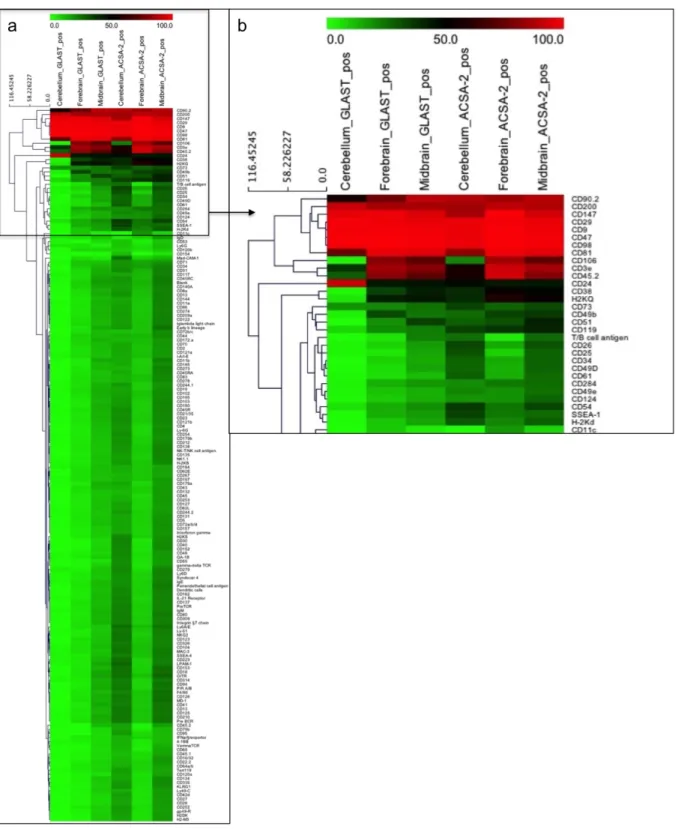
Dieser Ansatz basierte auf einem durchflusszytometrischen Screening einer Antikörperbibliothek. Es wurden ingesamt 232 Zelloberflächenmarker auf ihre Expression auf Astrozyten getestet. In diesem Experiment wurden 15 Marker identifiziert, die anschließend mit Hilfe von Immunhistochemie (IHC) und Multidimensional In Situ Cytometry Survey (MICS) validiert wurden. In einem Parallelansatz wurde das Expressionsprofil und das Antigen des Anti-ACSA-2 (ACSA: Astrozytenzell- oberflächenantigen) Antikörpers untersucht. In Zusammenarbeit mit der Arbeitsgruppe von Dr. Harold Cremer am Institut de Biologie du Développement de Marseille wurde das Expressionsprofil von ACSA-2 über IHC, Immunzytochemie (ICC), MICS sowie per Durchflusszytometrie untersucht. Durch die Koexpression von ACSA-2 mit diversen Astrozytenmarkern konnte ACSA-2 als Astrozyten spezifischer Marker bestätigt werden, der nicht von Neuronen, Oligodendrozyten oder anderen nicht- astrozytären Zellen exprimiert wird. Western Blot Analysen, Immunprezipitationen und Deglykosylierungs assays deuteten darauf hin, dass der Anti-ACSA-2 Antikörper eine Zuckerstruktur auf Astrozyten erkennt. In weiterführenden Untersuchungen wurde das Expressionsprofil von GLAST und ACSA-2 auf embryonalem, neonatalem und adultem Maushirn mittels IHC, ICC und per Durchflusszytometrie getestet. ACSA-2 und GLAST zeigten ein unterschiedliches Expressionsprofil in verschiedenen Hirnregionen, in der Retina und im Rückenmark von neonatalen Mäusen. Es folgten weitere Expressionsanalysen von GLAST und ACSA-2 in Stammzellregionen. Die Expression von ACSA-2 in der subventrikulären Zone (SVZ) wurde anhand von IHC, in vivo Elektroporation und Durchflusszytometrie untersucht. Hierbei konnte gezeigt werden, dass GLAST und ACSA-2 in der adulten SVZ koexprimiert vorliegen. Unterschiedliche Expressionsmuster wurden ferner im rostral migratory stream und im Hippocampus von adulten Maushirnschnitten detektiert. Jedoch war es nicht möglich, die Unterschiede auf Einzelzellebene zu bestätigen. Signifikante Expressionsunterschiede zwischen GLAST und ACSA-2 wurden im Cerebellum von adulten Maushirnschnitten identifiziert. In anschließenden Untersuchungen wurde die Herkunft dieser Zelltypen in der cerebellaren Entwicklungsphase (E13-P12) analysiert. Hierbei wurde eine ACSA-2-/GLAST+ und eine ACSA- 2+/GLAST+/- Zellpopulation identifiziert. Die größten Expressionsunterschiede lagen zwischen E17 und P3 vor. Zudem sank die Frequenz an ACSA-2-/GLAST+ Zellen in der weiteren Entwicklung. Beide Zellpopulationen wurden per magnetischer Zellsortierung mit hoher Reinheit angereichert und anschließend mittels ICC, Neurosphere assay, Genexpressionsanalysen und Zelltransplantation untersucht. Zudem wurde ein in vivo Zelldifferenzierungs Assay mit isolierten ACSA-2-/GLAST+ und ACSA-2+/GLAST+/- Zellen aus dem Cerebellum von β-actin GFP Mäusen durchgeführt. Diese Experimente fanden in Kollaboration mit der Arbeitsgruppe von Dr. Annalisa Buffo am Neuroscienze Institute Cavalieri Ottolenghi in Turin statt. Die Analyse zeigte, dass ACSA-2-/GLAST+ Zellen in Interneurone, Bergmann glia, Astrozyten and Oligodendrozyten differenzieren konnten. Im Gegenzug, konnten ACSA-2+/GLAST+/- Zellen nur in Astrozyten und Oligodendrozyten differenzieren.
Genexpressionsanalysen beider Zellgruppen zeigten, dass Stammzellfaktoren und Gene für einen GABAergen Phänotyp in den ACSA-2-/GLAST+ Proben angereichert waren (Gli1, Wif1, Nestin, Ptf1a, Ascl-1). Gene, die Zellinteraktion und Zelladhäsion kodieren (Gjb2, Gjb6, Gja1, Vitronectin), waren in den ACSA-2+/GLAST+/- Proben angereichert.
Zusammenfassend zeigt diese Studie eine regionale, funktionale und molekulare Charakterisierung von Astrozyten anhand von Zelloberflächernmarkern. Anhand dieser Charakteristika wurde eine neue ACSA-2-/GLAST+ multipotente Zellvorläuferpopulation im Cerebellum identifizieren.
Abstract
Abstract:
Astrocytes, a member of the glial cell family, are the most abundant neural cell type of the central nervous system (CNS) and present functional and morphological heterogeneity. Among other characteristics, astrocytes provide trophic functions, are essential for the formation of the blood-brain barrier, regulate ion homeostasis, synaptogenesis and synaptic plasticity. Despite their diversity astrocyte subpopulations are not well characterized mainly due to the lack of markers that could classify functional subclasses. This study aimed at the phenotyping of astrocyte subpopulations based on cell surface marker expression, their prospective isolation and subsequent molecular and cellular characterization of the identified subsets. The glutamate aspartate transporter (GLAST) is a common astrocyte marker expressed by astrocytes in the CNS. As the GLAST antibody allows for the isolation of astrocytes it was chosen as principal component for the identification of astrocyte subpopulations.
One approach, which was not successful, was the generation of novel monoclonal astrocyte subpopulation specific antibodies using GLAST isolated astrocytes and hybridoma techniques. Another approach was the identification of novel astrocyte markers by flow cytometry. 232 cell surface markers were tested for their expression on astrocytes. In total, 15 candidates were identified and subsequently validated by immunohistochemistry (IHC) and the recently established in house technology:
Multidimensional In Situ Cytometry Survey (MICS). The expression profile and the nature of the target of the novel Anti-ACSA-2 (ACSA: astrocyte cell surface antigen) antibody were addressed. In co- operation with the group of Dr. Harold Cremer at the Institut de Biologie du Développement de Marseille, the expression profile of the ACSA-2 antigen was analyzed systematically using IHC, immunocytochemistry (ICC), MICS and flow cytometry. Co-expression of ACSA-2 with common astrocyte markers such as GFAP, S100β and GLAST and missing expression on neurons, oligodendrocytes validated ACSA-2 as an astrocyte specific marker. Additional Western Blot analysis, immunoprecipitation and deglycosylation assays pointed out the ACSA-2 antibody addresses a glycosylation structure expressed by astrocytes. In the next step, the expression profile of ACSA-2 and GLAST was investigated in comprehensive analyses of embryonal, neonatal and adult mouse brain using IHC, ICC, MICS, and flow cytometry. ACSA-2 and GLAST demonstrated differential marker expression in distinct brain regions, retina and spinal cord of neonatal mice. Additional studies addressed ACSA-2 and GLAST expression on neural stem cells. The expression of ACSA-2 in the subventricular zone (SVZ) was analyzed using IHC, in vivo electroporation and flow cytometry. ACSA- 2 was demonstrated to be co-expressed with GLAST in the adult SVZ as shown by IHC and flow cytometry. Differences between ACSA-2 and GLAST expression were also identified in the rostral migratory stream (RMS) and hippocampus of the adult mouse brain. However, these differences could not be confirmed on the single cell level by flow cytometry. Cerebellar glia revealed significant differences in GLAST and ACSA-2 expression in the adult mouse brain. To address the origin of these potential subpopulations GLAST and ACSA-2 expression was monitored between E13 and P12 in the developing cerebellum. Thereby, ACSA-2-/GLAST+ and ACSA-2+/GLAST+/- cells were identified.
Populations were most discriminative between E17 to P3 and frequencies of ACSA-2-/GLAST+ cells decreased with the onset of development. Both fractions were isolated to high purities by magnetic cell sorting. Cell characteristics were addressed by ICC, neurosphere assay, gene expression profiling and transplantation assay. Cell differentiation in vivo was investigated in collaboration with the group of Dr.
Annalisa Buffo at the Neuroscienze Institute Cavalieri Ottolenghi in Turin. ACSA-2-/GLAST+ and ACSA-2+/GLAST+/- cells were isolated from the cerebellum of P0-P3 β-actin GFP mice and grafted into cerebellum of homochronic wild-type mice. ACSA-2-/GLAST+ cells were able to differentiate into interneurons (stellate and basket cells), Bergmann glia, astrocytes and oligodendrocytes. In contrast, ACSA-2+/GLAST+/- cells only differentiated into astrocytes and oligodendrocytes. Gene expression profiling was performed to address differences on the transcriptome level. Genes related to a multipotent and GABAergic phenotyp were enriched in the ACSA-2-/GLAST+ sample set (Gli1, Wif1, Nestin, Ptf1a, Ascl-1). Genes involved in cell-cell interactions as well as cell matrix proteins (Gjb2, Gjb6, Gja1, Vitronectin) were enriched in the ACSA-2+/GLAST+/- sample set.
In conclusion, this study presents a regional, functional and molecular discrimination of astrocyte subpopulations based on specific cell surface markers. As a result it describes the identification of a novel ACSA-2-/GLAST+ subpopulation of astrocytes which act as multipotent progenitors in the cerebellum.
Abbreviations
Abbreviations:
ACSA Astrocyte cell surface antigen
Aldh1l1 Aldehyde dehydrogenase
BSA Bovine serum albumin
BLBP Basic lipid binding protein
BMP Bone morphogenic protein
CD Cluster of differentiation
CHO Chinese Hamster Ovary cell line
CNS Central nervous system
DAPI 4′,6-Diamidin-2-phenylindol
DMEM Dulbecco's Modified Eagle's Medium
DMSO Dimethylsulfoxide
DPBS Dulbecco’s Phosphate Buffered Saline
E Embryonic day
EGFP Enhanced green fluorescent protein
EGL External granule layer (Cerebellum)
EDTA Ethylen-diamine-tetra-acetate
EGF Epidermal growth factor
FGF Fibroblast growth factor
FSC Forward scatter
Gcl Granule cell layer
GFAP Glial fibrillary acidic protein
GLAST (ACSA-1) Na+-dependent L-glutamate/L-aspartate transporter
GS Glutamine synthetase
h Hour
ipl Inner plexiform layer (olfactory bulb)
HEPES 4-(2-hydroxyethyl)-1-piperazineethanesulfonic acid HBSS Hank’s Balanced Salt Solution
IBDM Institut de Biologie du Développement de Marseille
LB Luria-Bertani medium
LRC Ligand receptor capturing
MAP2 Microtubule-associated protein 2
Mcl Molecular cell layer
mi Mitral cell layer (olfactory bulb)
min Minutes
MICS Multidimensional in situ Cytometry Survey
mL Milliliter
mM Millimolar
MOPS 3-(N-morpholino)propanesulfonic acid
ms Millisecond
NPC Neural precusor cell
NSC Neural stem cell
NTDK (T) Neural Tissue Dissociation Kit (Trypsin) NTDK (P) Neural Tissue Dissociation Kit (Papain)
NTDK (PN) Neural Tissue Dissociation Kit (Postnatal neurons)
OB Olfactory bulb
o/n Over night
P Postnatal day
PAGE Polyacrylamid gel elektrophoresis
PB PBS buffer supplemented with 5%BSA
PC Purkinje cell
Pcl Purkinje cell layer
PBS Phosphate buffered saline
Pen/Strep Penicillin/Streptomycin
PI Propidium Iodid
PNS Peripheral nervous system
PSA-NCAM Polysialyated cell-adhesion molecule
PVZ Periventricular Zone
PWM Prospective white matter
SDS Sodiumdodecylsulfa
Abbreviations
µm Micrometre
µM Micromolar
RT Room temperature
RIN RNA Integrity number
RMS Rostral migratory stream
RPMI-1640 Medium composed at the Roswell Park Memorial Institute
SHH Sonic hedgehog
SSC Side scatter
SVZ Subventricular zone
TAE Tris base; EDTA; acetic acid containing buffer
TBS Tris Based Saline
v/v Volume concentration (voume/volume)
VZ Venricular Zone (4th ventricle)
WGA Wheat germ agglutinin
wt wild-type
Contents
Table of contents:
Zusammenfasssung in deutscher Sprache Abstract
Abbreviations
Contents
1. Introduction
1.1 Function and heterogeneity of astrocytes 1
1.2 Radial glia 3
1.3 Neural stem cells 4
1.4 Identification of astrocyte subpopulation specific markers 5 1.5 Reactive astrocytes 6
1.6 Cerebellum
1.6.1 Development of the cerebellum 7
1.6.2 Cerebellar progenitors 8 1.6.3 Composition of the cerebellum at adult state 9
1.8 Aim of this study 11
2. Material and Methods
2.1.1 Antibodies for immunohistochemistry or immunocytochemistry 13
2.1.2 Secondary antibodies and nuclear stain 13
2.1.3 Antibodies for flow cytometry 14
2.1.4 Chemicals 14
2.1.5 Instruments and equipment 16
2.1.6 Reagents 17
2.1.7 Software 17
2.1.8 Animal strains, bacteria strains and cell lines 18
2.2 Cell and biological methods 19
2.2.1 Ethical guidelines for animal research 19
2.2.2 Tissue dissociation 19
2.2.2.1 Dissociation of embryonic brain tissue 19
2.2.2.2 Dissociation of neonatal brain tissue 19
2.2.2.3 Dissociation of adult mouse brain tissue 20
2.2.2.4 Dissociation of SVZ tissue 20
2.2.2.5 Dissociation of retinal tissue 20
2.2.2.6 Dissociation of spinal cord 21
2.2.3 Cultivation of neural cells 21
2.2.3.1 Cultivation of primary neural cells 21
2.2.3.2 Cultivation of isolated astrocytes 21
2.2.3.3 Neurosphere assay 21
2.2.4 Cell lines 22
2.2.4.1 Murine myeloma cell line (Sp20) 22
2.2.4.2 CHO cells 22
2.2.4.3 HEK 293 cells 23
2.2.4.4 1881 cells 23
2.2.5 Flow cytometry analysis 23
2.2.5.1 Flow cytometry - Gating strategy 24
2.2.6 Magnetic cell separation 24
2.2.7 Cell surface marker screen 25
Contents
2.2.8 Immunization 26
2.2.9 In vivo electroporation 27
2.2.10 Cell transplantation of ACSA-2+/GLAST+/-, ACSA-2- /GLAST+ cells into the neonatal
cerebellum 27
2.3 Histochemical methods 28
2.3.1 Preparation of mouse brain 28
2.3.2 Immunohistochemistry 28
2.3.3 Immunohistochemistry of Afg31l1 d/d Afg3l2 fl/fl GFAP cre mice 29
2.3.4 Immunocytochemistry 29
2.3.5 Live cell staining 29
2.3.6 Multidimensional in situ Cytometry Survey (MICS) 29
2.3.7 Imaging 30
2.4 Protein biochemical Methods 30
2.4.1 Ligand receptor capturing 30
2.4.2 Western Blot Analysis 30
2.4.3 Immunoprecipitation 31
2.4.4 Immunoprecipitation upon surface biotinylation 31
2.5.5 Deglycosylation Assay 32
2.5 Molecular Biological Methods 32
2.5.1 Transformation in E.coli 32
2.5.2 Plasmid preparation 33
2.5.3 Control restriction 33
2.5.4 Transfection of mammalian cells 33
2.5.5 Gene expression profiling (Microarray) 33
2.5.5.1 Strategies to analyse gene expression data (Microarray) 34 3 Results
3.1 Identification of novel astrocyte cell surface markers by cell surface marker screening 35 3.2 Generation of astrocyte subpopulation specific antibodies by immunization 39
3.3 ACSA-2 is an astrocyte specific cell surface marker 40
3.3.1 General expression profile of the ACSA-2 antigen 40
3.3.2 ACSA-2 expression on different neuronal cell populations 40
3.3.3 Identification of the ACSA-2 antigen 46
3.3.3.1 Identification of ACSA-2 antigen candidates by ligand receptor capturing 46
3.3.3.2 Identification of the ACSA-2 epitope by Western Blot analyses and IP 46
3.3.3.3 Transfection of Slc6a11 into HEK cells 48
3.3.3.4 Deglcosylation assay 50
3.4 Comparison of GLAST and ACSA-2expression 51
3.4.1 Expression of GLAST and ACSA-2 in the embryonic mouse brain 51 3.4.2 Expression of GLAST and ACSA-2 in neonatal mouse brain tissue 53 3.4.3 Comparison of GLAST and ACSA-2 expression in the olfactory bulb, RMS and
cortical hemispheres 55
3.4.4 Comparison of GLAST and ACSA-2 expression in neurogenic regions of adult
and neonatal mice 58
3.4.5 Comparison of GLAST and ACSA-2 expression in neuronal retina and spinal cord 63
3.5 GLAST and ACSA-2expression on reactive astrocytes 64
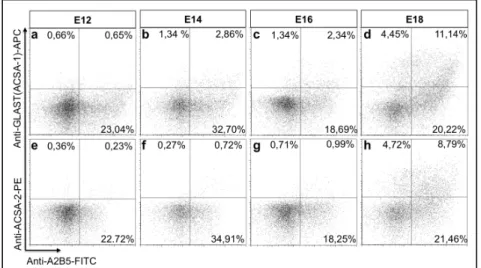
3.6 GLAST and ACSA-2expression in the cerebellum 66
3.6.1 Immunohistochemical analyses of GLAST and ACSA-2 in the adult cerebellum 66 3.6.2 Immunohistochemical analyses of GLAST and ACSA-2 in the neonatal cerebellum 68 3.6.3 Analyses of GLAST and ACSA-2 in the embryonic and neonatal cerebellum by flow
cytometry 70
3.6.4 Analyses of GLAST and ACSA-2 by NSP assay and cell transplantation 72
Contents
3.6.5 Analyses of GLAST and ACSA-2 positive astrocytes of the neonatal cerebellum by gene
expression profiling 77
4 Discussion
4.1 Mapping astrocyte heterogeneity by cell surface marker expression 84 4.2 Generation of novel astrocyte subpopulation specific antibodies and identification
of novel astrocyte subpopulation markers 84
4.3 Identification of the ACSA-2 antigen 87
4.4 Investigations of SVZ stem cells by surface marker expression 89 4.5 GLAST and ACSA-2: A versatile tool to discriminate astrocyte precursors of
the neonatal cerebellum 90
4.5.1 Gene expression profiling of ACSA-2+/GLAST-,ACSA-2+/GLAST+/-2 and
ACSA-2-/GLAST+ sorted cells from neonatal cerebellum 91
5 Outlook 95
I Supplementary data I
II Cell surface marker screen – Antibody list IX
III Appendix II: List gene expression profiling XVI IV Literature
V Acknowledgement Erklärung
Lebenslauf
Introduction
1 Introduction
1.1 Function and heterogeneity of astrocytes
During very early embryonic development three primary cell sources, the three germ layers are formed: the ectoderm, mesoderm and endoderm. The central nervous system (CNS) arises from the ectodermal germ layer. In this step of neural induction, organizers trigger the development of the neuro-ectoderm 1. Further differentiation of the neuro-ectoderm forms the neural plate. In a subsequent step, called neurulation, the lateral edges of the neural plate fold dorsally, fuse and give rise to the neural tube. Through formation of three primary brain vesicles, that give rise to five secondary brain vesicles, various structures of the adult brain are obtained 2.
This work focuses on the most abundant cell type of the CNS: the astrocytes, also named astroglia. They comprise 25-50% of total brain volume and outnumber neurons by the ratio of 10:1 in rodents 3,4. Astrocytes are non-neuronal cells that compose the subset of glia together with myelin-forming oligodendrocytes and microglia, derivatives of hematopoietic cells also named as macrophages of the brain. Further members of the glial cell family are NG2 cells, and Schwann cells, the oligodendrocyte equivalents of the peripheral nervous system (PNS).
Historically, glia have been considered as passive supporters that act like a glue for the brain.
At time of their discovery, in the 18th century by Rudolf Virchow, astrocytes were classified as non-excitable cells. Indeed, in comparison to neurons astrocytes are electrically non-excitable 5,6. However, their excitability is executed by the propagation of calcium signals as a mediator of bi-translational interactions between neurons and glia at the tripartite synapse 7. Astrocytes are coupled to each other by gap junctions 5. These gap junctions are permeable for ions, such as potassium, sodium or calcium, small molecules, including nucleotides, cAMP, sugars, and peptides 8. The flow of small molecules and ions serves as a mean of intercellular communication. Communication between astrocytes is initiated by a calcium influx and propagated to neighboring cells through calcium waves 9,10. Hippocampal astrocytes were demonstrated to be connected to up to 100,000 synapses 11. Furthermore, intracellular signaling in astrocytes is triggered by cell-cell contact with neuronal dendrites and synapses 12,13. Astrocytes also promote synaptogenesis and are an essential element of the tripartite synapse, which is composed of astrocytes, the pre- and the postsynaptic terminal at the synaptic cleft 14,15. Furthermore, astrocytes have housekeeping functions and are involved in several metabolic pathways. They do not only facilitate the clearance of neurotransmitters at the synaptic cleft 16 but also actively secrete metabolites, called gliotransmitters 17,18 such as D-serine, ATP 18,19, thrombospondins15 cholesterol20 as well as growths factors such as
Introduction
brain-derived neurotrophic factor (BDNF) or nerve growth factor (NGF) 21. Another important metabolic function is the conversion of glutamate into glutamine catalyzed by the enzyme glutamine synthetase (GS). Astrocytes release glutamine at the presynaptic terminal.
Glutamine is subsequently taken up by surrounding neurons and converted into glutamate 22,23. In addition, astrocytes regulate the pH level and brain homeostasis 24,25,26 through ion channels such as sodium, potassium 27,28 and water channels called aquaporins 11,12. Furthermore, astrocytes are the major source of extracellular matrix (ECM) proteins and cell adhesion molecules 29. They secrete different matrix metalloproteases (MMPs) which are important for the remodeling of the ECM 30. In addition to their functional divergence, astrocytes represent morphological and functional heterogeneity linked to different brain regions or developmental time-points 31. The most prominent example for morphologically different astrocytes are fibrous astrocytes of the white matter and protoplasmic astrocytes of the gray matter 32. The functions of astroglia during development were first addressed by Rakic et al. 32. They demonstrated that radial glia, primary progenitors of the developing CNS, a subgroup of astroglia, are essential for the differentiation and guidance of neurons during development 33. Similar observations were made in the developing cortex and the rostral migratory stream (RMS) 34,35. In this context it was demonstrated that neuronal guidance is mainly driven by the interaction between members of the Slit family, expressed by astrocytes, and Robo receptors, expressed by neurons 36,37. Furthermore, astrocytes are an essential element of the blood-brain barrier, a physical barrier around blood-vessels formed by astrocyte perivascular endfeet, endothelial cells and pericytes 38. In a healthy organism, this barrier separates the blood system from the CNS. Only certain gas molecules such as oxygen or carbon dioxide and small lipophilic agents can pass this barrier by passive diffusion. Some other small molecules such as glucose or amino acids can be selectively transported 38.
Apart from these general properties of astrocytes their functions in the diseased brain were addressed. On the one hand, astrocytes were reported to be mediators of neurodegenerative disorders. On the other hand, astrocytes are considered to be targets for translational or regenerative medicine. Their self-renewing capacities and the possibility to use them for trans- differentiation 39,40 are valuable for clinical approaches. In respect to all the distinct functional properties of astrocytes, it is apparent that astrocytes are not a uniform subset but rather composed of various subpopulations.
Introduction
1.2 Radial glia
The CNS of vertebrates consists of neurons and glia that derive from multipotent neuroepithelial progenitor cells of the neural tube. During initiation, multipotent neuroepithelial cells of the ectoderm undergo rapid symmetric cell division. They fold and form the neural plate, which then forms the neural tube via Sonic hedgehog (SHH), bone morphogenic protein (BMP) and Wnt signaling 41. In the next step, neuroepithelial cells polarize and divide asymmetrically. These cells give rise to radial glia a more restricted progenitor population in the early prenatal state 42-44 (Figure (Fig.) 1.1 a). Radial glia are characterized by a long, radial, bipolar shape and span from the ventricular zone towards the pial surface 45. This cell type was first described by Golgi in 1873. Later, Ramon y Cajal and Magini reported about the significance of radial glia during development 46. Radial glia belong to the astroglia subset. They express a panel of astrocyte specific markers such as GFAP (absent in rodents) 47, Tenascin C, GLAST 48, glycogen granules 49, and BLBP 50. Besides, radial glia express neuroepithelial cell markers such as Nestin 51 and the intermediate-filament associated protein RC2 52,53. In the first period of development, radial glia serve as scaffold for migrating neuroblasts 54 and as a source for new neurons 55 (Fig. 1.1 b,c). At the end of developmental neurogenesis radial glia disappear or transform into astroglia 56,57. However, it could be demonstrated that some radial glia subtypes persist as the Bergmann glia in the cerebellum, as Müller glia in the retina and as neural stem cells in the SVZ and hippocampus 58-62.
Figure 1.1: Radial glia during development.
Early in development cells of the neuroepithelium, the mutipotent neuroepithelial cells undergo rapid symmetric cell division. During embryonic development and around birth neuroepithelial cells transform into radial glia cells (a). By asymmetric division, radial glia generate pools of transit amplifying cell types or transform into neurons (b). Radial glia postnatally and transform directly into cortical astrocytes. Some radial glia transform intoneuronal stem cells residing in the SVZ and behave as stem cells that can self- renew and give rise to neurons (c). Modified from Alvarez-Buylla et al. (2001) 45.
Introduction
1.3 Neural stem cells
Among the features of astrocytes, their stem cell characteristics are of great interest.
Since their discovery in the 1980s 63, research on NSCs has increased dramatically due to the potential of NSCs to cure neurodegenerative diseases. Pioneering studies on neurogenesis were conducted by Altman et al. and Kaplan et al 64,91.The classical model of adult neurogenesis is restricted to two neurogenic niches. However, recent studies demonstrated that neurogenesis is not restricted to these two niches and occurs also in other brain areas as demonstrated for the striatum in human brain 65. One classical neurogenic niche is the subventricular zone (SVZ) of the lateral ventricle. The other one is the dentate gyrus of the hippocampus. Both areas are bilaterally located in the fore- and midbrain. In contrast to other germinal layers, the SVZ of the forebrain persists after birth. In the 1990s several groups discovered multipotent neural stem cells in the developing mouse brain 63,66,67. In 1999 Doetsch et al.68 characterized these multipotent stem cells of the SVZ as astrocytes based on morphology and marker expression.
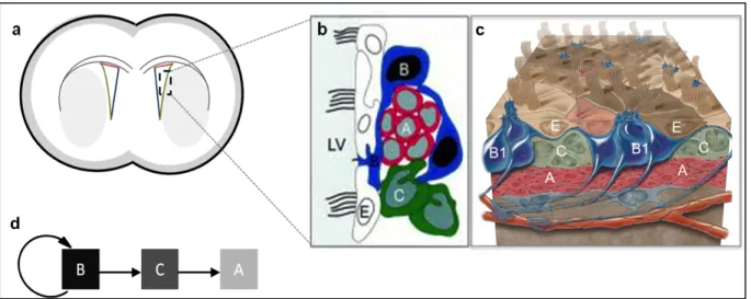
In a coronal section, the SVZ is shaped like a triangle composed of three walls as depicted in (Fig. 1.2 a). The medial wall contacts the septum. The dorsal wall adjoins the corpus callosum and contacts the lateral wall at the dorsal horn. The lateral wall flanks the striatum. Neural stem cells are commonly isolated from the lateral wall of the SVZ, which is assembled by different cell layers and distinct cell types classified as Type A, B, C, and E cells. The edge of the ventricle is composed of ependymal cells that derive from radial glia 69 (Type E cells), as seen in Figure 1.2 b,c. The ependymal cell layer is tightly connected with slowly proliferating Type B1 cells and a further source of astrocytes 69 the B2 cells that regulate local environment.
Type B1 cells are the neural stem cells and are characterized by the expression of common astrocyte markers 70,71. Since neural stem cells and ependymal cells express mutual astrocyte markers such as epidermal growth factor receptor (EGFR) or GFAP 71 there is a demand to identify a specific stem cell marker. So far, the isolation of neural stem cells is executed using at least two markers in combination, such as GLAST, GFAP, EGFR and Prominin-1 72,73-75. In vivo, Type B cells of the lateral wall of the SVZ generate transit amplifying precursors (Type C cells), that generate neuroblasts (Type A cells) (Fig. 1.2 b,d). These neuroblasts are characterized by the expression of polysialyted-neural cell adhesion molecule (PSA- NCAM)76,77. In the SVZ, neuroblasts are surrounded by Type B and Type C cells. After their generation in the SVZ, neuroblasts follow the RMS along a posterior-anterior axis towards the olfactory bulb 78. The RMS is composed of ‘tunneling astrocytes’ that guide the way towards the olfactory bulb. Astrocytes support the migration progress by the secretion of soluble molecules (e.g. growth factors), the expression of matrix proteins, cell-cell contacts as well as
Introduction
receptor-ligand interactions (e.g. Slit and Robo) 37,79. In the olfactory bulb neuroblasts migrate to their final location and differentiate into olfactory bulb interneurons.
Figure 1.2: The lateral ventricle of the adult mouse brain.
One neurogenic area in the adult brain is the subventricular zone (SVZ). The SVZ is composed of a dorsal (red) a lateral (green) and a medial wall (blue)(a). Cells of the lateral wall are classified as Type A, B, C and E cells (b,c)80,81. Type B cells are the neural stem cells that give rise to transit amplifying (Type C cells) that generate migratory neuroblasts (Type A cells) (d).
Modified from Alvarez-Buylla et al. (2002) and Mizadeh et al. (2008) 80,81.
1.4 Identification of astrocyte subpopulation specific markers
Astrocytes are phenotypically diverse and their tasks differ in respect to their localization. Hence, the marker expression profile of astrocytes is strongly correlated to their localization, developmental status and interaction with other cell types. The first sub- classification study was carried out by immunohistochemistry in 1984 82. This study was based on morphology, regional distribution and marker expression. The study identified two major astrocyte populations: the protoplasmic astrocytes and the fibrous astrocytes. Protoplasmic astrocytes are located in the grey matter, have short processes, many branches typically ensheath synapses and express the intermediate filament glial fibrillary acidic protein (GFAP) 11,83,84. Fibrous astrocytes are located in the white matter, have long unbranches processes and are characterized by a weaker expression of GFAP 32. Besides GFAP, other common astrocyte markers such as glycogen granules, glutamate transporter (GLT-1) and GS were identified 85-87. In 2008, aldehyde dehydrogenase (Aldh1l1) was identified as a novel astrocyte specific marker by comprehensive microarray analysis 88. Aldh11 is a more robust marker than GFAP, as GFAP is exclusively found on terminally differentiated astrocytes and not expressed by quiescent astrocytes in healthy tissue 89,90. In addition, first subtype specific markers like calcium-binding protein, S100β, or potassium channel, Kir4.1,
Introduction
were identified 91-93 using single-cell gene expression profiling 94 or electrophysiological studies 95. Another subtype marker, Aquaporin-4, a water channel, was identified by transcriptome analysis comparing primary and cultivated astrocytes, neurons and neural stem cells (NSCs) from distinct ages and distinct brain regions 96. Aquaporin-4 was demonstrated being exclusively expressed on astrocyte endfeet 97. Unfortunately, the majority of the mentioned markers are intracellular proteins. Therefore, these markers cannot be used for direct isolation and further characterization of living cells. To this aim, several transgenic mouse lines were generated. These mouse lines enable for the isolation of potential astrocyte subpopulations through expression of a reporter gene under an astrocyte specific promoter (e.g. Aldh1L1, BLBP, S100β, GFAP, GLT-1, GLAST 98,99,98,100-103). The first approach to isolate astrocytes from primary tissue of wild-type (wt) mice was realized in 2012 by the generation of a novel Anti-GLAST antibody 104.
1.5 Reactive Astrocytes
Trauma, neurodegeneration or other CNS insults cause tissue damage which influences neurons and glia and hampers their interaction 105-108. Upon tissue damage, activated microglia migrate to the affected tissue and initiate first immune response 109-112. Microglia also produce reactive oxygen species (ROS)113 and nitric oxide (NO) which cause oxidative stress that leads to damage and aging of cells 114,115. While NO species can be neutralized by the glutathione metabolites produced by astrocytes, ROS and reactive nitrogen species (RNS) lead to the phosphorylation of STAT6, a stress sensor. This phosphorylation up-regulates expression of cyclooxygenase-2 (COX-2) and induces release of pro-inflammatory cytokines. In addition, the STAT6 phosphorylation induces the release of PGE2 and prostacyclin. COX-2 activates neighboring cells e.g. astrocytes 116-118. Activation of astrocytes is called reactive gliosis 119. Upon reactive gliosis astrocytes undergo biochemical, morphological and physiological changes. They start to proliferate, modify the expression of cellular markers, become hypertrophic 120, increase the production of intermediate filaments 121 and migrate to the side of injury to form a glial scar. This is a cellular attempt to prevent further tissue damage and spread of the disease as the scar separates healthy tissue from injured tissue 122,123.
There is currently no exclusive cell-surface marker is described that allows for the discrimination of reactive astrocytes from non-activated astrocytes in the diseased brain.
Furthermore, several markers of reactive astrocyte address intracellular epitopes or transcription factors. Therefore, these markers do not allow for further isolation and characterization of viable reactive astrocytes. The most common marker to study reactive
Introduction
astrocytes is GFAP 119,121. Other astrocyte markers, that are upregulated on reactive astrocytes are S100β, BLBP 122, vimentin 124 or Nestin are also used to discriminate reactive astrocytes.
1.6 Cerebellum
1.6.1 Development of the cerebellum
The brain is composed of three main areas named as Prosencephalon (Forebrain), Mesencephalon (Midbrain) and Rhombencephalon (Hindbrain). Each region itself consists of region specifc areas. Major part of this study was performed in the cerebellum, which is part of the Rhombencephalon (Hindbrain).
The cerebellum is important for voluntary movement and motor function, as demonstrated in the early 19th century by three neurophysiologists: Luigi Roland, Marie-Jean-Pierre Flourens and John Call Dalton, Jr. 125. Further reports also showed that the cerebellum has an impact on higher cognitive functions, cognitive progression and behavior 126. Besides, the cerebellum is affected in several diseases like ataxia, schizophrenia, and autism 127. In addition, uncontrolled growth of cerebellar precursors results in medullablastoma the most common malignant brain tumor in children 128.
Although the cerebellum comprises only 10% of the total brain volume, granule neurons account for 50% of all brain neurons 129. The cerebellum is one of the best-studied brain region in respect to circuitry 130, development 54, anatomy and fiber connectivity 131. During ontogeny the cerebellum arises from the met-mesencephalic junction of the neural tube 132 around embryonic day 9 (E9) in mice 133. At the initiating phase (E9- E12) the cerebellum conducts a 90° rotation from the anterior-posterior axis towards the medial-lateral axis. This results in a bilateral wing-like morphology 134. Early developmental steps are driven by specific proteins such as the transcription factors Hoxa2 (posterior) and Oxt2 (anterior) are further accompanied by soluble molecules of the Wnt and FGF families (especially fgf8) 129.
After several structural modification steps, the cerebellum obtains its foliated structure.
Anatomically, the mammalian cerebellum is composed of 10 primary lobules (Fig. 1.4 a), first characterized by Olof Larsell (1970) 135,136. The cardian lobes are formed at embryonic stage while lobules and sub-lobules are formed at postnatal stage 137. Although the foliation process starts around E14 in mice 138, the maturation of the architecture lasts until adulthood.
Introduction
1.6.2 Cerebellar progenitors
The progenitor pools of the cerebellum derive from two distinct germinal territories:
the ventricular zone of the fourth ventricle and the rhombic lip, as demonstrated by Alvarado- Mallart and Le Dourian 132,139. It is suggested that cells of the deep cerebellar nuclei (DCN) arise from both germinal matrices. The ventricular zone (VZ) is located at the inner germinal layer, where interneurons arise around E12.5. These interneurons can be distinguished by the expression of Pax-2 140, and Pft1-a which is exclusively expressed by GABAergic projection neurons 141,142. Cells of the deep nuclei arise from the cerebellar plate around E10.25 143. These cells become post mitotic and form a „mantle“ above the VZ 144,145. Between E11 and E14.5 cortical precursor cells of the VZ exit the cell cycle. These are Purkinje cell precursors that start to express calbindin 146 and migrate radially along radial glia processes 147, compare Figure 1.3. These precursors pass the „mantle“ of deep cerebellar nuclei and settle down in the prospective white matter just beneath the developing granule cell layer (Gcl) 148. In this region, Purkinje cell precursors form a multilayer of immature neurons (E14-E17). Then, Purkinje cell precursors differentiate and start to interact with parallel fibers 146. By maturation, the multilayer disperses and a Purkinje cell monolayer appears (Fig. 1.3 c). Once the Purkinje cell monolayer is formed, interneurons such as stellate, basket and Golgi cells are generated from precursors of the deep (prospective) white matter 129,149.
The second major germinal matrix is the rhombic lip, an external layer located close to the roof plate of the neuroepithelium. This external germinal layer (EGL) arises around E12.5 and gives rise to all types of glutamatergic cells. A pool of migrating cells, which derives from this germinal matrix, contributes to the cerebellar nuclei 37.The EGL is a highly proliferative cell source, which produces granule neurons 150,151 regulated by SHH and Wnt signaling pathways 152,153. Cells of the EGL are characterized by the expression of Math1, a helix-loop- helix transcription factor 154. EGL cells proliferate rapidly between E13 and P15, highest at P8 143. Upon birth until P14, cells at the EGL exit cell cycle and start to migrate inwards along Bergmann glia. Migrating granule cells pass through the developing Purkinje cell layer and settle down at the developing internal Gcl 54,146,155,156. An important molecule involved in this migration process is Reelin 157 and the extracellular protein Astrotactin, which is expressed by migrating neurons in the cerebral cortex and the cerebellum 158-160. The migration stops soon after the disappearance of the EGL. The final differentiation step of Purkinje cells and granule cells is depending on their interplay and results in the arborisation of Purkinje cells 31,32.This interaction is also depending on Reelin, BDNF and SHH signaling 161.
Glial cells of the cerebellum were first described by Ramon y Cajal in 1911. The majority of cerebellar glia derives from the ventricular neuroepithelium (VN) 162 and shares a common
Introduction
ancestor with neurons 163. The key regulator for glial versus neuronal fate in the VN compartment is considered to be Notch and BMP dependent 164. Two minor fractions of glial cells originate from the rhombic lip. One is the unipolar brush cell, which migrates towards the white matter 165. The other fraction migrates tangentially over the cerebellar surface. This cell type is remained postnatally and expresses GFAP 166. In addition, this cell type expresses Math1 and can differentiate into granule cells 167. Upon isolation and SHH treatment these progenitors differentiate into astrocytes in vitro showing a multipotent potential 168.
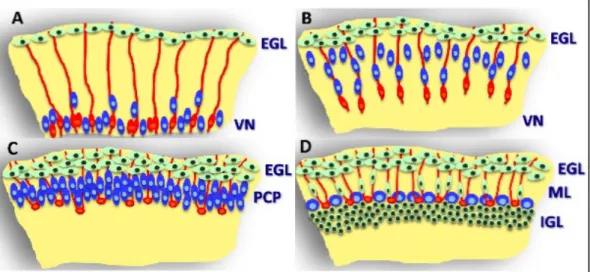
Figure 1.3: Bergmann glia in cerebellar corticogenesis.
Purkinje cells (blue) in the embryonic cerebellum (A) are generated by radial glia (red) in the
ventricular neuroepithelium (VN) and migrate along radial glia procresses (red). In the external granular layer (EGL) granule cell precursors (green) are generated. Around birth radial glia transform into Bergmann glia (B). Thereby loosing their connection to the cortex (B).
Next, Bergmann glia spread their processes towards the pial surface (B). Purkinje cells cluster in the Purkinje cell plate (PCP) beneath the EGL (C). At postnatal development (D), granule cells migrate inwards along Bergmann glia. Purkinje cells acquire a monolayer arrangement. The interaction between granule cells and Bergmann glia leads to the expansion of the molecular layer (ML) and the internal granule cell layer (IGL). Modified from Buffo et al. (2013) 169.
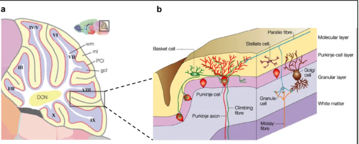
1.6.3 Composition of the cerebellum at adult state
A fully developed cerebellum comprises a four-layer-structure (Fig. 1.4 b) composed of distinct cell types 170: The inner cell layer of the adult cerebellum is the white matter, which is a myelin-rich area. Above the white matter is the Gcl, where Golgi cells and the most numerous kind of neurons of the brain the granule cells reside. Granule cells are major excitatory neurons, which transmit glutamate 171. Furthermore, they co-ordinate the afferent excitatory input they receive from mossy fibers. Mossy fibers originate from nuclei in brain stem and spinal cord 172. These fibers connect granule and Golgi cells and build synaptic complexes, called cerebellar glomeruli (also recognizable as rosettes) 173. The next cell layer is
Introduction
the Purkinje cell layer. The cell bodies of the Purkinje cells are surround by Bergmann glia171. Purkinje cells are inhibitory and secrete the neurotransmitter GABA. Bergmann glia derive from radial glia and are located around the Purkinje cells soma after maturation. Their processes spread from the Purkinje cell layer towards the subpial basement membrane 174. During early development, radial glia serve as a scaffold for migrating Purkinje cells (Fig. 1.3).
Postnatally, Bergmann glia serve as a scaffold for granule cells 175. In the superficial layer, the molecular layer, dendrites of the Purkinje cells are located. Furthermore, axons of granule cells (called parallel fibers) but also stellate and basket cells are found in this layer 171. Parallel fibers of the molecular cell layer (Mcl) derive from the granule cells of the Gcl. Parallel fibers are in contact with stellate and basket cells as well as dendrites of the Purkinje cells. While Purkinje cells are in close contact to Bergmann glia, basket and stellate cells are partially in contact with Bergmann glia whereas Golgi and granule cells are not in contact with Bergmann glia 176. Cerebellar glia are categorized into different subtypes: glia of the white matter, astrocytes of the Gcl 177, unipolar brush cells 165 and Bergmann glia. Several studies suggest that cerebellar glia of the developing cerebellum have stem cells or progenitor cells characteristics 178,179.
Figure 1.4: The architecture of the cerebellum.
After foliation the cerebellum is composed of 10 lobules (a). The cerebellum is comprised of four layers (b): the white matter, the Gcl, the Purkinje cell layer and the molecular layer. Granule cells and Golgi cells are located in the Gcl. The soma of Purkinje cells and Bergmann glia are located in the Purkinje cell layer. Inhibitory interneurons, stellate and basket cells, the processes of Bergmann glia and dendrites of Purkinje cells are located in the Mcl. Modified from Kobielak et al. (2004) and Takana et al. (2008) 180,181.