Research Collection
Doctoral Thesis
Identification of the TRPV4 ion channel as a mechanotransducer and therapeutic target in low back pain
Author(s):
Cambria, Elena Publication Date:
2021-03
Permanent Link:
https://doi.org/10.3929/ethz-b-000474936
Rights / License:
In Copyright - Non-Commercial Use Permitted
This page was generated automatically upon download from the ETH Zurich Research Collection. For more information please consult the Terms of use.
ETH Library
DISS. ETH NO. 27377
Identification of the TRPV4 ion channel as a mechanotransducer and therapeutic target in low
back pain
A thesis submitted to attain the degree of DOCTOR OF SCIENCES of ETH ZURICH
(Dr. sc. ETH Zurich)
presented by Elena Cambria
MSc in Bioengineering, Ecole polytechnique fédérale de Lausanne (EPFL)
born on 02.12.1989 citizen of
Switzerland (Bagnes, VS) and Italy
accepted on the recommendation of Prof. Dr. Stephen J. Ferguson, examiner Prof. Dr. Karin Wuertz-Kozak, co-examiner
Prof. Dr. Mauro Alini, co-examiner
2021
Dedicated to those who believed in me and supported me during the realization of this doctoral project
i
Table of contents
Acknowledgements ... iii
Summary ... v
Résumé ... vii
1. Introduction ... 1
1.1 Thesis motivation ... 2
1.2 Thesis aims ... 4
1.3 Thesis outline ... 5
1.4 References ... 7
2. Background ... 9
2.1 Anatomy of the intervertebral disc ... 10
2.2 Aging and pathologies of the intervertebral disc ... 12
2.3 Current treatments of low back pain ... 14
2.4 Preclinical therapeutic strategies ... 17
2.5 Biomechanics and mechanobiology of the intervertebral disc ... 19
2.6 References ... 25
3. Cell-laden agarose-collagen composite hydrogels for mechanotransduction studies ... 33
3.1 Abstract... 35
3.2 Introduction ... 35
3.3 Materials and methods ... 37
3.4 Results ... 42
3.5 Discussion ... 49
3.6 Conclusion ... 51
3.7 References ... 52
4. TRPV4 mediates cell damage induced by hyperphysiological compression and regulates COX2/PGE2 in intervertebral discs ... 55
4.1 Abstract... 57
4.2 Introduction ... 57
4.3 Materials and methods ... 59
4.4 Results ... 62
4.5 Discussion ... 68
ii
4.6 References ... 70
5. TRPV4 inhibition and CRISPR-Cas9 knockout reduce inflammation induced by hyperphysiological stretching in human annulus fibrosus cells ... 75
5.1 Abstract... 77
5.2 Introduction ... 77
5.3 Materials and methods ... 78
5.4 Results ... 82
5.5 Discussion ... 89
5.6 Conclusions ... 91
5.7 References ... 92
5.8 Supplementary information ... 97
6. The potential of CRISPR/Cas9 genome editing for the study and treatment of intervertebral disc pathologies ... 99
6.1 Abstract...101
6.2 Disc degeneration: the need for novel treatments ...101
6.3 Targeted genome editing by CRISPR/Cas9 ...102
6.4 Conclusion ...113
6.5 References ...113
7. Synthesis ... 121
7.1 Contributions to the field ...122
7.2 Limitations ...124
7.3 Future perspectives ...125
7.4 Conclusion ...127
7.5 References ...127
Appendix ... 129
TRPC6 in simulated microgravity of intervertebral disc cells ...130
List of abbreviations ...145
Curriculum vitae ...149
iii
Acknowledgements
This thesis would not be the same without the contribution of many talented and supportive people. My sincere gratitude goes to all of them. I will highlight some here hoping that I will not forget too many.
I would like to start by thanking Prof. Karin Wuertz-Kozak and Prof. Stephen Ferguson for welcoming me into their groups, supervising the projects in this thesis, and mentoring me during the past years. This doctoral project has been a very formative experience that shaped me both as a scientist and as a person. I am profoundly grateful to Karin and Stephen for providing the resources and the environment that were essential for the realization of this project.
I would like to express my gratitude to Prof. Peter Loskill for hosting me in his group in Stuttgart during a 6-month research stay and for his mentoring.
Many thanks further go to Karin and Stephen, as well as Prof. Mauro Alini, for reading and evaluating this thesis.
I would like to thank all of our collaborators and co-authors at ETH Zurich and internationally for their significant scientific contributions to the work described in this thesis: Prof. Jess Snedeker, Prof. Ralph Müller, Prof. Oliver Hausmann, Prof. Victor Leung, Prof. Robert Bowles, Prof. Alfredo Franco‑Obregon, Prof. Marcel Egli, Prof. Miho Sekiguchi, Prof. Norbert Boos, Prof. Hiroshi Kobayashi, Dr. Wolfgang Hitzl, Dr. Olga Krupkova, Dr.
Matthias Arlt, Dr. Ariane Scheuren, Dr. Wai Kit Tam, Dr. Agnieszka Karol, Dr. Lenka Besse, Philipp Fisch, Fabian Passini, Timon Wernas, Andrej Besse and Helen Greutert. Many thanks also go to Prof. Thomas Voets, Prof. Rudi Vennekens, and Silvia Pinto for their contributions, and to Justine Kusch-Wieser, Joachim Hehl, and Aymone Lenisa for technical assistance. In addition, I acknowledge the financial support of the Swiss National Science Foundation and Eurospine, the Spine Society of Europe.
I am deeply thankful for the work, efforts and motivation of my Master students and co-authors: Silvio Brunner, Sandra Wandel, Sally Heusser and Rahel Friedrich.
Supervising them has taught me many important lessons on the scientific and personal level and gave me the opportunity to improve my teaching and leadership skills over the years. For this, I am very grateful.
I would like to express my thanks to all the past and present members of the Wuertz- Kozak and Ferguson groups. Whether it was an introduction to a new technique, scientific advice, a lunch, a coffee break, or a “töggeli” game, these interactions have immensely contributed to my well-being and the realization of my doctoral project. I would like to particularly thank Oddny Björgvinsdottir, Dr. Aleksandra Sadowska, Helen Greutert, Dr.
Enrico de Pieri, Dr. Fabio D’Isidoro, Dr. Thomas Zumbrunn, Dr. Dmitriy Alexeev, Dr.
Dominika Ignasiak, Dr. Yabin Wu, Pascal Behm, Stephanie Huber, and Matthias Santschi.
Special thanks go to Dr. Olga Krupkova and Dr. Matthew Randall, whose scientific guidance and friendship have encouraged me to persevere over the years. I would also like to thank the members of the technical and administrative staff of the Institute for
iv
Biomechanics, including Anina Eglin, Joanne Lim, Peter Schwilch, Catherine Palmer, Sofia Delamanis, and Ingrid Okanta.
This doctoral project has not only generated scientific achievements, but also personal ones. I would like to sincerely thank all the people whom I had the opportunity to meet over the past years and who supported me in one way or another. Many thanks go to the members of the Peer Mentoring group supported by the Fix the Leaky Pipeline program for women in science. I would like to particularly thank Dr. Ariane Scheuren, who invited me to this group, as well as Dr. Penny Atkins and Dr. Caitlyn Collins for their advice and support. I am grateful to Dr. Florian Formica for his scientific advice and our personal discussions. Warm thanks go to Dr. Rong Chen, Dr. Julia Steiger, Julia Günter, Dr. Enrico de Pieri, Dr. Fabio D’Isidoro, Dr. Matthew Randall and Dr. Thomas Zumbrunn, whom I am proud to call my friends. I would like to express my deepest gratitude to Dr. Ingmar Fleps, whose love and encouragements over the years have been instrumental to the completion of my doctoral project and thesis.
Meeting new friends in Zurich certainly did not make me forget my old ones. I affectionately thank all the wonderful people, who have supported me until now: my friends met in Verbier, Sion, Lausanne, and Boston, including but not limited to Lisa Greve, Cassandre Vergère, Coralie Fournier-Neurohr, Aline Zbinden, Caroline Odermatt, Dr.
Claudio Hail, and Andreas Reichmuth.
Finally, I would like to express my profound gratitude to all my family, and especially to my mother Cettina, my father Sebastiano, and my sister Veronica, for their unconditional love and endless support in all my endeavors.
Infine, vorrei esprimere la mia profonda gratitudine a tutta la mia famiglia, e in particolare a mia madre Cettina, a mio padre Sebastiano e a mia sorella Veronica, per il loro amore incondizionato e l'infinito sostegno in tutti i miei sforzi.
Verbier, December 2020 Elena Cambria
v
Summary
Low back pain (LBP) is the leading cause of disability worldwide and a huge global socio-economic burden. The costs related to LBP are projected to further increase in the coming decades due to the growth and aging of the global population. Degenerative disc disease, which is characterized by intervertebral disc (IVD) degeneration, inflammation, and nerve ingrowth, principally contributes to LBP. One of the main contributors to IVD degeneration is excessive or aberrant mechanical loading. Current treatments of LBP, including physical, psychological, pharmacological, and surgical approaches, have unclear mechanisms of action, low effect sizes, and are not beneficial in the long term. Targeted therapeutic strategies in preclinical development, such as molecular and gene therapy, selectively address the biological changes that occur in IVD degeneration. Nevertheless, despite the mechanical nature of LBP, mechanotransduction pathways are currently not targeted. This is mainly due to the very limited information on mechanosensing and mechanotransduction mechanisms in the IVD. Transient receptor potential (TRP) ion channels are promising therapeutic targets to treat LBP, as they can sense and transduce a variety of signals, including mechanical stress. The TRP vanilloid 4 (TRPV4) channel is especially interesting, as it was shown to mediate mechanical, inflammatory and pain signals. Its clinical potential is further highlighted by ongoing preclinical and clinical trials.
The overall goal of this thesis was to investigate the potential role of TRPV4 in mediating hyperphysiological mechanical signals in the IVD, and its relevance as a therapeutic target to treat LBP.
As a first step towards the investigation of TRPV4, we developed a novel in vitro compression model for mechanotransduction studies. Agarose-collagen composite hydrogels were fabricated and characterized in terms of material and mechanical properties. Bovine nucleus pulposus (NP) cell phenotype and mechanotransduction ability after dynamic compression were further analyzed. Agarose-collagen composite hydrogels combined the mechanical strength of agarose with the biofunctionality of collagen, which enhanced cell adhesion and the activation of focal adhesion kinases.
Moreover, agarose-collagen scaffolds recapitulated the extracellular matrix (ECM) of the IVD, with their non-fibrillar matrix and collagen fibers, and allowed the exploration of mechanotransduction mechanisms in a reproducible system.
In a second study, NP cell-laden agarose-collagen hydrogels and a mouse model were used to investigate the role of TRPV4 in transducing hyperphysiological dynamic compression. Degenerative changes and the expression of the inflammatory mediator cyclooxygenase 2 (COX2) were examined in mouse IVDs that were dynamically compressed at a hyperphysiological regime (versus sham). Cell damage and inflammation (prostaglandin E2 (PGE2) release) were measured in bovine NP cells embedded in agarose-collagen hydrogels and dynamically compressed at a hyperphysiological regime with or without TRPV4 inhibition. The activation of the mitogen-activated protein kinase (MAPK) pathways was analyzed. Finally, degenerative changes and COX2 expression were further evaluated in the IVDs of trpv4 knockout (KO) mice (versus wild-type). TRPV4 was shown to regulate the COX2/PGE2 inflammatory factors and mediate cell damage induced by hyperphysiological dynamic compression, possibly via the extracellular signal- regulated kinases 1/2 (ERK) pathway.
vi
In a final step, we investigated the role of TRPV4 as a transducer of hyperphysiological cyclic stretching and a potential therapeutic target. Human primary annulus fibrosus (AF) cells were seeded on silicone chambers and cyclically stretched at a hyperphysiological magnitude in the presence or absence of a TRPV4 inhibitor.
Clustered regularly interspaced short palindromic repeats (CRISPR)-Cas9 TRPV4 KO AF cells were generated, hyperphysiologically stretched, and compared to control cells. Gene and protein expression of inflammatory and catabolic mediators, as well as activation of MAPK pathways, were analyzed. This study identified TRPV4 as a mediator of stretch- induced inflammation in human AF cells. Moreover, it revealed TRPV4 pharmacological inhibition and gene editing as potential future therapeutic approaches to rescue mechanoflammation.
In this thesis, in vitro and in vivo models of hyperphysiological compression and stretching were established and used to identify TRPV4 as mechanotransducer and therapeutic target in the IVD. A novel in vitro compression model was developed to mimic the ECM of the IVD and other native tissues composed of non-fibrillar matrix and collagens, and to investigate their mechanotransduction mechanisms. This system was instrumental to investigate the function of TRPV4 in IVD cells. The novel findings obtained with this model, together with those obtained in the mouse compression model and the stretching system demonstrate that TRPV4 mediates a mechanism leading from mechanical hyperphysiological loading to IVD degeneration and inflammation, which eventually lead to chronic LBP. TRPV4 modulation might thus constitute a promising therapeutic strategy to treat patients suffering from IVD pathologies caused by aberrant mechanical stress. Future studies should clarify the exact mechanism of action of TRPV4 inhibition and gene editing and examine their potential to mitigate chronic inflammation and LBP in preclinical and clinical trials.
vii
Résumé
La lombalgie est la principale cause d'invalidité dans le monde et représente un énorme fardeau socio-économique. Les coûts liés à la lombalgie vont encore augmenter dans les prochaines décennies en raison de la croissance et du vieillissement de la population mondiale. La discopathie dégénérative, qui se caractérise par la dégénérescence du disque intervertébral (DIV), l'inflammation et la croissance des nerfs dans le DIV, contribue principalement à la lombalgie. Un des principaux facteurs de dégénérescence discale est une sollicitation mécanique excessive ou aberrante. Les traitements actuels de la lombalgie, y compris les approches physiques, psychologiques, pharmacologiques et chirurgicales, ont des mécanismes d'action peu clairs, des effets de faible ampleur et ne sont pas bénéfiques à long terme. Des stratégies thérapeutiques ciblées en développement préclinique, telles que la thérapie moléculaire et génique, s'attaquent de manière sélective aux changements biologiques qui se produisent dans la dégénérescence discale. Néanmoins, malgré la nature mécanique de la lombalgie, les voies de mécanotransduction ne sont actuellement pas ciblées. Cela est principalement dû au fait que les informations sur les mécanismes de mécanodétection et de mécanotransduction dans le DIV sont très limitées. Les canaux ioniques TRP (de l’anglais transient receptor potential) sont des cibles thérapeutiques prometteuses pour traiter la lombalgie, car ils peuvent détecter et transduire divers signaux, y compris le stress mécanique. Le canal TRP vanilloïde 4 (TRPV4) est particulièrement intéressant, car il a été démontré qu'il transmet des signaux mécaniques, inflammatoires et de douleur. Son potentiel clinique est encore mis en évidence par des essais précliniques et cliniques en cours. L'objectif général de cette thèse était d'étudier le rôle potentiel de TRPV4 dans la médiation des signaux mécaniques hyperphysiologiques dans le DIV et sa pertinence en tant que cible thérapeutique pour traiter la lombalgie.
Dans une première étape vers l'étude de TRPV4, nous avons développé un nouveau modèle de compression in vitro pour les études de mécanotransduction. Des hydrogels composites agarose-collagène ont été fabriqués et caractérisés en termes de propriétés matérielles et mécaniques. Le phénotype de cellules bovines de noyau pulpeux (NP) et la capacité de mécanotransduction après compression dynamique ont aussi été analysés.
Les hydrogels composites agarose-collagène ont combiné la résistance mécanique de l'agarose avec la biofonctionnalité du collagène, ce qui a permis d'améliorer l'adhésion cellulaire et l'activation de la protéine FAK (de l’anglais focal adhesion kinase). De plus, les hydrogels agarose-collagène ont récapitulé la matrice extracellulaire du DIV, avec leur matrice non fibrillaire et leurs fibres de collagène, et ont permis l'exploration des mécanismes de mécanotransduction dans un système reproductible.
Dans une seconde étude, les hydrogels d’agarose-collagène chargés de cellules NP et un modèle de souris ont été utilisés pour étudier le rôle de TRPV4 dans la transduction de la compression dynamique hyperphysiologique. Les changements dégénératifs et l'expression du médiateur inflammatoire cyclooxygénase 2 (COX2) ont été examinés dans des DIV de souris qui ont été comprimés dynamiquement à un régime hyperphysiologique (en comparaison à des contrôles négatifs). Les dommages cellulaires et l'inflammation (libération de prostaglandine E2 (PGE2)) ont été mesurés dans des cellules bovines de NP
viii
incorporées dans des hydrogels d'agarose-collagène et comprimées dynamiquement à un régime hyperphysiologique avec ou sans inhibition de TRPV4. L'activation des voies MAPK (de l’anglais mitogen-activated protein kinase) a été analysée. Enfin, les changements dégénératifs et l'expression de COX2 ont été évalués dans les DIV de souris knockout (KO) pour le gène trpv4 (par rapport au type sauvage). Nous avons démontré que TRPV4 régule les facteurs inflammatoires COX2/PGE2 et est un médiateur des dommages cellulaires induits par la compression dynamique hyperphysiologique, probablement par la voie ERK (de l’anglais extracellular signal-regulated kinases 1/2).
Dans une dernière étape, nous avons étudié le rôle de TRPV4 comme transducteur de l'étirement cyclique hyperphysiologique et comme cible thérapeutique potentielle. Des cellules humaines de l'anneau fibreux (AF) ont été cultivées sur des chambres de silicone et étirées cycliquement à une magnitude hyperphysiologique en présence ou en l'absence d'un inhibiteur de TRPV4. Des cellules de l’AF KO pour le gène TRPV4 ont été générées par la technique CRISPR-Cas9 (de l’anglais clustered regularly interspaced short palindromic repeats Cas9), étirées de façon hyperphysiologique, et comparées à des cellules témoins. L'expression des gènes et des protéines des médiateurs inflammatoires et cataboliques, ainsi que l'activation des voies MAPK, ont été analysées. Cette étude a identifié TRPV4 comme un médiateur de l'inflammation induite par étirement dans les cellules d’AF humaines. De plus, elle a révélé que l'inhibition pharmacologique et la modification du gène de TRPV4 sont des approches thérapeutiques potentielles futures pour réduire la mécanoflammation.
Dans cette thèse, des modèles in vitro et in vivo de compression et d'étirement hyperphysiologiques ont été établis et utilisés pour identifier TRPV4 comme mécanotransducteur et cible thérapeutique dans le DIV. Un nouveau modèle de compression in vitro a été développé pour imiter la matrice extracellulaire du DIV et d'autres tissus natifs composés de matrice non fibrillaire et de collagènes et pour étudier leurs mécanismes de mécanotransduction. Ce système a permis d'étudier la fonction de TRPV4 dans les cellules du DIV. Les nouvelles découvertes obtenues avec ce modèle, ainsi que celles obtenues dans le modèle de compression de DIV de la souris et le système d'étirement, démontrent que TRPV4 intervient dans un mécanisme qui mène de la sollicitation mécanique hyperphysiologique à la dégénérescence et à l'inflammation des DIV, qui finissent par entraîner une lombalgie chronique. La modulation de TRPV4 pourrait donc constituer une stratégie thérapeutique prometteuse pour traiter les patients souffrant de pathologies des DIV causées par un stress mécanique aberrant. Les études futures devraient clarifier le mécanisme d'action exact de l'inhibition de TRPV4 et de la modification des gènes et examiner leur potentiel pour atténuer l'inflammation chronique et la lombalgie dans les essais précliniques et cliniques.
1
Chapter 1
Introduction
1. Introduction
2
1.1 Thesis motivation
Low back pain and impact on society
Low back pain (LBP) is a very frequent condition with a lifetime prevalence of up to 84% [1, 2]. It spans low- to high-income countries, and although uncommon in the first decade of life, affects people from all age groups [3]. The highest prevalence of LBP is observed among women and people between 40-80 years of age [4]. LBP is the leading cause of invalidity worldwide [5]. It is estimated that years lived with disability caused by LBP have augmented by 54% between 1990 and 2015, globally [3, 5]. This change is mainly due to the growth and aging of the global population [4]. While most LBP episodes resolve within weeks, recurrence is common [3]. The prevalence of chronic low back pain is about 23%, with 11–12% of the population being disabled by it [1]. LBP is the most frequent cause of medically certified sick leave and early retirement in Europe [6].
Consequently, LBP generates enormous expenses, including direct (healthcare) and indirect (productivity loss and work absence) costs. These costs differ between countries according to the local culture, social norms, and healthcare methods [3]. In Switzerland, direct costs of LBP were estimated at 2.6 billion €, and productivity losses between 2.2 and 4.1 billion € for 5.7 million individuals over the age of 20 in 2005 [7]. Costs related to LBP are projected to further increase in the coming decades. Intensified research efforts are thus sorely needed to tackle LBP as a public health and societal issue.
Current treatments of low back pain
Existing treatments of LBP are divided into physical, psychological, and pharmacological therapies, as well as surgical interventions. As the first line of management, the patient should be reassured, educated about the nature of LBP, and advised to remain active. Although early referral to physical therapy was shown to reduce healthcare use and total costs [8, 9], there is variable evidence for specific physical approaches [8, 10]. Exercise therapy appears to be as effective as no treatment or other treatment in acute LBP and to be slightly effective at reducing pain and improving function in chronic LBP [8, 11]. Guidelines further recommend considering the use of psychological therapies, such as cognitive behavioral therapy and mindfulness-based stress reduction, and combined physical and psychosocial approaches especially for patients who do not respond to first-line treatments [10]. Pharmacotherapy should be applied only in case of a lack of response to first-line non-pharmacological approaches.
Current pharmacological treatments have unclear mechanisms of action, low effect sizes [1], and can provoke side effects in the long term. The use of paracetamol is no longer recommended due to evidence of absence of effectiveness and potential harm [10, 12].
Non-steroidal anti-inflammatory drugs (NSAIDs) should be administered after taking into account risks such as gastrointestinal, liver, and cardiorenal toxicity [10]. Routine use of opioids is not recommended due to the small benefit and risk of misuse, abuse, and dependence [10]. Clinical guidelines regarding surgical interventions vary. These are not only more costly and prone to adverse events compared to non-surgical approaches, but their (long-term) benefit is also challenged [8, 10]. Considering the variable evidence in
3
favor of physical therapy and the high risks associated with surgery, efforts should be deployed to improve pharmacological therapies.
Mechanical loading as a contributor to low back pain
Key contributors to LBP include comorbidities, genetic, psychological, social, and biophysical factors that are generally inter-related [3]. The term LBP is usually accompanied by the adjectives “non-specific” or “mechanical” [8]. This is due to the fact that it is often impossible to identify a specific nociceptive cause, such as a vertebral fracture, malignancy, or infection [3]. Non-specific/mechanical LBP stems intrinsically from the spine, IVDs, or surrounding soft tissues [8]. The spine is constantly subjected to muscle activation, as well as flexion, extension, and torsion motions during daily activities [13]. Therefore, the IVDs are continuously mechanically stimulated. The central gel-like nucleus pulposus (NP) sustains compressive loads, hydrostatic and osmotic pressures, while tensile stress predominates in the ring-shaped annulus fibrosus (AF) [13]. A physiological level of mechanical loading is beneficial for IVD homeostasis, as it promotes solute transport and cell metabolism [14]. However, hyperphysiological mechanical stressors, such as impact, heavy lifting, muscle activations, and work/lifestyle factors (e.g., vibration exposure, gait, and posture) [15], contribute to IVD degeneration, characterized by increased cell death, reduced anabolic gene expression, increased catabolism and extracellular matrix degradation [13, 14]. IVD degeneration can ultimately lead to IVD structural failure and cause degenerative disc disease (DDD), which is accompanied by inflammation and nociception [16]. DDD can further cause nerve ingrowth and compression, which principally contribute to LBP [17, 18]. Despite the mechanical nature of LBP, current drugs do not specifically target mediators of mechanosensing and mechanotransduction pathways, as these are poorly understood. A better understanding of the molecular mechanisms leading from hyperphysiological mechanical loading to IVD degeneration, inflammation, and nociception would reveal novel, and possibly more effective, therapeutic targets.
Transient receptor potential channels as therapeutic targets
Transient receptor potential (TRP) channels have recently emerged as potential therapeutic targets to treat several diseases [19]. They are a super-family of non-selective calcium-permeable transmembrane channels composed of six subfamilies in mammals:
TRPC (canonical), TRPV (vanilloid), TRPM (melastatin), TRPP (polycystin), TRPML (mucolipin), and TRPA (ankyrin) [20]. TRP channels have sparked interest among researchers due to their polymodal nature. They can sense various stimuli, including changes in temperature, pH, osmolarity, as well as oxidative and mechanical stress [20].
The mechanosensitive member 4 of the vanilloid subfamily (TRPV4) is especially intriguing for the study of IVD degeneration and LBP. TRPV4 is known to transduce mechanical, inflammatory and pain signals in joint tissues [21] but is poorly explored in the IVD. TRPV4 was shown to mediate dynamic compression in chondrocytes [22].
Moreover, cartilage-specific TRPV4 knockout in mice reduced age-related osteoarthritis [23]. In the IVD, reduced osmolarity increased the expression of TRPV4 and pro- inflammatory cytokines [24]. The potential of TRPV4 as a therapeutic target is further
4
highlighted by several preclinical trials using TRPV4 inhibitors as a treatment for diverse conditions [25-27]. Furthermore, a selective antagonist of TRPV4 was recently administered to healthy volunteers and heart failure patients without any safety concern [28]. Nevertheless, the role of TRPV4 as a potential mechanotransducer and therapeutic target has never been investigated in the context of DDD and LBP. This thesis aims to fill this knowledge gap in the IVD field.
1.2 Thesis aims
The overall goal of this thesis was to investigate the potential role of the TRPV4 ion channel in mediating hyperphysiological mechanical signals in the IVD, and as therapeutic target. To this end, several in vitro and vivo models, as well as different types of mechanical loading, were established and applied. The specific aims of the thesis are described below.
Aim 1: To develop a novel 3D hydrogel that combines mechanical strength and biofunctionality for mechanotransduction studies
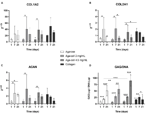
Mechanotransduction studies with physiological relevance require 3D in vitro models that: (i) recapitulate the in vivo environment of the tissue of interest; (ii) allow a reliable and reproducible application of the mechanical signals (e.g. dynamic compression); and (iii) allow the conduction of cellular and molecular assays. Agarose hydrogels have often been used to encapsulate chondrocytes and to study the effect of compression due to their strong mechanical properties [29, 30]. However, agarose is a bio-inert material that does not present any cell-adhesion motifs and therefore does not recapitulate the cell-matrix interactions that are crucial for mechanotransduction. We aimed to develop novel agarose-collagen composite hydrogels that: (i) mimic native tissues constituted of non-fibrillar matrix and collagens; (ii) combine the mechanical properties of agarose and the biofunctionality of collagen I; and (iii) allow the exploration of mechanical loading and cellular mechanotransduction. Specifically, we hypothesized that the physical blending of collagen I into agarose would enhance the biofunctionality of the construct by promoting cell adhesion and the activation of focal adhesion kinases (FAK). Agarose, collagen, and agarose-collagen composite hydrogels were compared in terms of structural homogeneity, rheological properties, and size stability. Bovine NP cell viability, proliferation, morphology, gene expression, glycosaminoglycan (GAG) production, adhesion, and FAK phosphorylation after dynamic compression were analyzed.
Aim 2: To investigate the role of TRPV4 in transducing hyperphysiological dynamic compression
Mechanical loading is a known contributor to IVD degeneration and LBP. However, current treatments of LBP do not target IVD mechanosensors and mechanotransducers, as they are poorly understood. The mechanosensitive TRPV4 ion channel is an interesting therapeutic target in the context of joint diseases, as it transduces mechanical,
5
inflammatory, and pain signals [21]. TRPV4 was shown to mediate the transduction of dynamic compressive loading in chondrocytes [22]. Moreover, reduced osmolarity was shown to increase TRPV4 expression and pro-inflammatory cytokines in bovine NP cells [24]. We aimed to investigate the potential role of TRPV4 in hyperphysiological dynamic compression of NP cells in vitro and IVDs in vivo. We hypothesized that: (i) hyperphysiological dynamic compression induces IVD degeneration, cell damage, and increased cyclooxygenase 2 (COX2) and prostaglandin E2 (PGE2) expression; and that (ii) TRPV4 mediates these effects. To test these hypotheses, mouse IVDs were dynamically compressed at a short repetitive hyperphysiological regime, and degenerative changes and the expression of the inflammatory mediator COX2 were examined (versus sham).
Bovine NP cells were embedded in our previously developed agarose-collagen hydrogels and dynamically compressed at a hyperphysiological regime with or without selective TRPV4 inhibition. Lactate dehydrogenase (LDH) and PGE2 release, as well as phosphorylation of mitogen-activated protein kinases (MAPKs), were analyzed.
Degenerative changes and COX2 expression were further evaluated in the IVDs of trpv4- deficient mice (versus wild-type).
Aim 3: To investigate the role of TRPV4 in transducing hyperphysiological cyclic stretching
AF disruption is commonly linked to LBP [31]. While the NP sustains hydrostatic and osmotic pressures generated by compressive forces from the body weight and spinal motions, the AF is subjected to tensile stresses [13] that could lead to AF disruption. We aimed to investigate the role of TRPV4 as a mechanotransducer and a potential therapeutic target in a more clinically relevant model by analyzing human primary AF cells that were cyclically stretched at a hyperphysiological strain. We hypothesized that:
(i) hyperphysiological cyclic stretching induces inflammation; and that (ii) TRPV4 inhibition and knockout (KO) reduce stretch-induced inflammation. TRPV4 pharmacological inhibition and gene editing were thus tested as potential therapeutic approaches to rescue the induced mechanoflammation. To test our hypotheses, human AF cells were stretched at a 20% hyperphysiological cyclic strain. TRPV4 was either inhibited with the selective TRPV4 antagonist GSK2193874 or knocked out via CRISPR- Cas9 gene editing. The gene expression, inflammatory mediator release and MAPK pathway activation were analyzed.
1.3 Thesis outline
This thesis is divided into seven chapters and an appendix. The content of each chapter is described below.
Chapter 1 introduces the clinical and economical motivations for this thesis and presents the aims and the outline of the thesis.
Chapter 2 provides background information on the anatomy of the IVD, IVD pathologies, and state-of-the-art therapeutic approaches. It further summarizes the
6
current knowledge on biomechanics and mechanobiology of the IVD, as well as TRP channels.
Chapter 3 addresses the first aim of the thesis and introduces novel agarose- collagen composite hydrogels as 3D in vitro models for mechanotransduction studies. We showed that agarose-collagen hydrogels retain strong mechanical properties while outperforming agarose hydrogels in terms of matrix production, cell adhesion, and mechanotransduction ability. This unique composite hydrogel scaffold thus mimics the natural extracellular matrix of the IVD, allowing the direct exploration of the influence of mechanical loading in a highly biomimetic and reproducible model system. This system can further be used to model other native tissues composed of non-fibrillar matrix and collagens and to investigate their mechanotransduction mechanisms.
Chapter 4 addresses the second aim of the thesis and describes a study that explores the role of TRPV4 in transducing hyperphysiological dynamic compression in NP cells in vitro and mouse IVDs in vivo. We identified TRPV4 as a regulator of the COX2/PGE2 inflammatory factors and as a mediator of cell damage induced by hyperphysiological dynamic compression, possibly via the extracellular signal-regulated kinases 1/2 (ERK) pathway. Targeted TRPV4 modulation might thus constitute a promising therapeutic strategy to treat patients suffering from IVD pathologies caused by aberrant mechanical stress.
Chapter 5 addresses the third aim of the thesis and presents a study that further investigates the role of TRPV4 in transducing hyperphysiological cyclic stretching in human AF cells in vitro. We first developed a model with an inflammatory response to hyperphysiological stretching representative of early-stage annulus fibrosus injury.
Moreover, we established the first successful CRISPR-Cas9 KO in degenerated human AF cells (TRPV4 KO). Through CRISPR-Cas9 gene editing and selective pharmacological inhibition experiments, we identified TRPV4 as a mediator of stretch-induced inflammation in human AF cells. This study and the one described in Chapter 4 highlight a novel link between mechanical hyperphysiological loading and injury mechanisms leading to degeneration of the IVD and elucidate an important pathway in the complex process that eventually leads to chronic LBP.
Chapter 6 expands on the use of the CRISPR-Cas9 gene editing technology in the IVD field and presents a review on this topic. This chapter provides an overview of the relevant recent advances, proposes potential treatment strategies for DDD and LBP, and discusses the current limitations that may hamper the clinical translation of the CRISPR- Cas9 technology.
Chapter 7 concludes the thesis by highlighting the contributions to the field and discussing the limitations and futures perspectives of this work.
In the Appendix, the reader will find a list of abbreviations, the curriculum vitae, and an additional study that investigates the TRP canonical 6 (TRPC6) ion channel in simulated microgravity of human IVD cells. In this study, we showed that simulated microgravity and TRPC channel inhibition led to reduced proliferation and increased senescence. Furthermore, simulated microgravity reduced TRPC6 expression. IVD cell
7
senescence and mechanotransduction may hence potentially be regulated by TRPC6 expression.
1.4 References
1. Balague, F., et al., Non-specific low back pain. Lancet, 2012. 379(9814): p. 482-91.
2. Airaksinen, O., et al., Chapter 4. European guidelines for the management of chronic nonspecific low back pain. Eur Spine J, 2006. 15 Suppl 2: p. S192-300.
3. Hartvigsen, J., et al., What low back pain is and why we need to pay attention. Lancet, 2018.
391(10137): p. 2356-2367.
4. Hoy, D., et al., A systematic review of the global prevalence of low back pain. Arthritis Rheum, 2012. 64(6): p. 2028-37.
5. Vos, T., et al., Global, regional, and national incidence, prevalence, and years lived with disability for 310 diseases and injuries, 1990-2015: a systematic analysis for the Global Burden of Disease Study 2015. Lancet, 2016. 388(10053): p. 1545-1602.
6. Bevan, S., et al., Fit for work. Musculoskeletal disorders in the European workforce. London:
The work foundation, 2009. 2009.
7. Wieser, S., et al., Cost of low back pain in Switzerland in 2005. Eur J Health Econ, 2011. 12(5):
p. 455-67.
8. Will, J.S., D.C. Bury, and J.A. Miller, Mechanical Low Back Pain. Am Fam Physician, 2018.
98(7): p. 421-428.
9. Childs, J.D., et al., Implications of early and guideline adherent physical therapy for low back pain on utilization and costs. BMC Health Serv Res, 2015. 15: p. 150.
10. Foster, N.E., et al., Prevention and treatment of low back pain: evidence, challenges, and promising directions. Lancet, 2018. 391(10137): p. 2368-2383.
11. Hayden, J.A., et al., Exercise therapy for treatment of non-specific low back pain. Cochrane Database Syst Rev, 2005(3): p. CD000335.
12. Qaseem, A., et al., Noninvasive Treatments for Acute, Subacute, and Chronic Low Back Pain:
A Clinical Practice Guideline From the American College of Physicians. Ann Intern Med, 2017. 166(7): p. 514-530.
13. Fearing, B.V., et al., Mechanotransduction and cell biomechanics of the intervertebral disc.
JOR Spine, 2018. 1(3).
14. Chan, S.C., S.J. Ferguson, and B. Gantenbein-Ritter, The effects of dynamic loading on the intervertebral disc. Eur Spine J, 2011. 20(11): p. 1796-812.
15. Setton, L.A. and J. Chen, Cell mechanics and mechanobiology in the intervertebral disc. Spine (Phila Pa 1976), 2004. 29(23): p. 2710-23.
16. Adams, M.A. and P.J. Roughley, What is intervertebral disc degeneration, and what causes it? Spine (Phila Pa 1976), 2006. 31(18): p. 2151-61.
8
17. Freemont, A.J., et al., Nerve ingrowth into diseased intervertebral disc in chronic back pain.
Lancet, 1997. 350(9072): p. 178-181.
18. Kos, N., L. Gradisnik, and T. Velnar, A Brief Review of the Degenerative Intervertebral Disc Disease. Med Arch, 2019. 73(6): p. 421-424.
19. Moran, M.M., et al., Transient receptor potential channels as therapeutic targets. Nat Rev Drug Discov, 2011. 10(8): p. 601-20.
20. Krupkova, O., J. Zvick, and K. Wuertz-Kozak, The Role of Transient Receptor Potential Channels in Joint Diseases. European Cells & Materials, 2017. 34: p. 180-201.
21. McNulty, A.L., et al., TRPV4 as a therapeutic target for joint diseases. Naunyn Schmiedebergs Arch Pharmacol, 2015. 388(4): p. 437-50.
22. O'Conor, C.J., et al., TRPV4-mediated mechanotransduction regulates the metabolic response of chondrocytes to dynamic loading. Proc Natl Acad Sci U S A, 2014. 111(4): p.
1316-21.
23. O'Conor, C.J., et al., Cartilage-Specific Knockout of the Mechanosensory Ion Channel TRPV4 Decreases Age-Related Osteoarthritis. Sci Rep, 2016. 6: p. 29053.
24. Walter, B.A., et al., Reduced tissue osmolarity increases TRPV4 expression and pro- inflammatory cytokines in intervertebral disc cells. Eur Cell Mater, 2016. 32: p. 123-36.
25. Dong, Q., et al., Blockage of transient receptor potential vanilloid 4 alleviates myocardial ischemia/reperfusion injury in mice. Sci Rep, 2017. 7: p. 42678.
26. Michalick, L., et al., Transient Receptor Potential Vanilloid 4 and Serum Glucocorticoid- regulated Kinase 1 Are Critical Mediators of Lung Injury in Overventilated Mice In Vivo.
Anesthesiology, 2017. 126(2): p. 300-311.
27. Thorneloe, K.S., et al., An orally active TRPV4 channel blocker prevents and resolves pulmonary edema induced by heart failure. Sci Transl Med, 2012. 4(159): p. 159ra148.
28. Goyal, N., et al., Clinical Pharmacokinetics, Safety, and Tolerability of a Novel, First-in-Class TRPV4 Ion Channel Inhibitor, GSK2798745, in Healthy and Heart Failure Subjects. Am J Cardiovasc Drugs, 2019. 19(3): p. 335-342.
29. Anderson, D.E. and B. Johnstone, Dynamic Mechanical Compression of Chondrocytes for Tissue Engineering: A Critical Review. Frontiers in Bioengineering and Biotechnology, 2017.
5(76).
30. Bougault, C., et al., Investigating conversion of mechanical force into biochemical signaling in three-dimensional chondrocyte cultures. Nature Protocols, 2009. 4(6): p. 928-938.
31. Torre, O.M., et al., Annulus fibrosus cell phenotypes in homeostasis and injury: implications for regenerative strategies. Ann N Y Acad Sci, 2019. 1442(1): p. 61-78.
9
Chapter 2
Background
2. Background
10
2.1 Anatomy of the intervertebral disc
The human spine is composed of 32 to 34 vertebrae, 23 intervertebral discs (IVDs), ligaments, the rib cage, and the spinal musculature [1]. The functions of the spine include:
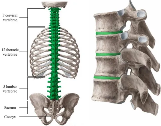
(i) housing and protecting the spinal cord and the efferent and afferent nerves; (ii) supporting the body mass and withstanding external forces; and (iii) allowing for mobility and flexibility. The spine is divided into 5 distinctive regions: cervical (with 7 vertebrae numbered C1 to C7), thoracic (T1-T12), lumbar (L1-L5), sacrum (S1-S5), and the coccyx (3 to 5 vertebrae) (Figure 1) [2]. Vertebrae in the cervical, thoracic, and lumbar regions are separated by IVDs, while the vertebrae of the sacrum and the coccyx are fused (Figure 1) [2].
Figure 1: Left: The human spine and its 5 regions. The green section highlights the part of the spine that contains individual vertebrae and IVDs. Right: Structure of the vertebrae with the IVDs in green. Reprinted from [2] with permission from the Multidisciplinary Digital Publishing Institute (MDPI) and Kenhub. Originally adapted from © Kenhub (www.kenhub.com); illustrator:
Begoña Rodriguez.
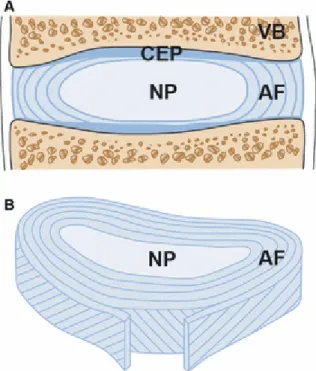
The IVD is composed of 3 main structures: the cartilaginous endplates (CEPs) at the superior and inferior faces of the IVD, the gelatinous nucleus pulposus (NP) in the center, and the fibrous and highly structured ring called annulus fibrous (AF) at the outer periphery (Figure 2 A, B) [3].
11
Figure 2: A) Schematic representation of the sagittal section of an IVD with the adjacent vertebrae (VB) and cartilaginous endplates (CEPs) at the superior and inferior faces, and the annulus fibrosus (AF) surrounding the nucleus pulposus (NP). B) Transverse section showing the structure of the lamellae and the alignment of the fibers of the AF. Reprinted from [4] with permission from John Wiley and Sons.
Cartilaginous endplates
The CEPs are thin, strong, and porous structures that separate the IVD from the adjacent vertebrae. They keep the NP pressurized and prevent it to bulge into the relatively soft center of the vertebrae [2]. Moreover, the CEPs are vascularized and ensure nutrient and fluid transport via diffusion between the vertebrae and the IVD [2]. The CEPs consist of hyaline cartilage composed of chondrocytes, proteoglycans, and a network of thin and closely packed fibrils of collagen (mainly type II) [2].
Nucleus pulposus
The NP is found in the center of the IVD. This highly hydrated (80-90% water [2]) and incompressible structure sustains compressive loads, as well as hydrostatic and osmotic pressures [3]. The NP is composed of a loose network of randomly distributed collagen II fibers (15–20% dry weight), radially dispersed elastin fibers, and proteoglycans (50% dry weight) [2]. Proteoglycans, like the abundantly present aggrecan, carry negative electric charges on their sulfated glycosaminoglycans (GAGs), which provide the NP with its osmotic and swelling properties [3]. NP cells are round chondrocyte-like cells that are interspersed in the NP at a low cellular density of around 4000 cells/mm3 [5]. The microenvironment at the center of the IVD is characterized by low oxygen tension, leading to anaerobic metabolism and low pH [6]. NP cells maintain tissue homeostasis by synthesizing and degrading large amounts of extracellular matrix (ECM) composed of collagens and proteoglycans [2]. Large vacuolated notochordal cells
12
can be present in young IVDs, but they are almost all completely replaced by chondrocyte- like cells in adulthood in humans [2].
Annulus fibrosus
The AF is a highly organized fibrocartilaginous structure that surrounds the NP. It is often subdivided into inner and outer AF. The main functions of the AF are to confine the relatively fluid NP, withstand compressive and tensile stress, and allow joint mobility [2].
The AF is composed of 15-25 concentric lamellae made of 20-60 collagen fiber bundles aligned at angles ranging from 55° to 20° that alternate direction at every layer (Figure 2 B) [2]. The ECM of the AF varies radially and mainly consists of water (70–78% inner and 55–65% outer wet weight), collagens (type I and type II collagen, 25–40% inner and 60–
70% outer dry weight), and proteoglycans (11–20% inner and 5–8% outer dry weight) [2]. The collagen types I and II further substitute each other in a radial gradient, transitioning from 100% type II in the innermost lamellae to 100% type I in the outermost lamella [2]. The outermost layers of the AF are superficially vascularized and innervated by afferent and efferent branches of the spinal nerves [2]. While cells of the inner AF tend to be rounder, outer AF cells display an elongated morphology and align with the fibers of the lamellae [3].
2.2 Aging and pathologies of the intervertebral disc
Intervertebral disc aging
Compared to the young healthy IVD, the aging IVD undergoes natural metabolic, biochemical, and structural changes, which do not necessarily provoke any symptoms [7, 8]. These changes usually start in the CEPs and sequentially affect the NP and the AF [6, 8]. The blood supply to the CEPs and the IVD cellularity start decreasing already during childhood [6]. The ECM synthesis is reduced, leading to a loss in NP hydration due to the decrease in proteoglycan and hence in osmolarity [6]. ECM turnover decreases as well, causing an increase in collagen content and cross-linking [6]. Moreover, collagen type II in the inner AF tends to be replaced by collagen I [6]. Microstructural defects, such as cleft and tears, are already visible in the CEPs and NP by the age of 15 years, further extending into the AF with increasing age [6, 9].
Intervertebral disc degeneration
Pathological IVD degeneration is defined as an accelerated aging process including structural failure [6]. Causes of IVD degeneration are various and include genetics, aging, scarce metabolite diffusion, and loading history [6]. They all cumulatively contribute to weakening the IVD. However, mechanical loading is thought to precipitate IVD degeneration [6]. Excessive mechanical loading causes structural defects and cellular catabolic and inflammatory responses that strongly affect the matrix composition of the IVD and its biomechanical properties [6]. This, in turn, induces further aberrant mechanical loading [8], thus triggering a vicious circle [10].
13
IVD degeneration mostly affects lower lumbar IVDs [6]. Their bigger size impairs an efficient nutrient diffusion and they are subjected to the highest mechanical load. The degenerated IVD is characterized by several structural defects, such as annular tears, height loss, and disruption (Figure 3) [8]. Moreover, the CEPs become calcified and sclerotic (Figure 3) [8].
Figure 3: Various stages of IVD degeneration observed in sagittal sections of human lumbar IVDs. Reprinted from [8] with permission from Springer Nature.
Structural changes are believed to alter the mechanical properties of the IVD and, consequently, the way it withstands mechanical loading [8]. Abnormal mechanical stress causes increased cell death [11, 12]. IVD cell senescence and death are thought to play an important role in the pathogenesis of IVD degeneration [12, 13]. Moreover, aberrant mechanical loading initiates catabolic and inflammatory cascades [10]. Degenerated IVDs show increased levels of matrix-degrading enzymes, such as matrix metalloproteinases (MMP 1, 3, 13) [14], “a disintegrin and metalloproteinase with thrombospondin motifs”
(ADAMTS 4) [14], and (pro-)inflammatory mediators, such as interleukins (IL 1β, 6, 8, 15), as well as cyclooxygenase 2 (COX2) and its enzymatic product prostaglandin E2 (PGE2), to name a few [15, 16]. Catabolic and inflammatory responses might be mediated by the nuclear factor-kappa B (NF-κB) and mitogen-activated protein kinase (MAPK:
extracellular signal-regulated kinases 1/2 (ERK 1/2), p38 and Jun-N-terminal kinase (JNK)) signaling pathways [17]. Detrimental cell-mediated responses further result in reduced ECM synthesis and proteoglycan content, and thus loss of osmotic and swelling
14
pressure in the NP [8]. Moreover, increased ECM degradation and inflammation exacerbate structural defects, leading to IVD collapse and structural failure [6, 8].
Intervertebral disc herniation
When radial fissures in the AF allow the bulging of the NP into the AF, herniation arises [6]. Depending on the level of NP bulging, it may result in protrusion, when the NP migrates through the lamellae of the AF, or extrusion, when the NP exits the AF [6].
Extrusion of the nuclear material can further cause inflammation and/or compression of a spinal nerve or its root, thus provoking so-called radicular pain [6, 18].
Degenerative disc disease and low back pain
Like IVD physiological aging, IVD degeneration is not necessarily symptomatic [8].
Degenerative disc disease (DDD) is characterized by IVD degeneration accompanied by pain [6]. Pain mechanisms can start with IVD degeneration, which induces anatomical and functional changes, such as NP prolapse, which further cause the irritation or compression of spinal nerves [18]. This is for example the case of radicular pain stemming from IVD herniation, as explained above. Alternatively, pain can arise from the IVD itself and can stem from the structural defects and inflammation that characterize IVD degeneration [18]. During severe disc disruption, the blood vessels and nerves that normally populate the surface of the AF grow into the diseased IVD [19]. Ingrown nerves express substance P, a nociceptive neurotransmitter, and are associated with pain [19]. It is believed that noxious inflammatory stimuli from the degenerated disc environment, as well as mechanical stimulation of ingrown (or superficial) nociceptors, provoke nerve sensitization and pain [18].
2.3 Current treatments of low back pain
Current treatments of LBP aim to manage pain but do not address the causes of the condition. Treatment strategies are divided into physical, psychological, and pharmacological therapies, as well as surgical interventions. Clinical guidelines have recently changed based on novel evidence and recommend against routine use of imaging as a diagnostic method, pharmacotherapy, and surgery [20]. However, there is an enormous gap between evidence-based recommendations and the current practice [20].
For example, IVD imaging including radiography and magnetic resonance imaging (MRI) is still frequently used, generating high direct costs [20]. Nevertheless, its use should be limited to cases where specific serious conditions, such as vertebral fracture, malignancy, or infection, are suspected [20]. The MRI-based Pfirrmann scale, which grades IVD degeneration from grade 1 (healthy IVD) to grade 5 (highly degenerated and collapsed IVD) [21] is useful to compare data in basic and clinical research. However, imaging findings do not correlate with pain [22] and patients with degenerative changes can be asymptomatic [23].
15
In a first consultation, the patient should be physically examined and questioned about occupational and psychosocial factors, as well as the severity and duration of pain [24]. Acute LBP is defined by a duration below 6 weeks and becomes chronic when it lasts over 12 weeks [20]. The results of this first evaluation will allow to rule out specific serious causes of LBP and select the appropriate treatment strategies in case of non- specific pain [24].
Physical and psychological therapies
As the first line of management, the patient should be reassured and told that LBP is not a serious condition and the prognosis is good in most of the cases [20]. The patient should also be advised to remain physically active and resume professional activities [20].
Early referral to physical therapy was shown to reduce healthcare use and total costs [24, 25]. Recommended physical therapies, especially for chronic LBP, include a graded exercise program aiming for functional improvement [20]. However, there is variable evidence for specific physical approaches [20, 24]. Exercise therapy appears to be as effective as no treatment or other treatment in acute LBP and to be slightly effective at reducing pain and improving function in chronic LBP [24, 26]. Passive therapies, such as acupuncture, massage, and spinal manipulation, are either optional or not recommended depending on the guidelines [20]. Despite insufficient evidence, guidelines further recommend considering the use of psychological therapies, such as cognitive behavioral therapy and mindfulness-based stress reduction, and combined physical and psychosocial approaches especially for patients who do not respond to first-line treatments [20].
Pharmacological therapies
Current guidelines recommend the use of pharmacotherapy only in case of a lack of response to first-line non-pharmacological approaches [20]. This recommendation is based on the fact that current pharmacological treatments have unclear mechanisms of action, low effect sizes [27], and can provoke side effects in the long term. The most common pharmacological therapies for LBP include paracetamol, non-steroidal anti- inflammatory drugs (NSAIDs), and opioids.
Paracetamol
Paracetamol (also called acetaminophen) used to be a first-line medicine for the management of LBP [20]. It is an analgesic with an unclear mechanism of action that does not possess anti-inflammatory properties [28]. Today, the use of paracetamol is no longer recommended for LBP due to evidence of absence of effectiveness and potential harm [20, 29].
Non-steroidal anti-inflammatory drugs
NSAIDs, such as ibuprofen and aspirin, inhibit the COX2 enzyme [28]. They were shown to be slightly effective at providing short-term relief in acute and chronic LBP without radicular pain [24, 30]. However, there was no significant difference between
16
NSAID treatment and placebo in the case of radiculopathy [30]. Moreover, NSAIDs were not more effective than paracetamol or other drugs [30]. NSAIDs should be administered after taking into account risks like gastrointestinal, liver, and cardiorenal toxicity [20].
Opioids
Opioids, typified by morphine, are potent analgesics that act on neuronal receptors in the central nervous system and peripheral nerves [28]. There is evidence of moderate short-term efficacy of opioids in treating chronic LBP compared to placebo [31]. However, rates of misuse average between 21% and 29%, and rates of addiction between 8% and 12% [32]. Routine use of opioids is therefore not recommended due to the small benefit and risk of misuse, abuse, and dependence [20].
Interventional therapies and surgery
Clinical guidelines regarding interventional therapies and surgery vary [20].
Increasingly invasive procedures can be applied based on the condition of the patient.
They range from epidural injections to microdiscectomy, spinal fusion, and total disc replacement (TDR). Surgery should be used as a last resort in disabling chronic LBP in case of failure of conservative therapies, as it is more costly and prone to adverse events compared to non-surgical approaches [20].
Epidural injections
The first more invasive approach involves injections of drugs, such as corticosteroids or anesthetics, into the epidural space that surrounds the spinal cord and nerve roots. Epidural injections are not recommended by recent guidelines for LBP, but rather for severe radicular pain [20]. They alleviate pain by delivering an increased drug concentration locally, but their effect is short-lived (<4 weeks) and there is no effect on long-term surgery risk [20, 33]. Moreover, epidural injections can cause rare but serious adverse events, including loss of vision, stroke, paralysis, and death [20].
Microdiscectomy
Microdiscectomy is a minimally invasive surgical procedure aimed to relieve pain stemming from disc herniation. The herniated tissue is surgically removed to relieve the pressure on the spinal nerves. Early surgery is associated with faster pain relief compared to initial conservative treatment and delayed surgery, but benefits decrease with longer follow-up (>1 year) [20, 34].
Spinal fusion
Spinal fusion is a surgical procedure that consists in the complete removal of the diseased IVD, replacement with a titanium fusion cage or a bone-like material, and connection of the two adjacent vertebrae with titanium pedicle screws and rods [2]. The aim is to prevent motion between the fused vertebrae. The benefits of spinal fusion for discogenic LBP are similar to those of intense rehabilitation and only slightly higher than non-surgical treatments [20, 35]. Long-term issues can further arise because spinal fusion
17
changes the biomechanical behavior of the spine, which may cause degeneration of the adjacent spinal segments [2].
Total disc replacement
In contrast to spinal fusion, TDR aims to preserve the motion of the diseased segment by removing the diseased IVD and replacing it with a synthetic implant. TDR is a relatively recent type of surgery compared to spinal fusion, with only a few disc implants approved so far for the lumbar level [2]. There is low-quality evidence that there are no clinically relevant differences between TDR and fusion techniques, with small overall success rates in both groups [36]. The safety and effectiveness of TDR need to be further evaluated in high-quality randomized controlled trials with long-term follow-up [36].
2.4 Preclinical therapeutic strategies
Considering the variable evidence in favor of physical therapy and the high risks and costs associated with surgery, efforts are currently deployed to develop targeted pharmacological and biological regenerative therapies with a clear mechanism of action and limited side effects. Targeted therapeutic strategies mainly include molecular and gene therapy. They may complement other promising but broader therapeutic approaches such as IVD tissue engineering and cell therapy.
Tissue engineering
Tissue engineering aims to replace either a part (the NP or AF) or the whole degenerated IVD with engineered scaffolds that may or not contain cells. Both natural and synthetic biomaterials are investigated and several state-of-the-art manufacturing techniques, such as electrospinning, composite hydrogel fabrication, and three- dimensional (3D) bioprinting, are applied [2]. Hurdles that need to be overcome by tissue- engineered scaffolds include scalability, integration with the native tissue, as well as sufficient load-bearing capacity [2].
Cell therapy
In cell therapy, cells are transplanted in the degenerated IVD with the aim of repairing the deteriorated environment. The current strategies are mainly directed at regenerating the NP, with only a few approaches targeting the AF or the CEPs [37]. Several issues remain open, including the choice of cell source and type (e.g. autologous or allogeneic; differentiated cells or mesenchymal stem cells), the delivery mode (e.g. in suspension or embedded in a biomaterial), and the selection of patients (e.g. early or later stages of IVD degeneration) [37]. The main challenge faced by cell therapy is the survival of the transplanted cells in the harsh and inflamed microenvironment of the degenerated IVD, which is characterized by low nutrient transport, oxygen and, pH, as well as high mechanical stress [37].
18 Molecular therapy
The goal of molecular therapy is to selectively address the biological changes that occur in IVD degeneration and DDD via the administration of specific molecules.
Molecular therapeutic approaches can be divided into those that promote IVD anabolism and regeneration (mainly via growth factors) and those that inhibit catabolic and inflammatory processes.
Growth factors
Growth factors are proteins that target specific cell receptors and play a role in cell proliferation, differentiation, and ECM synthesis [38]. The potential of several growth factors (bone morphogenic proteins (BMP 2 and 7), transforming growth factor beta (TGFβ), epidermal growth factor (EGF), basic fibroblast growth factor (bFGF), growth/differentiation factor 5 (GDF5), and insulin-like growth factor 1 (IGF1)) for IVD regeneration has been tested in vitro and in vivo with positive outcomes including the increase of ECM synthesis (studies reviewed in [38]). However, the main disadvantage of growth factors, besides their high cost, is their short half-life, which limits the duration of their therapeutic effects and requires multiple injections [38].
Anti-catabolic and anti-inflammatory approaches
Two of the major signaling pathways that are targeted by anti-catabolic and anti- inflammatory molecular therapies to treat DDD are the NF-κB and MAPK pathways [39].
Local administration of NF-κB decoy oligodeoxynucleotides to the DRG in a rat lumbar disc herniation model was shown to significantly suppress pain [40]. Systemic inhibition of NF-κB activity via pharmacological inhibition with the Nemo Binding Domain (8K- NBD) peptide was shown to mitigate age-associated IVD degeneration in a mouse model of accelerated aging by increasing proteoglycan synthesis and improving the loss of cellularity [41]. Modulation of the MAPK pathways, especially of the p38 MAPK, has further yielded promising results. Inhibition of p38 in cytokine-activated rabbit NP cells reduced gene expression and production of factors associated with ECM catabolism, inflammation, and pain [42]. Moreover, IL1-induced downregulation of ECM gene expression and proteoglycan synthesis were reversed [42].
Gene therapy
Gene therapy is an experimental technique that aims to modify genes in order to treat a disease. This approach is interesting in the context of DDD, as genetic inheritance is one of the multiple factors that can cause IVD degeneration. Gene polymorphisms affecting genes such as aggrecan, collagen 1, 9, and 11, MMP 1, 2 and 3, IL 1 and 6, and COX2 have been associated with IVD degeneration [43]. Gene therapy can not only correct unfavorable gene polymorphisms, but also control the dysregulated molecular pathways in DDD by activating anabolic genes and growth factors and repressing catabolic and inflammatory genes [44]. Several gene therapy studies targeting genes like osteogenic protein-1 (OP1), sex-determining region Y box 9 (SOX9), TGFβ1, IGF1, BMP2, and IL1 receptor antagonist (IL1Ra) have been performed in vitro and in vivo with encouraging
19
regenerative results [45-47]. Gene therapy can complement cell therapy by editing cells to be transplanted ex vivo so that they can survive the harsh environment of the degenerated IVD and secrete reparatory molecules. Another approach is to perform gene editing in vivo by delivering the transduction agents locally to IVD cells or dorsal root ganglion (DRG) neurons [48]. Cells can be transduced via non-viral (e.g. liposomes) or viral vectors (e.g. lentivirus, adenovirus, or adeno-associated-virus) [38]. Non-viral vectors have low transduction efficiency, while viral vectors are very efficient but raise the issue of immunogenicity [39]. Although promising, gene therapy needs to overcome technical, safety, and ethical issues before it can be applied in clinics in the future.
CRISPR-Cas9
The most recent gene editing technique is the Nobel prize-winning clustered regularly interspaced short palindrome repeats (CRISPR)-Cas9 technique (explained in detail in Chapter 6). The CRISPR-Cas9 system is composed of a Cas9 endonuclease that is directed to the target deoxyribonucleic acid (DNA) sequence by a single-guide ribonucleic acid (sgRNA) [48]. It can be used to perform gene knockout, editing, as well as transcriptional regulation (gene activation or interference) [48]. Recently, CRISPR-Cas9 was used for epigenome editing to repress the inflammatory cytokine receptors tumor necrosis factor receptor 1 (TNFR1) and IL 1 receptor 1 (IL1R1) in human adipose tissue- derived stem cells to improve cell therapy for musculoskeletal diseases [49]. This technique was further used to modulate the A-kinase anchoring protein 150 (AKAP150) in DRG neurons and abolish the nociceptive neuron activity as a targeted strategy for LBP [50].
RNA interference
Another recent form of gene therapy is RNA interference (RNAi). RNAi aims to silence genes by interfering with their transcription or translation via small non-coding RNAs like small interfering RNAs (siRNAs) and microRNAs (miRNAs) [38]. siRNA approaches aiming to silence ADAMTS5 and caspase 3, an enzyme that plays a role in apoptosis, have yielded promising results in rabbit models of IVD degeneration [51-53].
2.5 Biomechanics and mechanobiology of the intervertebral disc
Despite the mechanical nature of LBP, current drugs and biological therapies do not specifically target mediators of mechanosensing and mechanotransduction pathways, as these are poorly understood. A better understanding of the molecular mechanisms leading from hyperphysiological mechanical loading to IVD degeneration, inflammation, and nociception might reveal novel, and possibly more effective, therapeutic targets.

![Figure 4: Schematic representation of the mechanical deformation of the IVD. Adapted from [3] with permission from John Wiley and Sons](https://thumb-eu.123doks.com/thumbv2/1library_info/3901484.1524456/33.892.125.750.428.732/figure-schematic-representation-mechanical-deformation-adapted-permission-wiley.webp)