Analysis of cell-cell communication during repro- ductive processes by EA1-like peptides in maize
DISSERTATION
ZUR ERLANGUNG DES DOKTORGRADES DER NATURWISSENSCHAFTEN (DR. RER. NAT.) DER FAKULTÄT FÜR BIOLOGIE UND VORKLINISCHE MEDIZIN
UNIVERSITÄT REGENSBURG
Vorgelegt von
Susanne Uebler
aus Vilshofen an der Donau
im September 2015
Das Promotionsgesuch wurde eingereicht am 24.09.2015 Die Arbeit wurde angeleitet von Dr. Mihaela-Luiza Márton
Unterschrift: _____________________________________
Susanne Uebler
Meinen Eltern.
Meiner Liebe.
„Coming back to where you started is not the same as never leaving.”
Terry Pratchett
i
This thesis is partially composed of the manuscripts listed below:
C
HAPTER2
Uebler, S., Márton, M. L. and Dresselhaus, T. (under review in Plant Reproduction).
Classification of EA1-box proteins and new insights into their role during reproduction in grasses.
S. Uebler conducted bioinformatical analysis and performed all experimental procedures ex- cept RT-PCR of rice genes and generation of constructs for transient transformation of ZmEA1, AtEAG1, OsEAL1 and OsEAL3. The manuscript was written by S. Uebler and edited by T. Dresselhaus. The paragraphs about generation and analysis of transgenic maize lines and promoter analysis are excluded from the publication.
C
HAPTER3
Márton, M. L., Fastner, A., Uebler, S. and Dresselhaus, T. (2012). Overcoming hybridi- zation barriers by the secretion of the maize pollen tube attractant ZmEA1 from Ara- bidopsis ovules. Current Biology 22, 1194–1198.
S. Uebler performed the immunoblot experiments (Figure 3.4 E) and wrote the corresponding paragraph in the section of experimental design (3. 2. 6).
C
HAPTER4
Uebler, S., Dresselhaus, T. and Márton, M. L. (2013). Species-specific interaction of EA1 with the maize pollen tube apex. Plant Signaling & Behavior 8, e25682.
S. Uebler performed all experiments and wrote the manuscript except the introduction para- graph. The manuscript was edited by T. Dresselhaus and M. L. Márton.
C
HAPTER5
Uebler, S. and Dresselhaus, T. (2014). Identifying plant cell-surface receptors: combin- ing “classical” techniques with novel methods. Biochemical Society Transactions 42, 395–
400.
The manuscript was written by S. Uebler and edited by T. Dresselhaus.
ii CONTENTS
SUMMARY... 1
CHAPTER 1-GENERAL INTRODUCTION TO SEXUAL REPRODUCTION IN ANGIOSPERMS ... 3
1. 1 Development of gametophytes in flowering plants ... 3
1. 1. 1 Development of the female gametophyte ... 3
1. 1. 2 Development of the male gametophyte ... 5
1. 2 Progamic phase ... 6
1. 2. 1 Growth of the pollen tube through sporophytic tissue ... 6
1. 2. 2 Control of pollen tube growth by male-female communication ... 7
1. 3 Embryogenesis in model flowering plants ... 9
1. 3. 1 Developmental steps of embryo formation ... 9
1. 3. 2 Peptide signaling during embryogenesis ... 11
1. 4 Aims of this work ... 12
CHAPTER 2 - CLASSIFICATION OF EA1-BOX PROTEINS AND NEW INSIGHTS INTO THEIR ROLE DURING REPRODUCTION IN GRASSES ... 13
2. 1 Introduction ... 13
2. 2 Experimental procedures ... 15
2. 2. 1 Bioinformatic analysis ... 15
2. 2. 2 Generation of constructs for transformation of BMS cells ... 15
2. 2. 3 Transient transformation of maize BMS suspension cells ... 16
2. 2. 4 Extraction of total RNA and generation of cDNA ... 16
2. 2. 5 Semi-quantitative reverse transcriptase-PCR (RT-PCR) and quantitative real– time PCR (qRT-PCR) ... 17
2. 2. 6 Immunoblot analysis and generation of anti-ZmEAL2 antiserum ... 17
2. 2. 7 Immunohistochemical detection of ZmEAL2 ... 18
2. 2. 8 Generation of stable transgenic maize lines ... 18
2. 2. 8. 1 Constructs for Agrobacterium-mediated transformation of maize ... 18
2. 2. 8. 2 Agrobacterium-mediated transformation of maize... 19
iii
2. 3 Results and discussion ... 21
2. 3. 1 EA1-box containing proteins can be separated into three classes ... 21
2. 3. 2 EA1-box proteins show a highly variable expression pattern... 26
2. 3. 3 EALs and EACs enter the secretory pathway, while EAGs localize to the cytoplasm and nucleus ... 28
2. 3. 4 ZmEAL2 potentially plays a role during late embryogenesis... 32
2. 4 Conclusions ... 39
CHAPTER 3 - OVERCOMING HYBRIDIZATION BARRIERS BY THE SECRETION OF THE MAIZE POLLEN TUBE ATTRACTANT ZMEA1 FROM ARABIDOPSIS OVULES ... 41
3. 1 Introduction ... 41
3. 2 Experimental procedures ... 41
3. 2. 1 Plant material and growth ... 41
3. 2. 2 Generation of constructs and transgenic plants... 42
3. 2. 3 Chemical labeling of synthetic ZmEA1 peptide ... 43
3. 2. 4 In vitro pollen tube guidance and competition assays and ZmEA1 bioassay ... 43
3. 2. 5 Microscopy ... 44
3. 2. 6 Immunoblot analysis ... 45
3. 3 Results and discussion ... 46
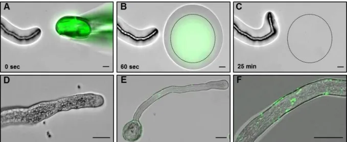
3. 3. 1 Mature ZmEA1 attracts and arrests maize pollen tubes in vitro and binds to the subapical region of their tips ... 46
3. 3. 2 ZmEA1-GFP fusion proteins are predominantly localized to the filiform apparatus, the secretory zone of synergid cells in Arabidopsis ovules ... 50
3. 3. 3 Maize PTs are attracted in vitro by Arabidopsis ovules expressing ZmEA1- GFP fusion protein in synergid cells ... 53
3. 4 Conclusions ... 57
CHAPTER 4 - SPECIES-SPECIFIC INTERACTION OF EA1 WITH THE MAIZE POLLEN TUBE APEX ... 58
4. 1 Introduction ... 58
4. 2 Experimental procedures ... 59
iv
4. 2. 1 Chemical labeling of synthetic peptides ... 59
4. 2. 2 Pollen tube binding assay ... 59
4. 3 Results and discussion ... 60
4. 4 Conclusions ... 64
CHAPTER 5-IDENTIFYING PLANT CELL-SURFACE RECEPTORS: COMBINING “CLASSICAL” TECHNIQUES WITH NOVEL METHODS. ... 66
5. 1 Introduction ... 66
5. 2 Genetic and bioinformatic approaches are limited to identify peptide ligand interaction partners ... 67
5. 3 Cross-linking: Panacea for receptor-fishing? ... 68
5. 4 Innovative novel method: Ligand-based receptor-capture technology ... 71
5. 5 Alternative “classical” method: mRNA-display libraries ... 73
5. 6 Concluding remarks ... 73
CHAPTER 6 -IDENTIFICATION OF CANDIDATE INTERACTION PARTNERS OF THE POLLEN TUBE ATTRACTOR ZMEA1 ... 75
6. 1 Introduction ... 75
6. 2 Experimental procedures ... 78
6. 2. 1 Cultivation of organisms ... 78
6. 2. 1. 1 Plant material and growth conditions ... 78
6. 2. 1. 2 Bacterial strains and culture conditions ... 78
6. 2. 2 Transformation of bacteria ... 79
6. 2. 3 Molecular cloning and PCR ... 79
6. 2. 3. 1 PCR and quantitative real-time PCR (qRT-PCR) ... 79
6. 2. 3. 2 Determination of UTR sequences ... 80
6. 2. 3. 3 Constructs for heterologous expression in E. coli ... 80
6. 2. 3. 4 Constructs for transient expression in N. benthamiana ... 81
6. 2. 3. 5 Constructs for in vitro transcription/translation (IVT/T) ... 81
6. 2. 4 Heterologous expression in E. coli and protein purification ... 82
6. 2. 4. 1 Purification of His-tagged protein ... 82
v
6. 2. 4. 2 On-column refolding of His-tagged protein ... 83
6. 2. 4. 3 Native protein purification of GST-tagged protein ... 83
6. 2. 5 Heterologous expression in N. benthamiana leaves and protein purification .... 83
6. 2. 5. 1 Transient transgene expression in tobacco ... 83
6. 2. 5. 2 Extraction of recombinant protein produced in tobacco ... 84
6. 2. 5. 3 Purification of sEA1-GFP produced in tobacco ... 84
6. 2. 5. 4 Pull-down of recombinant Myc-WSL3 with sEA1-biotin ... 85
6. 2. 5. 5 Pull-down of candidate proteins fused to GFP with sEA1-biotin ... 86
6. 2. 6 Protein analysis ... 86
6. 2. 6. 1 Generation of polyclonal peptide antibodies against EA1, WSL1a/b and WSL3 ... 86
6. 2. 6. 2 SDS-PAGE, gel staining and immunoblot analysis ... 86
6. 2. 7 In vitro transcription/translation (IVT/T) of proteins ... 87
6. 2. 8 Protein extraction from maize pollen tubes ... 88
6. 2. 9 Pull-down with sEA1-biotin and germinated maize pollen ... 88
6. 2. 9. 1 With crosslinking, analysis by immunoblot ... 88
6. 2. 9. 2 No crosslinking, analysis by mass spectrometry ... 89
6. 2. 10Bioinformatic analysis ... 89
6. 3 Results ... 90
6. 3. 1 Heterologous expression of predicted mature ZmEA1 (sEA1) ... 90
6. 3. 1. 1 Expression of sEA1 in Escherichia coli ... 90
6. 3. 1. 2 Expression of sEA1 in Nicotiana benthamiana leaves ... 97
6. 3. 2 Potential interaction partners of sEA1 ... 100
6. 3. 2. 1 Visualization of sEA1 binding to other proteins ... 100
6. 3. 2. 2 Identification of potential interaction partners of sEA1 ... 104
6. 3. 2. 3 Sequence analysis of the potential interaction partners ... 107
6. 3. 3 WSL1a/b and WSL3 exhibit structural similarities to DEFLs ... 110
6. 3. 4 Interaction studies with sEA1 and WSLs ... 111
6. 3. 4. 1 Expression of WSLs and ZmEA1/sEA1 in IVT/T systems ... 111
6. 3. 4. 2 Expression in Nicotiana benthamiana leaves ... 114
vi
6. 3. 5 Characterization of WSL peptides ... 116
6. 3. 5. 1 Expression profile of WSL peptides ... 116
6. 3. 5. 2 WSL1 is highly abundant in maize pollen ... 119
6. 3. 5. 3 WSL peptides enter the secretory pathway ... 121
6. 3. 5. 4 WSL peptides are exclusively found in maize and Sorghum bicolor ... 124
6. 4 Conclusions ... 125
6. 4. 1 Candidates for interaction with predicted mature ZmEA1 are secreted CRPs related to defensins ... 125
6. 4. 2 Future tasks to verify WSL peptides as interaction partners of ZmEA1 ... 126
6. 4. 3 Model for interaction of WSL peptides with ZmEA1 ... 129
CHAPTER 7-COMPREHENSIVE DISCUSSION AND OUTLOOK ... 132
7. 1 EA1-box as potential protein-protein interaction motive... 132
7. 2 Structure and posttranslational modifications of EALs: Open questions that need to be answered ... 134
7. 3 WSL-EA1 interaction – a novel reproductive isolation mechanism? ... 138
REFERENCES ... 140
SUPPLEMENTARY MATERIAL ... 165
Genomic locations of EA1-box proteins ... 165
Primer sequences ... 166
Vector maps ... 169
ABBREVIATIONS ... 174
FIGURES AND TABLES ... 176
ACKNOWLEDGEMENTS ... 179
1 SUMMARY
Signaling processes mediated by secreted peptides are of eminently importance in cell-cell- communication of plants. This work focuses on extracellular signaling peptides containing the EA1-box and their involvement in control of reproductive processes in maize (Zea mays L.).
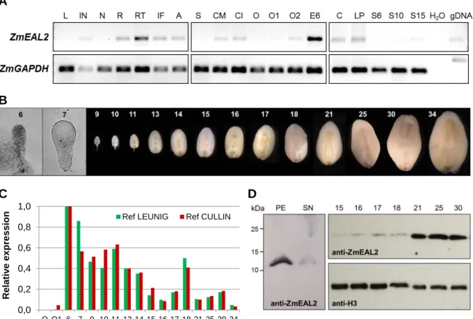
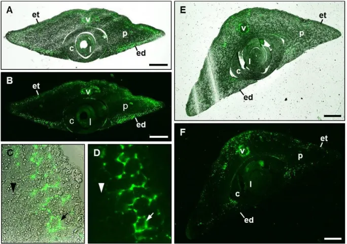
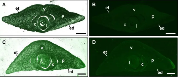
In maize, the EA1-box protein ZmEA1 was already demonstrated to act as a secreted signal- ing peptide in micropylar pollen tube guidance, whereas the closely related peptide ZmEAL1 is necessary for cell fate determination of the female gametophyte. To extent our knowledge about EA1-box proteins, several plant genomes of monocotyledonous and dicotyledonous plants were searched for this motif and EA1-box proteins could be identified in all of the ana- lyzed plant species. Based on similarities of structural features and subcellular localization, a new classification and phylogenetic analysis is presented, dividing these proteins into three classes, the EAL, EAG and EAC proteins. The EA1-like (EAL) proteins, including ZmEA1 and ZmEAL1, consist of less than 200 amino acids and are predicted to be secreted using ei- ther an N-terminal or internal signal sequence. Besides the EA1-box, they contain the so- called P-box and a C-terminal alanine-rich region as conserved motifs. All analyzed EAL proteins were demonstrated to enter the secretory pathway in transiently transformed plant suspension cells. In contrast, the EA1-box containing glycine-rich (EAG) proteins show cyto- plasmic localization and are expected to act as intracellular components. The third class, the EA1-box containing (EAC) proteins, includes EA1-box proteins that could not be classified as EALs or EAGs and contains proteins with up to several transmembrane domains. The ex- pression pattern of rice EALs was analyzed and comparison with maize EALs indicated that these proteins might act as orthologs. Furthermore, the final member of the small maize EAL family, ZmEAL2, was analyzed in detail and was demonstrated to exhibit a broader expression pattern compared to ZmEA1 and ZmEAL1, and showed a remarkable strong expression pattern in the embryo during later stages of development. Using immunohistochemical analysis, ZmEAL2 was localized in the scutellar parenchym and surrounding the vascular system of the embryo. Taken together, these results suggest a role of ZmEAL2 during embryogenesis.
To gain deeper insights into the role of ZmEA1 during micropylar pollen tube attraction, it was expressed as GFP-fusion protein in the synergids of Arabidopsis thaliana. Interestingly, dissected transgenic Arabidopsis ovules placed on solid media were demonstrated to attract in vitro germinated maize pollen tubes, whereas no impact on tube growth direction could be
2
detected for wild-type ovules. As this indicates the direct binding of secreted ZmEA1 protein to the pollen tube, germinated maize pollen was further incubated with predicted mature ZmEA1 (sEA1), labeled with a fluorophore to visualize the interaction. The peptide bound to the surface of the pollen tube apex and was internalized quickly, probably for degradation. To identify the interaction partner(s) located on the pollen tube surface binding to sEA1, a large number of pull-down experiments were performed. Immunoblot analysis of the isolated frac- tions indicated binding to one or more interaction partners, which should be identified by Or- bitrap mass spectrometry. Three different protein sequences were identified as candidates for interaction with sEA1 and named as WHY SO LATE (WSL) proteins. Contrary to the ex- pected membranous proteins, WSL proteins represent putative secreted cysteine-rich peptides.
They were shown to enter the secretory pathway in transiently transformed tobacco leaves.
WSL1 is encoded by two genes which were named as WSL1a and WSL1b. WSL1a/b and WSL3 protein sequences are highly similar, therefore they were classified to form a small protein family sharing structural similarities to defensin-like proteins (DEFLs). All WSL pep- tides are strongly and specifically expressed in pollen and are exclusively found in maize and the closely related grass Sorghum bicolor. First attempts were performed to confirm the inter- action of WSL peptides with sEA1. Taken together, potential factors acting in the pathway of ZmEA1-signaling could be identified and were made available for further research. In case, the direct binding between ZmEA1 and WSL peptides as well as the involvement of this complex in micropylar pollen tube guidance can be demonstrated in future, this would be the first report of a heterodimeric signaling ligand generated both by male and female gameto- phytes, which would provide a highly specific interaction representing a prerequisite for a molecular mechanism contributing to reproductive isolation barriers in plants.
3
CHAPTER 1 - GENERAL INTRODUCTION TO SEXUAL REPRODUCTION IN ANGIOSPERMS
Flowering plants (angiosperms) exhibit a life cycle with alteration of generations in which the diploid sporophyte represents the dominant generation and strongly reduced haploid male and female gametophytes are formed by meiosis within flowers of the sporophyte (for review see Reiser and Fischer, 1993). For sexual reproduction, the gametophytes produce male and fe- male gametes, which fuse to form a diploid zygote giving rise to the next diploid sporophytic generation containing both parental and maternal genetic information. A characteristic trait of flowering plants is double fertilization, in which the two male gametes, the sperm cells, fuse with two gametic cells of the female gametophyte, the egg cell and the central cell, respective- ly. Whereas fertilization of the egg cell gives rise to the embryo, fertilization of the central cell results in formation of the endosperm tissue, nourishing the embryo (Boavida et al., 2005). Both developments of the gametophytes as well as fertilization procedures follow a defined and precisely controlled pattern to ensure successful reproduction.
1. 1 Development of gametophytes in flowering plants
1. 1. 1 Development of the female gametophyte
The female gametophyte (FG), also called megagametophyte or embryo sac, develops in the ovules. Many different types of FG development exist in angiosperms, with the Polygonum- type pattern representing the most abundant one. Maize and Arabidopsis gametophytes stud- ied in this thesis exhibit this pattern, therefore FG development of both plants will be de- scribed here (for reviews see Evans and Grossniklaus, 2009; Drews and Koltunow, 2011). FG development can be distinguished into two phases called megasporogenesis and megagame- togenesis. Stages of the megagametogenesis are outlined in Figure 1.1. During megasporo- genesis in both species, one archesporial cell derived from a hypodermal cell differentiates into the megaspore mother cell, which enlarges before undergoing meiosis to give rise to four haploid megaspores. Three of these cells degenerate, whereas the last one forms the functional megaspore, representing the first stage of megagametogenesis (stage FG1). Subsequently, the functional megaspore enlarges by increasing vacuole volume. An eight-nucleated coenocyte is produced by three rounds of mitosis without cell division, during stages FG2 until FG5. Cel-
4
lularization takes place during stage FG5 and the seven-celled megagametophyte is formed. It consists of three antipodal cells at the chalazal pole of the FG, two synergid cells and one egg cell at the micropylar pole, and one central cell. Egg cell, antipodal and synergid cells are hap- loid, whereas the central cell contains two polar nuclei migrating towards each other to the micropylar pole of the central cell, defining stage FG6. During fertilization, both egg cell and central cell fuse with a sperm cell, giving rise to the diploid embryo (egg cell) and the triploid endosperm (central cell). In maize, the antipodal cells continue to divide during maturation of the embryo sac, to form a cluster of antipodal cells (Diboll and Larson, 1966). Contrary to this pattern, the three antipodal cells of Arabidopsis do not proliferate further (Christensen et al., 1998). In both plant species, the synergid cells are specialized at the micropylar pole by thick- ening and invagination of the cell walls, forming the secretory highly active filiform appa- ratus. Mutants with defects in filiform apparatus formation exhibit a lack of pollen tube attrac- tion during fertilization, suggesting that it represents a cell structure necessary for secretion of pollen tube attractant (Kasahara et al., 2005).
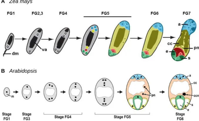
A Zea mays
B Arabidopsis
Figure 1.1 Megagametogenesis of Zea mays and Arabidopsis. Stages of megagametophyte development in (A) maize (image adapted from Evans and Grossniklaus, 2009) and (B) Arabidopsis (image taken from Drews and Koltunow, 2011) as detailed in the text, according to Christensen et al., 1998. Abbreviations: a = antipodal cells, cc = central cell, ccn = central cell nucleus, dm = degenerated megaspores, e = egg cell, m = megaspore, pn
= polar nuclei, s = synergid cell, va = vacuole.
5 1. 1. 2 Development of the male gametophyte
Successful seed formation requires a functional male gametophyte interacting with sporophyt- ic tissue and the female gametophyte during the fertilization process. During male gameto- phyte development, the pollen is formed by coordinated interaction with surrounding tissue, giving rise to haploid cells after meiosis. Maize is a valuable model for studying male game- togenesis and will represent the object of interest in the following paragraph. For review of pollen development in Arabidopsis see McCormick, 2004. In maize, each male flower within the tassels contains three stamen with four anther locules, in which the pollen develops (Vollbrecht and Schmidt, 2009). These developmental processes can be distinguished into three phases, first premeiotic development, followed by microsporogenesis and microgameto- genesis and finally postpollination events as the last phase (for review see Bedinger and Fowler, 2009). During premeiotic development, archesporial cells surrounded by a layer of primary parietal cells are formed, both originating from divided hypodermal cells (Figure 1.2 A). The archesporial cells further differentiate during microsporogenesis into pollen mother cells, also called microsporocytes. The primary parietal cell layer differentiates and gives rise to inner anther wall layers and the tapetum (Figure 1.2 B). Subsequently, the pollen mother cell undergoes meiosis to produce a tetrade of four haploid microspores, whereas the tapetal cells secretes substances for formation of the pollen coat as well as enzymes, like callase, to release the microspores from the tetrade (Figure 1.2 C) (Quilichini et al., 2015).
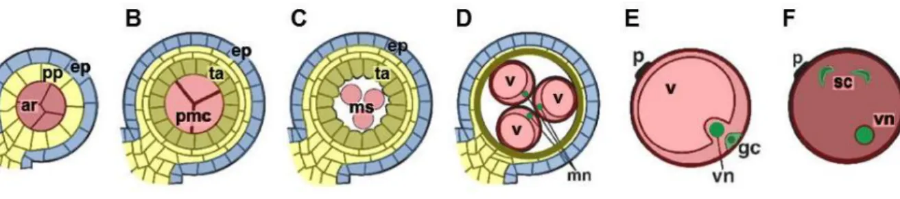
Figure 1.2 Development of the male gametophyte in maize. (A) Archesporial cells, embedded in a primary parietal cell layer, differentiate into (B) pollen mother cells, surrounded from the tapetum cells, which are de- rived from the primary parietal layer. (C, D) Pollen mother cells undergo meiosis to form enlarging microspores.
(E) Microspores undergo mitosis to produce a vegetative and a generative nucleus and cellularization gives rise to the generative cell. (F) Two sperm cells located in the cytoplasm of the vegetative cell are formed by mitosis of the generative cell. Abbreviations: ar = archesporial cells, ep = epidermis, gc = generative cell, mn = micro- spore nucleus, ms = microspores, pmc = pollen mother cells, pp = primary parietal layer, sc = sperm cells, ta = tapetum, v = vacuole, vn = vegetative nucleus. Image adapted from Bedinger and Fowler, 2009.
6
The enlarging microspores generate a vacuole and the surrounding sporophytic cell layers collapse. Tapetum cells undergo programmed cell-death (Solís et al., 2014) (Figure 1.2 D).
Asymmetric mitosis of the microspores marks the beginning of microgameteogenesis and produces a large transcriptionally active vegetative nucleus and a generative smaller nucleus with more compact chromatin. Cellularization around the generative nucleus leads to for- mation of the generative cell adjacent to the pollen wall, which later migrates into the cyto- plasm of the larger vegetative cell (Figure 1.2 E). A second mitosis event of the generative cell produces two sperm cells (Figure 1.2 F). The final steps of pollen maturation include starch accumulation, pollen wall maturation and dehydration, preparing the pollen to be re- leased out of the anthers after anthesis.
1. 2 Progamic phase
1. 2. 1 Growth of the pollen tube through sporophytic tissue
After the pollen is released from the anthers, it attaches to the silks of the female flower, rep- resenting the maize stigma. The vegetative cell germinates as pollen tube to transport the em- bedded sperm cells towards the FG. The journey of the maize pollen tube can be divided into different phases based on Lausser et al., 2010. During phase I, the pollen grains get attached on hairs covering the silk, hydrate and germinate (Figure 1.3 A). Growth of pollen tubes into the sporophytic silk tissue marks the beginning of phase II, in which the pollen tubes enter one of the two transmitting tracts inside of the silk to begin phase III of tube growth. Penetra- tion of the multicellular silk hairs by pollen tubes during phase II is thought to be necessary for directed growth of the pollen tube towards the basis of the silk during phase III (Booy et al., 1992; Lausser et al., 2010). Growth of pollen tubes through the transmitting tract causes degeneration of the silk abscission zone to prevent supernumerous pollen tubes reaching the female gametophyte (Heslop-Harrison et al., 1985). During phase IV, pollen tubes exit the transmitting tract and enter the ovular cavity to grow towards the embryo sac (Figure 1.3 B).
Finally, in phase V, the pollen tubes are directed to the micropylar region of the FG, which is covered with several cell layers of nucellus cells. This is contrary to the FG of Arabidopsis, which is covered by integuments, leaving a pore for penetration of the micropylar region with the pollen tube. In grasses, pollen tubes have to penetrate these cell layers to release the sperm cells for double fertilization, a unique feature of flowering plants. One sperm cell fuses with
7
the haploid egg cell to develop the embryo, whereas the second sperm cell fuses with the dip- loid central cell to arise the triploid endosperm.
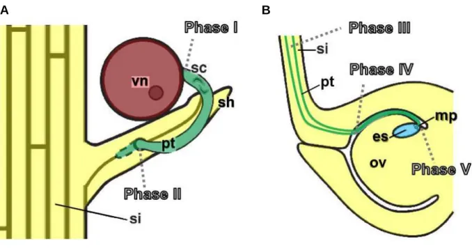
A B
Figure 1.3 Growth of pollen tube for fertilization of the female gametophyte in maize. In maize, the jour- ney of the pollen tube towards the female gametophyte can be distinguished into different phases (Lausser et al., 2010). (A) A pollen grain attaches to the silk hairs and germinates (phase I). In phase II, the pollen tube pene- trates the sporophytic tissue to enter the transmitting tract inside of the silk. (B) Pollen tubes pass through the transmitting tracts (phase III) and exit into the ovarial cavity in phase IV, to grow towards the female gameto- phyte. In phase V, one pollen tube enters the embryo sac to release the sperm cells for fertilization. Images adapted from Bedinger and Fowler, 2009.
1. 2. 2 Control of pollen tube growth by male-female communication
The growth of the pollen tube through the style towards the female gametophyte has to be controlled precisely, requiring communication between the pollen tube and the surrounding tissues. Several substances contributing to these communication processes were identified during the last years in different plant species (for review see Dresselhaus and Franklin-Tong, 2013; Higashiyama and Takeuchi, 2015). These factors are influencing pollen germination and growth rate as well as guidance of the growth direction mediated by sporophytic tissue or the female gametophyte, partially with overlapping functions. Germination of the pollen in Arabidopsis is stimulated for example by sulfinylated azadecalin, a substance isolated from pistil extract (Qin et al., 2011) as well as epibrassinolide, which also accelerates tube growth
8
(Vogler et al., 2014). With -aminobutyric acid (GABA), another substance was identified to promote pollen tube germination and growth. Additionally, GABA is involved in ovular guid- ance during the last steps of pollen tube growth (Palanivelu et al., 2003; Ling et al., 2013).
Growth of the pollen tube through the transmitting tract seems to be guided by an interplay of various factors with the tube cell, although also mechanical guidance of the tube growth was observed (Jauh et al., 1997; Higashiyama and Hamamura, 2008). An arabinogalactan protein named Transmitting-Tissue-Specific (TTS) from tobacco styles was reported to act as direc- tional cue (Cheung et al., 1995; Wu et al., 2000), just as several cysteine-rich peptides (CRPs) secreted by the sporophytic transmitting tract tissue like chemocyanin (Kim et al., 2003), plantacyanin (Dong et al., 2005) or the adhesine SCA, which was demonstrated to be in- volved in formation of the adhesive matrix necessary for pollen tube guidance through the transmitting tract (Mollet et al., 2000). As mentioned before, not only sporophytic tissue but also the female gametophyte contributes to the correct growth direction of the pollen tube, as functional female gametophytes are a prerequisite for the last steps of the pollen tube journey (Ray et al., 1997; Higashiyama et al., 2001). With ZmEA1, the first described secreted pep- tide with pollen tube attraction capability was isolated from maize (Márton et al., 2005).
ZmEA1 encodes a small 94 amino-acid propeptide which is secreted after passing the secreto- ry pathway from the egg apparatus (egg cell and synergids) towards the nucellus cells at the micropylar cone covering the female gametophyte. Down-regulation of ZmEA1 using the RNAi technique resulted in a loss of short-range guidance of the pollen tube, which failed to grow through the micropylar region (Márton et al., 2005). Later, pollen tube attracting pep- tides secreted from the synergids were also discovered in the dicotyledonous plants Torenia fournieri (TfLURE1-2; Okuda et al., 2009), Torenia concolor (TcCRP1; Kanaoka et al., 2011), Arabidopsis thaliana (AtLURE1.1 - 1.6; Takeuchi and Higashiyama, 2012) and Ara- bidopsis lyrata (AlLURE1.1 - 1.10; Takeuchi and Higashiyama, 2012). In contrast to ZmEA1, all of these attractants are defensin-like CRPs.
Communication between the pollen tube and the pistil is also required during the last steps of tube growth, the penetration of the female gametophyte to release the sperm cells. The pollen tube is perceived by the FG, its growth has to be arrested and tube burst has to be initiated.
These procedures require intensive cross-talk of both pollen tube and FG cells. As examples for female factors localized on the synergid surface, the small putative glucosylphsophatidyl- inositol-anchored protein LORELEI (LRE) (Capron et al., 2008) and the receptor-like kinase (RLK) FERONIA (FER), containing a putative extracellular carbohydrate-binding malectin-
9
like domain (Lindner et al., 2012), are necessary for pollen tube growth arrest (Escobar- Restrepo et al., 2007). Mutants lacking these proteins display overgrowing pollen tubes that fail to burst. Regarding the male side, the FER-related pollen tube derived RLKs ANXUR1 (ANX1) and (ANX2) seem to be involved in maintaining pollen tube integrity until it reaches the FG (Boisson-Dernier et al., 2009). To release the sperm cells for fertilization, the defen- sin-like cysteine-rich peptide ZmES4 from maize was identified to induce pollen tube burst. It is accumulating in the synergids and gets discharged upon pollen tube arrival (Amien et al., 2010).
To summarize, there are many key players involved in pollen-pistil communication to guide the pollen tube towards the FG. Many of them, like above described pollen tube attractants and ZmES4 (Amien et al., 2010), are thought to act in a species-preferential manner and pro- vide reproductive barriers between different plant species. In this paragraph, only a selection of them was described. Further factors mediating successful sperm cell delivery to the FG will be discussed in CHAPTER 6.
1. 3 Embryogenesis in model flowering plants
1. 3. 1 Developmental steps of embryo formation
Following fertilization, the basic plant body is formed during embryogenesis by establishing polarity, differentiation of primary tissue and accumulation of storage nutrients. As embryo development marks the first steps after changing from gametophytic to sporophytic phase of the plant life cycle, these processes are extensively studied. In general, the detailed pattern of embryo body formation is species-specific. The following paragraph will focus on morpho- logically described stages of embryogenesis of maize and will point out differences to the dicotyledonous model plant Arabidopsis (Figure 1.4) (for review see Vernoud et al., 2005 and Lau et al., 2012).
The zygote of maize divides asymmetrically into a small apical and a large basal cell giving rise to the proembryo. The proembryo consist of small cytoplasm-rich cells of the embryo proper and larger vacuolized cells forming the suspensor (Randolph, 1936). During the transi- tion stage, the protoderm is formed as external cell layer surrounding the embryo proper. The embryo proper cells representing the future scutellum enlarge during the following coleoptilar stage, whereas a group of cells facing towards the ear stays densely packed. At this position, formation of the shoot apical meristem (SAM) is initiated close to a protuberance marking the
10
future coleoptile. Additionally, the root apical meristem (RAM) is formed at a more basal position. Depending on genotype and environmental conditions, the coleoptilar stage lasts until 10 to 15 days after pollination. In contrast to Arabidopsis, in which the SAM does not generate leaf primordia before germination, the SAM of the maize embryo initiates the for- mation of 5-6 leaves, with the number of leaf primordia naming the developmental stage of the embryo (stage 1-6) (Nardmann and Werr, 2009). To protect the leaf primordia, they are covered by the coleoptile. During these stages, the vascular system is established in the em- bryo. With proceeding maturation, the embryo grows further, the suspensor degenerates and storage nutrients like lipids are accumulated (Giuliani et al., 2002).
Figure 1.4 Morphological stages of embryogenesis of Arabidopsis and Zea mays. The different embryo stag- es are indicated. (A) In Arabidopsis, asymmetric cell divisions of the zygote give rise to the embryo proper and suspensor, followed by protoderm formation in the dermatogen stage. After the globular stage, meristems are formed during the heart stage and a vascular system starts to develop during the torpedo stage. (B) Like in Ara- bidopsis, the maize zygote divides asymmetrically to form embryo proper and suspensor, respectively. The tran- sition stage is characterized by formation of the protoderm. At the coleoptilar stage, meristems start to develop and formation of the coleoptile is initiated. Up to six leaf primordia are initiated during the following stages 1-6 and a vascular system develops. The timeline marks approximate days after pollination (DAP). Images taken from Vernoud et al., 2005.
11
During early stages of embryogenesis, maize and Arbidopsis share some general features like the establishment of apical-basal axis polarity to form a proembryo consisting of embryo proper and suspensor formed after asymmetric zygote division, protoderm formation, organi- zation of the root and shoot meristem as well as accumulation of reserve substances (Vernoud et al., 2005). Nevertheless, there are some differences in embryo development of both plant species besides the aforementioned earlier activity of the maize SAM, embryo size and cell number. Cell divisions in Arabidopsis are stronger synchronized leading to a more geometric and stereotypic histological appearance than the maize embryo, as depicted in Figure 1.4. Ad- ditionally, SAM and RAM are arranged along the apical-basal axis by contrast to the oblique arrangement in maize.
1. 3. 2 Peptide signaling during embryogenesis
Several communication events have to take place to orchestrate embryogenesis. Besides plant hormones (Chen et al., 2014), secreted peptides were demonstrated to act as signaling com- ponents during embryo and endosperm development (for review see Ingram and Gutierrez- Marcos, 2015). In Arabidopsis, the cysteine-rich peptides of the EMBRYO SURROUNDING FACTOR 1 (ESF1) family are expressed in the central cell and endosperm cells of the em- bryo surrounding region and are involved in formation of the zygotic basal cell lineage (Costa et al., 2014). Furthermore, they act in non-cell autonomous signaling by promoting suspensor elongation in the signaling pathway of the receptor-like cytoplasmic kinase SHORT SUS- PENSOR (SSP) and YODA (YDA) mitogen-activated protein kinase (Bayer et al., 2009).
Another class of peptides controlling the embryo suspensor is the embryo-expressed KISS OF DEATH (KOD) peptide, which positively regulates controlled cell-death (Blanvillain et al., 2011). In maize, MATERNAL EXPRESSED GENE 1 (MEG1) peptides are involved in de- velopment of transfer cells of the basal endosperm transfer layer, mediating nutrient transloca- tion in seeds (Costa et al., 2012). Another signaling peptide involved in seed development is CLE8 from the CLE (CLAVATA3/ENDOSPERM SURROUNDING REGION) protein fami- ly, which can be found in young embryos and endosperm. Together with the transcription factor WUSCHEL-like homeobox 8 (WOX8), it acts by regulating the basal embryo cell divi- sion patterning, endosperm proliferation and endosperm differentiation (Fiume and Fletcher, 2012).
Like in many plant signaling pathways, receptors for most of the signaling peptides are not yet identified. Nevertheless, some putative receptors potentially representing interaction part- ners of secreted peptides were already demonstrated to be involved in signaling processes
12
during seed development, like the leucine-rich repeat (LRR) receptor protein kinase HAIKU2 (IKU2). Mutations of IKU2 result in reduced growth of the early endosperm (Luo et al., 2005). The LRR receptor protein kinases GASSHO1 (GSO1) and GASSHO2 (GSO2), locat- ed within the developing embryo, ensure formation of a functional embryonic cuticle in Ara- bidopsis (Tsuwamoto et al., 2008).
1. 4 Aims of this work
The aim of the following work was to examine peptide signaling during sexual reproduction of Zea mays by studying the EAL-like (EAL) gene family in more detail. The small peptide ZmEA1, secreted from the maize egg apparatus for short range growth guidance of the pollen towards the female gametophyte, was previously reported to be member of a small peptide family exclusively found in grasses (Dresselhaus et al., 2011). A detailed phylogenetic classi- fication of peptides related to ZmEA1 as well as investigations on yet uncharacterized family members should be performed and a manuscript was recently submitted for publication (Uebler et al., under review). These data are now shown in CHAPTER 2. Another task was to study the ability of ZmEA1 to overcome species-specific hybridization barriers by transgenic expression in Arabidopsis thaliana. These data are published in Márton et al., 2012, and my results of experiments contributing to this work are given in CHAPTER 3. Based on this work, the capability of ZmEA1 to directly bind the attracted pollen tubes should be tested and could be visualized in a species-specific manner as described in CHAPTER 4 and published in Uebler et al., 2013. Binding of ZmEA1 as ligand on the pollen tube surface requires interaction with surface located pollen tube protein(s) for initiating change in growth direction. As identifica- tion of membranous ligand binding partners is a highly challenging field of research, an over- view and discussion of current methods to isolate surface receptor proteins is given in CHAP- TER 5 (Uebler and Dresselhaus, 2014). Various methods to identify ZmEA1 receptors and interaction partners by biochemical approaches should be tested and applied. This work is described in CHAPTER 6 and includes the expression and purification of recombinant ZmEA1 protein in a variety of systems as well as isolation of potential ZmEA1 binding candidates.
Some candidates are further characterized regarding their classification, expression pattern, subcellular localization and protein level. Finally, a comprehensive conclusion is given in CHAPTER 7, which also provides an outlook for future research.
13
CHAPTER 2 - CLASSIFICATION OF EA1-BOX PROTEINS AND NEW IN- SIGHTS INTO THEIR ROLE DURING REPRODUCTION IN GRASSES
This CHAPTER is based on the manuscript Uebler et al., under review in Plant Reproduction, excluding the paragraphs about generation and analysis of transgenic maize lines and promot- er analysis. S. Uebler conducted bioinformatical analysis and performed all experimental pro- cedures except RT-PCR of rice genes and generation of constructs for transient transfor- mation of ZmEA1, AtEAG1, OsEAL1 and OsEAL3. The manuscript was written by S. Uebler and edited by T. Dresselhaus.
2. 1 Introduction
Peptide signaling plays an elementary role during various vegetative and reproductive pro- cesses in plants, including development of vegetative organs, fertilization and embryogenesis (Murphy et al., 2012; Costa et al., 2014; Qu et al., 2015; Ingram and Gutierrez-Marcos, 2015). During reproduction, the secretion and perception of peptides enables plants, for ex- ample, to distinguish self from non-self pollen, to support pollen tube growth and to mediate successful fertilization. The identification and characterization of signaling components is therefore of great importance to unveil the underlying communication mechanisms. Several thousand of potentially secreted peptides are predicted to exist in Arabidopsis (Lease and Walker, 2006), and many of them are indicated to play essential roles in reproductive pro- cesses (Huang et al., 2015). However, until now only a few of them could be successfully associated with signaling processes. Many signaling peptides can be grouped into protein families with shared conserved features. Based on single or several well-characterized found- ing peptides, sequence homology searches offer a possibility to identify more members of these families (Olsen et al., 2002; Lease and Walker, 2010). Classifications of yet unknown peptides into protein families might give further hints about their role in cellular processes.
One of the largest protein families of secreted peptides is formed by CLE (CLAVA- TA3/ENDOSPERM SURROUNDING REGION) proteins (Miyawaki et al., 2013). Members of this protein family are supposed to function as processed 12- or 13- amino acid peptides derived of larger precursor proteins containing the conserved CLE-motive (Ito et al., 2006;
Kondo et al., 2006; Ohyama et al., 2009). They are differentially expressed during plant de- velopment and are involved in regulation of the meristem activity in shoots (Fletcher et al.,
14
1999) and roots (Stahl et al., 2009) as well as in development of vascular tissues (Hirakawa et al., 2008). Originally, this protein family was assumed to consist of 65 members in both mon- ocotyledonous and dicotyledonous plants (Cock and McCormick, 2001; Oelkers et al., 2008).
Based on similarity to the CLE domain, 114 new members were identified in a variety of plants and could be classified into 13 groups, thus nearly tripling the number of previously known CLE proteins (Oelkers et al., 2008). A similar case can be depicted regarding the CEP (C-TERMINALLY ENCODED PEPTIDE) proteins, which are supposed to act as negative regulators that mediate environmental influences on root and shoot development in seed plants (Delay et al., 2013) and which were originally identified by an in silico approach (Ohyama et al., 2008). Additional bioinformatic analyses based on the highly conserved do- main in the predicted mature peptides of the CEP family tripled the number of classified CEP peptides in Arabidopsis (Delay et al., 2013; Roberts et al., 2013).
A protein family with members that were demonstrated to act as signaling peptides in repro- duction was named as EAL (EA1-like) protein family (Dresselhaus et al., 2011). The first identified and name-giving member was ZmEA1 (Zea mays Egg Apparatus 1) of Zea mays, which represents until now the only identified secreted attractant for pollen tube guidance in grasses. ZmEA1 is produced in the egg apparatus of maize and is secreted towards the micro- pylar region of the ovule. There it acts in short-range attraction of maize pollen tubes towards the female gametophyte (Márton et al., 2005; Márton et al. 2012) and binds in a very specific manner at the surface of growing maize pollen tube tips (Márton et al., 2012; Uebler et al., 2013). Another EAL protein with demonstrated activity in signaling is ZmEAL1 (Zea mays EA1-like 1) of Zea mays. Fused to GFP, ZmEAL1 was shown to accumulate in granules at the chalazal pole of the egg cell and is likely secreted towards the chalazal pole of the female gametophyte. RNAi-studies showed that ZmEAL1 is required to suppress gametic cell fate of antipodal cells (Krohn et al., 2012).
As EA1-box containing proteins are widely distributed throughout the plant kingdom, EA1 orthologs were wrongly predicted and EALs from other species misclassified. We therefore introduce here a new classification of EA1-box proteins. We further demonstrate that all ana- lyzed EAL peptides and larger EAC proteins enter the secretory pathway, whereas EAGs lo- calize to the cytoplasm and nucleus. ZmEAL2, the third EAL peptide in Zea mays was stud- ied in more detail. Although knock-down plants did not show a significant phenotype, our studies indicate that the peptide is involved in late embryogenic development where it is se-
15
creted to the extracellular space. Additionally, we introduce EALs of rice representing poten- tial functional orthologues to ZmEA1.
2. 2 Experimental procedures
2. 2. 1 Bioinformatic analysis
Protein sequences of EAL, EAC and EAG proteins were identified by BLASTP searches us- ing the EA1-box of ZmEA1 against the genomes of Zea mays, Sorghum bicolor, Oryza sativa ssp. japonica, Brachypodium distachyon, Arabidopsis thaliana, Arabidopsis lyrata, Glycine max and Populus trichocarpa, available at www.gramene.de (Monaco et al., 2014) and against protein sequences of the aforementioned plant species available at http://www.ncbi.nlm.nih.gov/. EA1-boxes of identified proteins were used for further BLASTP searches against the above plant species. A list of all identified genes including chromosomal location is attached (Supplement table 1). Protein sequence alignments and phy- logenetic tree were created using ClustalX 2.1 (Larkin et al., 2007). The phylogenetic tree was visualized by FigTree v1.4.2 (http://tree.bio.ed.ac.uk/software/figtree/) and protein alignments manually edited with GeneDoc V2.7.000 (Nicholas et al., 1997). Prediction of classical N-terminal signal peptides was performed using SignalP 4.1 (Petersen et al., 2011;
http://www.cbs.dtu.dk/services/SignalP/). Proteins entering the non-classical secretory path- way were predicted by SecretomeP 2.0 (Bendtsen et al., 2004, http://www.cbs.dtu.dk/services/SecretomeP/).
2. 2. 2 Generation of constructs for transformation of BMS cells
To generate constructs for transient transformation of BMS cells, the open reading frames (ORFs) of ZmEA1, sEA1, AtEAG1, OsEAL1 and OsEAL3 were amplified using the primer pairs EAF-GFP/EAR-GFP (ZmEA1), oSU26/oSU27 (sEA1), 3f/3r (AtEAG1), 2f/2r (OsEAL1) and 1f/1r (OsEAL3) and digested with SpeI/BamHI for ligation into the vector pLNU-GFP (DNA Cloning Service, Hamburg, Germany). The ORFs of ZmEAL2, ZmEAC1 and ZmEAC2 were amplified using the primer pairs oSU17/oSU19 (ZmEAL2), oSU38/oSU39 (ZmEAC1) and oSU40/oSU41 (ZmEAC2) for directional TOPO® cloning into the pENTR™/D-TOPO®
vector (Thermo Fisher Scientific Inc., Waltham, USA) followed by transfer of the ORFs into the destination vector pB7FWG2,0 (Karimi et al., 2002) using Gateway® LR clonase® II enzyme mix (Thermo Fisher Scientific Inc., Waltham, USA). The plasmid PMON30049
16
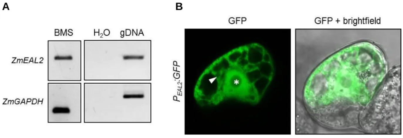
(Pang et al., 1996) was used as control for cytoplasmic GFP. To analyze ZmEAL2 promoter activity in BMS suspension cells, a fragment containing the putative ZmEAL2 promoter and ORF of GFP was cut out from the plasmid p7U-EAL2P-GFP described in 2. 2. 8. 1 using SfiI restriction sites. The fragment was cloned into the pLNU-GFP plasmid (DNA Cloning Ser- vice, Hamburg, Germany), after removing its GFP cassette with SfiI restriction sites, to gen- erate the plasmid pLG-EAL2P-GFP.
2. 2. 3 Transient transformation of maize BMS suspension cells
Black Mexican Sweet (BMS) maize suspension cells (Sheridan, 1982) were cultivated in liq- uid MS media (30 g/l sucrose, 4.4 g/l MS-salts [Duchefa Biochemie B.V, Haarlem, Nether- lands], 2 mg/l 2,4-Dichlorophenoxyacetic acid, pH 5.8, based on Murashige and Skoog, 1962). For transient transformation via microprojectile bombardement after Klein et al., 1987 and Klein et al., 1988, 2-4 ml of growing culture were harvested and transferred as a thin lay- er onto solid MS media (liquid MS media solidified with 0.3% Gelrite). Plates were incubated for 2 hours at 26°C. Gold particles (0.6 µM diameter) were washed and dissolved in ethanol (p.a.). 10 µg plasmid DNA (concentration 0.5-1 µg/µl) was precipitated on gold particles by adding 50 µl CaCl2 (2.5 M) and 20 µl spermidine (0.1 M) followed by incubation on ice. Gold particles were washed with absolute ethanol and finally resuspended in 150 µl ethanol (p.a.).
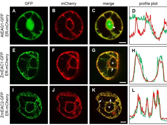
7.5 µl of the suspension was transferred on a macrocarrier for bombardment using the particle delivery system PDS1000/He (BioRad Laboratories, Inc., Hercules, USA) at a vacuum of 28 inch Hg, 1,100 psi rupture discs and a distance of 6 cm. All constructs were co-transformed with a marker for the endoplasmic reticulum (ER) (Nelson et al., 2007), composed of the sig- nal peptide of AtWAK2 at the N-terminus of mCherry and an ER-retention signal at its C- terminus (Gomord et al., 1997). The plate was incubated overnight at 26°C in the dark. Af- terwards, cells were transferred into liquid MS media and shaken at 26°C for at least 4 h in the dark. Microscopic analysis was performed using an Axiovert 200M inverted microscope (Carl Zeiss AG, Oberkochen, Germany), equipped with the confocal laser scanning unit LSM 510 META with GFP-excitation at 488 nm and detection using the BP 505-550 filter. Images were processed using the software ImageJ 1.43q (http://imagej.nih.gov/ij/).
2. 2. 4 Extraction of total RNA and generation of cDNA
Total RNA of various plant tissues was extracted as described by Logemann et al., 1987. To generate cDNA, 1 µg total RNA was digested with DNase I (Thermo Fisher Scientific Inc., Waltham, USA) to remove DNA contaminations. cDNA was synthesized by using Super-
17
Script® III Reverse Transcriptase (Thermo Fisher Scientific Inc., Waltham, USA) and Oli- go(dT)18 primers (Thermo Fisher Scientific Inc., Waltham, USA) according to the manufac- turer’s protocol.
2. 2. 5 Semi-quantitative reverse transcriptase-PCR (RT-PCR) and quantitative real–time PCR (qRT-PCR)
For semi-quantitative RT-PCR, rice and maize tissues were isolated and grounded in liquid nitrogen. Approximately 150 mg of the sample powder was used for RNA extraction and cDNA generation. RT-PCR was performed using the primer pairs OsP0493C06- fwd/OsP0493C06-rev (OsEAL1), OsEAL2a-fwd/OsEAL2b-rev (OsEAL2), OsEAL3-a(fwd- new)/OsEAL3-b(rev-new) (OsEAL3), oSU99/oSU100 (ZmEAL2), and GAPnew1/GAPnew2 (ZmGAPDH, Krohn et al., 2012). Standard PCR reactions were performed applying 1 µl of the cDNA as template and primer concentrations of 0.25 µM for each primer.
For qRT-PCR experiments, 20 to 30 maize embryos were dissected from pollinated maize cobs immediately after removing the cob from the plant. Embryo samples were collected and grounded in liquid nitrogen. Approximately 150 mg of the powder was used for RNA extrac- tion and cDNA generation. The cDNA was diluted 1:10 and directly used as template for qRT-PCR reactions with KAPA SYBR FAST Master Mix Kit (VWR International GmbH, Erlangen, Germany). For each sample, three technical replicate reactions were performed.
Primer pair oSU99/oSU100 was used to quantify the ZmEAL2 transcript level. For analysis of the reference genes, the primer pairs LUGfwd/LUGrev (LUG) and CULfwd/CULrev (CUL) (both derived from Manoli et al., 2012) were used. Final concentration of all primers was 200 nM. All reactions using the Realplex 2 cycler (Eppendorf, Hamburg, Germany) were per- formed with 20 s at 54°C for elongation and 10 s at 72°C for synthesis. Data were statistically processed by calculating the 2Ct value with normalization against LUG or CUL, respectively.
2. 2. 6 Immunoblot analysis and generation of anti-ZmEAL2 antiserum
Proteins smaller than 15 kDa were separated by SDS-PAGE using Tris/Tricine buffer system (Schägger and Jagow, 1987). The Tris/Glycine buffer system (Laemmli, 1970) was used for proteins with a higher molecular weight. Following SDS-PAGE, proteins were transferred for 35 minutes at 300 mAmp onto a nitrocellulose membrane using a tank blotting system. The membrane was blocked in blocking solution (5% milk powder, 50 mM Tris-HCl pH 7.5, 150 mM NaCl, 0.1% Tween-20). The 11 amino acid peptide NH2-CFLAKKELYFK-CONH2 spe- cific to ZmEAL2 (partially covering the EA1-box with an additional cysteine at the N-
18
terminus for coupling with a protein carrier) was commercially synthesized (Centic Biotec, Heidelberg, Germany) and used for immunization of rabbits (Pineda Antibody-Service, Ber- lin, Germany). Unpurified polyclonal anti-ZmEAL2-antiserum was diluted 1:1000 in block- ing solution for overnight incubation of the membrane at 4°C. Afterwards, the membrane was washed in washing buffer (50 mM Tris-HCl pH 7.5, 150 mM NaCl, 0.1% Tween-20) and subsequently incubated with anti-rabbit IgG antibody from goat conjugated with horse red- dish peroxidase (Sigma-Aldrich, St. Louis, USA) diluted 1:5000 in blocking buffer for 2 hours at room temperature. Commercially available H3-directed antibody was used as loading control. After washing, the membrane was incubated in luminol-based substrate (PJK GmbH, Kleinblittersdorf, Germany). Chemiluminescence was detected using Super RX X-ray films (Fujifilm, Minato, Japan). Estimation of the apparent molecular weight of detected proteins was carried out by comparison to commercially available molecular weight standards.
2. 2. 7 Immunohistochemical detection of ZmEAL2
Immunohistochemical experiments were performed based on a protocol by Stadler and Sauer, 1996, with adaption of incubation times. Embryos 18 DAP and 25 DAP were isolated and manually cut into 2-4 cross sections. Fixation was performed for 6-8 h. After fixation, the samples were washed and placed in 70% ethanol (v/v) containing 1 mM DTT for 2 days.
Samples were dehydrated for 1 day in increasing ethanol concentrations. For infiltration with methacrylate, samples were incubated for at least 1 day per step. Sections of 5-10 µm were prepared with a Reichert Om U2 microtome (Leica, Wetzlar, Germany). Unpurified polyclo- nal ZmEAL2-directed antibody from rabbit was diluted 1:50 for overnight incubation of sec- tions, followed by incubation for at least 2 h with anti-rabbit IgG antibody conjugated with Cy2 from goat (Dianova GmbH, Hamburg, Germany) diluted 1:80. Samples were analyzed microscopically at the Axioskop FL epifluorescence microscope (Carl Zeiss AG, Jena, Ger- many) using Zeiss filter set no. 46 and at the Eclipse TE2000-S inverted microscope (Nikon Corporation, Tokyo, Japan) using a HC Alexa 488/eGFP filter. Images were processed using the software ImageJ 1.43q (http://imagej.nih.gov/ij/).
2. 2. 8 Generation of stable transgenic maize lines
2. 2. 8. 1 Constructs for Agrobacterium-mediated transformation of maize
For analysis of ZmEAL2 promoter activity in maize, the putative promoter fragment described in 2. 3. 4 was amplified using primer pairs oSU44/oSU46 and cloned into p7U-GFP (DNA
19
Cloning Service, Hamburg, Germany) using SpeI and HindIII restriction sites. To generate the construct for ZmEAL2 expression under control of its endogenous promoter, the putative promoter fragment and the ORF of ZmEAL2 was amplified with the primer pair oSU44/oSU47 and ligated into p7U-GFP using SpeI and HindIII restriction sites. Additional- ly, two RNAi constructs were generated. For the first RNAi construct, a 219 bp fragment in- cluding the 5’-UTR and the N-terminus of the ORF of ZmEAL2 was amplified with the pri- mer pair oSU48/oSU49 and cloned in antisense direction into the pUBI-IF2 vector (DNA Cloning Service, Hamburg, Germany) using EcoRI and SbfI restriction sites. The same frag- ment was amplified with the primer pair oSU54/oSU55 and cloned in sense-direction into the pUBI-IF2 vector with the antisense fragment by using HindIII and MluI restriction sites. A fragment containing the UBI promoter and the RNAi cassette was cut out of the vector using SfiI restriction sites and transferred into the vector p7U (DNA Cloning Service, Hamburg, Germany) to generate the plasmid p7U-RNAi1(EAL2). For the second RNAi construct, a 208 bp fragment including the 3’-UTR and the C-terminus of the ZmEAL2 ORF was amplified with the primer pair oSU50/oSU51 and cloned in antisense direction into the pUBI-IF2 vector using EcoRI and SbfI restriction sites. The same fragment was amplified with the primer pair oSU56/oSU57 and cloned in sense-direction into the pUBI-IF2 vector with the antisense fragment by using HindIII and MluI restriction sites. A fragment containing the UBI promoter and the RNAi cassette was cut out of the vector using SfiI restriction sites and transferred into the vector p7U to generate the plasmid p7U-RNAi2(EAL2).
2. 2. 8. 2 Agrobacterium-mediated transformation of maize
Stable transgenic maize lines were generated based on the protocol of Frame et al., 2002. Me- dia were prepared as described for HiII transformation in Frame et al., 2011. HiIIA and HiIIB plants were cross-pollinated and harvested 11-13 DAP for transformation. Agrobacterium tumefaciens strain LBA4404 (background TiAch5, Ti plasmid: pAL4404; Hoekema et al., 1983) was transformed with binary plasmids like described in 6. 2. 2. 30 ml overnight culture supplemented with antibiotics was grown at 28°C until OD600 = 0.4, washed with 15 ml 10 mM MgSO4 and resuspended to OD600 = 0.5 in infection medium (1x infection medium with 100 µM acetosyringone; 2 x infection medium contains per liter: 200 ml 10x N6 macro salts [contains per 1 l: 4.63 g (NH4)2SO4, 28.3 g KNO3, 4 g KH2PO4, 1.85 g MgSO4 * 7 H2O, 1.66 g CaCl2 * 2 H2O], 2 ml 1000x N6 micro salts [contains per 100 ml: 387 mg MnSO4 * H2O, 150 mg ZnSO4 * 7 H2O, 160 mg H3BO3, 80 mg KI], 2 ml 1000x N6 vitamins [contains per 100 ml: 200 mg glycine, 100 mg thiamine-HCl, 50 mg pyridoxine-HCl, 50 mg niacin], 4 ml
20
NaFe-EDTA (50 mM), 1.4 g L-proline, 136.8 g sucrose, 72 g glucose, pH 5.2). Harvesting steps were performed at room temperature. Immature embryos at size of approximately 1.5 mm were dissected under sterile conditions and washed twice with infection medium. Embry- os were transferred into Agrobacterium suspension containing 15 µl/ml Silwet® L-77 and incubated for 5 min at RT. The embryos were distributed on co-cultivation medium plates (1 x co-cultivation medium [2 x co-cultivation medium contains per 1 l: 200 ml 10x N6 macro salts, 2 ml 1,000x N6 micro salts, 2 ml 1000x N6 vitamins, 4 ml NaFe-EDTA (50 mM), 1.4 g L-proline, 60 g sucrose, pH 5.8 ], 5 µM AgNO3, 100 μM acetosyringone, 300 mg/l L- cysteine, 1.5 mg/l 2,4-D, 0,3% Gelrite®), excess Agrobacterium suspension was removed and the plates were incubated overnight at 21°C in the dark. The day after, embryos were placed onto new co-cultivation plates with reverse orientation and incubated overnight at 21°C in the dark. Subsequently, the embryos were transferred onto resting medium plates (1 x resting me- dium [2 x resting medium was composed like co-cultivation medium supplemented with 1 g 2-(4-morpholino)-ethanesulfonic acid (MES)], 5 μM AgNO3, 100 mg/l cefotaxime, 100 mg/l vancomycin, 1.5 mg/l 2,4-D, 0.3% Gelrite®) with scutellum side upturned for incubation at 28°C in the dark. After 7 days, the embryos were placed onto selection medium I plates (1 x resting medium, 5 μM AgNO3, 100 mg/l cefotaxime, 100 mg/l vancomycin, 1.5 mg/L glufosinate, 0.3% Gelrite®) and incubated for 14 days at 28°C in the dark, followed by two times incubation for 14 days at the same conditions on selection media II plates (like selection medium I with doubled concentration of glufosinate) to induce embryogenic type II callus formation. Emerging coleoptiles were removed routinely by dissection. For regeneration of the plants, the embryos with callus were placed onto regeneration I medium plates (1 x regen- eration medium I, 100 mg/l cefotaxime, 3 mg/ml glufosinate, 0.3% Gelrite®; 2 x regeneration medium I contains per 1 l: 200 ml 10x MS macro salts (contains per 1 l: 16.5 g NH4NO3, 19 g KNO3, 1.7 g KH2PO4, 3.7 g MgSO4 * 7 H2O, 4.4 g CaCl2 * 2 H2O), 2 ml 1000x MS micro salts (contains per 100 ml: 1.69 g MnSO4 * H2O, 860 mg ZnSO4 * 7 H2O, 620 mg H3BO3, 83 mg KI, 25 mg Na2MoO4 * 2 H2O, 2.5 mg CoCl2 * 6 H2O, 2.5 mg CuSO4 * 5 H2O), 2 ml 1000x MS vitamins (contains per 100 ml: 200 mg glycine, 10 mg thiamine-HCl, 50 mg pyri- doxine-HCl, 50 mg niacin), 4 ml NaFe-EDTA (50 mM), 4 ml myo-inositol (50 mg/ml), 120 g sucrose, pH 5.8) for 21 days at 28°C in the dark. Afterwards, they were transferred onto re- generation II medium plates (like regeneration I medium, but without cefotaxime and with half of sugar concentration) for incubation at 28°C in a light chamber with a photoperiod of 16 hours per day. For analysis of regenerated maize plants, genomic DNA was extracted like described in Pallotta et al., 2000 and tested for insertion of the construct by standard PCR (6.
21
2. 3. 1). Positive plants were grown at greenhouse conditions (6. 2. 1. 1) and either self- pollinated or used for cross-pollination with wild type plants. Seeds were harvested at maturi- ty and analysis of the transgenic maize lines was performed with plants derived from this seed material.
2. 3 Results and discussion
2. 3. 1 EA1-box containing proteins can be separated into three classes
First experimental examinations of the EA1-like protein family were performed about one decade ago, showing that genes homologous to ZmEA1 exist also in other cereals (Márton et al., 2005). The genes encoding secreted peptides were therefore named as EA1-like (EALs).
Later, all proteins containing the conserved EA1-domain were classified as EALs (Gray- Mitsumune and Matton, 2006), disregarding the fact that many of these proteins are likely not secreted and contain a different overall structure. With respect to the increasing quality of genome-wide sequencing efforts and the appearance of new EA1-box proteins in maize (Chettoor et al., 2014), we now suggest to reclassify EA1-box containing proteins. These pro- teins were identified by using the sequence of the EA1-box of ZmEA1 (Dresselhaus et al., 2011) as query for BLASTP searches against the genomes of the monocotyledonous species Zea mays, Sorghum bicolor, Oryza sativa japonica and Brachypodium distachyon as well as the dicotyledonous plants Arabidopsis thaliana, Arabidopsis lyrata, Glycine max and Populus trichocarpa. BLASTP searches were performed on two platforms, the Gramene Database (http://www.gramene.org/) and the National Center for Biotechnology Information (http://www.ncbi.nlm.nih.gov/). Proteins containing the EA1-box were found in all of the analyzed monocotyledonous and dicotyledonous plants. By further classification, these pro- teins were clustered into three families: EAL, EAG (EA1-box glycine-rich) and EAC (EA1- box containing) (Figure 2.1 A-C). Protein sequences of the three classes were aligned to visu- alize conserved motives and protein domains.
Class 1 consists of EAL peptide precursors with a length of approximately 100 amino acids (Figure 2.1 A). All EAL proteins exhibit either a “classical” N-terminal signal peptide or an N-terminally located internal signal peptide domain preceding the conserved EA1-domain.
The C-terminal region contains an alanine-rich domain (A-box). The predicted N-terminal cleavage sites are located shortly downstream of a conserved motive that was named as P- box, based on a highly conserved proline at its last position. Cleavage of an EAL peptide pre-
22
cursor behind the P-box creates a peptide of about 50 amino acids starting with a variable sequence of 3-7 amino acids followed by the highly conserved EA1-box and the A-box. GFP- fusion protein experiments previously indicated that EAL peptides are not cleaved at their C- termini (Márton et al., 2005; Krohn et al., 2012; Márton et al., 2012). The only identified EAL protein with a different structure is BdEAL5 with a predicted length of 197 amino acids.
The first half of BdEAL5 exhibits high similarity with other EALs, while the second half after the A-box encodes for a different protein. As this region is also annotated as a second exon in the Gramene database, it might have been wrongly annotated or may represent a recent gene fusion event.
The second class of EA1-box proteins was named as EAG proteins and was found to be pre- sent in all analyzed species except Populus trichocarpa (Figure 2.1 B). All EAGs are short proteins with amino acid sequences ranging between 70 and 120 AS. Besides the highly con- served EA1-box at their C-termini, they possess a glycine content varying from 35.6% to 59.5%, spanning the whole protein outside the EA1-box on both sides. The C-termini of EAGs are very short with a length of maximum 8 amino acids behind the EA1-box and usual- ly end with a lysine. EAGs lack both A- and P-boxes as well as a classical signal peptide do- main for targeting to the secretory pathway. Proteins with a high content of glycine have been identified from several eukaryotic species. In insects, for example, they are known to act as antimicrobial peptides. As they are usually directed against fungi, gram-negative bacteria and cancer cells, they play an important role in immunity (Herbinière et al., 2005; Dang et al., 2009). Like insects, plants generate glycine-rich proteins involved in defense mechanisms against bacteria and fungi, but also in a wide variety of other independent physiological pro- cesses (Park et al., 2000; Bocca et al., 2005). Glycine-rich proteins (GRPs) can be grouped into different classes based on the nature of the glycine-repeats in combination with additional motives, their expression pattern, their modulation by external factors and their subcellular localization (for review see Mangeon et al., 2010). GRPs of several plants including Ara- bidopsis thaliana act as secreted signaling peptides involved, for example, in pollen hydration or as extracellular ligands in signal transduction processes (Mayfield and Preuss, 2000; Park et al., 2001) or act as cell wall constituents with different functions (Ryser et al., 2004; Ueki and Citovsky, 2005). However, considering that secreted and extracellular proteins usually contain an N-terminal signal peptide, which is lacking in all EAGs, it is unlikely that they are involved in extracellular signaling processes. Cytoplasmic GRPs were demonstrated to act, for example, as RNA-binding proteins involved in flowering time control and stress responses (Kim et al., 2007; Streitner et al., 2008). For some of the GRPs, additional domains, like