Neovascular glaucoma in a child: an unusual presentation of medulloepithelioma
Abstract
A healthy 12 month old infant without significant medical history presented with left eye redness for one week. Ophthalmic examination
Enoch Kassa
1Helen Li
1showed elevated intraocular pressure with iris neovascularization in
Yang Sun
1the affected eye with increased optic nerve cupping. Scleral depression revealed a ciliary body mass in the supratemporal quadrant. A large,
non-pigmented, vascular mass was noted; biopsy results showed mul- 1 Glick Eye Institute, Indianapolis, USA tilayered cords, tubules, and sheets resembling primitive medullary
epithelium arising from the ciliary body. The patient was diagnosed with medulloepithelioma. The patient underwent enucleation of the affected eye. Medulloepithelioma is a rare but important cause of neovascular glaucoma in the pediatric population. This case will focus on the char- acteristics of medulloepthelioma and the differential diagnosis for a non-pigmented ciliary body mass in a child.
Keywords:ciliary body, medulloepithelioma, neovascular glaucoma
Case
A previously healthy 12 month old infant without a signi- ficant past medical history presented with left eye redness for one week. The mother denied recent trauma, sick contact, or fever. Review of systems was negative. No past medical history and surgical history were reported.
The patient did not take any medications.
On physical examination, the patient was a well appearing child. She fixed and followed, was orthophoric in primary gaze. Intraocular pressures were 12 and 36 in the right and left eye, respectively. Gross examination of the left eye was positive for conjunctival injection and poorly re- active pupil. Examination under anesthesia showed left eye with mild corneal edema and shallow anterior chamber (Figure 1). Iris neovascularization was observed, along with a ciliary body mass in the supratemporal quadrant on scleral depression. Gonioscopy showed angle neovascularization and diffuse PAS. The right eye was unremarkable.
The patient underwent MRI to examine the extent of the lesion, which showed a gadolinium enhancing lesion that corresponded to the ciliary body mass. No extraocular or intracranial lesions were noted. Systemic blood tests were performed to rule out inflammatory or infectious causes of ciliary body mass; Lyme, treponemal Ab, RPR, toxoplas- mosmosis antibody, toxocara antibody were all negative.
CBC showed mild microcytic anemia without eosinophilia.
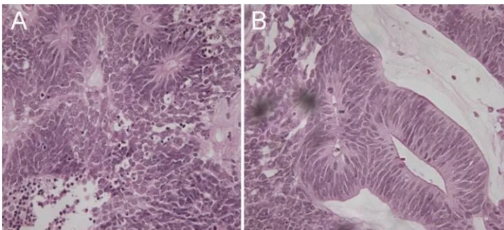
The patient underwent a biopsy of the lesion. The patho- logy reported neuroepithelial lesion with cords, tubules, and sheets arising from the medullary epithelium with mitotic activity most consistent with malignant medulloepithelioma (Figure 2). Due to the large size and vascularity of the lesion, an enucleation was performed.
Questions
• What is medulloepithelioma?
• What is the differential diagnosis for a non-pigmented ciliary body mass in a child?
• What is the management for medulloepithelioma?
What is medulloepithelioma?
Ciliary body medulloepithelioma is an intraocular neo- plasm derived from the primitive ciliary body medullary epithelium, which forms the non-pigmented ciliary body epithelium. First described in histologic detail by Verhoeff in 1904, this unusual lesion was named “teratoneuroma”
[1]. Fuchs in 1908 coined the term of “diktyoma” [1].
However, Grinker later on identified the cellular origin of the tumor by naming it “medullopithelioma” in 1931 [2].
Without any hereditary or racial predisposition, this childhood tumor is the second most common intraocular primary malignancy, following primary intraocular retino- blastoma [3]. Its exact population based incidence is undetermined because it is a very rare neoplasm [4].
Based on the incidence of retinoblastoma, which has been reported to be around 4.1 per million in the United States, we can infer that the incidence of medulloepithe- lioma is significantly lower [5]. The average onset of this disease is 4 years of age, with diagnosis typically in the first decade of life. In adults, ciliary body medulloepithe- lioma has only been reported in 12 cases [6]. The tumor often presents as a solitary nonpigmented lesion that occurs most commonly in the ciliary body, but can also rarely involve the iris, retina, or optic nerve. It also tends
1/4 GMS Ophthalmology Cases 2014, Vol. 4, ISSN 2193-1496
Case Report
OPEN ACCESS
Figure 1: (A) A large vascular non-pigmented mass of the ciliary body, (B) optic nerve cupping and vascular attenuation are noted.
Figure 2: (A) H&E staining of tumor showing Flexner-Wintersteiner rosettes; many mitotic figures can be seen. (B) Cellular mass extends from ciliary epithelium with multilayered cords, tubules, and sheets resembling primitive medullary epithelium.
to be locally aggressive toward neighboring intraocular structures, but it rarely metastasizes [7].
Most cases of medulloepithelioma are diagnosed after the lesion has enlarged to cause anatomic displacement on neighboring structures. As in this patient, unilateral neovascular glaucoma secondary to neoplasmic prolifer- ation is a common feature. Other presentations include lens subluxation, eye pain, conjunctival injection, ectopia lentic, secondary angle closure, uveitis, ectropion uvea, vision loss or vitreous hemorrhage [2].
Gross examinations often reveal a grey flesh colored, vascular, amelanotic lesion. Cysts can be seen on ultra- sound, which are most suggestive of medulloepithelioma but biopsy is required for diagnosis [8]. Pathologic fea- tures of medulloepithelioma can be generally divided into teratoma and non-teratoma and can be further distin- guished as either benign or malignant [6]. Teratoid medulloepithelioma contains heteroplastic elements such as hyaline cartilage, skeletal muscle or neuroglial tissue.
On the other hand, non-teratoid medulloepithelioma is a pure proliferation of the medullary epithelium [8].
What is the differential diagnosis for ciliary body mass in a child?
A ciliary body mass in a child can include various neo- plastic, infectious, or inflammatory processes. The lesion
identified in this patient, which is associated with neovascularization of iris, suggests neoplastic cause.
However, non-neoplasmic etiologies must be ruled out.
The presence of the ciliary mass may be secondary to chronic inflammatory lesion. Granuloma forming infection like ocular toxocariasis must be considered. Ocular toxo- cariasis, estimated to cause about 1 to 2% of uveitis in children, resembles retinoblastoma, coats’ disease, tox- oplasmic retinochoroiditis and other intraocular abnor- malities [9]. Other less likely infections in this case include syphilis. Several case reports have identified masquer- ades of medulloepithelioma including trauma and staphyl- oma [10], [11], [12], [13]. Therefore, neoplastic growth must be considered high in the differential especially if symptoms do not resolve after institution of treatment.
Neoplastic lesions include retinoblastoma, medulloepithe- lioma, as well as Fuch’s adenoma [4]. Other possibilities are more aggressive tumors like adenocarcinoma, metastatic carcinoma and amelanotic melanoma [4].
Histologic differentiation between retinoblastoma and medulloepithelioma is the presence of calcifications.
Clinical and pathologic features that suggest retino- blastoma are bilaterallity (70%), calcification on pathology, genetics (40%). Medulloepithelioma is almost exclusively unilateral and generally not genetically inherited [6].
Fuchs adenoma is a reactive proliferation of the nonpig- mented ciliary epithelium (NPCE) that is associated with
2/4 GMS Ophthalmology Cases 2014, Vol. 4, ISSN 2193-1496
Kassa et al.: Neovascular glaucoma in a child: an unusual presentation ...
aging and is found in the pars placate of the ciliary body [14]. It does not present in the pediatric population.
What is the treatment?
Medulloepithelioma can be treated with local excision, enucleation, and/or radiation depending on the size of the tumor [12], [15], [16]. Attempts to treat locally have been complicated by a high rate of recurrences. Previous studies have published up to 83% recurrence in eyes with local conservative management [16]. Case studies have also reported no recurrence for 7 years and counting after local resection followed by iodine 125 brachytherapy [16].
In general, if neovascular glaucoma is noted with medulloepithelioma, then localized iridocyclectomy may not be sufficient.
Discussion
Early diagnosis of medulloepithelioma is challenging be- cause of the difficulty of direct visualization, the rarity of the neoplasm and early age of presentation. As a result, high proportions of cases are misdiagnosed and manage- ment is delayed. Kaliki et al report that 88% of cases were initially misdiagnosed and 39 % had an additional history of treatment for erroneous diagnosis. Ultrasono- graphic biomicroscopy can be useful for diagnosis; the heterogenic consistency of medulloepithelioma can be detected by UBM and be used to correctly differentiate it from other solid tumors [3], [8]. Furthermore, UBM can adequately demarcate the borders of the neoplasm to aid in resection during iridocyclectomy. Biopsy and histo- logic studies are critical to proper diagnosis and manage- ment.
After the diagnosis of medulloepithelioma, vision sparing management is possible but requires close follow up.
Recurrence of lesion may be secondary to incomplete excision. While small tumors can be locally excised and/or radiated, enucleation maybe required for large tumors and recurrent lesions.
In conclusion, medulloepithelioma is the second most common intra-ocular tumor in pediatric patients and a common presentation for neovascular glaucoma and re- quires a high index of suspicion to reduce the delay in diagnosis.
Final diagnosis:Ciliary body medulloepithelioma
Notes
Funding statement
This research received no specific grant from any funding agency in the public, commercial or not-for-profit sectors.
Competing interests
The authors declare that they have no competing in- terests.
Contributorship statement
All 3 authors were involved in preparing the manuscript.
References
1. Zimmerman LE. Verhoeff's “terato-neuroma” a critical reappraisal in light of new observations and current concepts of embryonic tumors. Trans Am Ophthalmol Soc. 1971;69:210-36.
2. Chua J, Muen WJ, Reddy A, Brookes J. The masquerades of a childhood ciliary body medulloepithelioma: a case of chronic uveitis, cataract, and secondary glaucoma. Case Rep Ophthalmol Med. 2012;2012:493493. DOI: 10.1155/2012/493493 3. Ayres B, Brasil OM, Klejnberg C, Moura LR, Fernandes BF, Burnier
MN Jr. Ciliary body medulloepithelioma: clinical, ultrasound biomicroscopic and histopathologic correlation. Clin Experiment Ophthalmol. 2006 Sep-Oct;34(7):695-8. DOI: 10.1111/j.1442- 9071.2006.01321.x
4. Saunders T, Margo CE. Intraocular medulloepithelioma. Arch Pathol Lab Med. 2012 Feb;136(2):212-6. DOI:
10.5858/arpa.2010-0669-RS
5. Age-adjusted and age-specific SEER cancer incidence rates, 2005-2009 by international classification of childhood cancer (ICCC). In: SEER Cancer Statistics Review, 1975-2009. Available from: http://seer.cancer.gov/csr/1975_2009_pops09/results_
merged/sect_29_childhood_cancer_iccc.pdf
6. Al-Salam S, Algawi K, Alashari M. Malignant non-teratoid medulloepithelioma of ciliary body with retinoblastic differentiation: a case report and review of literature.
Neuropathology. 2008 Oct;28(5):551-6. DOI: 10.1111/j.1440- 1789.2008.00886.x
7. García-Feijoó J, Encinas JL, Méndez-Hernández C, Ronco IS, Martínez de la Casa JM, García Sánchez J. Medulloepithelioma of the ciliary body: ultrasonographic biomicroscopic findings. J Ultrasound Med. 2005 Feb;24(2):247-50.
8. Zhou M, Xu G, Bojanowski CM, Song Y, Chen R, Sun X, Wang W, Chan CC. Differential diagnosis of anterior chamber cysts with ultrasound biomicroscopy: ciliary body medulloepithelioma. Acta Ophthalmol Scand. 2006 Feb;84(1):137-9. DOI: 10.1111/j.1600- 0420.2005.00542.x
9. Pivetti-Pezzi P. Ocular toxocariasis. Int J Med Sci. 2009;6(3):129- 30. DOI: 10.7150/ijms.6.129
10. Kamal MM. Intraoperative cytodiagnosis of ciliary body medulloepithelioma masquerading as anterior ciliary staphyloma.
Acta Cytol. 2010 Sep-Oct;54(5):726-30.
11. Virji MA. Medulloepithelioma (diktyoma) presenting as a perforated, infected eye. Br J Ophthalmol. 1977 Mar;61(3):229- 32. DOI: 10.1136/bjo.61.3.229
12. Kaliki S, Shields CL, Eagle RC Jr,Vemuganti GK, Almeida A, Manjandavida FP, Mulay K, Honavar SG, Shields JA. Ciliary body medulloepithelioma: analysis of 41 cases. Ophthalmology. 2013 Dec;120(12):2552-9. DOI: 10.1016/j.ophtha.2013.05.015 13. Correa ZM, Buffe F, Odashiro AN, Burnier MN. Tumor intraocular
diagnosticado dos años después de un trauma perforante [Intraocular tumor diagnosed two years after perforating trauma].
Arch Soc Esp Oftalmol. 2009 Sep;84(9):469-72.
3/4 GMS Ophthalmology Cases 2014, Vol. 4, ISSN 2193-1496
Kassa et al.: Neovascular glaucoma in a child: an unusual presentation ...
14. Razzaq L, Marinkovic M, Swart W, van Duinen SG, Keunen JE, Luyten GP. Fuchs' adenoma of the choroid simulating a choroidal hemangioma. Case Rep Ophthalmol. 2012 Jan;3(1):83-6. DOI:
000336989
15. Zhao HS, Wei WB. Dilemma in management of ocular medulloepithelioma in a child. Chin Med J (Engl). 2012 Jan;125(2):392-5.
16. Cassoux N, Charlotte F, Sastre X, Orbach D, Lehoang P, Desjardins L. Conservative surgical treatment of
medulloepithelioma of the ciliary body. Arch Ophthalmol. 2010 Mar;128(3):380-1. DOI: 10.1001/archophthalmol.2009.404
Corresponding author:
Yang Sun, MD
Department of Ophthalmology, Glick Eye Institute, Indiana University School of Medicine, 1160 West Michigan Street, Indianapolis, IN 46202, USA, Phone: +1 317 274 2020, Fax: +1 317 278 1007
sunyo@iupui.edu
Please cite as
Kassa E, Li H, Sun Y. Neovascular glaucoma in a child: an unusual presentation of medulloepithelioma. GMS Ophthalmol Cases.
2014;4:Doc03.
DOI: 10.3205/oc000016, URN: urn:nbn:de:0183-oc0000165
This article is freely available from
http://www.egms.de/en/journals/oc/2014-4/oc000016.shtml Published:2014-04-17
Copyright
©2014 Kassa et al. This is an Open Access article distributed under the terms of the Creative Commons Attribution License
(http://creativecommons.org/licenses/by-nc-nd/3.0/deed.en). You are free: to Share — to copy, distribute and transmit the work, provided the original author and source are credited.
4/4 GMS Ophthalmology Cases 2014, Vol. 4, ISSN 2193-1496
Kassa et al.: Neovascular glaucoma in a child: an unusual presentation ...