Management of macular edema with branch retinal vein occlusion in a case of secondary polycythemia
Abstract
Purpose:We report a case of polycythemia with an ocular complication of branch retinal vein occlusion associated with macular edema that
Sumeer Singh
1Srividya Neriyanuri
1was managed by anti-vascular endothelial growth factor (VEGF) and
systemic management.
Rajiv Raman
2Methods:A 43-year-old, one-eyed male, a known case of polycythemia
presented with complaints of decreased vision in the right eye. He un- 1 Elite School of Optometry, Chennai, India
derwent comprehensive eye evaluation, fundus photography and optical coherence tomography at the baseline visit and post intravitreal ranibi-
zumab 1-, 3-, 4- and 11-month follow-up. 2 Shri Bhagawan Mahavir
Vitreoretinal Services, Results:A patient with polycythaemia was diagnosed in the right eye
with superotemporal branch retinal vein occlusion and macular edema, Sankara Nethralaya Hospital, Chennai, India
which clinically and on optical coherence tomography resolved after 1 intravitreal injection of ranibizumab. However, as he discontinued systemic management, macular edema reappeared and the edema resolved well after intravitreal ranibizumab. He then became more compliant to the systemic therapy and was asymptomatic for the last 7 months.
Conclusion:In a case of retinal vein occlusion with macular edema of recognizable cause, the management of systemic disease and anti-VEGF can give satisfactory results.
Keywords:polycythemia, macular edema, branch retinal vein occlusion, intravitreal injections
Introduction
Polycythemia vera or primary polycythemia is a myelopro- liferative disorder characterized by the presence of in- creased red blood cells, haemoglobin and total blood volume and occurs due to a JAK2 mutation [1]. Haemat- ological alterations in primary polycythemia give rise to prothrombosis, an event that results in serious ocular and systemic manifestations [2], [3]. Secondary poly- cythemia is a heterogeneous group of disorders which results in abnormal increase in blood cell mass due to a decrease in tissue oxygenation and alterations in produc- tion of erythropoietin. Previous reports have shown a presence of bilateral retinal vein occlusion as initial manifestation of polycythemia [4]. There has been paucity of information on ocular complications in patients with secondary polycythemia.
We report a case of secondary polycythemia who presen- ted to us with unilateral branched retinal vein occlusion with macular edema and was managed by phlebotomy and intravitreal ranibizumab. The case highlights the im- portance of treatment of the systemic disease in reducing the need of intravitreal injections and implications in re- currence of macular edema. To the best of our knowledge, macular edema with branch retinal vein occlusion (BRVO)
as a complication of secondary polycythemia was not re- ported earlier.
Case description
A 43-year-old male presented to a tertiary eye care centre with complaints of sudden onset (3 days) blurring of vision in the right eye. He was one-eyed and had lost his left eye 20 years ago following a traumatic injury. He was a heavy smoker and gave a positive history of alcohol consump- tion. He was a known case of polycythemia since 6 months; he was negative for JAK2 mutation and had high haematocrit values (Table 1) indicating a secondary polycythemia according to the guidelines set by the British Committee for Standards in Haematology [5]. He was on systemic anti-hypertensive treatment and was advised phlebotomy. However, he was non-compliant to phlebot- omy.
On clinical examination, his best corrected visual acuity (BCVA) was 6/7.5 for distance and N6 for near in the right eye. Anterior segment evaluation was normal and intraocular pressure by applanation tonometry was 14 mm Hg in the right eye. Dilated fundus evaluation re- vealed a superotemporal branch retinal vein occlusion (ST BRVO) with macular edema. Cirrus high definition op-
1/4 GMS Ophthalmology Cases 2019, Vol. 9, ISSN 2193-1496
Case Report
OPEN ACCESS
Table 1: Baseline laboratory evaluation for polycythemia
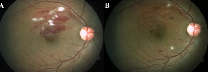
Figure 1: Fundus pictures
A: Fundus picture of the right eye showing a superotemporal branch retinal vein occlusion at the baseline visit.
B: Fundus picture at 1stfollow-up, a month after anti-VEGF showing resolved superotemporal branch retinal vein occlusion.
tical coherence tomography (Carl Zeiss Meditec, Germany) using macular cube (A scans 512, B scans 128) and 5- line raster scans (4096 A-scans) showed an increased central subfield foveal thickness (358 microns) with the presence of cystoid macular edema (CME) and subretinal fluid (SRF). As the patient was symptomatic, even though his vision was good, he was advised intravitreal anti-VEGF and a review with his physician for phlebotomy. The pa- tient underwent intravitreal injection of ranibizumab (0.5 mg) and was advised to undergo phlebotomy regu- larly.
On th consecutive 1stand 2ndmonthly follow-up visits, the patient was asymptomatic, his BCVA was 6/6 for distance and N6 for near. Optical coherence tomography (OCT) showed a normal central subfield foveal thickness (180 microns) with resolved CME and SRF (Figure 1).
At the 3rdmonthly visit, the patient complained of a sud- den drop in vision. He was noncompliant to the treatment advised by his physician for secondary polycythemia. On ocular examination, there was a drop in BCVA to 6/18 with recurrence of CME (increased central subfield foveal thickness – 521 microns). He underwent intravitreal in- jection of ranibizumab (0.5 mg) at this visit. He was ex- plained the need for compliance to systemic manage- ment. On the subsequent visit, the patient’s vision im-
proved to 6/6, N6 and OCT showed resolving macular edema.
Thereafter, he was followed up monthly for 3 months and bimonthly for 1 year. On, the 1-year follow-up, the patient maintained a BCVA of 6/6, N6 with no recurrence of vein occlusion or macular edema (Figure 2). He was under regular care of his treating physician for secondary poly- cythemia.
Discussion
Ocular complications of polycythemia include retinal and choroidal ischemia, retinal haemorrhages, papilloedema and cilioretinal artery occlusion, all of which pose a risk of visual loss and transient blindness [2], [6]. Phlebotomy, a bloodletting procedure for polycythemia, also reduces the risk of systemic and ocular morbidity [6], [7].
Systemic management together with an appropriate ocular management for macular edema and vascular occlusions plays a pivotal role in eye salvage. Previous studies demonstrate that intravitreal ranibizumab therapy is effective for the reduction of macular edema and im- provement in visual acuity. PRN (pro re nata) dosing, treat and extend regimen are also effective in treating macular
2/4 GMS Ophthalmology Cases 2019, Vol. 9, ISSN 2193-1496
Singh et al.: Management of macular edema with branch retinal vein ...
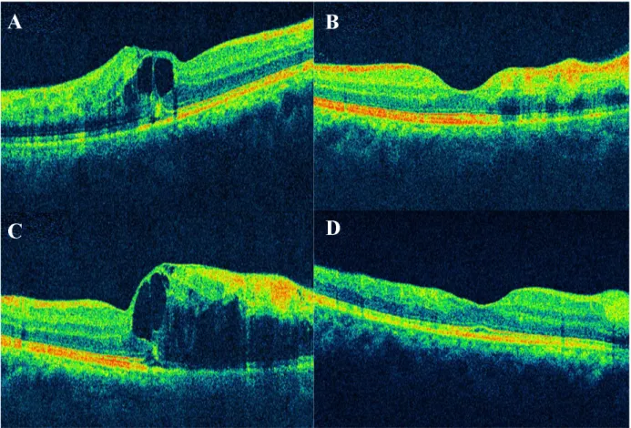
Figure 2: Optical coherence tomography (OCT)
A: OCT at the baseline visit shows an increased foveal contour with macular edema, photoreceptor layer and retinal pigment epithelium (RPE) layer are disrupted temporal to the fovea.
B: OCT at the 1stfollow-up, a month after anti-VEGF shows resolved macular edema.
C: OCT at the 3rdmonthly follow-up shows reoccurrence of the macular edema with sub retinal fluid noted.
D: OCT at the last visit, 11-month follow-up from the baseline shows completely resolved macular edema, normal inner and outer retinal layers noted.
edema in BRVO rather than having monthly injections [8]
[9]. In the present case, timely management of BRVO and strict adherence to the systemic management helped in the recovery of lost vision and halted the reoccurrence of macular edema. This case report brings out an import- ant observation that in cases where the cause of BRVO is certain, treatment of the underlying cause is crucial as it helps to reduce the number of ocular injections a pa- tient receives.
Thus, BRVO with macular edema secondary to hematolo- gical diseases like polycythemia can be managed by systemic control of the disease and as needed injection of intravitreal anti-VEGF agents. However, as there are issues of safety due to the hypercoagulable state of the blood, they need regular physician care and monitoring.
Notes
Competing interests
The authors declare that they have no competing in- terests.
References
1. Spivak JL. Polycythemia vera: myths, mechanisms, and management. Blood. 2002 Dec;100(13):4272-90. DOI:
10.1182/blood-2001-12-0349
2. Parija S, Mohapatra MM, Pattnaik BK. Polycythemia vera presenting with bilateral papilledema: a rare case report. Indian J Ophthalmol. 2008 Jul-Aug;56(4):327-9. DOI: 10.4103/0301- 4738.41418
3. Melamed E, Rachmilewitz EA, Reches A, Lavy S. Aseptic cavernous sinus thrombosis after internal carotid arterial occlusion in polycythaemia vera. J Neurol Neurosurg Psychiatry.
1976 Apr;39(4):320-4. DOI: 10.1136/jnnp.39.4.320 4. Rodriguez N, Eliott D. Bilateral central retinal vein occlusion in
Eisenmenger syndrome. Am J Ophthalmol. 2001 Aug;132(2):268- 9.
5. McMullin MF. The classification and diagnosis of erythrocytosis.
Int J Lab Hematol. 2008 Dec;30(6):447-59. DOI:
10.1111/j.1751-553X.2008.01102.x
6. Yang HS, Joe SG, Kim JG, Park SH, Ko HS. Delayed choroidal and retinal blood flow in polycythaemia vera patients with transient ocular blindness: a preliminary study with fluorescein angiography. Br J Haematol. 2013 Jun;161(5):745-7. DOI:
10.1111/bjh.12290
3/4 GMS Ophthalmology Cases 2019, Vol. 9, ISSN 2193-1496
Singh et al.: Management of macular edema with branch retinal vein ...
7. Streiff MB, Smith B, Spivak JL. The diagnosis and management of polycythemia vera in the era since the Polycythemia Vera Study Group: a survey of American Society of Hematology members' practice patterns. Blood. 2002 Feb;99(4):1144-9. DOI:
10.1182/blood.v99.4.1144
8. Campochiaro PA, Sophie R, Pearlman J, Brown DM, Boyer DS, Heier JS, Marcus DM, Feiner L, Patel A; RETAIN Study Group.
Long-term outcomes in patients with retinal vein occlusion treated with ranibizumab: the RETAIN study. Ophthalmology. 2014 Jan;121(1):209-19. DOI: 10.1016/j.ophtha.2013.08.038 9. Brand CS. Management of retinal vascular diseases: a patient-
centric approach. Eye (Lond). 2012 Apr;26 Suppl 2:S1-16. DOI:
10.1038/eye.2012.32
Corresponding author:
Dr. Rajiv Raman
Shri Bhagawan Mahavir Vitreoretinal Services, Sankara Nethralaya, 18 College Road, Chennai – 600 006, Tamil Nadu, India, Phone:+91-44-28271616, Fax:
+91-44-28254180 rajivpgraman@gmail.com
Please cite as
Singh S, Neriyanuri S, Raman R. Management of macular edema with branch retinal vein occlusion in a case of secondary polycythemia . GMS Ophthalmol Cases. 2019;9:Doc38.
DOI: 10.3205/oc000127, URN: urn:nbn:de:0183-oc0001272
This article is freely available from
https://www.egms.de/en/journals/oc/2019-9/oc000127.shtml Published:2019-11-20
Copyright
©2019 Singh et al. This is an Open Access article distributed under the terms of the Creative Commons Attribution 4.0 License. See license information at http://creativecommons.org/licenses/by/4.0/.
4/4 GMS Ophthalmology Cases 2019, Vol. 9, ISSN 2193-1496
Singh et al.: Management of macular edema with branch retinal vein ...