J. Perinat. Med.
15 (1987) 143
15-keto-13
914-dihydroprostaglandin F
2oand prolactin in maternal and cord blood during prostaglandin £
2or oxytocin therapy for labor induction
Katarina Bremme1, Peter Eneroth1, and Hans Kindahl2
department of Obstetrics and Gynecology, Karolinska Hospital, Stockholm, and
2Department of Obstetrics and Gynecology, Swedish University of Agri- cultural Sciences, Uppsala, Sweden
1 Introduction
In previous studies we have demonstrated a difference in the mode of action of prostaglan- din E2 versus oxytocin during induced parturi- tion: prostaglandin Ei gave a slower onset of contractions [2] and induced a parturition time dependent decrease in maternal serum prolactin levels [3]. Prostaglandin p2a elevates maternal serum prolactin concentrations [7] and is a po- tent stimulator of uterine contractions [5]. Some investigators suggest that prostaglandin Fia is formed in the uterus during contractions [16, 17,28]. If this prostaglandin reaches the circula- tion one would expect an increase in maternal serum prolactin. However, the scarce data available from spontaneous labor rather indi- cate a decrease in serum prolactin during partu- rition [9, 15, 31, 32].
For reasons discussed in detail by GRANSTRÖM et al. [14], primary prostaglandins cannot be measured accurately in blood plasma samples.
The major PGE
2metabolite, 15-keto-13,14-di- hydroprostaglandin Ei, is rearranged and its analysis requires special conditions [13]. The corresponding PGF2a metabolite, 15-keto- 13,14-dihydroprostaglandin F2a, can be deter- mined in plasma and reflects the formation of F-prostaglandins [12]. Accordingly, it was
Curriculum vitae KATARINA BREMME, . D., was graduated in 1969 and qualified as a specialist in Obstetrics and Gynecology in 1974. Since 1975 she has been on the staff of the De- partment of Obstetrics and Gynecology, Karolinska Hospital, Karolinska Insti- tute, Stockholm, Sweden.
selected for assay in the present communica- tion. To explore whether changes in 15-keto- 13,14-dihydroprostaglandin F2a levels in ma- ternal blood during induced parturition are re- lated to serum prolactin changes, the present study was undertaken.
2 Materials and methods
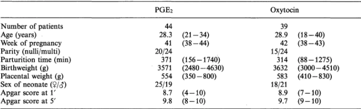
The study comprised 83 healthy women in whom labor was induced mainly for reasons of post-datism (table I). All patients had a vertex presentation and a normal pelvic outlet.
Patients with toxemia of pregnancy treated with
more than 100 mg hydralazine (Apresoline®,
Table I. Clinical data on patients in whom labor was induced with prostaglandin E2 (PGE2) or oxytocin.
Number of patients Age (years) Week of pregnancy Parity (nulli/multi) Parturition time (min) Birthweight (g) Placental weight (g) Sex of neonate (?/<}) Apgar score at Υ Apgar score at 5'
PGE2
44 28.341 20/24 3571371 554 25/19 8.7 9.8
Oxytocin
(21-34) (38-44) (156-1740) (2480-4630) (350-800) (4-10) (8-10)
28.939 15/2442 314 3632583 18/21 8.9 9.7
(18-40) (38-43) (88-1275) (3000-4510) (410-830) (7-10) (9-10) Data in the table are mean values and ranges.
CIBA) and/or 1 g chlortiazide (Chlortride®, MSD) daily were excluded from the study, as were patients with any other disease.
Labor was induced by low amniotomy and intravenous infusion of oxytocin (Syntocinon®, Sandoz) in 5.5% glucose, starting at 8.30 a.m.
with 5 mlU/min and increasing over three hours to a maximum of 20 mlU/min; or by low
amniotomy and oral administration of PGEi (Upjohn Co., Kalamazoo, Mich., USA): dosage 0.5 mg initially, followed by 1.0 mg every hour until delivery. Treatment was randomized. The registration of uterine contractility was done with external and internal tocography accord- ing to established procedures [27]. Start of labor was defined as the time when three contractions with an amplitude of more than 25 mm Hg
Table II. 15-keto-13,14-dihydro-PGF2a and prolactin in maternal blood during prostaglandin E2 (PGE2) or oxytocin therapy for labor induction. Mean values; 95% confidence limits.
Labor stage
Start of treatment
Immediately5) prior to the delivery
15-ketodihydro-PGF2a (pmol/1) PGE2 (n = 21) Oxytocin (n = 23)
473 347 (330-
*s
>
18
-675) (227-
:i) !
: ' :
90 8'
-526)
|e i\
s z)
|e '
(1602-2232) (588-1285)
n
Prolactin ^g/l) PGE2(n=32)
144 (119-176)
I 3 )
(71-119)92
Oxytocin (n = 30) (130-205)163
··>
(100-169)130
!) Paired t-test; t = -10.49 p < 0.001
2) Paired t-test; t = -5.70 p < 0.001
Contrast between 1) and 2): t = 2.23, p = 0.03.
Tested contrast: Diff (15-ketodihydro-PGF2a) PG - Diff (15-ketodihydro-PGF2a) OXY
3) Paired t-test; t = 3.90 p < 0.001
4) Paired t-test; t = 2.52 p = 0.017
Contrast between 3) and 4): t = 1.54, p = 0.13.
Tested contrast: Diff (prolactin) PG - Diff (prolactin) OXY
5) Time difference (mean and ranges) between blood sample and parturition: in PGE2 treated patients 33 (4—60) minutes; in oxytocin treated patients 34 (2 — 60) minutes.
J. Perinat. Med. 15 (1987)
were registered during 10 minutes [6]. The two patient categories were given the same type of analgesic treatment. Demerol® (Meperidine, Pethidine) 100 mg i. m. (AGO, Sweden), was administerd to 23 PGEi and to 29 oxytocin treated women. Epidural analgesia (bupivac- aine-Marcaine®, Bofors, Sweden) was given to 12 PGE2 and to 8 oxytocin treated patients, respectively.
Blood samples were drawn from the antecubital vein at the start of treatment, one hour into
treatment, immediately prior to delivery and after delivery. Mixed umbilical blood was col- lected at parturition by section of the cord.
Serum and plasma were isolated by centrifuga- tion and stored at — 20 °C until analyzed. The number of individuals from whom a complete set of analyzes could be obtained was few.
Therefore, the results given in tables II to IV refer to patients in whom the appropriate sam- ples were obtained to allow intra-patient com- parisons.
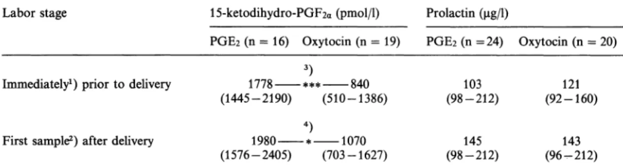
Table ΙΠ. 15-keto-13,14-dihydro-PGF2a and prolactin in maternal blood during prostaglandin £2 (PGE2) or oxyto- cin therapy for induction of labor. Mean values; 95% confidence limits.
Labor stage 15-ketodihydro-PGF2a (pmol/1) Prolactin
PGE2 (n = 16) Oxytocin (n = 19) PGE2 (n = 24) Oxytocin (n = 20)
Immediately1) prior to delivery
First sample2) after delivery
1778 *** 840 (1445-2190) (510-1386)
4)
1980 * 1070 (1576-2405) (703-1627)
(98-212)103
(98-212)145
121 (92-160)
(96-212)143
*) Time difference, mean and range, between blood sample and delivery: 33 (4—60) minutes (PGE2), and 34 (2 — 60) minutes (oxytocin).
2) Time difference, mean and range, between blood sample and delivery: 38 (2 — 60) minutes (PGE2), and 39 (2 — 60) minutes (oxytocin).
3) Independent t-test: p = 0.001.
4) Independent t-test: p = 0.016.
Table IV. 15-keto-13,14-dihydro-PGF2a and prolactin concentrations in blood in connection with partus induced by prostaglandin £2 (PGE2) or oxytocin. Mean values; 95% confidence limits.
Labor stage 15-ketodihydro-PGF2a (pmol/1) Prolactin
Immediately prior to delivery
PGE2 (n = 15) Oxytocin (n = 13) 1870 1058 (1467-2383) (714-1568)
PGE2 (n =23) Oxytocin (n = 24) 96 120 (67-137) (94-154)
I 2 )
Mixed umbilical 1467
(1151-1868) 913
(714-1123) 248
(204-301) 227
(176-292)
1) Paired t-test; t = -4.63 (p < 0.001).
2) Paired tested; t = -5.57 (p < 0.001).
Radioimmunoassay (RIA) of serum samples was performed in duplicate with commercial kits from Serono Diagnostics (prolactin) and Diagnostic Products Corp. (cortisol). Intraas- say and interessay coefficients of variation were all below 10%. Levels of 15-ketodihydro- PGF2a in plasma were determined by RIA as described in detail previously [12]. Specificity of the employed antiserum and accuracy data have been reported previously [26].
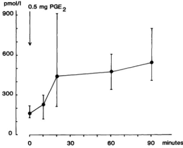
The study also included eight non-pregnant healthy women with normal menstrual cycles.
These women were given a single dose of 0.5 mg PGE2 orally. Blood was sampled after 10, 20, 30, 60 and 90 minutes. Levels of 15-ketodi- hydro-PGFia were determined as described above.
Statistical methods: Calculations were pro- formed on the logarithms of the measured va- lues. Two-tailed 95% conficence intervals of the mean in terms of logarithmic values were calculated. Probability values (p-values) were determined in two-tailed tests. Geometric means and interval limits were obtained from the antilogarithms. When variances of two groups differed significantly, a separate vari- ance estimate of Student's t-statistic was used, otherwise a pooled variance estimate, to test whether the means of the groups differed or not. Contrasts of means between more than two groups were also calculated. Student's t- statistic was used to test whether the formed contrasts differed from expectancy.
3 Results
The changes in maternal serum hormone levels during induced parturition are displayed in table II. Oxytocin and PGEi caused similar elevations in maternal serum cortisol (data not shown): whereas, decreases in the levels of pro- lactin seemed more pronounced in the PGE2 group (table II). The changes observed in 15- ketodihydro-PGFia throughout parturition were highly significant in both treatment groups. The PGEi group showed a significantly higher increase than the oxytocin group.
In terms of onset of contractions, the median time for women in the PGEi group was 62 minutes and for women in the oxytocin group 45 minutes (p = 0.004; median test). The total time to delivery did not differ between the groups (table I). If onset of contractions is related to elevation of 15-ketodihydro-PGF2a, this should be reflected one hour into treat- ment. Among women receiving oxytocin, the 15-ketodihydro-PGF2a level changed from 315 pmol/1 (226-439) to 361 pmol/1 (264-494) during the first hour of treatment (n = 31).
This increase is not significant. In the PGE2 group (n = 31) the corresponding figures were 396 pmol/1 (282-556) and 821 pmol/1 (614-1096). The elevation in the PGE2 group is highly significant (p < 0.001; paired t-test).
Tested for the contrast between the PGE2 and oxytocin groups, a significant difference was found (p < 0.001), i.e. the elevation in the PGE2 group was significantly higher than in the oxytocin group.
Following delivery, there was still a difference between PGE2 and oxytocin treated women in terms of 15-ketodihydro-PGF2a levels in ma- ternal plasma; whereas, no significant differ- ence was seen either in prolactin (table III) or in cortisol concentrations (data not shown).
The level of 15-ketodihydro-PGF
2ain the mixed umbilical plasma in each treatment group was almost the same as in the maternal plasma immediately prior to delivery. When the two experimental groups were compared for hor- mone levels in mixed umbilical blood (table IV), a clearly significant difference was observed in 15-ketodihydro-PGF2a concentration but not in prolactin.
The non-pregnant women responded to oral
PGE2 with an increase in levels of 15-keto-
13,14-dihydro-PGF2a which was already signifi-
cant at 10 (p = 0.034; Wilcoxon) and 20 min-
utes (p = 0.012; Wilcoxon) after PGE2 adminis-
tration. The mean basal level was 160 pmol/1
(129-218) and after PGE
2administration 228
pmol/1 (118-297) and 440 pmol/1 (213-907)
at 10 and 20 minutes, respectively. One hour
after treatment the average concentration was
476 pmol/1 (324 — 605), i.e. no significant in-
pmol/l 900 .
600
300
0.5 mg PGE2
30 60 90 minutes
Figure 1. Plasma levels of 15-keto-13,14-dihydro-PGF2a (geometric mean; 95% confidence limits for geometric mean) following a single oral dose of 0.5 mg PGE2 in eight healthy non-pregnant female volunteers.
crease occurred between 20 and 60 minutes.
None of the women experienced uterine con- tractions or complained of abnormal pain and after 90 minutes the average plasma level was still elevated, i.e. 543 pmol/l (409-789) (figure 1).
4 Discussion
The present report shows that oral PGE2 treat- ment leads to increases in the plasma content of 15-ketodihydro-PGF2a that are clearly above the levels induced by oxytocin and reportedly at hand during spontaneous labor [29, 36]. Yet, contractions appeared later in the PGEz treated group. The average time before contractions appeared in the PGE2 treated pregnant women was 62 minutes. The women receiving oxytocin had a mean time to onset of contractions of 45 minutes but no increase in plasma concentra- tions of 15-ketodihydro-PGF2a at 60 minutes.
From two hours into treatment to the time immediately before parturition there was no significant difference in the increase in plasma 15-ketodihydro-PGF2a between PGE2 and oxy- tocin treated women. Thus, the present study fails to relate increases in plasma levels of 15- ketodihydro-PGF2a to onset of uterine contrac-
tions [10, 21]. Nor was it possible to find a relation between the plasma concentration of the metabolite and delivery time [33]. The trig- gering of contractions may have been mediated by amniotomy and the locally formed prosta- glandins may not have been reflected in the circulation [34, 35]. Once labor had been estab- lished, increases in plasma 15-ketodihydro- PGF2a levels were similar in PGE2 and oxytocin treated women. In spontaneous labor it has been reported that 15-ketodihydro-PGF2a in- creased in parallel with cervical dilatation [16, 24], possibly reflecting formation of uterine PGP2a. However, it has been questioned whether local formation of PGF2a in the uterus is soon reflected by 15-ketodihydrp-PGF2a in plasma [25, 30]. Oral PGE
2obviously led to an increase in plasma levels of 15-ketodihydro- PGP2a. A possible explanation is that PGE2 itself may be converted to PGF2a in the course of, or following, absorption [8, 18, 19, 20] and later be metabolized into 15-ketodihydro- PGP2a. This metabolite lacks biological activity and cannot be related as such to the clinical events or to prolactin levels. Thus, high levels of this metabolite caused by exogenous PGE2 will shadow an endogenous production. Also in the case of non-pregnant women, a significantly elevated level of 15-ketodihydro-PGF2a metab- olite is seen 20 minutes after oral PGE2. The levels remained high for as long as 90 minutes and, considering the short half-lives of PGE2 and PGF2a as well as of 15-ketodihydro-PGF2a in the circulation, it seems likely that exogenous PGE2 is not only converted to PGF2a but may also have induced an ongoing endogenous prostaglandin — i.e. PGF2a — biosynthesis, perhaps in the uterus. An alternative hypothesis for our findings is that an excess of PGE2 and its circulating metabolites could well overload enzyme systems to such an extent that this results in a longer circulating half-life of the PGF
2ametabolites [11].
Since PGF2a and PGE2 exert antagonistic
effects on prolactin levels in pregnant women
[4, 7], the lowering of prolactin during oral
PGE2 therapy indicates that PGE2 effects domi-
nate over PGF2a effects, even though £2 to
some extent is converted to PGFia or induces PGFia biosynthesis. Since the stress of labor measured as serum cortisol (data not shown) was the same in both oxytocin and PGE2 treated women, this factor is likely not directly involved in prolactin release, as previously sug- gested [38].
The possibility exists that the epidural analgesia might have influenced circulating prolactin and 15-ketodihydro-PGF
2alevels [22, 23, 37]. But the relative frequency of this therapy was very similar in the two treatment groups. It is thus less likely that epidural analgesia influenced our results.
About half an hour after delivery, maternal serum prolactin levels were the same in both treatment groups, indicating that the PGEi in- duced effects on maternal pituitary prolactin release had stopped or been counteracted.
However, 1 S-ketodihydro-PGFia levels were at least as high some 30 minutes after as before delivery, which makes it less likely that 15- ketodihydro-PGF2a in itself participates in ma- ternal pituitary prolactin release. The levels of 15-ketodihydro-PGF2a in the mixed umbilical
cord blood seem to be close to the maternal plasma concentration in both treatment groups.
Since there is no artero-venous difference in umbilical blood for 15-ketodihydro-PGF
2a[1], a maternal origin for the 15-ketodihydro- PGF2<x measured in mixed umbilical blood may be suggested. The data also indicate that the placenta does not metabolize 15-ketodihydro- PGF2a to any major extent.
In conclusion, our data show that there was no correlation between 15-keto-13,14-dihydro- PGF2a concentration and onset of contraction or labor time in either the PGE2 or the oxytocin treated group. The higher values of plasma 15- keto-13,14-dihydro-PGF2a in women receiving PGE2 tablets as compared to those treated with oxytocin suggest that exogenous PGE2 is to some extent reduced to PGF2a. Such a reduc- tion seems also to take place in non-pregnant women, in whom administration of oral PGE2 gave rise to marked increases in plasma 15- keto-13,14-dihydro-PGF2a. The marked de- crease in serum prolactin in the PGE2 treated pregnant women, which normalized following parturition, suggests a PG involvement in serum prolactin control.
Summary
15-keto-13,14-dihydroprostaglandin p2a plasma levels were measured in pregnant women following labor in- duction with either oral PGE2 treatment or intravenous oxytocin, both combined with amniotomy. The median time to start of contractions was 62 minutes in the PGE2 treated group and 45 minutes in the oxytocin treated group (p < 0.01; median test). The increase in 15- ketodihydro-PGF2a appeared earlier in the PGEa group but not in the oxytocin group (p < 0.001 and p = 0.210, respectively). At delivery, the 15-ketodihydro- PGF2a values had further increased in both treatment groups. The increase was significantly higher in the PGE2 treated patients compared with oxytocin treated patients (p = 0.03; contrast test). Despite higher 15- ketodihydro-PGF2a concentrations throughout parturi-
tion, PGE2 women did not deliver more rapidly than oxytocin infused women. There was no correlation be- tween 15-ketodihydro-PGF2a blood concentrations and either onset of contrations or labor time. The decrease in maternal serum prolactin concentration during partu- rition was pronounced (p < 0.001) in the PGE2 group but occurred also in oxytocin treated patients (p < 0.02). A single oral dose (0.5 mg) of PGE2 taken by non-pregnant women led to significant (p < 0.05) increases in 15-ketodihydro-PGF2a levels in blood plasma after 10 minutes. This increase persisted for at least 90 minutes. It is suggested that oral PGE2 may be transformed into PGF2a and/or induce endogenous PGF2a biosynthesis.
Keywords: Cord blood, labor induction, maternal blood, prolactin, 15-keto-13,14-dihydroprostaglandin
Zusammenfassung
15-Ketodihydro-PGF2a- und Prolaktinspiegel im mütter- lichen Serum und Nabelvenenblut während der Geburtsein- leitung mit Prostaglandin £2 oder Oxytozin
Wir bestimmten die Plasmaspiegel von 15-Keto-13,14- dihydroprostaglandin F2a bei schwangeren Frauen nach Geburtseinleitung mit oraler PGEi-Gabe bzw. intrave- nöser Oxytozingabe und arteflzieller Blaseneröffnung.
In der PGEz-Gruppe setzten die Wehen nach durch- schnittlich 62 Minuten ein, in der Oxytozingruppe nach 45 Minuten (p < 0.01; Mediantest). In der PGE2- Gruppe kam es früher zu einem Anstieg des 15-Ketodi- hydro-PGFza. Eine Stunde nach Behandlungsbeginn war der PGFia-Spiegel in der PGEi-Gruppe signifikant er- höht, nicht aber in der Oxytozingruppe (p < 0.001 versus p = 0.210). Zum Zeitpunkt der Geburt waren die 15-Ketodihydro-PGF2a-Werte in beiden Gruppen weiter angestiegen, wobei der Anstieg in der PGE2-Gruppe
signifikant größer war als in der Oxytozingruppe (p = 0.03 bei Gegenüberstellung). Trotz höherer 15-Ketodihy- dro-PGF2a-Konzentrationen in der PGE2-Gruppe war der Geburtsverlauf hier nicht kürzer als in der Oxytozin- gruppe. Die Höhe des 15-Ketodihydro-PGP2a-Spiegels im Blut und das Einsetzen bzw. die Geburtsdauer korre- lierten nicht miteinander. Der Abfall des Serumprolak- tins war unter der Geburt in der PGE2-Gruppe beson- ders ausgeprägt (p < 0.001), zeigte sich aber auch in der Oxytozingruppe (p < 0.02). Eine einmalige Gabe von 0,5 mg PGE2 an nichtgravide Frauen führte nach 10 Minuten zu einem signifikanten Anstieg des 15-Keto- dihydro-PGF2a (p < 0.05), das über mindestens 90 Minuten erhöht blieb. Wir meinen, daß oral verabreich- tes PGE2 zu PGF2a metabolisiert wird und/oder die endogene PGF2a-Biosynthese induziert.
Schlüsselwörter: Geburtseinleitung, Nabelschnurblut, mütterliches Serum, Prolaktin, 15-Keto-13,14-dihydropro- staglandin F2a-
Resume
Taux de 15-ceto-13,14 dihydroprostaglandine Fza et de prolactine dans le sang maternel et dans le sang du cordon lors des declenchements du travail par prostaglandine £2 ou par ocytocine
On a mesure les taux plasmatiques de 15-13,14 di- hydroprostaglandine F2a chez des femmes enceintes apres induction tu travail avec soit PGE2 per os soit ocytocine intra-veineuse, les deux methodes etant asso- ciees ä la rupture des membranes. Le delai moyen du debut des contractions a ete de 62 minutes dans le groupe traite par prostaglandines et de 45 minutes pour le goupe traite par ocytocine (p < 0,01). L'elevation des 15-cetodihydro-PGF2a est apparue plus tot chez les femmes traitees par PGE2. Au bout d'une heure de traitement Faugmentation etait significative dans le groupe PGE2 mais pas dans le groupe ocytocine ( P <
0,001 et p = 0,210, respectivement). Au moment de Paccouchement, les valeurs de PGF2a se sont encore plus elevees et celä dans les deux groupes. L'elevation a ete
de fagon significative plus importante dans le groupe PGE2 que dans le groupe ocytocine (p = 0,03). Malgre des taux de 15-cetodihydro PGF2a plus eleves tout au long de 1'accouchement les femme sous PGE2 n'accou- chent pas plus rapidement que les femmes sous perfusion d'ocytocine. II n'y a pas de correlation entre les concen- trations seriques de 15-cetodihydro PGF2a ni avec le debut des contractions ni aved la duree du travail. La diminution des taux maternels de Prolactine serique au cours de 1'accouchement est nette (p < 0,001) dans le groupe PGE2 mais existe egalement chez les patientes sous ocytocine (p < 0,02). Une dose orale unique (0,5 mg) de PGE2 prise par une femme non enceinte entraine une augmentation significative (p < 0,05) des 15- cetodihydro-PGP2a plasmatiques au bout de 10 minutes.
Cette augmentation persiste au moins 90 minutes. Les auteurs suggerent que les PGE2 orales peuvent etre trans- formees en PGF2a et/ou induire la Synthese de PGF2 endogenes.
Mots-cles: Declenchement du travail, prolactine, sang du cordon, sang maternel, 15-ceto-13,14-dihydroprostaglan- dine, p2a-
Acknowledgements: The skilful assistance of MARIANNE OLSSON and ASTRID HÄGGBLAD as well as the midwifery staff at the maternity ward at the Karolinska Hospital is gratefully acknowledged. Expert help with the statistical calculations was obtained from Bo NILSSON, Epidemiologie Center, Karolinska Hospital. Financial support was received from the research funds of the Karolinska Institute and from The Swedish Council for Forestry and Agricultural Research. We are also grateful to the Upjohn Company for supplying PGE2 tablets.
References
[1] BIBBY JG, JD BRUNT, H HODGSON, MD MITCHELL, ABM ANDERSON, AC TURNBULL: Prostaglandins in umbilical plasma et elective cesarean section. Br Med J 86 (1979) 282
[2] BREMME K, M BYGDEMAN: A comparative pattern of uterine activity and fetal heart rate in labour induced with oral prostaglandin £2 or oxytocin.
Acta Obstet Gynecol Scand 92 (1980) 23
[3] BREMME K, P ENEROTH: Changes in serum hormone levels during labour induced by oral PGE2 or oxyto- cin infusion. Acta Obstet Gynecol Scand 92 (1980) [4] BREMME K: Induction of labour by oral administra-31 tion of prostaglandin £2. Thesis, Karolinska Insti- tute, Stockholm 1982
[5] BYGDEMAN M, S KWON, N WIQVIST: The effect of prostaglandin Ei on human pregnant myometrium in vivo. In: S BERGSTRÖM, B SAMUELSSON: Nobel Symposium II on Prostaglandins. Almqvist &
Wiksell, Stockholm 1967
[6] CALDEYRO-BARCIA R, H ALVAREZ, SRM REY- NOLDS: A better understanding of uterine contractil- ity through simultaneous recording with an internal and a seven channel external method. Surg Gynecol Obstet 91 (1950) 641
[7] CAMINITI F, A NASI, M DE MURTAS, G PARODO, GB MELIS, P FIORETTI: Effect of 15-methyl-prosta- glandin F2a on pituitary prolactin secretion. Ab- stract, 2nd International Symposium on Clinical Psycho-Neuro Endocrinology in Reproduction.
Venice, June 1979
[8] CRYSTAL AL, L LEVINE: Evidence of the presence of a prostaglandin E2-9-keto reductase in rat organs.
Biochem Biophys Res Commun 52 (1973) 717 [9] DE GEZELLE H, M DHONT, W PAREWYCK, M
THIERY: Prolactin levels during labor and the early post-partum period. IRCS 5 (1977) 475
[10] GHODGAONKAR RB, NH DUBIN, DA BLAKE, TM KING: 13,14-dihydro-15-keto-prostaglandin Fia concentrations in human plasma and amniotic fluid. Am J Obstet Gynecol 134 (1979) 265 [11] GOESCHEN K, AR FUCHS, F FUCHS, AB RASMUSSEN,
JV REHNSTRÖM, E SALING: Effect of ß-mimetic tocolysis on cervical ripening and plasma prosta- glandin F2a metabolite after endocervical applica- tion of prostaglandin £2. Obstet Gynecol 65 (1985) [12] GRANSTRÖM E, H KINDAHL: Radioimmunoassay of166 the major plasma metabolite of PGF2a, 15-keto- 13,14-dihydro-PGF2a. Methods Enzymol 86 (1982) 320
[13] GRANSTRÖM E, F FITZPATRICK, H KINDAHL: Ra- dioimmunologic determination of 15-keto-13,14- dihydro-PGE2: A method for its stable degradation product, 11 -deoxy-15-keto-13,14-dihydro-11 , 16 - cyclo-PGE2. Methods Enzymol 86 (1982) 306
[14] GRANSTRÖM E, H KINDAHL, ML SWAHN: Profiles of prostaglandin metabolites in the human circulation:
Identification of late-appearing long-lived prod- ucts. Biochim Biophys Acta 713 (1982) 46 [15] GREGORIOU O, S PITOULIS, B COUTIFARIS: Prolactin
levels during labor. Obstet Gynecol 53 (1979) 630 [16] GREEN K, M BYGDEMAN, M TOPPOZADA, N WIQV-
IST: The role of prostaglandin F2a in human parturi- tion. Am J Obstet Gynecol 120 (1974) 25
[17] HAMBERG M: Quantitative studies on prostaglandin synthesis in man. III. Excretion of the major urinary metabolites of prostaglandins Fia and F2a during pregnancy. Life Sei 14 (1974) 247
[18] HAMBERG M, U ISRAELSSON: Metabolism of prosta- glandin £2 in guinea pig liver. I. Identification of seven metabolites. J Biol Chem 245 (1970) 5107 [19] HAMBERG M, M WILSON: Structures of new metab-
olites of prostaglandin £2 in man. Adv Biosci 9 (1973) 39
[20] HILLIER K: Prostaglandins and thromboxanes:
Pharmacologic and biosynthetic aspects. Semin Perinatol 2 (1978) 197
[21] HUSSLEIN P, AR FUCHS, F FUCHS: Oxytocin and the initiation of human parturition. I. Prostaglandin release during induction of labour by oxytocin. Am J Obstet Gynecol 141 (1981) 688
[22] JOUPPILA R: The effect of segmental epidural anal- gesia on hormonal and metabolic changes during labour. Anaesthesiologica No 2, Series D, Medica No 16 (1977)
[23] JOUPPILA R, P JOUPPILA, K MOILANEN, A PAKAR- INEN: The effect of segmental epidural analgesia on maternal prolactin during labour. Br J Obstet Gynaecol 87 (1980) 234
[24] KEIRSE MJNC: Biosynthesis and metabolism of prostaglandins in the pregnant human uterus. Adv Prostaglandin Thromboxane Res 4 (1978) 87 [25] KEIRSE MJNC, MD MITCHELL, AC TURNBULL:
Changes in prostaglandin F and 13,14-dihydro-15- keto-prostaglandin F concentrations in amniotic fluid at the onset of and during labour. Br J Obstet Gynaecol 84 (1977) 743
[26] KINDAHL H, LE EDQVIST, E GRANSTRÖM, A BANE:
The release of prostaglandin F2a as refelcted by 15- keto-13,14-dihydro-prostaglandin F2a in the pe- ripheral circulation during normal luteolysis in heifers. Prostaglandins 11 (1976) 871
[27] KLAPHOLZ H: Techniques of fetal heart rate moni- toring. Semin Perinatol 2 (1978) 119
[28] MITCHELL MD, APF FLINT, J BIBBY, J BRUNT, JM ARNOLD, ABM ANDERSON, AC TURNBULL: Rapid increase in plasma prostaglandin concentrations after vaginal examination and amniotomy. Br Med J 2 (1977) 1183
[29] MITCHELL M, AP FLINT, J BIBBY, J BRUNT, JM ARNOLS, ABM ANDERSON, AC TURNBULL: Plasma
concentrations of prostaglandins during late human pregnancy: Influence of normal and preterm labour.
J Clin Endocrinol Metab 46 (1978) 947
[30] RASMUSSEN AB, P JOHANNESEN, J ALLEN, J REHN- STR M, AR FUCHS: Plasma prostaglandin ?2α and plasma 13,14-dihydro-15-keto-prostaglandin Fza levels in women during induction of labor with i. v.
infusion of prostaglandin Fia in relation to uterine contractions. J Perinatol Med 13 (1985) 15 [31] Di RENZO GC, V MAZZA, A VOLPE: Prolactin varia-
tion in maternal blood and in amniotic fluid during vaginal delivery and elective cesarean section. In:
A KLOPPER, A GENAZZANI, PG CROSIGNANI: The Human Placenta. Serono Symposium No 35. Aca- demic Press, London-New York 1980
[32] RIGG LA, SSC YEN: Multiphasic prolactin secretion during parturition in human subjects. Am J Obstet Gynecol 128 (1977) 215
[33] SATOH K, T YASUMIZU, H FUKUOKA, K KINOSHITA, Υ ΚΑΝΕΚΟ, M TSUCHIYA, S. SAKAMOTO: Prostaglan- din F2a metabolite levels in plasma, amniotic fluid and urine during pregnancy and labour. Am J Obstet Gynecol 133 (1979) 886
[34] SELLERS, SM, HT HODGSON, MD MITCHELL, ABM ANDERSON, AC TURNBULL: Release of prostaglan- dins after amniotomy is not mediated by oxytocin.
Br J Obstet Gynaecol 87 (1980) 43
[35] SELLERS, SM, MD MITCHELL, ABM ANDERSON, AC TURNBULL: The relation between the release of prostaglandins at amniotomy and the subsequent onset of labour. Br J Obstet Gynaecol 88 (1981) [36] WEPPELMAN B, HO HOPFEN, U GETHMANN, E1211 SCHUSTER: Plasma levels of prostaglandin metab- olites during spontaneous delivery. Adv Prostaglan- din Thromboxane Res 8 (1980) 1409
[37] WILLDECK-LUND G, G LINDMARK, ΒΑ NILSSON:
Effect of segmental epidural analgesia upon the uterine activity with special reference to the use of different local anaesthetic agents. Acta Anaesthesiol Scand 23 (1979) 519
[38] WLADIMIROFF JW, WM BRANDT: The relationship between maternal plasma prolactin and cortisol concentration during labour. Eur J Obstet Gynecol Reprod Biol 12 (1981) 13
Received May 24, 1985. Revised February 24, 1986.
Accepted March 10, 1986.
Dr. Katarina Bremme
Department of Obstetrics and Gynecology Karolinska Hospital
S-10401 Stockholm, Sweden
J. Perinat. Med. 15 (1987)