Distribution of Ih Channels and their Function in the Stomatogastric Ganglion.
I n a u g u r a l - D i s s e r t a t i o n zur
Erlangung des Doktorgrades
der Mathematisch-Naturwissenschaftlichen Fakultät der Universität zu Köln
vorgelegt von Marie-Luise Göritz
aus Marburg
(Köln 2008)
2
Berichterstatter: Prof. Dr. Peter Kloppenburg Prof. Dr. Joachim Schmidt Prof. Dr. Ron Harris-Warrick
Tag der letzten mündlichen Prüfung: 8. Januar 2009
3
1 INTRODUCTION ... 8
1.1 RHYTHMICALLY ACTIVE NETWORKS... 8
1.2 THE CRUSTACEAN STOMATOGASTRIC NERVOUS SYSTEM... 10
1.3 THE HYPERPOLARIZATION-ACTIVATED INWARD CURRENT IH... 16
1.4 HOMEOSTATIC RELATIONSHIP OF IH AND IA... 18
1.5 AIM OF THIS STUDY... 20
2 METHODS ... 21
2.1 STG DISSECTION AND PD CELL IDENTIFICATION...21
2.2 RNA MICROINJECTION INTO NEURONS... 21
2.3 ELECTROPHYSIOLOGY... 22
2.4 IMMUNOCYTOCHEMISTRY... 28
3 IA-IH HOMEOSTASIS IN PYLORIC NEURONS...30
3.1 PROPERTIES OF INCREASED IA AFTER SHAL-GFP OVEREXPRESSION...31
3.2 PROPERTIES OF THE HYPERPOLARIZATION-ACTIVATED INWARD CURRENT IH AFTER SHAL EXPRESSION...34
3.3 FIRING PROPERTIES OF SHAL-GFP EXPRESSING PYLORIC NEURONS...35
3.4 AN UNIDENTIFIED COMPONENT OF THE INWARD CURRENT... 36
3.5 EXPRESSION OF A NONFUNCTIONAL MUTANT OF SHAL-GFP IN PD NEURONS... 37
3.6 POSITIVE CORRELATION BETWEEN IA AND IH IN NON-INJECTED PD NEURONS... 39
3.7 LOCALIZATION OF IA AND THE IMPLICATIONS FOR NEURONAL CYCLING...40
3.8 DIRECTIONALITY... 41
DISCUSSION... 65
4 IH PROTEIN LOCALIZATION...76
4.1 OVERALL EXPRESSION PATTERN...78
4.2 IH PROTEIN IN THE SYNAPTIC NEUROPIL... 79
4.3 IH AND IA PROTEIN LOCALIZATION... 79
DISCUSSION... 107
5 ACTIVATION OF IH CHANNELS CAN SHUNT SYNAPTIC TRANSMISSION... 116
5.1 EFFECTS OF IHACTIVATION UNDER CONTROL CONDITIONS...118
5.2 IPSP AMPLITUDE DURING BLOCK OF IHCHANNELS...122
5.3 COMPARISON WITH CALCULATED EXPECTATIONS OF IPSP AMPLITUDES...129
4
DISCUSSION... 131
BIBLIOGRAPHY...138
ACKNOWLEDGEMENTS ...152
ERKLAERUNG... 153
TEILPUBLIKATIONEN ...154
LEBENSLAUF: ... 155
5 Zusammenfassung:
Stereotype Bewegungsmuster wie Fortbewegung, Verdauung und Atmung werden durch repetitive Entladungen von Motoneuronen in relativ autonomen neuronalen Netzwerken, sogenannten zentralen Rhythmusgeneratoren oder Central Pattern Generators (CPGs) generiert. Innerhalb eines solchen Netzwerkes erzeugt das Zusammenspiel einzelner Neurone rhythmische Aktivitaetsmuster, die typischerweise unabhaengig von synaptischen Eingaengen uebergeordneter Zentren generiert werden koennen. In der vorliegenden Studie wurde die Bedeutung der hyperpolarisations-aktivierten Ih Kationenkanaele im pylorischen Netzwerk des stomatogastrischen Ganglions untersucht. Dieses rhythmisch aktive Netzwerk steuert Bewegungen des Hummermagens. Von besonderem Interesse waren die Rolle von Ih bei der Aufrechterhaltung regelmaessiger Aktivitaetsmuster, die Lokalisation von Ih Kanaelen innerhalb des Ganglions, und die Regulation synaptischer Uebertragung durch Aktivierung von Ih.
Ich beschreibe eine kompensatorische Interaktion zwischen Ih und dem transienten Kaliumstrom IA in verschiedenen Motoneuronen, bei der die Ueber-Expression des IA Gens shal durch RNA-Injektion zu einer Zunahme von Ih fuehrte. Zusaetzlich zu Ih habe ich eine weitere Komponente des hyperpolarisations-aktivierten Einwaerts-Stromes gefunden, die im Vergleich zu unbehandelten Neuronen mit hoeherer Wahrscheinlichkeit nach Injektion von shal-RNA and gfp-RNA auftrat.
Weiterhin zeige ich, dass der Mechanismus der kompensatorische Zunahme von Ih
richtungsabhaengig ist; die Ueberexpression des Ih Gens PIIH fuehrte zu keiner messbaren Veraenderung des A-Stromes.
In einer immunocytochemischen Untersuchung charakterisiere ich die Verteilung von Ih Protein innerhalb des stomatogastrischen Ganglions. Ih Protein wurde in stomatogastrischen Neuronen vor allem in der synapsenreichen Region des feinen Neuropils gefunden.
Schliesslich demonstriere ich, dass Ih moeglicherweise an der Regulierung synaptischer Uebertragung beteiligt ist. In einer elektrophysiologischen Studie war
6
die Amplitude postsynaptischer Potentiale vom Aktivierungzustand der Ih-Kanaele abhaengig und wurde mit zunehmender Aktivierung von Ih verringert.
7 Abstract:
Generation of rhythmic patterns in the absence of descending commands is an essential and powerful trait of many motor networks. Cyclic rhythmic discharges of motoneurons in repeated motor activities like locomotion, mastication and respiration require underlying circuits of neurons, which are called central pattern generators (CPG). This study examined the possible roles of Ih cation channels in the pyloric network of the stomatogastric nervous system, a rhythmically active network of motoneurons that controls movements of the lobster foregut. Of specific interest were the H-current’s involvement in maintaining firing properties, the distribution of Ih channels within the stomatogastric ganglion, and a potential role for Ih in regulation of synaptic strength. I was able to confirm a homeostatic interaction of Ih with A-type potassium channels, where the over-expression of the IA shal gene after RNA injection evoked a compensatory increase of Ih in different motoneuron types. I observed an additional, non-Ih component of the hyperpolarization activated current, which was more likely to occur in shal-RNA and gfp-RNA injected neurons, compared to untreated neurons. Further, I showed that the homeostatic response of Ih increase is unidirectional; overexpression of the Ih protein PIIH did not lead to an increase of IA. In an immunocytochemical study, I found high concentrations of Ih protein localized in the fine neuropil of the stomatogastric ganglion, an area which is rich in synaptic contacts. Finally, I demonstrate a potential role for Ih in regulating synaptic transmission, for which I found evidence in electrophysiological experiments, where the amplitude of inhibitory postsynaptic potentials decreased with increasing activation of Ih.
8
1 Introduction
This study examined the possible roles of Ih cation channels in a rhythmically active motor network of the lobster foregut. Of specific interest were the role of Ih
in maintaining firing properties, the localization of Ih protein PIIH in neurons of this network, and its potential role in regulation of synaptic strength.
1.1 Rhythmically Active Networks
Generation of rhythmic patterns in the absence of descending commands is an essential and powerful trait of many motor networks. Cyclic rhythmic discharges of motoneurons in repeated motor activities like locomotion, mastication and respiration require underlying circuits of neurons, which are called central pattern generators (CPG), and can be as small as one single cell. Membrane oscillations or repetitive bursting in the absence of phasic sensory input are distinguishing features of such a network (Hooper, DiCaprio 2004, Kiehn 2006a, Lund, Kolta 2006, Selverston, Moulins 1985, Stein 2007a, Wyman 1976, Yamaguchi 2004).
The smallest CPG can be a single neuron, but it more often consists of a network of cells. Rhythm generating networks integrate multiple processes including the appropriately timed activation of ion channels and synaptically driven transmitter receptors within motor networks (Nistri et al. 2006a). A number of ionic currents contribute to the integrated function of motoneurons and CPG interneurons (Butt, Harris-Warrick and Kiehn 2002, Harris-Warrick 1993, Harris-Warrick 2002, Kiehn et al. 2000). Rhythm-generating networks exist to drive behaviors of different levels of complexity. In the spinal cord, CPG networks underlie neuronal activity of variable and complex behaviors like walking, crawling or swimming (Hooper, DiCaprio 2004, Kiehn 2006b, Kiehn, Kjaerulff 1998, Stein 2007b).
CPGs in the brainstem control rhythmic breathing (Ramirez, Richter 1996, Wyman
9 1976). Rhythmicity is also an important feature of non motor networks, where learning and recalling of memory involve synchronous activity within cortical and hippocampal networks (McCormick, Contreras 2001), and studies of CPG networks may provide insights into how these other rhythmic patterns are generated and controlled. Output from CPGs needs to be robust to prevent interruptions of essential motor behavior, but must also be modifiable in a context dependent way. Flexibility of rhythmic patterns produced by CPGs is provided by neuromodulation and sensory input (Dickinson 2006, Harris-Warrick 1993, Marder and Bucher 2007, McLarnon 1995, Nistri et al. 2006b, Pinsker 1982, von Euler 1981).
The net output of a rhythm-generating network therefore depends on:
1. The hardwiring of the network, which is set by the pattern of synaptic connectivity. Connections can be inhibitory or excitatory and can be realized through chemical or electrical synapses, or a combination of both. 2. Sub-cellular properties of individual cells, which are reflected in the diversity and distribution of ion channels, receptor proteins and secondary messenger pathways. 3.
Neuromodulation of these properties, which can shape the strength and direction of synapses, change the binding characteristics of receptors, as well as modifying the intrinsic firing properties of the component neurons by influencing gating of ion channels and controlling second messenger pathways. Sensory input can trigger neuromodulatory effects by acting through modulatory neurons and can affect the timing and phasing of rhythmic output. Current questions regarding rhythmic pattern generation focus on the underlying principles of these features: What mechanisms control the balance between robust stereotypic network activity and flexibility? How is the hardwired system adjusted for different behavioral tasks or developmental states? What homeostatic mechanisms exist to achieve consistent motor output? How is synaptic input of different origin weighted during different behavioral states or under varying external conditions?
Sub-threshold or subliminal currents appear to play important roles in generating rhythmic activity in a number of systems. These are currents that activate at or
10
below the firing threshold and shape the cell’s firing properties. Among the best known are the persistent sodium current INaP, the T-type calcium current ICA-T, subtreshold A-type potassium currents and the hyperpolarization activated inward current Ih (Hammond, 2001, Jerng et al., 2004, Harris-Warrick, 2002, Johnston et al., 2000, Magee 1999). Cell specific differences in channel expression help to create a neuron’s unique function within a network. Based on these differences, neuromodulation of sub-threshold channels changes the input-output properties of individual neurons and can alter the resulting network output. Here, I studied cellular and synaptic aspects of Ih function and Ih channel distribution in the pyloric motor network, which controls movements of the lobster stomach. I found a potential role for Ih in balancing network output and in regulation of synaptic strength.
1.2 The Crustacean Stomatogastric Nervous System
Invertebrate motor networks are model systems for rhythmic pattern generation.
The organization of cyclic motor activity has been well studied in the crustacean stomatogastric nervous system (STNS), in crayfish swimmeret movement, in the locust flight system, and in cockroach and stick insect walking behavior, among many others (Harris-Warrick and Marder, 1991, Hooper and DiCaprio, 2004, Duysens et al., 2000, Bueschges and ElManira, 1998, Clarac and Cattaert, 1996, Clarac et al., 2000, Wiersma and Hughes, 1960, Delcomyn, 1980, Selverston et al., 1976). One advantage of these networks compared to vertebrate preparations is the smaller degree of centralization and fusion of the nervous system, leading to relatively small circuits of functionally connected neurons. Further, these neurons are often large and repeatedly identifiable, which allows the use of intracellular recording techniques like two-electrode voltage clamp, facilitates molecular manipulation of protein levels through RNA injection, and allows evaluation of gene expression levels within single cells through RT-PCR. Many of these preparations can be isolated and studied in vitro.
11 Organization
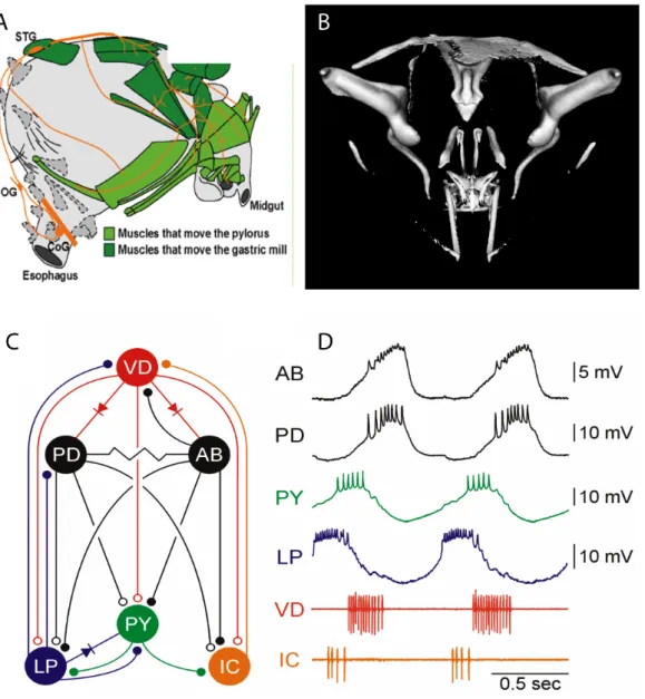
This study of Ih distribution and function was performed in pyloric neurons of the stomatogastic ganglion (STG) in the California Spiny Lobster Panulirus interruptus (Figure 1.1A). The stomatogastric nervous system (STNS) has been extensively studied as a model system for rhythm generation and neuromodulatory control of motor output (Maynard, 1972, Harris-Warrick and Marder, 1991, Hooper and DiCaprio, 2004, Marder and Bucher, 2006). It controls movement of the foregut (Figure 1.1 B, Figure 1.2 A and B) through repetitive activation of several groups of muscles, which attach externally to the stomach wall. The STNS consists of the connected paired commissural ganglia (CoGs), the esophageal ganglion (OG) and the stomatogastric ganglion.
Figure 1.1 A. Dorsal drawing of a California Spiny Lobster Panulirus interruptus (L.B.
Holthuis, Rathbun, 1884) B. Location of the foregut or stomach and parts of the nervous system in a stylized lateral view of the Maine Lobster Hommarus americanus (from Marder and Bucher, 2006)
The STG contains about 30 cells of 20 different cell types. With the exception of two identified interneurons (AB and INT1), most of these neurons are bi- functional: they are both members of CPGs that drive rhythmic foregut movements and motor neurons that innervate pyloric and gastric mill muscles and drive the movements evoked by CPG activity. The crustacean STNS can stay alive and
12
produce spontaneous rhythmic firing up to 5 days in culture. The pyloric network (Figure 1.2C,D) is one of three spontaneously active networks in the STG (Harris- Warrick et al., 1998, 1992) and controls rhythmic peristaltic and filtering movements of the posteror foregut to move food particles into the midgut at a frequency of 1-2 Hz. This network contains six different types of neurons, five of them motoneurons, which all have been well characterized (Hille, 2001, Turrigiano,1994, 1999, Harris-Warrick et al., 1992. Selverston and Moulins, 1986, Marder and Thirumalai, 1996, Marder and Calabrese, 2002). The pyloric network produces a stereotyped triphasic output with characteristic phases for each neuron (Figure 1.2D). Order of firing is determined both by synaptic interactions and by differences in intrinsic properties of the neurons, including rhythmic oscillatory bursting, bistability, post-inhibitory rebound and spike adaptation; these depend on the coordinated activity of different currents in the different neurons. In the presence of modulatory input from the CoGs, endogenous bursting of the AB interneuron plays the most important role to set the frequency of the pyloric rhythm. The two PD neurons are electrically coupled to the AB cell. This pacemaker group bursts together and inhibits all other pyloric neurons. Within the pyloric network, all known chemical synapses are inhibitory and mediated by glutamate, with the exception of the PD and VD neurons, which use acetylcholine in their inhibitory synapses onto other cells (Cleland and Selverston 1998, Eisen and Marder 1982, King 1976a, King 1976b, Marder and Eisen 1984, Marder and Paupardin-Tritsch 1978, Wyman 1976). Rectifying and non-rectifying electrical synapses also exist within the network.
13
Figure 1.2 Schematics of the crustacean stomach and nervous system. A. In situ position of the stomatogastric nervous system (orange) and its target muscles (green) in a lateral view of the stomach. B. Reconstruction of the oscicles which span the foregut. C.
Neuron types of the pyloric network: anterior burster interneuron (AB), lateral pyloric (LP), inferior cardiac (IC), ventral dilator (VD), two pyloric dilator (PD) and up to 8 pyloric (PY) neurons. D. Rhythmic activity of pyloric neurons in intracellular (top four) and extracellular (bottom two) recordings. (A from Marder and Bucher, 2007; B from K. H. Hobbs and S. L. Hooper, 2007,unpublished; C and D from Weaver, 2002)
14
The other neurons in the STG belong to the gastric-mill and the cardiac-sac networks. Chewing movements of the median and lateral teeth are caused by the slower and more variable gastric mill rhythm, which usually shows periods of 8-20 s (Clemens et al., 1998, Marder, Bucher 2007). Occasionally, a third monophasic rhythm can be observed in the STG that drives contraction of the cardiac sac. This slow rhythm with a period from 15-20sup to several minutes recruits the pyloric VD and PD neurons to elicit long bursts with characteristically large amplitudes of underlying depolarization in phase with the cardiac dilator neuron (CD2). Other cardiac-sac neurons are located in the commissural ganglia (CoGs) and in ivn fibers (Ayali, Harris-Warrick 1998, Dickinson 2006). Behavioral studies have confirmed the existence of similar nerve and muscle activity patterns in vivo in resting and feeding animals (Heinzel, Weimann and Marder 1993).
Modulation
Motor output from STG neurons can be altered through application of neuromodulators. Their effects on network activity can be slow or rapid and last from seconds to hours (Marder 1976, Marder and Eisen, 1984, Katz et al., 1990, Marder and Bucher, 2001, Nusbaum, 2001, Harris-Warrick et al., 1998, Dickinson 2006, Marder and Bucher 2006). In vivo, the CoGs and the OG provide over 100 axons which generate descending modulatory input to the STG, the loss of which temporarily abolishes spontaneous bursting of STG neurons (Marder and Bucher, 2006, Thoby-Brisson et al., 2002, Turrigiano 1995, Harris-Warrick et al., 1995, Golowasch et al. 1992). Sensory feedback from serotonin-containing mechanoreceptors provides another source of modulation is (Katz and Harris- Warrick 1989a,b,c, 1990, 1991, Beenhakker et al., 2004, Beenhakker et al., 2005, Beenhakker and Nussbaum, 2004, Blitz et al., 2004). Neuromodulators target both the intrinsic firing properties of pyloric network neurons and their chemical and electrical synapses. All synapses of the STG can be modulated, and the sensitivity to a large number of different neuromodulators has been shown (Ayali, Harris-Warrick 1998, Ayali, Harris-Warrick 1999, Johnson, Kloppenburg and
15 Harris-Warrick 2003, Kiehn, Harris-Warrick 1992, Marder, Bucher 2007, Marder, Eisen 1984, Peck et al. 2001a, Turrigiano, Marder 1993). Often, application of the same modulator causes opposite effects in different cells or even different synapses of the same neuron. For example, dopamine differently affects the mixed electrical-chemical synapse between pyloric (PY) and lateral pyloric (LP) neurons.
It reduces the excitatory electrical component of the synapse and strengthens the chemical inhibition, functionally inverting the sign of the synaptic interaction (Ayali, Johnson and Harris-Warrick 1998, Johnson, Harris-Warrick 1997, Johnson, Peck and Harris-Warrick 1995).
Cell-type specific modulation of intrinsic cellular firing properties can arise due to expression of modulatory receptor types with different second messenger mechanisms or the differential expression of ion channels which could be modulated. Recent data show that ion channel expression in pyloric neurons seems to follow a cell type-specific pattern. Differences in the expression of potassium currents like IA, IKV, and Ih, calcium activated IKCA, as well as L-type calcium and persistent sodium current have been characterized (Baro et al. 2000, French, Lanning and Harris-Warrick 2002, Gruhn et al. 2005, Harris-Warrick et al.
1995a, Ouyang, Goeritz and Harris-Warrick 2007b, Peck et al. 2001a, Schneider et al. 2000, Zhang, Wootton and Harris-Warrick 1995). For several channels, expression levels have been estimated based on the number of mRNA transcripts, which can been determined in individual cell types with the help of single cell RT- PCR (Baro et al., 1996, 1997, (Schulz, Goaillard and Marder 2006, Schulz, Goaillard and Marder 2007). Depending on the neuron type, these currents can respond oppositely to the same neurotransmitter. Dopamine for example increases the transient IA in PD neurons, while it reduces the same current in LP, AB, IC and PY neurons (Kloppenburg et al, 1999, Peck et al., 1995).
Of these currents, Ih is of special interest because of its involvement in homeostatic mechanisms and its interaction with the fast potassium outward current IA
(MacLean et al. 2003, Zhang et al. 2003b). Its susceptibility to neuromodulation
16
is well known, but its functional role in the STG is still poorly understood (Peck et al. 2006).
1.3 The Hyperpolarization-Activated Inward Current Ih
Ihis a hyperpolarization-activated, small inward current of mixed cations, mostly carried by Na+and K+ ions. It reacts very sensitively to changes of the external K+ concentration. The hyperpolarization-induced activation of Ih channels usually follows a very slow time course with time constants (!) on the order of seconds.
Because of its slow kinetics and its extremely hyperpolarized V1/2actof -70mV and below, it is likely that in many neurons Ih channels mainly function as a leak conductance within the neuron’s physiological range of membrane potentials. The channel is a tetramer, of which each subunit contains six trans-membrane domains.
All Ih subunits show an intracellular cyclic nucleotide (CN)-binding site close to the C-terminus.
Modulation and Pharmacological Interactions of Ih
Ih is found in the central and peripheral nervous system and in cardiac pacemaker tissue (Pape 1996, Kaupp and Seifert 2002, Robinson and Siegelbaum 2003). It is best known for contributing to the resting membrane potential and for being involved in rhythm generation. Ih helps set the resting potential as a leak current, plays a role in the generation of plateau potentials (Kiehn and Harris-Warrick, 1992, Robinson and Siegelbaum, 2003, Beaumont and Zucker, 2000,) and functions as a depolarizing pacemaker current in the heart (Baruscotti and Difrancesco 2004, DiFrancesco 2006, DiFrancesco and Ojeda 1980). Ih
contributes to postinhibitory rebound by generating depolarizing sag potentials upon hyperpolarization, which deactivate only slowly upon repolarization (Harris- Warrick et al. 1995a, Pape 1996, Robinson and Siegelbaum 2003). It is also involved in regulation of synaptic transmission, long term facilitation and integration of synaptic events through shaping temporal summation as well as
17 spatial normalization of distant synaptic events (Beaumont and Zucker 2000, Genlain, Godaux and Ris 2007, Harris-Warrick et al. 1995b, Magee, 1998, 1999, Williams and Stuart, 2000, Berger et al. 2003, Migliore et al., 2004). In layer V pyramidal cells of the somatosensory cortex, Ih channel activation at hyperpolarized membrane potentials disconnects somatic and dendritic spike innitiation zones and thus may prevent initiation of dendritic calcium action potentials in the absence of proximal input (Berger et al., 2003). In the STG and at the neuromuscular junction, Ih could be partially responsible for enhancement of synaptic strength during aminergic modulation (Beaumont and Zucker, 2000) (Harris-Warrick 2002, Johnson and Harris-Warrick 1997).
Neurotransmitters can facilitate Ihactivation by increasing the cAMP level (Rateau and Ropert 2006, Robinson and Siegelbaum 2003, Rosenkranz and Johnston 2006, Santoro and Baram 2003, Svoboda and Lupica 1998). Binding of cAMP at the CN-binding site shifts the voltage dependence of activation to more depolarized potentials and enhances the amplitude of maximal conductance. This site also binds cGMP with a lower affinity (Robinson and Siegelbaum 2003). During prolonged whole cell recordings, a dramatic run-down of Ih occurs, which can shift the voltage dependence of activation by 40-50 mV in the hyperpolarizing direction.
Reductions in cAMP levels account for less than half of the run-down effect, and it is still unknown what regulatory factor causes the largest part of this shift (Chen 2004, DiFrancesco 1986, Robinson and Siegelbaum 2003). Of the vertebrate isoforms of Ih, HCN2 - 4 are most sensitive to up-regulation by cAMP (Pape 1996, Robinson and Siegelbaum 2003). Ih has been found in all six pyloric neuron types in the STG, where it is subject to differential monoaminergic modulation (Harris- Warrick et al. 1995a; Peck et al. 2006; Thirumalai et al. 2006). For example, dopamine enhances Ih in the lateral pyloric (LP) neuron by shifting the voltage dependence of activation to more depolarized values and by accelerating its rate of activation. In motor-neurons of the feeding circuit of the snail Lymnaea, modulation by serotonin increases Ih, causing prolonged depolarization of the membrane potential, triggering conditional endogenous bursting properties, and
18
enhancing postinhibitory rebound (PIR) properties (Straub and Benjamin, 2001).
In higher brain regions like the hypothalamus, hippocampus and medulla oblongata, serotonin diminishes Ih by reducing the maximal conductance and causing a negative shift of the voltage dependence of activation. This effect is mediated by 5-HT2 receptors, which activate protein kinase C (Liu et al., 2003).
Pharmacologically, Ih can be blocked by bath application of 5-10 mM CsCl, 50-200 µM ZD7288 or, in the case of HCN1 channels, by the drug zatebradine and the VR1 receptor antagonist capsazepine (Gill et al. 2006, Kiehn, Harris-Warrick 1992, Ouyang, Goeritz and Harris-Warrick 2007). Forskolin increases Ih by upregulation of cAMP. The anticonvulsant drug lamotrogine enhances Ih in the dendrites of pyramidal neurons by shifting the voltage dependence of activation (Poolos et al., 2002). Gabapentin enhances Ih in pyramidal CA1 neurons by increasing the conductance without influencing the properties of activation (Surges et al., 2003).
Soma recordings of Ih in pyloric neurons show that the activation and deactivation of this current is most likely too slow to directly impact firing properties by changing its conductance on a cycle-by-cycle basis in the pyloric network. The large degree of Ih modulation in many rhythmically active systems suggests a functional role of Ih,, which may vary in a state-dependent way. Ihcould be simply help set the membrane resting potential, but it could also participate in more complicated mechanisms that affect regulation of the strength of synaptic transmission or temporal integration of synaptic events. Knowledge of Ih channel distribution in the STG might imply a functional role more clearly.
1.4 Homeostatic Relationship of Ih and IA
A compensatory homeostatic relationship between Ih and the fast transient outward current IA has been previously described (MacLean et al. 2003, MacLean et al.
2005, Zhang et al. 2003a). IA is a transient depolarization-activated potassium current whichrapidly inactivates during maintained depolarization but shows a fast
19 recovery from inactivation at hyperpolarized levels. IA is involved in several aspects of neuronal excitability, including the timing of action potentials after hyperpolarization, the time course of postinhibitory rebound during bursting, spike adaptation and control of the interspike interval (Graubard and Hartline, 1991, Golowasch and Marder, 1992, Tierney and Harris-Warrick, 1992, Harris- Warrick et al., 1995a,b, 1998, Kloppenburg et al., 1999, Peck et al., 2001, Hille, 2001).
Enhancing IA in the PD neuron during bath application of dopamine dampens its bursting properties. Increased IA reduces the rate of rebound after the post-burst hyperpolarization, increasing the first spike-latency and length of first inter-spike interval, and reducing the number of spikes per burst (Harris-Warrick et al. 1995a, Kloppenburg, Levini and Harris-Warrick 1999, Peck et al. 2001b, Tierney, Harris-Warrick 1992). Therefore, over-expression of IA by RNA injection of its gene, shal, would be expected to cause dramatic changes of firing properties.
Instead, previous work from our lab found that the firing patterns after IA over- expression to be unchanged, due to a compensatory up-regulation of Ih (MacLean et al., 2003; 2005). The homeostatic interaction of these two currents appeared to be independent of activity changes, as over-expression of a mutated, non- functional form of IA still caused an increase of the measured Ih. Therefore, a novel molecular mechanism was proposed to exist, which could recognize the presence of additional IA mRNA or protein, and trigger a compensatory increase of Ih.
This homeostatic response could be unique for PD neurons or might be a more general mechanism of maintaining stable levels of rhythmically active networks.
Knowledge of its presence in other STG neurons might indicate whether or not similar IA-Ih interactions could be expected to be found in other systems.
Similarly, a reverse interaction where overexpression of Ih leads to increased IA
would point to this response as a more general homeostatic mechanism. Ectopic expression of Ih by injection of RNA from a closely related species, Panulirus
20
argus, did not affect the expression of IA, but this could be an artifact of inter- species differences in the Ih sequence (Zhang et al. 2003).
1.5 Aim of this Study
In order to better understand the role of Ih in the pyloric network, I examined three aspects of Ih. First, homeostatic interactions of IA and Ih in pyloric neurons were examined in more detail, to determine whether this could be a general mechanism to regulate network activity. The directionality of the homeostatic response was studied by overexpressing Ih splice variants of P. interruptus in oocytes and PD neurons, to see if a species-specific form of Ih could lead to an up-regulation of IA. Second, the distribution of endogenous Ih protein in STG neurons was determined by immunocytochenistry and confocal imaging and compared to the expression pattern of a synaptic marker and of IA protein. Finally, a potential role of Ih in regulating graded synaptic transmission was examined by studying the effects of Ih
activation at the graded glutamatergic LP to PD synapse.
21
2 Methods
2.1 STG dissection and PD cell identification
Adult California spiny lobsters, P. interruptus, were obtained from Don Tomlinson Commercial Fishing (San Diego, CA) and maintained in artificial sea water at 16°C until use. Lobsters were anesthetized by keeping them on ice for 30 min before dissection. The STG, along with its motor nerves and associated commissural and esophageal ganglia, was dissected and pinned in a silicone elastomer (Sylgard)- coated dish containing saline, as described by Mulloney and Selverston (1974). The physiological saline solution consisted of (in mM): 479 NaCl, 12.8 KCl, 13.7 CaCl2, 3.9 Na2SO4, 10.0 MgSO4, 2 glucose, and 11.1 Tris base, pH 7.4 (Mulloney and Selverston 1974). The PD, VD and LP neurons were identified during intracellular recordings (3 M KCl, 10–25 M") by their typical membrane potential oscillation shapes and synaptic inputs (Kloppenburg et al.
1999), and IC neurons and gastric neurons were identified by extracellular recordings with suctions or pin electrodes of the respective nerves.
2.2 RNA microinjection into neurons Pyloric neurons
Capped RNA was transcribed from linearized DNA clones with a T3 (shal, shal- GFP, mshal) or SP6 (PIIH, GFP) mMessage mMachine kit (Ambion), using T3 or SP6 RNA polymerase. The capped transcripts were cleaned using the RNeasy mini kit (Qiagen).
The RNA solution contained 0.25– 0.5 µg/µl PIIH or GFP(control) cRNA and 0.08% Fast Green to monitor the injection and was centrifuged at 3600 rpm for 10
22
minutes prior to injection. After identification, pyloric neurons were injected with RNA using pressure pulses (40 psi; 0.2 Hz, 30- to 70-ms duration) driven by a homemade pressure injector and a pulse generator (Master-8; AMPI, Jerusalem, Israel). After injection, the whole preparation was incubated in sterilized recording saline without Tris base, but containing 5 mM HEPES, pH7.4, 2 g/l glucose, 100,000 unit/l penicillin, and 100 mg/l streptomycin at 16°C for 4–5 days to allow protein expression.
Xenopus oocytes
Stage V to VI oocytes were surgically removed from female frogs during anesthesia in 0.15% MS222 (3-aminobenzoic acid ethyl ester). The eggs were then treated with 1 mg/ml collagenase type IA in solution containing (in mM): 82.5 NaCl, 2 KCl, 1 MgCl2, and 5 HEPES (pH 7.5) for 60 min. The oocytes were defolliculated, but the vitelline membrane was not removed. Isolated oocytes were injected with 40 nl of shal or PIIH cRNAs (250–500 ng/µl) and cultured in ND96 solution containing (in mM): 96 NaCl, 2 KCl, 1.8 CaCl2( _2H2O), 1 MgCl2, and 5 HEPES (PH7.6) supplemented with 50 mg/l gentamicin, 2.5 mM Na pyruvate, and 5% horse serum for 3–4 days until recording.
Oocytes were injected with a Sutter Instrument microinjector (model NA-1)(San Rafael, CA). This was used to inject ~ 100nl of cRNA (concentration ~50 ng/µl) into Xenopus oocytes, which were isolated and maintained according to Quick et al. (1992). Recordings were made by two electrode voltage clamp 3 days later.
2.3 Electrophysiology Voltage clamp
Immediately after identification or after 4–5 days in organ culture, PD neurons were voltage clamped using an Axoclamp 2A amplifier and pClamp8 software (Axon Instruments, Foster City, CA). Microelectrodes were filled with 3 M KCl and had a tip resistance of 8–10 M" for voltage recording and <8 M" for current
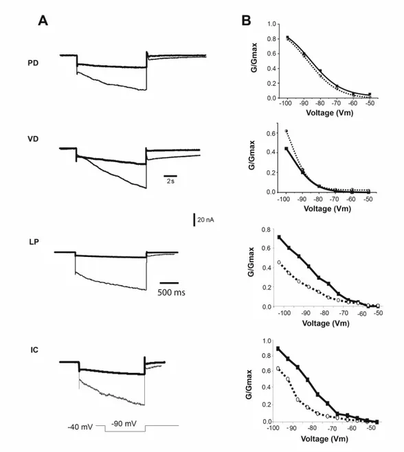
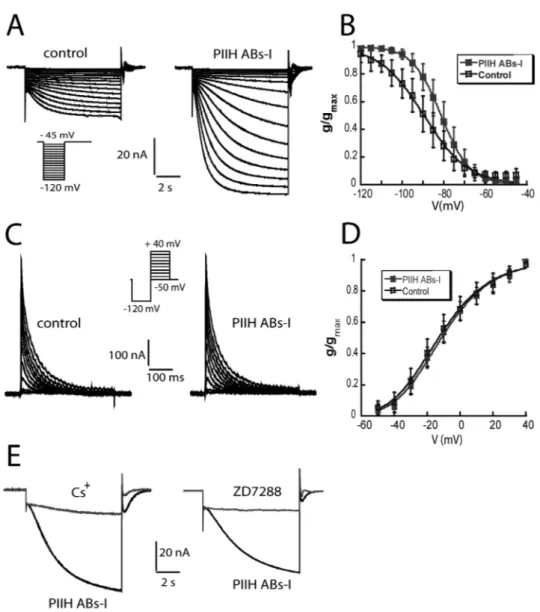
23 injection. To isolate neurons from most synaptic input and to isolate Ih and IA from most other currents, we superfused the ganglion with saline containing 10-7 TTX, 5 x 10⁻⁶ M picrotoxin and 20 mM tetraethylammonium chloride (TEA). For some experiments, 5 and 10 mM CsCl or 50 and 100 µM ZD7288 were added to block Ih. To measure Ih, the cells were held at –40 mV, and the voltage dependence of activation was measured with a series of 8-s hyperpolarizing voltage steps in 5-mV increments from –45 to –120 mV at 20-s intervals. Because the time constant of activation is slow, no leak subtraction was used, so any instantaneous leak current is detectable at the beginning of the step; this value was subtracted from the amplitude of Ih. The reversal potential of Ih was measured from the tail currents after a pre-activating pulse to –100 mV for 8 s with a series of 4-s pulses from –70 to +30 mV in 10-mV increments.
To measure IA, the cells were held at –50mV and the voltage dependence of activation was measured following a deinactivating prepulse to –120 mV for 400 ms and then a series of 400-ms voltage steps from –50 mV to +40 mV in 10-mV increments. A control protocol for activation of non-IA currents was the same as the activation but without the deinactivating step to –120 mV. Traces were leak subtracted using a P/6 protocol with steps opposite to the sign of activation. The control protocol currents were digitally subtracted from the activation protocol currents to isolate IA.
Synaptic transmission measurements
The PD and LP cells were impaled with two electrodes each to allow independent current injection and voltage recording in each cell. Synaptic measurements were recorded during current clamp with constant amplitude depolarizations of the pre- synaptic LP neuron to evoke IPSPs in the PD cell. Action potentials and transient potassium currents were blocked with 0.1 µM TTX, and 4 mM 4-AP. LP-evoked IPSPs were recorded in the PD during current injection to evoke a range of
24
hyperpolarizing steps in PD neurons that activated Ih to differing extents. The amplitudes of IPSPs were measured with and without Ih activation by taking advantage of the very slow activation rate of Ih channels. At the beginning of PD hyperpolarization Ih channels have only just begun to open, so Ih activation is low.
IPSP amplitudes at this point were compared to those recorded after 8 s of PD hyperpolarization, when Ih channels were more completely activated. Current injection protocols were generated by Clampex software (Molecular Devices, CA).
The PD membrane potential was changed by a series of 8-sec current injecting steps in 0.5 - 2 nA increments, at one minute intervals to allow recovery of Ih. If necessary, a bias current was injected into the PD cell to hold the membrane potential at -58mV, which was approximately the average PD membrane potential after blocker application. This is a relatively hyperpolarized potential compared to typical cycling PD neurons, which have a trough around -55 mV. It was chosen to include a maximal number of earlier recordings, when bias current injection was not applied. IPSPs were elicited by 200 ms depolarizing steps to -30 mV into the LP cell at the beginning or at the end (after7.8 sec) of the PD polarization. The LP cell was held at -58 to -60mV between steps. To avoid Cl- loading during the current steps, which would alter the Vrev of the IPSP, we used electrodes filled with 0.6M KSO4 + 20mM KCl. and relatively high resistance (20 M" or higher) electrodes were used for the voltage recording. IPSP amplitudes were measured and plotted against the membrane potential prior to the IPSP.
Xenopus oocytes
A standard two-microelectrode voltage clamp was used to measure the current properties of the PIIH splicing variants. The oocytes were voltage clamped using a Geneclamp amplifier driven by Clampex 8.0 software (Molecular Devices, CA).
All recording were made in standard ND96 solutions without gentamicin, Na pyruvate, and horse serum. For some experiments, 5-10 mM CsCl or 50-100 µM ZD7288 were included. Microelectrodes were filled with 3 M KCl and had a tip
25 resistance of 1–5 M". We measured Ih from a holding potential of –40 mV with steps from -50 to -120 mV, as described above. These protocols were used in all of the PIIH splice variants except PIIH-I. For PIIH-I, the voltage activation curve was measured with hyperpolarizing voltage steps from –70 to –140 mV, and the preactivating pulse for measuring the reversal potential was to –120 mV. The effect of cAMP was tested by recording the basic parameters before and after switching to a bath solution containing the membrane- permeable cAMP analog, 8- Br-cAMP. Perhaps due to the presence of the vitelline membrane on the oocytes, bath application of 1 mM 8-Br-cAMP caused only a subtle modulation of the activation kinetics and voltage-dependent activation of PIIH channels after 1.5 h.
To accelerate the rate of increase in intracellular cAMP in the large oocytes surrounded by a vitelline membrane, we increased the concentration of 8-Br-cAMP to 10 mM; responses to this larger dose were seen within 30 min.
Current analysis
To compare data with earlier work, Ih amplitudes were measured from single exponential fits of the data performed in Clampfit, version 9.0 (Molecular Devices), extrapolated back to the beginning of the hyperpolarizing step (at the point of the leak current) and forward to approximate the steady state at 10 s.
Currents were converted to conductances, using a reversal potential (Vrev) of -30 mV for STG neurons and -40 mV for oocytes. (Zhang et al. 2003). The conductance-voltage data were fit to a first order Boltzmann equation (1):
s n
mV V
g e g
) 1
(
1
2) / ( 1
max
!
!
+
=
26
where g is the conductance, gmax is the maximal conductance, V1/2 is the voltage of half-activation, s is the slope factor and n = 1 for Ih. The voltage dependence of activation of IA was determined by converting the peak current to a peak conductance, g, assuming Vrev = –86 mV (Hartline and Graubard, 1992). The resulting g/V curve was fitted to a Boltzmann relation (Eq. 1) but with n = 3.
Analysis of rhythmic activity
We analyzed rhythmic activity in PD neurons using Spike2 (Cambridge Electronic Design, Cambridge, UK). The minimal membrane potential (Vmin) was measured at the most hyperpolarized potential in the trough of the oscillation. The oscillation amplitude was the difference between Vmin and the most depolarized potential of the slow wave oscillation (at the base of the action potentials, Vmax).
The time to the first spike was the time from Vmin to the top of the first spike. The cycle duration was the time between the Vmin of two adjacent oscillations, and the duty cycle was the burst duration divided by the cycle duration. All measures were based on average measures of 40 cycles.
Statistics
All values are given as the mean ± SD. Statistical significances were determined using ANOVA and Student’s t-test. Regression lines were plotted, and R values determined using SigmaPlot and SigmaStat 10.0 (Systat Software).
Modeling of IPSP amplitudes.
The ratio of IPSP amplitudes at different membrane potentials was predicted by a simplified model, that takes into account the internal and external chloride and potassium activities and reflects the changing driving force for the IPSP at different membrane potentials (Hille, 2001, Hammond, 2001). In this model, IPSP amplitudes were considered proportional only to the changing driving force of the synaptic chloride currents through the inhibitory glutamate receptors (GluR), while
27 all other voltage-dependent changes of the membrane conductance were either blocked or insignificant. Ohm’s law (1) describes the amplitude of IPSPs recorded from the soma. The driving force for the synaptic chloride flux across the membrane changes as a function of electrochemical and concentration gradients according to the Goldman Hodgkin Katz current equation for a single ion species at constant field (2). Under these simplifying assumptions, f was determined as the voltage dependent factor of the synaptic current at a given membrane potential (3).
The experimentally derived IPSP amplitude at -90 mV in the presence of 5mM CsCl was chosen as reference, in order to calculate expected IPSP amplitudes at different membrane potentials in the absence of Ih activation (4). Ih is blocked under these conditions and most likely no other voltage dependent channels are active. The extracellular chloride concentration [Cl]o was 510mM, and the intracellular chloride activity [Cl]i was assumed to be 37mM at 20°C, based on the IPSP reversal potential and documented values in seawater crustaceans (Freel, 1978, Theander et al, 1999, Cleland and Selverston, 1995, 1998, Marder and Eisen, 1982, Hashemzadeh-Gargari and Freschi, 1992, Doolin et al., 2001).
(1)
m IPSP
IPSP g
V = I
VIPSP : postsynaptic inhibitory potential, recorded in the soma; IIPSP:synapticcurrent; gm : overall membrane conductance.
28
(2)
RT Vm zF
RT Vm zF i o
m
e e Cl Cl
RT V zF
I p ( )
) ( 2
1 ] [ ] [ ) ( ) (
!
!
!
"
" !
=
I: ionic flux; p: opening probability of GluR; F: Faraday number; z: charge; Vm: membrane potential; [Cl]o and [Cl]i: extracellular and intracellular chloride concentrations;
(3)
RT Vm zF
RT Vm zF i o
m Vm
e
e Cl Cl
f V ( )
) ( )
(
1
) ]
([
] ([
) (
!
!
!
"
!
"
=
(4)
) (
) 90 ( ) 90 ( )
(
Vm IPSP Vm
IPSP f
f
V V ! " !
=
2.4 Immunocytochemistry
Selected neurons were filled with 4% neurobiotin (NB) in 50mM Tris and 0.5M KCl. For neurobiotin injection, tips of low resistance electrodes (3-5 M" when filled with 3M KCl) were backfilled with the NB solution for 10 minutes. The shaft was then filled with 2 M KCl, leaving a small (1cm) gap between the NB in the tip and the KCl in the shaft. The resistance of the filled electrode was 25-90 M". Neurobiotin was injected for about 40 minutes with 500ms,+5nA pulses at 1Hz. Preparations were left for 1 hour for individual neuron staining, or for 3-16 hours for additional gap junction-mediated staining of electrically coupled neurons.
For example, after three hours of incubation of a neurobiotin injected VD cell ,the VD, AB, both PD neurons and INT1 were strongly stained and both LPG neurons were more weakly stained. Interestingly, neurobiotin crosses rectifying electrical synapses only in the direction of the rectification. In PD neurons, thirty minutes to one hour of incubation after neurobiotin injection caused no staining of other
29 neurons, whereas longer periods of incubation resulted in additional filling of the other PD and the AB neuron.
STGs were fixed in 3.5% or 2% paraformaldehyde in phosphate buffered saline (PBS) for 90 minutes at room temperature. The fix was washed out with 8 changes of PBST (PBS + 0.3-1 % triton X100) over 2-8 hours. The tissue was then blocked for 3 hours with 5% normal goat serum and 1% BSA in PBST at room temperature and incubated overnight in a rabbit anti-shal (1:2000) , rabbit anti-synaptotagmin (1:1000), or mouse anti-pentaHis (1:20) primary antibody in PBST +5% normal goat serum and 0.1% BSA. The primary antibody was washed out with PBST for 2 hours. The tissue was then incubated for 2 hours with alexa 488-, alexa 568- or alexa 635-conjugated goat anti-rabbit, goat anti-mouse or streptavidin secondary antibodies (Molecular Probes) at 1:500 dilution in PBST + 5% normal goat serum and 0.1% BSA. The secondary antibody was washed out with PBS for 2 hours.
All incubations were performed at room temperature with constant shaking. The STG was mounted and cleared on a slide with Vectashield mounting media (Fluka). In several experiments, fixed ganglia were imbedded in 40°C warm, 4%
low melting point agarose (Sigma) in Panulirus saline prior to antibody treatment.
Slices (40-70 #m) were made with a vibrating microtome (Leica Microsystem, speed 4; frequency 9) and transferred to PBST filled wells. Antibody treatment was performed on the floating agarose sections or individual ganglion slices on a slide. Antibody staining in images of x-y planes or series of z-stacks were visualized and collected with a Leica TCS SP2 confocal system. For multiple staining, sequential imaging and narrow emission settings were used to prevent bleed-through effects. Image analysis and 3D reconstructions were performed with Volocity Visualization and Classification Software.
30
3 IA-Ih Homeostasis in Pyloric Neurons
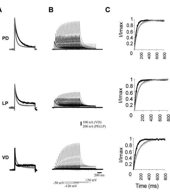
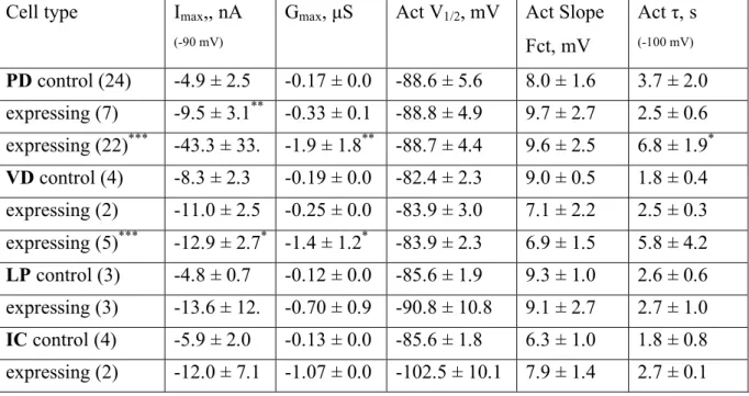
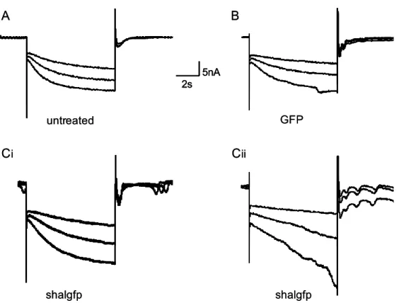
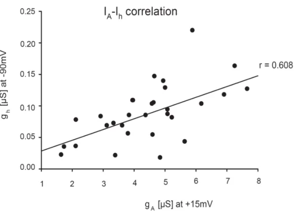
Previously, our lab found a homeostatic relationship between expression of the slow hyperpolarization-activated inward current Ih and the fast transient outward current IA (MacLean et al. 2003, Zhang et al. 2003a). In the lobster, the shal gene encodes IA in all six classes of pyloric neurons (Baro et al., 1996; Baro et al., 2000). An artificial increase of IA in one of the two electrically coupled Pyloric Dilator (PD) neurons, by intracellular injection of shal-GFP RNA, was accompanied by a compensatory increase of Ih, such that the firing properties of the PD neurons did not change despite significant increases in both IA and Ih. This homeostatic up-regulation of Ih appeared to be independent of activity changes, as overexpression of a mutated, non-functional form of IA still caused an increase of Ih current. A novel molecular mechanism was proposed which could detect the presence of additional IA RNA or protein and consequently trigger the compensatory increase of Ih.
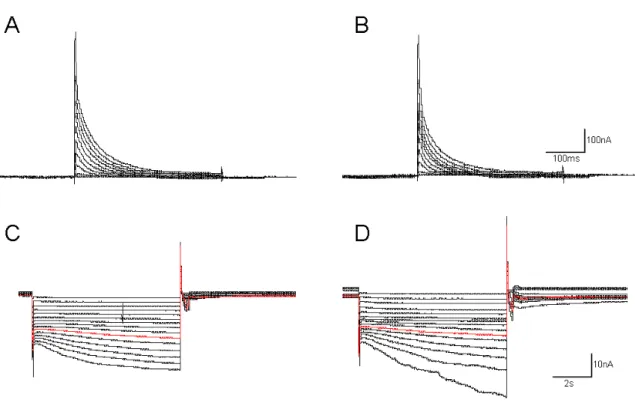
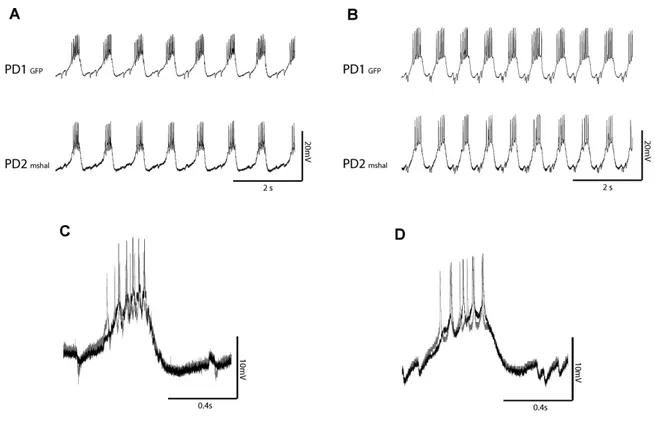
To understand this interaction, and to determine if it is unique to PD neurons, in collaboration with Dr Jason MacLean, I over-expressed IA via shal-GFP RNA injection in three pyloric neurons, the Pyloric Dilator (PD), the Lateral Pyloric (LP) and the Ventricular Dilator neuron (VD), and performed preliminary studies of the Inferior Cardiac (IC) neuron. This allowed me to examine in detail several properties of the system. 1) I studied the voltage dependence and kinetic properties of IA and Ih in each pyloric neuron type, to determine whether the newly inserted channels were modified to generate neuron-specific IA current properties, or whether a similar current is generated in all neuron types. 2) I studied the overall effect of shal-GFP overexpression on pyloric network activity patterns. 3) I characterized a low threshold, non-Ih component of the hyperpolarization