genome-wide data in cancer research
Inaugural-Dissertation zur
Erlangung des Doktorgrades
der Mathematisch-Naturwissenschaftlichen Fakultät der Universität zu Köln
vorgelegt von
Châu Nguyễn
aus Namdinh, Vietnam
Köln, 2014
Prof. Joachim Krug Prof. Leonid Mirny
Tag der letzten mündlichen Prüfung: 16.01.2015
Diese Arbeit beschäftigt sich mit der Maximum-Entropie-Methode im Zusammenhang statistischer Modellierung. Anwendungsbeispiele sind dem aufkommenden Forschungs- bereich der Krebsgenomik entnommen.
Wir beginnen mit einer kurzen Einführung in die Biologie von Krebserkrankungen in Kapitel 1. In Kapitel 2 diskutieren wir die Grundlagen der statistischen Modellierung, wo- bei wir eingehend die Maximum-Entropie-Methode besprechen. Insbesondere zeigen wir, dass viele statistische Modelle in einen einheitlichen Rahmen, basierend auf der Maximum- Entropie-Methode, gebracht werden können, der diese auf Probleme der statistischen Me- chanik abbildet. In Kapitel 3 befassen wir uns mit einem bestimmten Maximum-Entropie- Modell, dem Ising-Modell, im Kontext des inversen Ising-Problems. Wir führen eine Bethe–
Peierls-Näherung für das inverse Ising-Problem ein. Des Weiteren schlagen wir eine modi-
fizierte Version der Molekularfeld-Näherung vor, welche auch für niedrige Temperaturen
funktioniert. Die folgenden Kapitel wenden Maximum-Entropie-Modelle auf verschiede-
ne Probleme aus dem Bereich der Krebsgenomik an. Eine direkte Anwendung des inver-
sen Ising-Problems auf Daten zur Anzahl von Genkopien in Krebszellen wird in Kapitel 4
beschrieben. In Kapitel 5 erweitern wir die Konzepte der indirekten Korrelationen und
der direkten Kopplungen des inversen Ising-Problems um den Einfluss der Anzahl von
Genkopien auf die Expression von Genen in Krebszellen zu untersuchen. Wir zeigen, dass
die Korrelationen in der Genexpression nicht unbedingt durch regulatorische Wechselwir-
kung zwischen Genen hervorgerufen werden müssen. Stattdessen können die Korrelatio-
nen in der Genexpression durch die Korrelationen in der Anzahl von Genkopien hervor-
gerufen werden, was auf der geometrischen Organisation des Genoms beruht. Wir zeigen,
dass ein einfaches Maximum-Entropie Modell die Korrelationen in der Anzahl von Gen-
kopien von den sogenannten “blanken Korrelationen” in den Genaktivitäten, welche nur
den Effekt der regulatorischen Wechselwirkungen beschreiben, trennen kann. Kapitel 6 ist
der Klassifizierung von Krebs gewidmet. Wir führen einen einfachen semi-überwachten
Lernalgorithmus ein um eine Mischung aus paramagnetischen Modellen mit Ising-Spins
dahingehend zu trainieren, Krebsmutationsprofile zu klassifizieren. Wir zeigen, dass die-
ser Lernalgorithmus, mit der Möglichkeit sowohl von den nicht zugeordneten Proben zu
ten als auch die unüberwachten Lernalgorithmen übertrifft. Die zwei Anhänge A und B fassen die jüngeren Studien über die Sensibilität und die Widerstandsfähigkeit von Krebs- zellen gegenüber Therapien zusammen.
Die Ergebnisse von Kapitel 3 wurden in H. C. Nguyen and J. Berg (2012a). “Bethe–
Peierls approximation and the inverse Ising problem”. J. Stat. Mech. P03004; and H. C.
Nguyen and J. Berg (2012b). “Mean-field theory for the inverse Ising problem at low tem-
peratures”. Phys. Rev. Lett. 109, p. 50602 publiziert. Einige der Resultate aus Kapitel 6 wur-
den als Teil von The Clinical Lung Cancer Genome Project (CLCGP) and Network Genomic
Medicine (NGM) (2013). “A genomics-based classification of human lung tumors”. Science
Transl. Med. 5.209, 209ra153 publiziert.
This thesis studies the maximum entropy principle in statistical modelling. Applications are taken from the emerging field of cancer genomics.
We start with a short introduction to the biology of cancer in chapter 1. In chapter 2, we discuss general principles of statistical modelling. We discuss in detail the maximum entropy principle in statistical modelling. In particular, we show that many statistical mod- els can be put in a unified framework based on the principle of maximum entropy, which maps them into problems of statistical mechanics. In chapter 3, we consider a particular maximum entropy model, the Ising model, in the context of the inverse Ising problem. We introduce a Bethe–Peierls approximation to the inverse Ising problem. We then also sug- gest a modification for the mean-field approximation to work at low temperatures. The following chapters apply maximum entropy models to different problems of cancer ge- nomics. A direct application of the inverse Ising problem to gene copy-number data of cancer cells is described in chapter 4. In chapter 5, we extend the concepts of indirect cor- relations and direct couplings of the inverse Ising problem to investigate the influence of gene copy-numbers on gene expressions in cancer cells. We show that the correlations in gene expression need not be due to regulatory interactions between genes. Instead, corre- lations in gene expression of cancer cells can be induced by the correlations in their copy- numbers, which is due to the geometrical organisation of the genome. We show that a simple maximum entropy-model can disentangle copy-number-induced correlations and the so-called “bare-correlations” in gene expression, which capture the effect of regula- tory interactions alone. Chapter 6 is devoted to cancer classification. We introduce a sim- ple semi-supervised learning algorithm to train a mixture of paramagnetic models with Ising spins to classify cancer mutation profiles. We show that, with the capability of both learning from unlabelled samples and correcting mislabelled samples, this learning algo- rithm outperforms both the supervised and unsupervised learning algorithms. The two appendices A and B summarise recent studies on sensitivity and resistance of cancer cells to therapy.
The results of chapter 3 were published in H. C. Nguyen and J. Berg (2012a). “Bethe–
Peierls approximation and the inverse Ising problem”. J. Stat. Mech. P03004; and H. C.
peratures”. Phys. Rev. Lett. 109, p. 50602. Some results of chapter 6 were published
as a part of The Clinical Lung Cancer Genome Project (CLCGP) and Network Genomic
Medicine (NGM) (2013). “A genomics-based classification of human lung tumors”. Science
Transl. Med. 5.209, 209ra153.
In writing these lines, I am very grateful to my supervisor Johannes Berg. Over the years, I have learned from him many things, from how to pose a scientific problem to how to make scientific figures. I have also learned to be patient and to be humorous. Looking back, he has listened to my ideas however innocent they were. He has been very patient with my mistakes (sometimes repeatedly). His sense of humour has expanded in our group and has relaxed the stress of Ph.D. students.
Together with Johannes Berg, my mentors, Prof. Joachim Krug and Prof. Hartmut Monien also had encouraging advice. Comments and advice from my former supervisors, Prof. Markus Müller and Prof. V. Lien Nguyen, have been always very helpful. I also thank Prasanna, Nico, Nina, Nhung, Van-Anh for discussions and comments on different parts of this thesis. Nico also helped me with translating the abstract.
Our collaboration with Roman Thomas was very enjoyable. I have learned much about clinical biology through discussions with him and people in his group, Martin, Julie, Danila, Sandra, any many others. The contribution from the patients in the Clinical Lung Cancer Genome Project (CLCGP) and Network Genomic Medicine (NGM), who have con- tributed their data, played a vital role in my thesis. I consider my thesis as dedicated to them.
My friends in our group, Nico, Prasanna, Simon, Nina, Nandita, Filippos, Joachim, Bhavin have made my time in Cologne memorable. Our scientific and non-scientific chats during lunches and dinners, together with Michael Lässig and members of his group, Mara, Simone, Christian, Daniel, Torsten, Armita, Donate, Stephan and Stephane were very joyful. I am thankful to Frau Christa Stitz for her enthusiastic help, which saved me from a lot of bureaucratic problems.
The financial support from the DFG through the SFB-680 project, the emed-SMOOSE
project and the Bonn–Cologne Graduate School are acknowledged.
1 Introduction to the biology of cancer 3
1.1 The human body homeostasis . . . . 3
1.2 Tumours in the human body . . . . 7
1.2.1 Tumours are of genetic nature . . . . 8
1.2.2 Somatic mutations and the tumour founder cells . . . . 9
1.2.3 Genetic instability and tumour hypermutations . . . 10
1.2.4 Somatic selection and the evolution of a tumour . . . 11
1.2.5 Tumour heterogeneity, clonal interactions and cancer stem cells . . . 15
1.3 Cancer genomics and statistical modelling . . . 18
2 Probability, information and statistical modelling 23 2.1 Probability and information . . . 23
2.1.1 Probability . . . 23
2.1.2 Information: entropy and relative entropy . . . 24
2.2 Statistical modelling . . . 26
2.2.1 Assumptions . . . 26
2.3 Maximum entropy principle and linear models . . . 31
2.3.1 Gaussian model . . . 33
2.3.2 Linear regression . . . 36
2.3.3 Logistic regression . . . 42
Binary logistic regression . . . 42
Categorical logistic regression . . . 44
2.3.4 Mixture model classification . . . 45
Gaussian mixture model . . . 45
Paramagnetic Ising mixture model . . . 47
2.4 Beyond parametric and probabilistic models . . . 49
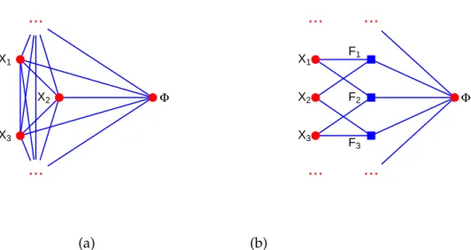
3 The inverse Ising problem 51 3.1 The inverse Ising problem . . . 51
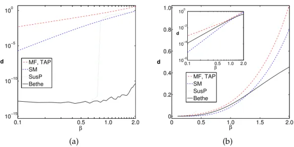
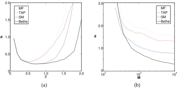
3.2 Bethe–Peierls approximation and mean-field-like methods . . . 54
3.2.1 Mean-field approximation . . . 55
3.2.2 Bethe–Peierls approximation . . . 57
3.3 The inverse Ising problem at low temperatures with mean-field approximations . . . 64
4 Gene copy-number correlations in cancer cells 73
4.1 The gene copy-number variation in cancer cells . . . 73
4.2 Correlation and interaction: the inverse Ising problem . . . 77
4.2.1 The inverse Ising problem in mean-field approximation . . . 79
4.2.2 Analysing the copy-number data of lung cancers . . . 79
4.3 Conclusions and outlook . . . 83
5 Gene expression correlations in cancer cells 85 5.1 Gene expressions of cancer cells and the influence of copy-number alterations . . . . 85
5.2 Elimination of the copy-number-induced correlation . . . 88
5.2.1 Maximum entropy model for induced-correlation elimination . . . 88
5.2.2 Analysing the copy-number and gene expression data of lung cancer patients 91 5.3 Conclusions and outlook . . . 94
6 Semi-supervised classification of lung cancers 97 6.1 Cancer classification based on mutation profiles . . . 97
6.2 Semi-supervised model to classify cancer mutation profiles . . . 100
6.2.1 Semi-supervising the mixture of paramagnetic Ising models . . . 100
6.2.2 Analysing the lung cancer mutation profiles . . . 108
6.3 Conclusions and outlook . . . 110
A Drug sensitivity of cancer cells 113 A.1 The drug responses of cancer cells . . . 113
A.2 Turning the regression problem to a data-recovery problem . . . 117
A.2.1 Drug response as a regression problem . . . 117
A.2.2 Analysis of the drug-response library . . . 120
A.3 Conclusions and outlook . . . 122
B The emergence of drug resistance in an expanding tumour 125 B.1 Evolution of drug resistance in cancer . . . 125
B.2 The mergence of a mutated clone in an expanding population . . . 127
B.2.1 Birth-death process with arrivals . . . 127
B.2.2 A model of tumour expansion and the emergence of resistant cells . . . 133
B.3 Conclusions and outlook . . . 137
Introduction to the biology of cancer
More is different.
P. W. Anderson
In this chapter, we discuss some aspects of cancer biology to set the basis for the quan- titative discussions in the following chapters. We keep the contents at the minimal level and more detailed discussions are given as separate additional remarks so that interested readers may have quick references. We close the chapter with a brief introduction to sev- eral data types emerging in cancer genomics research that are most relevant to our analysis throughout this thesis.
1.1 The human body homeostasis
In many aspects, a human body can be regarded as a spatial system of some trillions of cells of different types living in a common environment. These cells are organised in tissues, tissues make organs and organs make systems; all are working in harmony to maintain the body as a whole.
Remark: Cells in the human body
Cells are the building blocks of the human body, and arguably of all organisms. A cell is sur- rounded by the cell membrane, which separates the intracellular space from the extracellular space.
Inside the intracellular space is the cytoplasm, where billions of biochemical processes are taking place at any time.
Proteins are the main elements of all biochemical reactions
Proteins are sequences of amino acids folded in specific spatial conformations. There are mil-
lions of them in a cell. Proteins can interact with each other, changing their chemical details or
their conformations, or forming protein complexes. Virtually all biological processes in a cell
are performed by proteins and protein complexes: they break up nutrient molecules for energy;
they carry other substances to places where they are needed; they also catalyse the synthesis of fundamental materials for the cell, including the proteins themselves.
Genes are the templates for protein synthesis
Proteins are not very stable molecules; old proteins are degraded, and new proteins are constantly synthesised. A protein is synthesised according to a very stable template – the genes. Genes are specific sequences of nucleotides lying on a great polymer chain, the DNA molecule (deoxyri- bonucleic acid), which (in eukaryotes such as humans) are protected in the nucleus of the cell, separated from the cytoplasm by the nuclear membrane.
A DNA molecule is a double-stranded sequence of four nucleotides, or bases, namely adenine (A), thymine (T), guanine (G) and cytosine (C). Along a single strand, the nucleotides are linked via covalent bonds and the two strands are bound by hydrogen bonds complementarily, A-T, G-C.
In normal conditions, together with some special proteins, the double stranded DNA molecules are twisted and folded multiple times in a very condensed structure, appearing as chromosomes, which were first observed by biologists of the eighteenth century. Chromosomes appear in pairs.
There are 23 pairs of chromosomes in the nucleus of a human cell, known as the human genome.
Protein synthesis
The sequence of a gene on the DNA molecule is first copied to a shorter messenger RNA sequence via special proteins – the RNA polymerases – in a process known as transcription. Similar to DNA, a messenger RNA is also a sequence of the four nucleotides, but with thymine (T) replaced by uracil (U). Messenger RNA are single-stranded, much shorter and more dynamic than DNA.
After transcription, the messenger RNA sequences are further processed and transported out of the nucleus to cellular organelles called ribosomes in the cytoplasm. At the ribosomes, they are translated into the amino acid sequences, where every three nucleotides (a codon) make an amino acid. The amino acid sequences then fold into a specific spatial conformations, giving rise to mature proteins. When a gene is actively making proteins, we say the gene is expressed or active, otherwise it is said to be silenced or inactive.
Gene expressions are controlled by gene regulation mechanisms at multiple levels. The ex- pression of gene A (i.e., protein A) can seek and bind specifically to a short sequence before the starting point of gene B on the DNA, blocking (or promoting) the RNA polymerases from tran- scribing the gene. This is called transcriptional regulation. Alternatively, a protein can degrade the mRNA sequence of another gene or modify another protein in a way to deactivate or activate its function; the former mechanism is called post-transcriptional regulation and the latter mechanism is called post-translational regulation.
Cell stability and flexibility
Protein reactions, including those with DNA and with other substrates in the cell plasma form a complex network of biochemical reactions. The network has multiple alternative pathways, complicated feedback and feedforward pathways, which aim to give the cell both stability and flexibility. Stability means that the network is stable under small disturbances or fluctuations from inside the cell (intrinsic fluctuations) or from the environment outside (extrinsic fluctuations).
Flexibility means the network can change to an other state with other active genes in a necessary
response to large disturbances. Importantly, in a multicellular organism, cell flexibility also im-
plies the ability to response to external signals from the other cells of the body. An external signal
is usually a protein (factor) generated by some specific cell in the body that binds onto a protein
on the surface of another cells (receptor). Upon the binding of the factor, the receptor changes its
characters and ignites a cascade of biochemical reactions into the cells (called signalling pathway), which changes the expression profile and therefore the biological state of the cell. We will see that such signalling pathways are of great importance for the integration of a multicellular organism.
Cells of the body are embedded in a slowly flowing extracellular fluid, which is con- nected throughout the body, forming the living environment of the cells. The concentra- tions of small dissolved molecules in the extracellular fluid are highly homogeneous and stable. The narrow ranges of allowed variations of the concentrations of different elements in the extracellular fluid in table 1.1 illustrate this stability.
normal value normal range short-term
nonlethal limit unit
Oxygen 40 35–40 10–1000 mm Hg
Carbon dioxide 40 35–45 5–80 mm Hg
Sodium ion 142 138–146 115–175 mmol/l
Potassium ion 4.2 3.8–5.0 1.5–9.0 mmol/l
Calcium ion 1.2 1.0–1.4 0.5–2.0 mmol/l
Chloride ion 108 103–112 70–130 mmol/l
Bicarbonate ion 28 24–32 8–45 mmol/l
Glucose 85 75–95 20-1500 mg/dl
Body temperature 37.0 37.0 18.3–43.3 °C
Acid-base 7.4 7.3–7.5 6.9–8.0 pH
Table 1.1: The composition of the extracellular fluid as from Guyton and Hall 2006.
The stability of the extracellular fluid is maintained by the cells themselves through the activities of all organs and systems of the body. The circulating system keeps the extra- cellular fluid in flowing state; the respiratory system regulates oxygen and carbon dioxide concentrations; the kidneys control the water volume of the body and the ion concen- trations; etc. Either directly or indirectly, virtually every cell contributes to maintaining constant living conditions for the whole population of cells, ensuring the stability of the tissues and therefore the whole body.
The cell populations in tissues normally remain in local equilibrium, but this equilib-
rium is very dynamic. Cells age and die; other cells continuously divide giving rise to new
cells to replace the old ones. The cell renewal process needs to be very strictly regulated to
ensure the tissue equilibrium. Cells can only grow and divide when they receive signals
to grow and divide. They have to die when they receive signals to die. Even the ways they
die is carefully controlled and organised, which are usually very soft deaths, avoiding any
harm to the other cells (apoptosis). These control signals are often the responses of the body
to the deviations of the cell population from equilibrium.
Remark: Tissue architectures and cell renewal
Tissues usually consist of cells of several types. Cells can be glued together by direct contacts via proteins hanging in their membrane. They can also secrete special proteins (collagens) to form a network of filaments called extracellular matrix which mechanically supports the tissues. Tissue architectures are classified into two classes: epithelial and connective.
Epithelial architecture and connective architecture
Epithelial architecture appears in organ surfaces. Skin, gut lumen and lung squamous are exam- ples of epithelial architecture. They are usually thin and dense layers of cells. The cell walls are directly attached to each other via transmembrane proteins. In the upper layer, cells usually have enzyme production activities. At the base of epithelial tissues, cell walls have integrins, which go through the cell membrane and connect to the cytoskeleton (which are the “bones” that support the whole mechanical shape of the cells) at one end and attach to the extracellular matrix of the connective architecture lying below.
Below the epithelial layer is the connective architecture, which consists largely of extracellu- lar matrix with cells scattering among. The extracellular matrix generates the tensile forces of the tissue. The space in the network is filled with proteins and polysaccharide, which provide com- pression resistance. The cells are attached to the collagen filaments of the extracellular matrix via integrins.
Cells in the connective architectures are of very different types. The endothelial cells and pericytes form blood vessels which bring nutrients and oxygen in, carbon dioxide and waste products out of the tissue, maintaining the local stability of the extracellular fluid. Nerve cells provide a way of sensing and feedback control on the tissue from the nervous system. Cells from the immune system protect the tissue from invasion by aliens such as bacteria, or digest old dying non-function cells to clean up the tissue environment.
Cell renewal
Cells age and die. Aged cells are usually prone to apoptosis, where a special series of reactions is activated resulting in cell destruction without releasing toxic proteins that are harmful to the tissue. Dying cells or their debris are also taken up by the immune system. Dying cells of the epithelial layers such as the skin or the gut lumen are usually directly detached and washed out.
New cells to replace the dying ones come about by cell divisions. Interestingly, not all cells in a multicellular organism can divide. Cells are organised into classes of differentiated cells and stem cells. Differentiated cells are functional, but they do not divide. The population of these differentiated cells is relied on the divisions of stem cells. A stem cell can perform a symmetric division, giving rise to two new stem cells, or an asymmetric division, giving rise to one stem cell and one differentiated cell. In reality, one type of stem cells can often differentiate into different types of differentiated cells. The differentiation process is also usually multi-step with cells of different levels of “stemness".
To maintain the homeostasis of the population, cell renewals are strictly regulated. Cells can only divide when they received signals that allow them to divide; new cell can only survive when they received signals that allow them to survive. There are also inhibitors that block the cell divisions or induce cell deaths. Cell renewal is strictly controlled by the balance of many positive and negative control signals.
The dynamical equilibrium of the body is usually known as the human body homeostasis .
Depending on the context, the homeostasis may refer to the stability of the extracellular fluid, or the equilibrium of the cell population. On the other hand, these are ultimately two sides of the same problem. In any case, a disturbance in the human body homeostasis is always either a cause or a consequence of some disease (Guyton and Hall 2006). In particular, the disturbance in cell population homeostasis is the ultimate nature of cancer, the topic of the following sections.
1.2 Tumours in the human body
Tumour as a disease is the disturbance of cell population homeostasis. Tumours are abnor- mal tissue bulks developing within normal tissues. Such tissue bulks contain uncontrolled growing cells.
Tumours are very diverse; they differ enormously in their levels of abnormality. Some tumours are slightly hyper-growing bulks of cells; other than that they are rather normal and can integrate well to the tissue. These are called hyperplastic. When the cells in a tumour ap- pear abnormal in their shapes, their nuclei and their cytoplasmic activities, the tumour is called dysplastic. When such tumours archive macroscopic sizes, they are called polyps or adenomas. Adenomas however do not invade nearby tissues, respecting their boundary;
they are thus called benign tumours, which can normally be removed by surgery. This is to distinguish from malignant tumours, which invade into nearby tissues, destroying the basal layers and can migrate to distal parts of the body via the blood streams or lymph streams to seed new tumours – a process called metastasis. Surgery and other treatments usually fail to treat malignant tumours because of their “roots” in the nearby tissues and because of their metastatic seeds. The explosive growth of metastasis tumours all over the body seriously disrupts the functions of different tissues of the body, causing pain and death of the organism (Weinberg 2007).
The word cancer refers to malignant tumours in clinical contexts. However, in cancer research, the terms “cancer” and “cancer cells” are usually also used for benign tumours and their cells. Following the convention of cancer research, we will also use “cancer” and
“cancer cells” for a general tumour and tumour cells.
It is generally believed that tumours progress through at least some of the different levels of abnormality. Consistent evidence is available for some well-studied cases such as colorectal tumours; extension to all types of tumours is very difficult to verify due to the diversity and the complexity of the disease: any tumour is different from any other (Weinberg 2007).
A tumour does usually resemble, to some extent, the tissue and even the cell type from which it
originates. Tumours and cancers can therefore be named according to their original tissues
and their original cell types.
Tumours that develop in epithelial layers are called carcinomas, which make up 90%
of clinical cases (Weinberg 2007). We can also distinguish two cell types in the epithelial layers: cells that do not have enzymatic secretory functions and those do have. Carcinomas of the former are called squamous cell carcinomas, while carcinomas of the latter are called adenocarcinomas .
There are also non-epithelial tumours. Tumours that form in the connective architec- tures such as the cartilage are called sarcomas . Leukemias are tumours in the blood-forming system (hematopoietic); lymphomas are tumours in the immune system. Tumours that form in the nervous systems are collectively called neuroectodermal tumours, which include, for example, gliomas and glioblastomas.
These classifications are by no mean clear-cut. In fact, they are sometimes not applica- ble, for example, when the original cell type of a tumour cannot be definitely determined.
Small cell lung cancer is one example. Moreover, more often than not, many tumours such as those grow in lungs contain mixtures of squamous cell carcinomas and adenocarcino- mas, rather than a simple homogenous type (Weinberg 2007).
1.2.1 Tumours are of genetic nature
If we zoom-in to look at the cells of a tumour, we find that they are physiologically different from normal cells. They are different in volumes, stiffnesses, shapes, surface structures, etc. In fact, it is always found that tumour cells either express abnormal proteins, or fail to express important proteins, or accumulate normal proteins at abnormal levels. As a result, different pathways of biochemical reactions are seriously disturbed, changing the behaviours of the cells in the tissue.
Today, it is clear that the alterations in the physiology of tumour cells are down to the alterations in their genetic contents. Indeed, it was found that in tumour cells, many genes that code for proteins that are crucial to the cell physiology are modified, or mutated . In fact, the genetic contents of tumour cells are usually very seriously damaged: not a few, but thousands of genes are modified – a phenomenon called genetic instability , which we will discuss in more detail in a following section.
Today, it is also clear that a tumour is generally initiated by a single cell. The accumula- tion of the mutations in some key genes of a cell in the body during the living time of the organism alters the cell physiology in a way that allows it to escape from the cell homeostasis regulation of the body. The cell divides and divides, initiating a clonal population of tumour cells; and there a primary tumour emerges and grows.
The life of a tumour is however not easy. Indeed, as we have learned, cells in a human
body suffer from very strict regulations. During the growth of a tumour, the genetic materials
of the tumour cells need to change many times to adapt to and overcome such strict constraints. In other words, a tumour keeps evolving by means of mutation and selection in the somatic environment ( somatic evolution ).
1.2.2 Somatic mutations and the tumour founder cells
It is worth noting that essentially all normal cells of a human body share nearly identi- cal genetic content. Indeed, starting from a single fertilised egg, cells continuously divide forming the whole organism. Cell divisions continue during the whole life of the organ- ism: new cells are continuously born balancing old dying ones during cell renewals. A crucial point is that during such processes the genetic contents are highly conserved: the DNA molecules of new cells are meant to be the exact copies of the DNA molecules of the old ones. 1
In reality, little changes in the genetic contents in the cells of a human body – or somatic mutations – nevertheless do occur. Somatic mutations can be due to the chemical instability of DNA molecules under environmental damages, or errors in DNA replications during cell divisions, which somehow survive the DNA repair mechanisms. In most cases, so- matic mutations have no important effects on the cells, and the cells and their descendants will be continuously washed out from the body during cell renewals. In some other cases, somatic mutations are accumulated until some fatal mutations alter a cell in a way that allows it to proliferate; a tumour then starts.
It has been difficult to measure the somatic mutation rate, which is now estimated to be 10 − 6 –10 − 7 per gene per division (Frank and Nowak 2004). Suppose a human body con- sists of some 10 13 cells, which all originated from a single cell, then there have been no less than 10 13 cell divisions (when cell deaths are ignored). Then on the way to get to our ma- ture body, we should have encountered 10 6 – 10 7 mutations for each gene (!) This is already very large despite that the number of cell divisions was underestimated. Luckily a muta- tion in one gene is usually not sufficient to cause tumour; several mutations are required.
The requirement for several simultaneous mutations is essential, which radically changes the picture. Indeed, if we assume that some three specific mutations are independently needed to initiate a primary proliferation, the above estimation would yield less than 10 − 5 cells that may have all the three mutations. 2
The somatic mutation rate is significantly elevated through the exposure to mutagens.
1
One may also wonder how different cell types in a human body arise despite their identical genetic con- tent. This is due to a process called cell differentiation, during which the genomes of the cell are epigenetically modified, i.e., some chemical groups are attached to the nucleotide without changing the basic sequence. The result of these epigenetic modifications is that selective set of genes are silent in specific cell-types.
2
Nevertheless, there is a speculation that most of us do indeed develop some covert tumours during our
life-time (Greaves 2014).
Physical mutagens include ionising radiation and ultraviolet radiation. Chemical muta- gens are very diverse, ranging from soot to some insecticides. Ionising radiations may di- rectly cause double-stranded breaks, a very fatal type of damages to the DNA molecules.
Ultraviolet radiation and chemical mutagens on the other hand chemically modify the bases of DNA molecules. The results of these modifications may be the conversion of the bases to other bases, or inducing interactions between the bases. The interactions between the bases can form obstacles which interfere with the movements of the replication ma- chinery on the DNA molecule during replication, causing insertions, deletions or single- and double-stranded breaks in the synthesised sequences (Alberts et al. 2010, Weinberg 2007).
As the somatic mutation rate increases, tumours are more likely to occur – a fact that has been well-confirmed in tumour epidemiology. Thus, mutagens are also regarded as car- cinogens – agents that cause cancer. Tobacco is well-known to cause tumours, in particular lung tumours. In the US, it was estimated that tobacco causes 90% of lung cancer deaths in male, and 75%-80% in female (Hecht 1999). There are some 55 mutagens in the tobacco smoke which are known to cause tumours when administrated on laboratory animals.
The mutagens usually form covalent bonds with the bases of the DNA molecules, called DNA adducts, which normally block the replication machinery causing disruption of the synthesised DNA molecules (Hecht 1999).
1.2.3 Genetic instability and tumour hypermutations
It can be seen under a microscope that the chromosomes of a tumour cell usually appear in very different abnormal conformations. Here and there, parts of a chromosome or whole chromosomes are missing; other parts present in multiple copies; the chromosome frag- ments have strange shapes, sometimes stuck at the ends, see Hanahan and Weinberg 2011 and the references therein. Given the chaotic picture, it is obvious that thousands of genes may be deleted, other thousands may be amplified. When we zoom into their sequences, we also find excessive point mutations (i.e., alterations of single bases), short and long insertions, deletions all over the genome. Tumours are said to express genomic instability. 3 There are two proposed mechanisms by which genomic instability may arise (Negrini et al. 2010). The mutator hypothesis is rather natural: at some point sooner or later in the development of the tumour, the tumour cells confer mutations which render its DNA care- taker machinery defective, of which the normal function is to keep the DNA integration
3
Chromosomal instability is the major class of genomic instability. There are also other forms of genomic instability such as micro-satellite instability, which we will not discuss here, see Popat et al. 2005 for example.
Moreover, it was also found that large parts of the DNA chains are usually also epi-genetically modified. We also
skip the discussion of epigenetic modifications; interested readers are redirected to Dawson and Kouzarides
2012, Virani et al. 2012.
in check and to perform reparation when necessary. The obvious consequence of such defections is that an enormous number of DNA damages or DNA replication errors are left un-repaired. In reality, it was however found that in many cases, mutations in the DNA repair machinery are rare and late, despite the well established chromosomal instability of the tumour cells. This called for a second hypothesis: oncogene-induced genomic instability , where abnormal proteins made by mutated genes in tumour cells may directly interfere with the replication process, blocking of the replication machinery and causing replication errors (Negrini et al. 2010).
Regardless of the origin of genomic instability, it is generally believed that the high mutation rate it implies is necessary for the tumour to find its evolutionary path to escape from the strict constraints imposed by the human body environment.
1.2.4 Somatic selection and the evolution of a tumour
As we have already mentioned, there are multiple mechanisms that allow the body to regulate the cell homeostasis, which directly suppress or eradicate tumours. On its way to malignancy, a tumour keeps evolving to overcome the regulation of the body. Interestingly, in many aspects the evolution of a tumour resembles the familiar evolutionary process of a normal biological population (Okasha 2012): on one hand, somatic mutations continu- ously give rise to new genotypes; on the other hand, the cells carrying genes that allow them to overcome the body regulation divide faster than the others, driving the dynamics of the genetics of the whole population toward escaping the regulation. In that sense, tumour de- velopment is an evolutionary process by means of mutation and selection in somatic context (Frank 2007, Greaves and Maley 2012).
The evolution model of tumour development forms a very comprehensive picture of tumourigenesis, clarifying and systemising the hallmarks of cancer. One may recall the words from Theodosius Dobzhansky (Dobzhansky 1973),
“Nothing in biology makes sense except in the light of evolution.”
Having conceptually identified the tumour development with an evolution process, we may attempt to extrapolate the sentence to the case of tumour biology,
“Nothing in tumour biology makes sense except in the light of tumour evolution.”
We do not try to push such an extrapolation to extreme. What it implies here is that a hallmark of cancer should have some rationale under the light of tumour evolution.
The hallmarks of cancer have been summarised in the landmark papers by Hanahan
and Weinberg (Hanahan and Weinberg 2011, Hanahan and Weinberg 2000). The hallmarks
of cancer in the context of tumour evolution consists of sustaining proliferative and sur- vival signals; resistant to anti-growth and apoptosis signals; modifying stress responses;
modifying cellular metabolism; invading the immune system; activating indefinite replica- tions; inducing angiogenesis and activating invation and metastasis. Interested readers may find a more detailed discussion of these hallmarks in the following additional remark, or in Hanahan and Weinberg 2011, Hanahan and Weinberg 2000 and the references therein.
Remark: The hallmarks of cancer
Sustaining survival and growth signals
Starting from a normal cell, tumour cells will not be able to survive and grow if they do not receive special signal proteins that allow them to survive and grow. To promote their development, tumour cells implement several strategies to sustain the proliferative and survival signals. For example, they can activate the genes that code for these signals, which are often silent in normal differentiated cells. Alternatively, they can distort the signalling pathways that sense and relay the signals into the cell nucleus in a way that the pathway is consecutively activated.
Resistant to anti-growth and apoptosis signals
Anti-growth and apoptosis signals are examples of negative regulation of cell proliferation. In order to develop, tumours evolve to resist to these anti-growth and apoptosis signals. More in- terestingly, while being resistant to, tumour cells may actively produce these anti-growth and apoptosis signals as a mean of suppressing the normal cells in the neighbouring tissues.
Modifying stress responses
Tumour cells have abnormal genetic contents and express abnormal proteins. This is necessary for the tumour cells to invade the body regulation, but also seriously disturbs the normal living physiology of the cells. These disturbances may lead to cell cycle arrest or cell apoptosis as stress responses. As an example, protein p53 (coded by gene TP53) is activated as a result of DNA damages. Activated p53 proteins act as transcription factors which mediate the DNA repair ma- chinery. When the damage is so serious that repair is not possible, p53 alternatively activates the apoptosis program. In order to develop, a tumour needs to invade these lethal stress responses.
In particular, to avoid the destiny driven by p53, many tumours often evolve to mutate the gene TP53 so that the protein can no longer function properly. In fact, TP53 is non-functional in more than half of tumours of all kinds.
Modifying cellular metabolism
Cellular metabolism of glucose is a process which produces energy and necessary materials for
the cell. Glucose after being transported into the cell is converted to pyruvate via a process called
glycolysis. Pyruvates are then transported to the mitochondria where they are oxidised by oxy-
gen. Both of these two steps produce energy in the form of highly energetic substances such
as ATP (adenosine triphosphate). The latter process gives much more energy in comparison to
the former; the price to pay is that it requires oxygen (aerobic metabolism) while the former does
not (anaerobic metabolism). Anaerobic metabolism is important in hypoxia environment (i.e., lack-
ing oxygen). However, it has been observed long ago that anaerobic metabolism is favoured in
tumour cells even in the oxygen-sufficient environment. The phenomenon is known as aerobic
glycolysis, which can happen quite early in tumour development. The exact rationale of tumour
aerobic glycolysis is yet to be clarified, but according to an old hypothesis, glycolysis is impor- tant to tumour cells perhaps because it gives rise to many other intermediate products which are important for synthesising diverse biomolecules necessary to build a new cell (Hanahan and Weinberg 2011).
Activating indefinite replications
Already in the 1960s it was known that a normal cell can divide only a finite number of times.
A cell culture after a number of generations would come into a senescence state, where the cells remain alive but can no longer divide. Genetic techniques can kick them back into cell-cycles for some more generations. But soon after that, the culture comes to a second state called crisis, where most of the cells die with heavily damaged genomes. Few of them, however, survive the crisis and become immortal in the sense that they can divide indefinitely many times.
Today, it is known that the repeated sequences that cap the two ends of chromosomes – the telomeres – play a central role in this phenomenon. Normal DNA replication machineries cannot replicate those very ends of the chromosomes. As a result, the telomeres are shortened and shortened after each replication. When the caps can no longer be formed, the chromosome ends are unprotected and exposed to damage.
Stem cells are the source of cells for cell renewal; we expect that they must be able to divide indefinitely. How do they do that? In stem cells, there is a special protein, the telomerase, that takes the role of adding the repeated sequences to the DNA molecules after replications. Telomerase is deactivated at some point in cell differentiation.
Many tumours emerge from normal differentiated cells, where telomerase has been deac- tivated. It can be argued that without overcoming the cell senescence and the crisis, a tumour can never get to the sizes big enough to threaten the life of the organism. Although a normal differentiated cell can live up to some 70-80 generations before senescence, which already gives rise to some 2
70cells – larger than the number of cells of any body, we should not forget that cells in a tumour are dying and washing out very fast. A simple correction to the above calculation shows that with such a limit in the number of generations, tumours are indeed limited to very small sizes. Suppose the birth rate is b = 1.4 divisions per cell per day (Chmielecki et al. 2011), the time it takes for 70-80 generations is T = 70/1.4 = 50 days. If the death rate is d = 1.3 deaths per cell per day (Chmielecki et al. 2011), by the 50th day the tumour reaches e
(b−d)T≈ 150 cells – far too small for any harm.
Cancer genomics research found that telomerase is (re)activated in 80-90% of tumours (Harley 2008). It can be argued that the reactivation of telomerase is a frequently used strategy of cancer cells to become immortal. This raises the hope of targeting telomerase for cancer treatment (Harley 2008, Williams 2013).
Invading the immune system
Tumour is different from infectious diseases in that the tumour cells are from the organisms themselves, and not from the outside. While the infectious factors such as bacteria or viruses are usually easily recognised and destroyed by the powerful immune system of the body, we expect that tumour cells are not recognisable. One may be surprised when first learning that the immune system can also pinpoint and destroy tumour cells, sometimes very efficiently.
Evidences from laboratory experiments and clinical records have confirmed that the immune
system have important prevention of tumours from developing in the body (Hanahan and Wein-
berg 2011, MacKie et al. 2003, Servan-Schreiber 2009). It is believed that the immune system
recognises tumour cells because of their abnormal features on the surface of tumour cells (abnor- mal proteins, missing immune suppressing complexes...) In considering the immune response as a promising cancer therapy, a large body of research has enumerated abnormal surface proteins (surfaceome) as targets for the immune system (Scott et al. 2012). Tumour-specific targets, defined as those antigens only expressed in tumour cells but not in normal cells, are yet to be found.
However, there are a number of non-specific targets that invoke stronger immune responses in tumour cells than in normal cells.
Inducing angiogenesis
Cells require a connection to the blood streams for nutrients and waste transport; in fact, a cell in the human body cannot live at more than few micrometers away from a micro blood vessel.
Because tumour cells grow and replicated faster, nutrients are even more essential for tumour cells than for normal cells. Indeed, it is now clear that solid tumours need to recruit blood vessels, without which they cannot grow to a macroscopic size and would be of no harm.
To recruit blood vessels (angiogenesis), tumours utilise the mechanisms by which blood ves- sels develop in newly forming organ or in wound healing processes (Jain and Carmeliet 2001, National Cancer Institute 2011). These involve mutual interactions between tumour cells and the cells that form the blood vessels. One of the well-known molecules which realises such interac- tions is the VEGF signalling protein (vasculature epidermal growth factor). The tumour cells activate and produce VEGF proteins; or they can also elicit other cells to do so. VEGF signalling proteins from the region of the tumour diffuse and bind to the VEGF receptors on the surface of the cells that form the blood vessels (endothelial cells). These endothelial cells are therefore excited and divide to form new blood vessels toward the tumour.
Tumour angiogenesis is conceptually promising for cancer therapy. By inhibiting, for ex- ample, VEGF, we expect that the development of tumour is called to a halt. Angiogenesis is a character of neoplastic tissues, therefore we also expects that angiogenesis inhibitors do no or little harm to normal stable tissues. In fact, it was hoped that cancer will be cured in few years after the discovery of angiogenesis molecules (Jain and Carmeliet 2001). Unfortunately, things were more complicated. Angiotherapy did not show high efficacy in clinical trials and/or tumours developed resistance to anti-angiogenesis agents. More importantly, after treatments, anti-angiogenesis resistant tumours were even more aggressive than before therapy, which led to the withdrawal of some FDA-approved angiogenesis inhibitors (Bergers and Hanahan 2008, Bottsford-Miller et al. 2012). Nevertheless, angiotherapy continues to be an active area of research and there is hope that such obstacles may be overcome at some point in the future, see also Berg- ers and Hanahan 2008, Bottsford-Miller et al. 2012, Cook and Figg 2010, Hanahan and Weinberg 2011.
Activating invasion and metastasis
Most of tumours eventually turn malignant, aggressively invading the nearby tissues and send- ing out tumour cells to colonise other parts of the body. Invasion and metastasis are highly com- plicated, of which many aspects are still elusive (Chambers et al. 2002).
Cells in a malignant tumour lose cell-cell adhesion molecules which held the cells together,
and which also transduce antigrowth control signals from cell to cell. Alterations in integrins
which help the cell attach to the extracellular matrix are also observed. Inside the cells, the cy-
toskeletons and other related proteins are altered. These changes are believed to be necessary for
the cells to perform the very complicated metastasis process: damaging the basal layer, detach-
ing from the tumour, crawling through the extracellular matrix, leaking into the blood vessels, circulating throughout the body, leaking out of the blood vessels, and starting a new tumour at a new organ (Chambers et al. 2002, Wirtz et al. 2011).
Although metastasis is ubiquitous, the exact evolutionary forces that drive a tumour into the metastasis state are still a subject of debate (Chen et al. 2011). Note that if a cell of a non-malignant tumour at some point gains the ability to migrate (for metastasis), it is likely to move out of the tumour and leaves behind the static core of the tumour. In terms of tumour evolution, metastatic cells in a tumour have lower fitness than the other non-metastatic cells. Therefore, they cannot expand to dominate the tumour. Then how does a tumour become metastatic? The solution may lie in the fact that the transformation from non-metastasis to metastasis is more complicated than a one-step transformation sketched above: the complicated multi-step nature of the process and the spatial heterogeneity may be important factors that influence the evolution of a tumour toward metastasis (Quail and Joyce 2013).
1.2.5 Tumour heterogeneity, clonal interactions and cancer stem cells
The picture of the evolution of a tumour above is simple and clear. However there are com- plications behind the scene, which make tumour evolution a hard evolutionary problem.
These complications are of multi-faceted: the exceedingly high mutation rate of tumour genomes, weak competence between clones, the environment heterogeneity, etc. One of the very important consequences is that there are often multiple clones with complicated interactions living together in the bulk of a tumour.
Tumour cell cooperation seems to be popular. Cooperation in sustaining growth sig- nals and angiogenesis signals are prototype examples: few cells in a tumour produce those signals which bring the benefit not only to themselves but also to all the cells in the pop- ulation. Cooperation in metabolism is another example: some tumours are divided into subpopulations with different metabolism schemes, where waste products of this subpop- ulation can serve as nutrients for the other subpopulation, see Hanahan and Weinberg 2011 and the references therein. There is also direct interference between tumour cell clones:
it has been known that a primary tumour can secrete signals to suppress the growth of metastasised tumours; as a result, removing the former may boost the growth of the latter (Chambers et al. 2002).
It has been difficult to access the tumour heterogeneity experimentally. Molecular measure- ments performed on tumours are normally at the level of population-average. Any tumour specimen contains thousands of cells which are different from each other. By sequencing the specimen, for example, we are somehow accessing the population-averaged sequence;
this calls for care in analysis and interpretation of the experiment data. Although there
have been attempts in bioinformatics to infer the sequences of different clones in a tumour
based on population-averaged measurements, this is still a difficult problem (Carter et al.
2012, Fischer et al. 2014, Oesper et al. 2013).
At the heart of cancer heterogeneity is the cancer stem cell hypothesis. The starting point of the hypothesis was the observation that only a minority of cells in a tumour can initiate new tumours when injected in laboratory animals (xenograft); the vast majority cannot (Beck and Blanpain 2013, Nguyen et al. 2012, Reya et al. 2001). The former was defined as can- cer stem cells. Different surface markers for cancer stem cells were subsequently identified in some tumour types. The cancer stem cell hypothesis ventures that a tumour consist- ing of millions of cells is maintained only by a much smaller population of cancer stem cells, much in a similar way that normal tissues are maintained by normal stem cells. This hypothesis clearly has a great implication for cancer research. Since the cancer stem cell population is small, population-average measurements or even multiple-sampling mea- surements could not reveal them. This hinders understanding the genetics of cancer stem cells; and difficulties in designing therapies that address cancer stem cells follow. Much of current therapies are believed to address normal tumour cells instead of cancer stem cells.
Cancer stem cells are less sensitive or hidden from therapy by the natural organisation of a tumour, which may also explain the invariable resistance of tumours to therapies. The analysis of cancer stem cells in different tumours and deeper understanding of their na- ture are waiting for future research, which may be critical to advances in cancer research and cancer therapy in the near future.
Remark: Towards a more complete picture: tumour ecosystem
The concept of tumour evolution shapes a very comprehensive picture of tumour development.
Yet, there is an important conceptual limitation. In particular, the evolution model of tumours sets the tumour cells in the centre of the picture, which may lead to underestimate of the role of the responses of the body to the development of the tumour. Here, by the body, we mean not only the normal cells in the tumour or in its neighbourhood (which form the tumour stroma), but also the body as a whole. Recently, there seems to be a shift from the evolution model of tumours toward the ecosystem model of tumours (Quail and Joyce 2013): instead of thinking of a tumour as a population of tumour cells, we think of a tumour as a composite, co-evolving system of tumour cells and the body. This view is supported by abundant evidences of the mutual interactions between tumours and different elements of the body, of which the results are both promoting and preventing tumour development. It also changes the approach to cancer therapy: instead of addressing the tumour cells, one may target different components of the tumour ecosystem. In fact, the angiotherapy, which inhibits endothelial cells, already uses this approach. With the view of tumour ecosystem, therapeutic targets are now extended to include many other elements, e.g., the cells of the immune system or the fibroblasts.
Among the different cells that influence the development of a tumour, the cells from the im-
mune system with their paradoxical roles are being of central interest (Visser et al. 2006). We
learned that the immune system prevents a tumour from emergence and growth, but it also pro-
motes tumour development in several ways. It has been known for a long time that a tumour
causes inflammation, followed by the infiltration of innate immune cells such as macrophages, neutrophils and mast cells into the neoplasia region. Macrophages and neutrophils are phago- cytes, which are important to protect the body from the infection of alien agents such as bacteria or viruses. It was believed that the infiltration of the immune cells into a tumour reflects the at- tempt of the immune system in eradicating the tumour. Surprisingly, this belief turned out not quite true! There is evidence showing that the activities of the cells also promote tumour devel- opment in many ways. The recruited immune cells generate many important signalling proteins such as growth factors (e.g., TNFα) and vascular epidermal growth factor (VEGF), which are essential for the tumour growth. These cells also secrete MMP proteins (matrix metalloproteinase) that modify the extracellular matrix and cell adhesion proteins, supporting angiogenesis and cell migration in metastasis. Their highly reactive products are also thought to contribute to tumour genomic instability. Moreover, through multiple feedback interactions, the macrophages and the mast cells (which belong to the innate arm of the immune system) also inhibit the attempt of other components of the immune system in eradicating the tumour (which are often found to be of the adaptive arm of the immune system). Experiments have confirmed that suppressing the innate arm of the immune system does indeed slow down tumour progression, see Visser et al. 2006 and the references therein.
Further research is going on to reveal the exact activities of different immune components and their mutual interactions in response to a tumour (Gajewski et al. 2013). Once we under- stand and are able to harness the tumour immune response, the next generation immunotherapy will appear to be among the most promising approaches for tumour treatment and prevention (Couzin-Frankel 2013, Gajewski et al. 2013, Hanahan and Weinberg 2011, Scott et al. 2012, Visser et al. 2006).
Let us come back to the story of the blood vessels of tumours. We learned that angiogen- esis inhibitors founded the basis for angiotherapy. Things go further than that. It was known long ago that tumour blood vessels are different from normal blood vessels. Tumour vessels are highly irregular. The fact that the slots on the walls of tumour blood vessels are much larger than that of normal blood vessels inspires the idea of nano-medicine. The idea is as follows: if the drug molecules are larger than normal vessel slots and smaller than the tumour vessel slots, they can get out of tumour blood vessels but not of normal blood vessels. Using nano-particles with appropriate sizes as drug transporters therefore may lead to the accumulation of drugs in the tumour but not in normal tissues, thereby increasing drug specificity (Grossman and McNeil 2012).
Pericytes are cells that form the outer cover to support the endothelium of normal blood ves- sels. Recent research reveals diverse functions of pericytes with close interactions with the en- dothelial cells (Bergers and Song 2005). In particular, pericytes have growth signalling activity, playing important roles in angiogenesis and the stabilisation of neovasculatures. Interestingly, in normal tissues, pericytes still function when the density is reduced by even 90%. In tumours, pericytes are normally sparse, and only loosely attached to the endothelia. Still, they are believed to be essential to the tumour blood vessels. Indeed, inhibition of pericytes leads to the destabili- sation of tumour blood vessels (while normal blood vessels can cope with such slight reduction of pericytes), making them more susceptible to vessel destroying therapies. In experiments, peri- cyte inhibitors indeed improve the efficacy of endothelial-targeted agents and angiogenic therapy, see Bergers and Song 2005 and the references therein.
Fibroblasts are cells imbedded in and maintaining the extracellular matrix in normal tissues.
Fibroblasts are recruited to tumours in very early stages. Under the interactions with tumours and other cells in the tumour stroma, fibroblasts become active, proliferating and synthesising a large amount of extracellular matrix. The active fibroblasts secrete MMPs (matrix metallopro- teinases) which degrade the extracellular matrix, thereby remodelling the extracellular matrix.
These activities are thought to be important to tumour angiogenesis and metastasis activation.
Fibroblasts also secrete growth factors, which directly affect tumour growth and cell motility. In addition, they are also sources of signalling molecules that regulate the activity of the immune system. Therapies that address fibroblasts are also under current investigation (Kalluri and Zeis- berg 2006).
Many other cell types in the neighbourhood of the tumour also contribute to the tumour development, see Pienta et al. 2008, for example. Many other signalling interactions between different cell types in a tumour are still unknown. We are waiting for a more complete picture and great potential advances in cancer therapy fostered by the on-going researches in the field (Gatenby 2009, Gatenby and Gillies 2008, Merlo et al. 2006).
As a last note for this section, I would like to mention that given the complex nature of the in- teractions between the tumour and the body, high level responses of the body to tumours should be seriously considered. Likewise, the effects of alternative and complement therapies such as diets, traditional medicines, sports, meditations, etc. should be looked into scientifically. In the future, highly integrative therapies are perhaps to be expected.
1.3 Cancer genomics and statistical modelling
The above discussed picture of tumour development is the knowledge of centuries of can- cer research in conjunction with biology and other scientific areas. Most significantly, one should mention the influence of molecular biotechnology in the last two, three decades.
During the time, molecular biotechnology has been developing in a very fast pace in many different aspects, a very important consequence of which was the starting of the era of ge- nomic research. A molecular biology measurement nowadays can consist of thousands of parallel specific simpler measurements. We are now able to measure the expression levels of a large part of a genome (in terms of messenger RNA concentrations). We are also able to measure the copy-numbers of most genes in parallel, or even to access to the sequences of the whole genome of an organism. These genomic measurements have provided a way for researchers of cancer research to look into the defects in the molecular machinery of cancer cells.
Cancer genomic research comes with big data. Here we will discuss several data types
that are most relevant to our analysis. In particular, we summarise the ideas of a gene
expression measurement (by microarrays) and a copy-number measurement. Although
the exact protocol and detailed technology vary from experiment to experiment, these
basic ideas remain relatively the same.
The gene expression measurement
In this thesis, gene expression measurement refers to the measurement of messenger RNA concentrations. Here we are interested in gene expression measurements by microarrays.
In these measurements, messenger RNA are first extracted from a tumour sample and reverse-transcribed to the short complementary DNA sequences (oligonucleotides) by spe- cific enzymes. The DNA sequences are then labelled by fluorescent agents. These labelled DNA short sequences are subsequently hybridised with their complementary sequences that have been prepared at specific places on an array (microarray). By scanning the fluo- rescent intensities on the array, the concentrations of the messenger RNA can be inferred.
For the details of the measurement of gene expressions by microarrays, see Parmigiani and Garett 2003 for example.
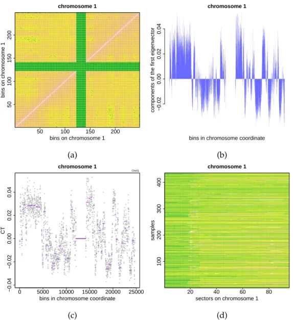
The gene copy-number measurement
Copy-numbers refer to the numbers of copies of genes in the genome of a cancer cell, which are often different from two (diploid) because of genetic instability. Microarrays have been adapted to measure the copy-numbers of cancer cells. The procedure remains very similar to that of the gene expression measurement. Generally, DNA molecules are first cut into oligonucleotides by special enzymes and amplified by polymerase chain reactions (PCR).
The oligonucleotides are then labelled by fluorescent agents and these labelled oligonu- cleotides are then hybridised with their complementary sequences prepared on an array.
The array is then scanned to measure the fluorescent intensities. By this construction, these fluorescent intensities are directly related to the numbers of copies of the short sequences present in the sample. For the details of the measurement of copy-number by microarrays, see Huang et al. 2004, Pinkel et al. 1998.
DNA sequencing
Besides gene expressions and copy-numbers of cancer cells, we will also work with se- quencing data. However, we will be concerned only with gene-specific sequencing in- stead of who genome sequencing. One of the classic technologies for sequencing is called Sanger sequencing. The DNA segment to be sequenced may first be amplified if necessary.
One then lets the sequences replicate under the activity of polymerases in a medium with
sufficient nucleotides. A small fraction of the nucleotides in the medium are modified so
that they do not allow DNA elongation. The replicated sequences therefore are stopped
at a random base if by chance a modified nucleotide is added at that position. Sequences
of different lengths can be separated using the so-called gel-electrophoresis effect. If the
modified nucleotides are labelled with fluorescent agents of four different colours which correspond to four different nucleotides, then the ending bases of the random-stopped sequences can be identified. From these data, the sequence of the DNA segment can be inferred. For a more detailed description of the basis of Sanger sequencing, see Alberts et al. 2010.
Although we already mentioned in the last section, it is also worth emphasising again that often both the gene expression measurement and the copy-number measurement mentioned above only capture the population-averages of the messenger RNA concentra- tions and the DNA copy-numbers as they both require biological samples which contain thousands of cells. This calls for care in the interpretation of the results. 4
Together with these molecular techniques, analytical methods to analyse the emerg- ing data have been developing in a very fast pace. We will have short reviews of such analytical methods in the specific contexts of the following chapters before describing our corresponding approaches.
References
Alberts, B., D. Bray, K. Hopkin, A. D. Johnson, J. Lewis, M. Raff, K. Roberts, and P. Walter (2010).
Essential cell biology. Garland Science.
Beck, B. and C. Blanpain (2013). “Unravelling cancer stem cell potential”. Nat. Rev. Cancer 13.10, pp. 727–38.
Bergers, G. and D. Hanahan (2008). “Models of resistance to anti-angiogenic therapy”. Nat. Rev.
Cancer 8, pp. 592–603.
Bergers, G. and S. Song (2005). “The role of pericytes in blood-vessel formation and maintenance”.
Neuro. Oncol. 7.4, pp. 452–64.
Bottsford-Miller, J. N., R. L. Coleman, and A. K. Sood (2012). “Resistance and escape from aniangio- genesis therapy: clinical implication and future stategies”. Journall Clin. Oncol. 30.32, pp. 4026–
4034.
Carter, S. L. et al. (2012). “Absolute quantification of somatic DNA alterations in human cancer”.
Nat. Biotechnol. 30.5, pp. 413–21.
Chambers, A. F., A. C. Groom, and I. C. MacDonald (2002). “Dissemination and growth of cancer cells in metastatic sites”. Nat. Rev. Cancer 2, pp. 563–572.
Chen, J., K. Sprouffske, Q. Huang, and C. C. Maley (2011). “Solving the puzzle of metastasis: the evolution of cell migration in neoplasms”. PLoS One 6.4, e17933.
Chmielecki, J. et al. (2011). “Optimization of dosing for EGFR-mutant non-small cell lung cancer with evolutionary cancer modeling”. Sci. Transl. Med. 3.90, 90ra59.
Cook, K. M. and W. D. Figg (2010). “Angiogenesis inhibitors: current strategies and future prospects”.
CA Cancer J. Clin. 60, pp. 222–243.
4