idiopathic epilepsy syndromes
Inaugural - Dissertation
zur
Erlangung des Doktorgrades
der Mathematisch-Naturwissenschaftlichen Fakultät
der Universität zu Köln
vorgelegt von
Dennis Lal
aus Bremen
Prof. Dr. Peter Nürnberg Prof. Dr. Bernd Neubauer Prof. Dr. Markus Nöthen Vorsitz der Prüfung:
Prof. Dr. Peter Kloppenburg Beisitzerin:
Dr. Birgit Budde
Tag der letzten mündlichen Prüfung:14.01.2014
1 Introduction ... 1
1.1 Epilepsy ... 1
1.2 Idiopathic epilepsies investigated ... 2
1.2.1 Idiopathic generalized epilepsies (IGE) ... 2
1.2.2 Rolandic epilepsy (RE) and the RE spectrum ... 4
2 Goals and objectives ... 6
2.1 Discovery cohorts ... 7
2.2 Molecular genetic screening methods ... 7
3 Publications ... 9
3.1 Publications on IGE ... 9
3.1.1 RBFOX1 in IGE (published) ... 9
3.1.2 GPHN in IGE (in revision) ... 20
3.2 Publications on RE ... 67
3.2.1 RBFOX genes in RE (published) ... 67
3.2.2 GRIN2A in RE (published) ... 76
3.2.3 16p11.2 duplications in RE (submitted; revised manuscript) ... 101
3.2.4 DEPDC5 in RE (in revision) ... 145
3.3 Publications derived from additional projects ... 160
3.3.1 NDUFV1 in IBSN (published) ... 160
4 Overall discussion of the studies presented ... 166
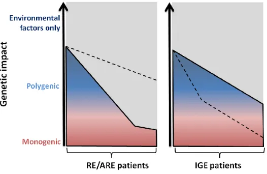
4.1 Identified variants associated with IGE ... 166
4.2 Identified variants associated with RE-spectrum epilepsies ... 168
4.3 Epilepsy genetics ... 169
5 References ... 172
6 Summary / Zusammenfassung ... 182
6.1 Summary ... 182
6.2 Zusammenfassung ... 184
7.1 Declaration of contribution as co-author ... 186
7.2 Curriculum Vitae ... 188
7.3 List of publications ... 189
1 Introduction
1.1 Epilepsy
The term “epilepsy” encompasses a number of different syndromes whose key feature is defined by recurrent, unprovoked seizures (Berg et al. 2010). Epilepsy is one of the most common chronic neurologic diseases worldwide with a prevalence of 0.5-1% and a lifetime incidence of 3% (Hauser et al., 1993). Recurrent seizures critically change quality of life, particularly of those 30% of epilepsy patients who are drug-resistant (Berg et al., 2009). The economic burden of epilepsies is high, it has been estimated that the 6 million people with active epilepsy in Europe cost over €20 billion per year (Cross et al., 2011; www.who.int).
Epilepsy syndromes are highly heterogeneous in their clinical manifestation. In 1989, the International League Against Epilepsy (ILAE) proposed a classification scheme (Commission on Classification and Terminology of the International League against Epilepsy, 1989.) which has been revised in recently (Berg et al., 2010). Epilepsy syndromes are classified by their etiology and symptomatology. Symptomatic epilepsy is considered an acquired disorder, developing in response to congenital or acquired brain insults such as malformations, tumors or infections. Genetic (formerly:
idiopathic) epilepsies represent about 40% of all epilepsies in childhood and approximately 20% of epilepsies in adulthood (Steinlein et al., 1999). Off note, due to our previous studies on the same patient cohort, before classification change, we keep in the presented follow up studies in this dissertation the term "idiopathic"
(Commission et al.,1989) epilepsies instead of "genetic" (Berg et al., 2010)
epilepsies. Idiopathic (genetic) epilepsies are characterized by recurrent seizures in
otherwise healthy individuals. The absence of known or suspected cerebral lesions
and their familial aggregation implicated a genetic basis (Ottman, 2005). Given the
strong impact of genetic factors in the pathogenesis of idiopathic epilepsies,
molecular genetic studies provide a promising approach to dissect the responsible
susceptibility genes and to determine molecular pathways in epileptogenes. The
most common forms of idiopathic epilepsies include the generalized i) idiopathic
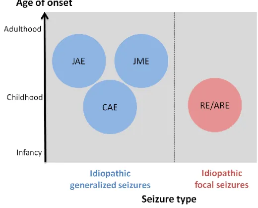
generalized epilepsy (IGE) and the focal ii) rolandic epilepsy (RE) (Figure 1).
Figure 1 Overview of common idiopathic epilepsy types. The epilepsy types are distributed by age of onset as well as seizure type. Abbreviations: JAE= Juvenile absence epilepsy; JME= Juvenile myoclonic epilepsy; CAE= Childhood absence epilepsy; RE= Rolandic epilepsy; ARE= Atypical rolandic epilepsy.
1.2 Idiopathic epilepsies investigated
1.2.1 Idiopathic generalized epilepsies (IGE)
The group of idiopathic generalized epilepsies (IGEs) represent about one-third of all
epilepsies (Jallon et al., 2011). These epilepsies are characterized by their age-
related manifestation of generalized seizure types (Nordli et al., 2005). Generalized
spike-wave discharges are the characteristic electroencephalographic signature,
which reflects a synchronized hyperexcitable state of thalamocortical circuits
(Blumenfeld et al., 2005). IGEs can be clinically subdivided by leading seizure types
and age-of-onset into four major IGE syndromes: childhood absence epilepsy (CAE),
juvenile absence epilepsy (JAE), juvenile myoclonic epilepsy (JME) and epilepsy with
generalized tonic–clonic seizures alone (EGTCS). In addition, a large variety of other
genetic epilepsies exist, however, quantitatively they represent a small minority of
less than 10% (Nordli et al., 2005). The characteristic seizure types of the common
twin studies suggest a predominant genetic predisposition of common IGE syndromes with heritability estimates up to 80% (reviewed in Helbig et al., 2008).
1.2.1.1 Genetics
A significant complex genetic predisposition is indicated by concordance rates of 70–
80% for IGE in monozygotic twin pairs (Berkovic et al., 1998) compared to rapidly declining recurrence risks of IGEs in siblings ranging from four to 10% depending on the IGE subtype (Beck-Mannagetta et al., 1991). IGEs follow a complex mode of inheritance, suggesting that several genetic factors contribute to generalized seizures (Greenberg et al., 1992; Sander et al., 1996; Berkovic et al., 1998). The recurrence risk for IGE ranges from 70% to 95% in monozygotic twins and is 10- to15-fold higher than for first-degree relatives (5–8%), and more than 100-fold greater than the prevalence of 0.6% in the general population (ratio of sibling risk to population prevalence: λ
S≈ 8) (Sander et al., 1996). However, CAE, JAE and JME cluster in families, and, frequently, absence seizures are followed by myoclonic seizures in the same patient in an age-dependent manner. These observations support the neurobiological concept that the common IGE subtypes share an overlapping genetic predisposition (Berkovic et al., 1987; Beck-Mannagetta et al., 1991; Reutens et al., 1991; Wirrell et al., 1991; Janz et al., 1997). Furthermore, supporting this concept, the age-related expression of various seizure types arises from a genetically determined impairment of brain maturation or is influenced by the stage of brain maturation (Shinnar et al., 1991). In rare monogenic types of idiopathic epilepsies, several genes have been identified of which the majority of genes encode voltage- gated or ligand-gated ion channels (e.g. SCN1A, GABRA1, KCNQ2, KCNQ3, CHRNA4 and many others) (Reid et al., 2009). The molecular dissection of genes conferring susceptibility to the genetically complex idiopathic epilepsies remains a challenge. In contrast to gene mapping strategies applied in rare monogenic epilepsies, a large number of linkage and candidate gene association studies failed to identify susceptibility genes for common IGE syndromes with complex genetic predisposition (Gardiner, 2005; Hempelmann et al., 2006; Kasperaviciute et al., 2010;
Greenberg and Subaran, 2011). The failure to identify replicable risk genes for
common epilepsies most likely reflects the underestimated degree of genetic
complexity and heterogeneity in human epilepsies.
1.2.2 Rolandic epilepsy (RE) and the RE spectrum
Benign epilepsy with centrotemporal spikes (BECTS) was first described by Martinus Rulandus in 1597 (Van Huffelen, 1989). The syndrome is now named rolandic epilepsy (RE) because of the characteristic features of partial seizures involving the brain region around the lower portion of the rolandic fissure. RE is one of the most common epilepsy syndromes in children, accounting for about 15 % of epilepsies beginning before the age of 16 years (Freitag et al., 2001; Shinnar et al., 2002).
Onset typically occurs from 4–12 years in otherwise healthy children (Lerman et al., 1986). Seizures have a focal origin and frequently start with one-sided sensorimotor symptoms of lips tongue, and face, resulting in hypersalivation and speech arrest (summarized by Strug et al., 2009). The prognosis of RE is benign. It resolves spontaneously and by far of most affected children remain intellectually normal.
There is, however, an increased comorbidity with attention deficit hyperactivity disorder (ADHD) and specific mild cognitive deficits (approx. 20%) (Smith et al., 2012).
RE is related to much rarer, and less benign epilepsy syndromes, including atypical benign partial epilepsy of childhood (ABPE), Landau-Kleffner syndrome (LKS) and epileptic encephalopathy with continuous spike-and-waves during sleep (CSWSS) (Doose et al., 2001; Gobbi et al., 2006; Guerrini and Pellacani, 2012; Hahn et al., 2001), referred to as RE related syndromes, or atypical rolandic epilepsy (ARE) by some authors (Fejerman, 2009). RE and the ARE share blunt, high-voltage, characteristically shaped centrotemporal spikes (CTS) of characteristic morphology in the EEG. However, ARE denote more severe forms of focal childhood epilepsies with various additional seizure types, developmental language delay or regression, speech dyspraxia and variable neuropsychiatric deficits in up to 50% of the affected (Gobbi et al., 2006; Hughes et al., 2011).
1.2.2.1 Genetics
The majority of RE patients have a complex genetic predisposition as implicated by
the relative decline of the recurrence risks of seizures in 9.8% of first-degree
relatives, 3% in second- and 1.5% in third-degree relatives as well as the intriguing
phenotypic variability observed among family members (Vears et al., 2012). The age-
clinically unaffected family members and was earlier reported to be inherited in an autosomal-dominant manner (Heijbel et al., 1975; Bali et al., 2007). However these findings were questioned for methodological reasons (Ottman et al., 1989) ARE belongs to an extended spectrum of Rolandic epilepsy, a rate of 40% EEG concordance for siblings investigated at the age of maximum expression (3 to 10 years) is observed (Doose et al., 2001). Despite the strong genetic predisposition of the CTS-EEG trait, the etiology of epilepsies with centrotemporal spikes is largely unknown. Various loci have been reported for classic or rare forms of RE (Scheffer et al., 1995; Neubauer et al., 1998; Guerrini et al., 1999; Roll et al., 2006; Strug et al., 2007; Strug et al., 2009).
Furthermore, rare variants in KCNQ2, KCNQ3 and SRPX2 have been associated
with the RE or ARE in small subsets of patients (Neubauer et al., 2008; Roll et al.,
2006). Recently, rare structural variations at 16p13 deleting the genomic region of
GRIN2A have been observed for patients with ARE (Reutlinger et al., 2010). All these
variants and loci have never been replicated and might account for only a small
fraction of the genetic risk for the rolandic epilepsies, whereas the major cause(s)
remain unknown.
2 Goals and objectives
The aim of the present studies was the molecular genetic dissection of risk factors
with strong epileptogenic effects in idiopathic epilepsies (Figure 2) and the
elucidation of their molecular pathways in epileptogenesis. Taking into account the
low power of our case-control study for genome-wide scans, our current strategy
focused on a candidate gene/locus approach of genes and CNVs that have either
been implicated in the pathogenesis of epilepsy or represent plausible candidate
genes with a presumed impact on neuronal excitability. This candidate gene/locus
approach was restricted to the identification of genetic risk factors with a strong
impact on epileptogenesis according to the common disease-rare variant (CD-RV)
model (Pritchard, 2001; McClellan et al., 2007). The CD-RV model implies that
multiple, heterogeneous rare variants with strong effects contribute to the genetic
architecture underlying common idiopathic epilepsies. In line with the CD-RV model,
we had previously demonstrated that large recurrent microdeletions in the
chromosomal regions 15q11.2, 15q13.3 and 16p13.11 constitute individually rare but
collectively substantial genetic risk factors of IGE (Helbig et al., 2009; De Kovel et al.,
2010).
Two strategies were applied to identify genetic risk factors for idiopathic epilepsies: 1) copy number variation (CNV) analysis using high-resolution SNP arrays and 2) whole exome sequencing (WES) to detect mutations in the coding regions. Taking into account our cohort sizes and the amount of expected variants, the study power was small. Therefore, our analyses focused on genetic variants affecting genes involved in neuronal excitability.
2.1 Discovery cohorts
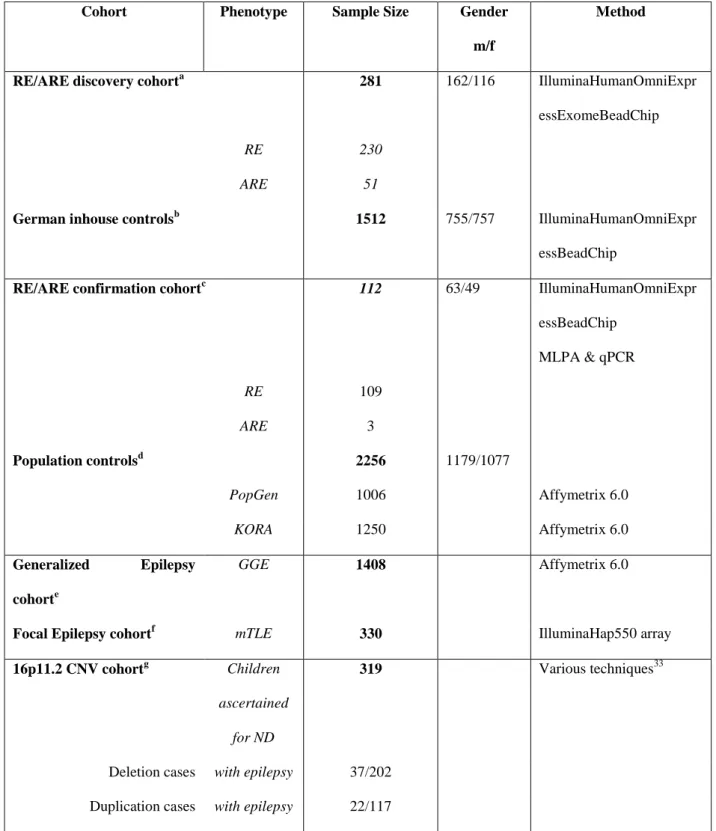
The discovery cohorts of idiopathic epilepsies included two case-control cohorts: i) 1582 unrelated IGE and 2795 populations controls of North-Western European descent, and ii) 308 patients with RE-spectrum epilepsies and 1512 controls of European descent. In addition, we performed candidate gene sequence analysis in 242 patients with RE-spectrum epilepsies. The IGE study cohort was collected by a European concerted action organized by the EPICURE Consortium (EPICURE GWAS discovery cohorts; EPICURE et al., 2012). The standardized ascertainment scheme, clinical protocols and diagnostic criteria are available at http://portal.ccg.uni- koeln.de/ccg/research/epilepsy-genetics/sampling-procedure/. The cohort of 281 patients with RE–spectrum epilepsies were recruited in the framework of the European collaborative research project EuroEPINOMICS. In a multi-centre effort, RE/ARE patients were recruited from Germany, Austria, Canada and Australia.
Ninety-eight of the patients were ascertained through multiplex-families with at least two affected siblings. Diagnosis of RE was performed according to the International Classification of Seizures and Epilepsies (Commission on Classification and Terminology of the International League against Epilepsy 1989; Berg et al., 2010).
The various syndromes classified as RE-spectrum epilepsies fulfilled the diagnostic criteria as specified previously (Aicardi and Chevrie, 1982; Doose et al., 2001; Hahn et al., 2001). In total, the discovery cohort comprised 230 patients affected by typical RE and 51 patients with ARE (165 males and 116 females). Both screening cohorts represent the largest assemblies of IGE and RE/ARE patients reported so far.
2.2 Molecular genetic screening methods
The present studies applied two molecular genetic screening techniques to identify
genetic risk factors with strong effects: i) copy number variation (CNV) analyses
using high-resolution SNP arrays (IGE and RE cohorts), and ii) candidate gene
sequence analysis based on an ongoing whole exome sequencing project (RE/ARE, n =242).
2.3 Selection of candidate genes
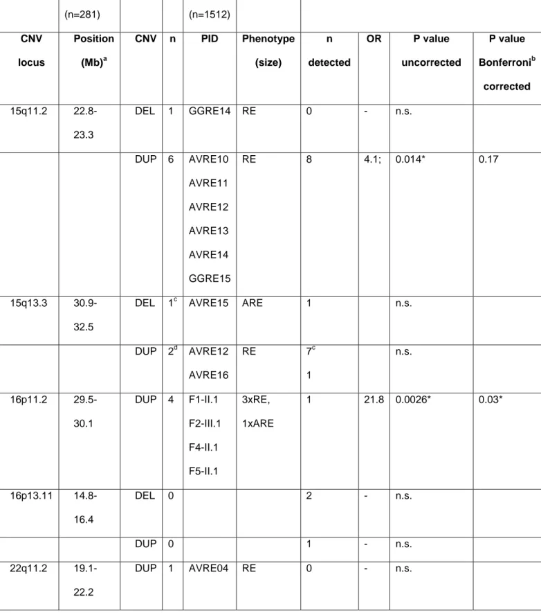
We investigated large recurrent CNVs at 1q21, 15q11.2, 15q13.3, 16p11.2, 16p13.11 and 22q11.2 (Helbig et al., 2009; Coe et al., 2012; De Kovel et al., 2013); as well as the high-ranking candidate genes GRIN2A (Reutlinger et al., 2010; Lesca et al., 2012), GPHN (Förstera et al., 2010; Lionel et al., 2013), RBFOX1 and RBFOX3 (Bhalla et al., 2004; Martin et al., 2007; Gallant et al., 2011; Gehman et al., 2011) as well as DEPDC5 (Dibbens et al., 2013; Ishida et al., 2013). The investigated CNV loci have been implicated in the pathogenesis of epilepsies or other neurodevelopmental disorders, whereas the candidate genes are presumed to have a strong functional impact in pathways controlling neuronal excitability.
2.4 Scope of the ongoing research activities
In the presented cumulative thesis I report the results of our current CNV and candidate gene sequence analyses resulting in three articles already published in peer review journals (Lal et al., 2013a; Lal et al., 2013b, Lemke et al., 2013). Another three papers are currently in revision (Lal et al., Dejanovic et al., Reinthaler et al.,).
Our ongoing studies identified CNVs at 16p11.2, RBFOX1, RBFOX3 and GRIN2A
and putative deleterious sequence mutations in GRIN2A and DEPDC5 in patients
with idiopathic epilepsies. These results offer new insight into the genetic architecture
of common epilepsy syndromes by: i) the delineation of the phenotypic spectrum
associated with these epilepsy genes, ii) the characterization of their allelic spectra
and modes of inheritance (de novo, inherited, estimates of the effect size,
penetrance, expressivity), and iii) a first step in the elucidation of the underlying
molecular pathomechanisms in epileptogenesis. In the long term, our study results
might accelerate the prospects to translate the rapidly increasing knowledge into
clinically relevant actions.
3 Publications
3.1 Publications on IGE
3.1.1 RBFOX1 in IGE (published)
Lal D, Trucks H, Møller RS, Hjalgrim H, Koeleman BPC, et al. (2013) Rare exonic deletions of the RBFOX1 gene increase risk of idiopathic generalized epilepsy. Epilepsia 54: 265–
271. doi:10.1111/epi.12084
3.1.2 GPHN in IGE (in revision)
Dejanovic B*, Lal D*, Catarino BC*, Arjune S et al. Exonic microdeletions of the gephyrin gene exert a dominant-negative effect on GABAergic synaptic inhibition in two patients with idiopathic generalized epilepsy. Human Molecular Genetics (in revision)
*These authors contributed equally to this work
Exonic microdeletions of the gephyrin gene impair GABAergic synaptic inhibition in patients with idiopathic generalized epilepsy
Borislav Dejanovic
1*, Dennis Lal
2,3,4*, Claudia B. Catarino
5*, Sita Arjune
1, Ali A. Belaidi
1, Holger Trucks
2,8, Christian Vollmar
5, Rainer Surges
6,8, Wolfram S. Kunz
6,8, Susanne Motameny
2, Janine Altmüller
2, Anna Köhler
1, Bernd A. Neubauer
4, EPICURE Consortium
8, Peter Nürnberg
2,3,7,8, Soheyl Noachtar
5, Günter Schwarz
1,3,7#and Thomas Sander
2,8#1. Department of Chemistry, Institute of Biochemistry, University of Cologne, 50674 Cologne, Germany
2. Cologne Center for Genomics (CCG), University of Cologne, 50931 Cologne, Germany
3. Cologne Excellence Cluster on Cellular Stress Responses in Aging-Associated Diseases (CECAD), University of Cologne, 50674 Cologne, Germany
4. Department of Neuropediatrics, University Medical Center Giessen and Marburg, 35392 Giessen, Germany
5. Epilepsy Center, Department of Neurology, University of Munich, 81377 Munich, Germany
6. Department of Epileptology, University Clinics Bonn, 53105 Bonn, Germany
7. Center for Molecular Medicine (CMMC), University of Cologne, 50931 Cologne, Germany
8. EPICURE Consortium
* Authors contributed equally
# Equal senior authorship
EPICURE Consortium participants are listed in Appendix
To whom correspondence should be addressed at:
Günter Schwarz, Institute of Biochemistry, University of Cologne, Zülpicher Str. 47, 50674 Cologne, Germany, Phone: +49-221-470-6441, Fax: +49-221-470-5092, Email:
gschwarz@uni-koeln.de
Thomas Sander, Cologne Center for Genomics, University of Cologne, Weyertal 115b, 50931 Cologne, Germany, Phone: +49-221-478-96800, Fax: +49-221-478-96866, Email:
thomas.sander@uni-koeln.de
ABSTRACT
Gephyrin is a postsynaptic scaffolding protein, essential for the clustering of glycine and γ-aminobutyric acid type-A receptors (GABA
ARs) at inhibitory synapses. An impairment of GABAergic synaptic inhibition represents a key pathway of epileptogenesis. Recently, exonic microdeletions in the gephyrin gene (GPHN) have been associated with neurodevelopmental disorders including autism spectrum disorder, schizophrenia and epileptic seizures. Here we report the identification of novel exonic GPHN microdeletions in two patients with idiopathic generalized epilepsy (IGE), representing the most common group of genetically determined epilepsies. The identified GPHN microdeletions involve exons 5-9 (Δ5-9) and 2-3 (Δ2-3), both affecting the gephyrin G-domain. Molecular characterization of the GPHN Δ5-9 variant demonstrated that it perturbs the clustering of regular gephyrin at inhibitory synapses in cultured mouse hippocampal neurons in a dominant-negative manner, resulting in a significant loss of
2-subunit containing GABA
ARs. GPHN Δ2-3 causes a frameshift resulting in a premature stop codon (p.V22Gfs*7) that most likely leads to haplo-insufficiency of the gene. Our results demonstrate that structural exonic microdeletions affecting the GPHN gene constitute a rare genetic risk factor for IGE and other neuropsychiatric disorders by an impairment of the GABAergic inhibitory synaptic transmission.
KEYWORDS
Idiopathic generalized epilepsy; Microdeletion; GPHN; gephyrin;
ABBREVIATIONS
GABA
AR, γ-aminobutyric acid type-A receptors; CNV, copy number variations; IGE,
diffusion tensor imaging; HEK293, Human Embryonic Kidney 293; TLE, temporal lobe epilepsy; qPCR, quantitative polymerase chain reaction;
INTRODUCTION
Epilepsy is characterized by recurrent spontaneous seizures due to a neuronal hyperexcitability and an abnormal cortical synchronization. Approximately 3% of the general population are affected by epilepsy until the age of 40 years
1. The idiopathic generalized epilepsies (IGEs) represent the most common group of genetically determined epilepsies, accounting for 20-30% of all epilepsies
2. Their clinical features are characterized by age-related recurrent unprovoked generalized seizures, in the absence of detectable brain lesions or metabolic abnormalities
3,4. Genetic factors play a predominant role in the etiology of IGE with heritability estimates of 80%. However, the vast majority of IGE syndromes have an oligo-/polygenic predisposition and their genetic basis remains elusive
5.
Structural genomic copy number variations (CNVs) account for a substantial fraction of the genetic variance in about 3% of patients with idiopathic epilepsies
6-9. Recurrent microdeletions at 15q11.2, 15q13.3, 16p13.11
6,7and exonic deletions in NRXN1
10and RBFOX1
11increase the risk of IGE and a wide range of neurodevelopmental disorders
12. Recently, exonic microdeletions in the gene encoding the synaptic scaffolding protein gephyrin (GPHN) have been found to represent a rare cause of neurodevelopmental disorders, including autism spectrum disorder (ASD), schizophrenia and epileptic seizures
13. Two out of six patients carrying exonic GPHN deletions exhibited seizures
13.
Gephyrin is a postsynaptic scaffolding protein, essential for the clustering and
Gephyrin is composed of three domains - the C-terminal E-domain binds directly to the glycine and GABA
Areceptor subunits, whereas the N-terminal G-domain is crucial for gephyrin’s oligomerization. Both domains are connected by the central (C) domain
18. Aberrant function of gephyrin impairs GABAergic synaptic inhibition, which thereby may promote neuronal hyperexcitability and seizure susceptibility.
Previously, we have reported a gephyrin-specific effect in temporal lobe epilepsy (TLE), where stress-induced irregular splicing of GPHN resulted in the expression of truncated gephyrin variants that impaired the function of regular gephyrin by dominant-negative interaction
20. Additionally, reduced gephyrin expression has been detected in temporal lobe surgical specimens of patients with medically refractory TLE, as well as in rat models
21. These accumulating lines of evidence support the hypothesis that gephyrin dysfunction impairs GABAergic synaptic inhibition and thereby contributes to epileptogenesis
21,22. In the present study, we screened the GPHN gene for microdeletions in 1469 European patients with common IGE syndromes and 2256 population controls
11. We identified exonic GPHN deletions affecting the gephyrin N-terminal G-domain in two IGE patients and characterized the underlying molecular mechanism causing functional alterations in gephyrin clustering thus promoting epileptogenesis.
MATERIALS AND METHODS Study cohort and CNV screening
This study has been approved by the local Research Ethics Committees. All study
participants including the family members of the GPHN deletion-carriers provided an
informed written consent. CNV screening of the genomic GPHN sequence
(chr14:66,974,125-67,648,525; human genome build 37/hg19) was carried out in
and 2,256 German population controls
11. DNA samples were investigated by the Affymetrix Genome-Wide Human SNP Array 6.0 (Affymetrix, Santa Clara, CA, USA).
CNV analysis was performed, using the Birdsuit algorithm implemented in the Affymetrix Genotyping Console version 4.1.1. Regional log2 ratios of the signal intensities and the SNP heterozygosity state were visualized in the Chromosome Analysis Suite v1.2.2 (Affymetrix, Santa Clara, CA, USA). The copy number state of the GPHN microdeletions identified by the array-based CNV analysis were examined by real-time quantitative PCR (qPCR), using a TaqMan® CNV assays located in GPHN exon 7 (Assay ID: Hs01711518_cn) and intron 2 (Assay ID: Hs07084813_cn;
Life Technologies, Carlsbad, CA, USA). Genome-wide CNV screening beyond the GPHN locus was restricted to CNVs with a segment size > 500kb
23and a minimum of 50 markers to achieve a high accuracy and reliability. Again, regional log2 ratios of the signal intensities and the SNP heterozygosity state were visualized for all CNVs in the Chromosome Analysis Suite v1.2.2 (Affymetrix, Santa Clara, CA, USA) and were manually inspected.
Transcript analysis
RNA was isolated from whole blood samples using the PAXgene Blood RNA Kit (Qiagen, Hilden, Germany). For amplification of the Δ5-9 deletion flanking primers were designed: Forward (within exon 4) AGGAACAGGATTTGCACCAC and reverse (within exon 13) GCGATGTCTTCTAGCCACCT. Amplification of the Δ2-3 deletion was performed with primers: Forward (within exon 1)
CCGAGGGAATGATCCTTACTAA and reverse (within exon 7)
TCAAGTTCATCATGCACCTCC. The sizes of the amplified DNA fragments were
assessed using a 1.5% agarose gel electrophoresis and the Agilent 2200
USA). Bidirectional Sanger sequencing of the identified GPHN cDNA amplicons was done following standard protocols.
Paired-pulse transcranial magnetic stimulation
For Family 1, the patient, his father and six healthy controls with no personal history of seizures or other neurological disorders and no medication (three females, median age 38 years, range 24-44 years) had transcranial magnetic stimulation (TMS). After evaluating the resting motor threshold (MT), paired-pulse TMS was used to study intra-cortical inhibition (ICI; interstimulus interval [ISI] of 3 ms) and intra-cortical facilitation (ICF; ISI of 13 ms). The protocol used was adapted from Werhahn and colleagues
24. IBM SPSS Statistics 21.0 software was used for statistical analysis.
Expression construct
Regular enhanced green fluorescent protein (EGFP)-tagged gephyrin
25served as the basis for Δ5-9geph expression constructs. Fragments encoding gephyrin exons 1-4 and 10-30 were amplified from regular gephyrin construct and connected by fusion PCR. The two fragments were connected by fusion PCR and introduced into the pEGFP-C2 vector (Clontech) using XhoI and HindIII restriction sites. To create myc-tagged gephyrin, EGFP-tagged gephyrin was amplified with primers that introduce the sequence for the myc-tag (EQKLISEEDL) at the N-terminus of gephyrin and a stop-codon after the last amino acid of gephyrin. Fragment was introduced into pEGFP-N2 the vector (Clontech, Mountain View, CA, USA) using XhoI and HindIII restriction sites.
Hippocampal neuron cultures and transfection
Primary neuron cultures were prepared from hippocampi of C57BL/6 mice and plated
on poly-L-lysine coated coverslips at a density of 75,000/24-well dish. Neurons were
Neurons were transfected after 11 days in vitro (DIV) unless otherwise stated.
Transfection was carried out with Lipofectamin 2000 (Invitrogen) according to manufactures manual. Constructs were expressed for 48 hours unless otherwise stated.
Human embryonic kidney cell culture and transfection
Human embryonic kidney (HEK293) cells were grown in Dulbecco’s modified Eagle’s medium supplemented with 10% fetal calf serum and 2 mM L-glutamine at 37 °C and 5% CO
2. For confocal laser-scanning microscopy, 1 × 10
5HEK-293 cells were seeded on collagenized coverslips in 12-well plates and immediately transfected with polyethylenimine (1 mg/ml, diluted in H
2O, pH 7.0) using standard protocols. Cells were grown for 24-48 h depending on the assay.
Immunostaining of cultured cells
Cells were fixed with 4% PFA for 10 min at room temperature. Unspecific binding sites were blocked with blocking solution (2% BSA, 10% goat serum, 0.2% Triton X- 100) for 1h and primary antibodies were applied for 1 h in 10% goat serum. After three washing steps in PBS, cells were incubated with secondary antibodies in 10%
goat serum for 1 h at room temperature. After the final three washing steps in PBS, slides were mounted on cover slips with Fluoro gel II containing DAPI (Science Service, Munich, Germany) to stain the nuclei. Antibodies used for the staining:
mouse anti-gephyrin (1:50 cell culture supernatant, clone 3B11); rabbit anti-gephyrin
(1:1,000, rGeph-C, epitope EDLPSPPPPLSPPP, Eurogentec, Belgium); guinea-pig
anti-GABA
A-gamma2 receptor (Jean-Marc Fritschy, ETH, Zurich); rabbit anti-VGAT
(1:500, Synaptic Systems, Göttingen, Germany); mouse anti-myc-tag (1:10 cell
Image analysis and quantification
Images were acquired with a confocal laser scanning microscope (Nikon A1) at 60x using a resolution of 1024x1024. Images were processed using the software NIS Elements (NIKON). For analysis of neuronal cultures at least two independent preparations per condition were used. Images were acquired as a z-stack with three optical sections with 0.5 μm steps. Maximum intensity projections were created and analyzed using NIS Elements software. Usually two 20 x 5 μm region of interests (ROI) were placed on dendrites and clusters were counted using the analyze particles option in NIS Elements. For statistical analysis, gephyrin cluster were compared pairwise between EGFP and Δ5-9geph expressing neurons. Mean values were compared for significance using Student’s t-test with the software SigmaPlot (Systat Software, Chicago, IL, USA). Errors are presented as standard error of the mean (SEM). Significance levels are indicated as *P < 0.01, **P < 0.05, ***P < 0.001.
Images were processed with the software ImageJ (NIH).
Co-immunoprecipitation
Protein extracts were prepared in an IP-buffer (25 mM Tris, pH 7.4, 150 mM NaCl,
1% Triton X-100, protease and phosphatase inhibitor cocktail (Roche, Mannheim,
Germany)). The post-nuclear fraction was incubated with primary antibodies for 1 h at
room-temperature or overnight at 4 °C. 20 μl Protein-G Sepharose beads were
incubated with the extracts for 2 hours at room temperature. After a brief
centrifugation (500 g for 3 min), the immune-beads were washed three times with IP
buffer. The adsorbed proteins were eluted from the immune-beads by boiling in 50 μl
SDS-loading buffer. Immunoprecipitated samples were subjected to SDS-PAGE
followed by immunoblotting.
Western Blot analysis and antibodies
For immunoblotting, a standard protocol was followed and detection was carried out using chemiluminescence and an ECL system with a cooled CCD camera (Decon Science Tec, Germany). The following primary antibodies were used and diluted in TBS-Tween containing 1% dry-milk: anti-Gephyrin (1:50, clone 3B11 cell culture supernatant), rabbit anti-ß-tubulin (1:100, Santa Cruz, Santa Cruz, CA, USA), rabbit anti-GFP (1:5,000, Abcam, Cambridge, UK), mouse anti-myc-tag (1:10 cell culture supernatant, clone 9E10). As secondary antibodies anti-mouse or anti-rabbit conjugates were used in a 1:10,000 dilutions in TBS-Tween containing 1% dry milk.
RESULTS
Detection of exonic GPHN microdeletions and mRNA transcription analysis
We screened the genomic GPHN sequence (14q23.3:66,974,124-67,648,524,
reference coding sequence: NM_020806, CCDS9777.1; hg19) for microdeletions
(size > 40 kb, number of probe sets > 20) using high coverage microarrays from
1,469 unrelated European patients with common IGE syndromes and 2,256 German
population controls. We identified two hemizygous exonic GPHN microdeletions in
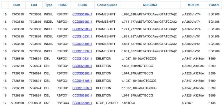
the IGE cohort: i) a 129 kb microdeletion (chr14:67,314,917-67,443,729) affecting
GPHN exons 5-9 (Δ5-9) in a German male patient with juvenile myoclonic epilepsy
(JME, Family 1), and ii) a 158 kb microdeletion (chr14:67,136,658-67,295,196)
encompassing GPHN exons 2-3 (Δ2-3) in a German male patient with myoclonic
astatic epilepsy (Doose syndrome, Family 2) (Fig. 1A). None of the 2,256 German
controls carried a microdeletion at the GPHN locus. The hemizygous copy number
state of the GPHN deletions was validated by TaqMan quantitative PCR (qPCR).
Exome sequencing of the GPHN coding regions and exon/intron boundaries did not reveal any additional indels and rare missense mutations in the parent-offspring trio of Family 1. Direct blood cDNA amplification of GPHN exons 4-13 for Family 1 and exons 1-7 for Family 2 followed by Sanger sequencing of the major amplicons confirmed the transcription of the truncated GPHN variants and revealed a paternal transmission of both microdeletions (Fig. 1B-D and Supplementary Material S1).
Genome-wide screening for CNVs previously associated with epilepsy
6,8,9,26,27, revealed no additional known pathogenic CNVs in both index patients.
On protein level, the GPHN Δ5-9 microdeletion leads to the truncation of the gephyrin G- and C-domain (hereafter termed “Δ5-9geph” - Fig. 1E). Given that residue 22 is encoded by a splitted codon composed by exons 1 and 2, the GPHN Δ2-3 variant results in a frameshift and a premature stop codon (p.V22Gfs*7, Fig. 1F). We assume that, although through first strand synthesis we were able to amplify the GPHN Δ2-3 variant, most of the transcript will be degraded by nonsense-mediated mRNA decay, a translation-dependent posttranscriptional process that selectively recognizes and degrades mRNAs whose open reading frame is truncated by a premature translation termination codon
28, resulting in negligible amounts of the protein.
Clinical features of the GPHN deletion carriers and segregation analysis Family 1 (GPHN Δ5-9 deletion)
The pedigree of Family 1 is presented in Fig. 1A. The non-consanguineous German
parents (57-years-old father, 55-years-old mother) and the patient’s 30-year-old sister
had no unequivocal history of seizures. The GPHN Δ5-9 deletion was paternally
inherited. The mother showed a diploid copy number status at the GPHN locus. DNA
from the clinically unaffected sister was not available. The 33-year-old male index IGE patient was born at term after an uneventful pregnancy and delivery. He had recurrent simple febrile seizures from the age of six months to six years, unprovoked generalized tonic-clonic seizures since the age of five years and myoclonic seizures recorded by video-EEG starting at the age of 16 years. Complete remission of epileptic seizures by monotherapy with levetiracetam was reached since the age of 24 years. Neurological examinations and brain MRIs (3-T) were normal. The epilepsy diagnosis was classified as JME. Neuropsychiatric and developmental comorbidities were mild deficits in motor coordination, hyperactivity, and learning difficulties during childhood. Neuropsychological testing at the age of 19 years showed normal cognitive abilities with average performance scores. Recurrent episodes of major depression with suicide attempts started at the age of 16 years, accompanied by generalized anxiety, panic attacks and phobic vertigo. Non-epileptic psychogenic attacks became evident since the age of 32 years. The patient’s father experienced three unprovoked events of loss of consciousness between 4 and 6 years of age.
There is a strong family history of migraine affecting the maternal branch of the family. Any history of psychiatric disorders, dysmorphism, developmental delay, intellectual disability or miscarriages was absent.
Neurophysiological examinations
To evaluate cortical excitability, we performed paired-pulse transcranial magnetic stimulation (TMS) in Family 1
29. For both, the JME index-patient and his father, intra- cortical inhibition was significantly reduced as compared to six healthy controls (P <
0.001), while intra-cortical facilitation was more pronounced than in controls (P <
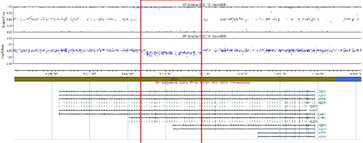
0.001- Fig. 2). Likewise, diffusion tensor imaging (DTI) showed an enhanced
pathways in the index patient and his father that was beyond the range seen in healthy controls (Supplementary Material Fig. S3). For the patient the peak voxels reached statistical significance for the connectivity to the motor pathways (P < 0.001), whereas the father's values showed only a trend but did not reach statistical significance.
Family 2 (GPHN Δ2-3 deletion)
The pedigree of Family 2 is presented in Fig. 1A. The non-consanguineous parents (50-year-old father, 48-year-old mother) are of German ancestry. The GPHN Δ2-3 deletion was paternally inherited. The mother and the brother had diploid copy numbers at the GPHN locus. The 16-year-old male index IGE patient was affected by febrile seizures at the age of 15 months and by myoclonic astatic seizures starting at the age of 1.5 years. Since antiepileptic treatment with valproate, which was started shortly after epilepsy-onset, he is seizure-free. The EEG exhibited irregular sharp- slow wave discharges at 3-Hz. The patient showed a normal physical but delayed cognitive development with persistent learning disability. His epilepsy was classified as idiopathic myoclonic-astatic epilepsy (Doose syndrome). The father had no history of seizures and a normal psychomotor development. The mother experienced three seizures during infancy with spontaneous remission and normal psychomotor development. Her EEGs displayed interictal epileptiform discharges. The 18-year-old brother of the index patient had two febrile seizures during childhood while no subsequent history of afebrile seizures, normal psychomotor development and EEG were reported. None of the family members had a history of psychiatric disorders.
Molecular and biochemical characterization of the GPHN Δ5-9 variant
Gephyrin forms postsynaptic clusters along the neurites as well as soma, while
aggregates (“blobs”, Fig. 3A,B). To examine the effects of the GPHN Δ5-9 deletion, we expressed GFP-tagged Δ5-9geph in cultured hippocampal neurons and HEK293 cells. Δ5-9geph-GFP was homogenously distributed in the cytosol of HEK293 cells and neurons including all morphological structures, i.e. spines (Fig. 3A,B), suggesting its inability of form postsynaptic clusters. To see how clustering deficient Δ5-9geph influences regular gephyrin, we first co-expressed myc-tagged gephyrin and Δ5- 9geph-GFP in HEK293 cells. We found that gephyrin blobs were dissolved resulting in either diffuse regular gephyrin or the formation of microclusters co-localizing with Δ5-9geph-GFP (Fig. 3C,D). This demonstrates a dominant-negative effect of Δ5- 9geph on the oligomerization of regular gephyrin. Notably, Δ5-9geph-GFP was stably expressed at levels equivalent to regular gephyrin-GFP (Fig. 3E). The observed dominant-negative effect on regular gephyrin clustering is mediated by direct interaction with Δ5-9geph, as shown by co-immunoprecipitation (Fig. 3F).
To evaluate the consequences of dominant-negative Δ5-9geph action on neuronal
gephyrin clustering, we expressed Δ5-9geph-GFP or GFP alone in cultured
hippocampal neurons. To distinguish between endogenous gephyrin and Δ5-9geph-
GFP, we used a monoclonal antibody against a peptide in the C-domain that is
deleted in Δ5-9geph (rGeph-C, Fig. 4A). Expression of Δ5-9geph-GFP significantly
reduced the number of endogenous gephyrin clusters confirming the previously
stated dominant-negative effect (Fig. 4B,C). Moreover, remaining gephyrin clusters
were significantly reduced in size indicating that Δ5-9geph disturbs the clustering of
regular gephyrin (Fig. 4D). To determine how Δ5-9geph-GFP expression influences
the clustering of GABA
ARs, we visualized the γ
2-subunit (Fig. 4E), which plays a
central role in gephyrin-dependent postsynaptic clustering of GABA
ARs
30,31. Number
presence of Δ5-9geph-GFP (Fig. 4F,G). Together with the observed reduced quantity of gephyrin clusters, our data imply that receptor homeostasis responds to decreased amounts of clustered gephyrin.
To investigate whether Δ5-9geph-mediated reduction in GABAergic innervation influences synaptic spines, typical morphological structures of the excitatory synapse
32
, we visualized spines in control and Δ5-9geph-GFP-expressing neurons by thresholding the EGFP-signal to an identical value. Spine head morphology and amount was similar in EGFP and Δ5-9geph-expressing neurons, suggesting that diminished GABAergic synapses did not interfere with synaptic spines (Fig. 4H-J).
Besides its synaptic function, gephyrin catalyzes the last step of the molybdenum
cofactor (Moco) biosynthesis being essential for the activity of four molybdenum-
dependent enzymes
33-35. We measured typical urinary biomarkers of two Moco-
dependent enzymes, xanthine oxidase (uric acid, xanthine) and sulfite oxidase (S-
sulfocysteine) as well as the Moco degradation product urothion
36,37in the patient
and his parents of Family 1. Concentrations of all analyzed metabolites were
inconspicuous, suggesting that the hemizygous GPHN Δ5-9 variant did not cause
any subclinical Moco deficiency phenotype in the affected patient (Supplementary
Material Fig. S3).
DISCUSSION
We present a comprehensive clinical, genetic, functional and neurophysiological characterization of two IGE patients with novel hemizygous microdeletions encompassing GPHN exons that code for the N-terminal gephyrin G-domain. Our finding strengthens a previous report of six unrelated subjects with a wide range of neurodevelopmental disorders (ASD, schizophrenia, seizures) who carried hemizygous GPHN microdeletions affecting the gephyrin G-domain
13, and extends the phenotypic spectrum related to N-terminal GPHN microdeletions by IGE syndromes (Supplementary Material Fig. S4). The study reported by Lionel et al.
deduced evidence of pathogenicity of GPHN microdeletions from the significant association with neurodevelopmental disorders (P = 0.009; 6/8,775 cases versus 3/27,019 controls) and found that three out of five GPHN microdeletions tested for inheritance arose as de novo events
13. In this study, we present additional functional data elucidating the molecular mechanisms by which the N-terminal GPHN deletions cause dysfunctional GABAergic synaptic inhibition and thereby increase susceptibility of IGE.
Molecular characterization demonstrates that the truncated GPHN Δ5-9 variant
(Family 1) exerts a dominant-negative effect on the structural synaptic organization of
gephyrin and GABA
AR clusters. Gephyrin oligomerization is thought to be a
concerted interaction mediated by G-domain trimerization
38, G- and E-domain
interaction
18, E-domain dimerization
25and C-domain binding to other proteins
controlled by posttranslational modifications
39. The truncated Δ5-9geph protein itself
is not able to form clusters and impairs the oligomerization process of regular
gephyrin (Fig. 4), which can be explained by the loss of the G-domain trimerization
Thereby, it stochastically affects oligmerization of regular gephyrin in a dominant- negative manner. Importantly, the size of remaining gephyrin and GABA
AR clusters was significantly reduced, which will probably impair the recently described homeostatic regulation of gephyrin scaffolds and normal development of the neuronal inhibitory GABAergic circuits
40. Consistently with our findings, it was speculated that cellular stressors such as elevated temperature or alkalosis, which can arise from seizure activity, might induce irregular exon skipping in GPHN mRNA leading to deletions within the G- and C-domain thus causing a dominant-negative effect on the oligomerization of synaptic gephyrin clusters
20. Moreover, given the constitutional expression of the truncated Δ5-9geph protein, it is also likely that during development Δ5-9geph expression negatively affects the establishment of certain networks which cumulatively contribute to the genetic variance of the IGE phenotype.
In Family 2, the GPHN Δ2-3 variant causes a frameshift with premature stop codon,
resulting in a truncated GPHN protein consists of the first 21 amino acids encoded by
exon 1, followed by seven residues encoded by out of frame codons before the
premature stop codon (p.V22Gfs*7 – Fig. 1F). Notably, five out of six GPHN multi-
exon deletions (Δ2-5 (n = 2), Δ3-8, Δ3-11 and Δ3-12) reported by Lionel et al.
13in
patients with neurodevelopmental disorders result in frameshifts and premature stop
codons, causing truncated GPHN proteins including the first 21 or 47 amino acids
encoded by exon 1 or exons 1-2, respectively. Although truncated gephyrins
comprising the first 47 amino acids have the capacity to impair the oligomerization of
regular gephyrin
20, the expression of these frameshift-causing GPHN multi-exon
deletions will probably be negligible due to nonsense-mediated mRNA decay
28.
Considering the fact that we were able to amplify the GPHN Δ2-3 transcript in cDNA
derived from blood cells (Supplementary Material Fig. S2), at least a partial
expression of the GPHN Δ2-3 transcript is possible. However, we assume that in these patients the pathogenic effect of the truncated GPHN results predominantly from haplo-insufficiency rather than from effects on the oligomerization of regular gephyrin.
Given the intriguing impact of an impaired GABAergic synaptic transmission in epileptogenesis
41, the reduced amount of synaptic GABA
ARs in the presence of truncated gephyrins exerts a functionally convergent effect similar to that observed by mutations of genes encoding GABA
AR subunits (GABRA1, GABRB3, GABRG2 and GABRD) in rare families with dominantly inherited IGE syndromes (for reviews see
42,43
). On molecular level, GABR gene mutations generally lead to a reduced surface expression of GABA
ARs, which results in the reduction of the amplitude of GABA- evoked currents. Although GPHN deletions also should affect glycine receptor clustering, this seems to have no significant pathogenic consequences considering that typical symptoms of glycinergic deficits, such as hyperekplexia, are missing.
Interestingly, in Family 1, the clinically unaffected father as well as the JME-affected
index patient carrying the GPHN Δ5-9 deletion exhibited an impaired cortical
inhibition leading to hyperexcitability, as demonstrated by TMS, which mainly reflects
the functional state of GABAergic interneuronal circuits
44. This imbalance of the
inhibitory/excitatory ratio corresponds well with the experimentally validated
dominant-negative effect of the GPHN Δ5-9 deletion on the reduced size and number
of GABA
AR clusters at inhibitory synapses. Moreover, diffusion tensor imaging
showed a statistically significant increase in the structural connectivity of the mesial
frontal region and the descending motor circuits in the JME-affected index patient
explain the predominant manifestation of myoclonic seizures in the JME-index patient of Family 1. Altogether, our present findings add to convergent lines of evidence that dysfunctional GABAergic synaptic inhibition represent a key pathogenic process shared by a wide spectrum of neurodevelopmental disorders, such as epilepsy, ASD, schizophrenia, and intellectual disability
46. Consistently, a high prevalence of epilepsy has been found in autism spectrum disorder (ASD), ranging from 21% in ASD subjects with an intellectual disability to 8% in those without intellectual disability
47