EXTENDED REPORT
Disease progression in systemic sclerosis-overlap syndrome is signi fi cantly different from limited and diffuse cutaneous systemic sclerosis
Pia Moinzadeh,
1Elisabeth Aberer,
2Keihan Ahmadi-Simab,
3Norbert Blank,
4Joerg H W Distler,
5Gerhard Fierlbeck,
6Ekkehard Genth,
7Claudia Guenther,
8Ruediger Hein,
9Joerg Henes,
10Lena Herich,
11Ilka Herrgott,
12Ina Koetter,
13Alexander Kreuter,
14Thomas Krieg,
1Kathrin Kuhr,
11Hanns-Martin Lorenz,
15Florian Meier,
16Inga Melchers,
17Hartwig Mensing,
18Ulf Mueller-Ladner,
16Christiane Pfeiffer,
19Gabriela Riemekasten,
20Miklós Sárdy,
21Marc Schmalzing,
10Cord Sunderkoetter,
12Laura Susok,
22Ingo H Tarner,
16Peter Vaith,
23Margitta Worm,
24Gottfried Wozel,
8Gabriele Zeidler,
25Nicolas Hunzelmann,
1and all participating DNSS centers
Handling editorTore K Kvien
▸Additional material is published online only. To view please visit the journal online (http://dx.doi.org/10.1136/
annrheumdis-2013-204487).
For numbered affiliations see end of article.
Correspondence to Dr Pia Moinzadeh, Department of Dermatology and Venereology, University of Cologne, Kerpener Str. 62, Köln, Cologne 50937, Germany;
pia.moinzadeh@uk-koeln.de Received 19 August 2013 Revised 12 November 2013 Accepted 10 December 2013 Published Online First 3 January 2014
To cite:Moinzadeh P, Aberer E, Ahmadi-Simab K, et al.Ann Rheum Dis 2015;74:730–737.
ABSTRACT
Background Systemic sclerosis (SSc)-overlap syndromes are a very heterogeneous and remarkable subgroup of SSc-patients, who present at least two connective tissue diseases (CTD) at the same time, usually with a specific autoantibody status.
Objectives To determine whether patients, classified as overlap syndromes, show a disease course different from patients with limited SSc (lcSSc) or diffuse cutaneous SSc (dcSSc).
Methods The data of 3240 prospectively included patients, registered in the database of the German Network for Systemic Scleroderma and followed between 2003 and 2013, were analysed.
Results Among 3240 registered patients, 10% were diagnosed as SSc-overlap syndrome. Of these, 82.5%
were female. SSc-overlap patients had a mean age of 48
±1.2 years and carried significantly more often‘other antibodies’(68.0%; p<0.0001), including anti-U1RNP, -PmScl, -Ro, -La, as well as anti-Jo-1 and
-Ku antibodies.
These patients developed musculoskeletal involvement earlier and more frequently (62.5%) than patients diagnosed as lcSSc (32.2%) or dcSSc (43.3%) ( p<0.0001). The onset of lungfibrosis and heart involvement in SSc-overlap patients was significantly earlier than in patients with lcSSc and occurred later than in patients with dcSSc. Oesophagus, kidney and PH progression was similar to lcSSc patients, whereas dcSSc patients had a significantly earlier onset.
Conclusions These data support the concept that SSc-overlap syndromes should be regarded as a separate SSc subset, distinct from lcSSc and dcSSc, due to a different progression of the disease, different
proportional distribution of specific autoantibodies, and of different organ involvement.
INTRODUCTION
Systemic sclerosis (SSc) is a heterogeneous, multi- system, chronic disorder, leading tofibrosis of the
skin and many internal organs. To classify patients with established disease, the American College of Rheumatology published in 1980 preliminary cri- teria.1 Currently a subclassification developed by LeRoyet al, this is the most widely used classifica- tion system for limited and diffuse SSc in clinical practice,2and is the basis for many registries world- wide.3 In these registries, it became apparent that in a sizeable number of patients, symptoms of SSc occur in combination with those of other connect- ive tissue diseases (CTD),4–10 also described by some authors as SSc-overlap syndrome.4 11–13 Up to now, no firm classification criteria for SSc-overlap syndromes are established, but it is gen- erally considered when musculoskeletal involve- ment (myositis, arthritis) or clinical signs of other rheumatic diseases are substantially greater than usually found in SSc patients.12
Clinical features of overlap syndrome patients are very heterogeneous, and epidemiological studies report divergent frequencies of overlap sub- groups as well as of organ manifestations.11 14 15
Musculoskeletal involvement, including joints, tendons and muscles, is the most frequent clinical feature, highlighting the difference to other SSc forms. Inflammatory joint involvement is reported to be the second most frequent manifestation in patients with musculoskeletal involvement and overlap syndromes. These patients are often identi- fied by typical clinical symptoms (usually limited skin involvement) together with high titres of antic- yclic citrullinated peptides (CCP/ACPA) and/or higher rheumatoid factors (RF).16 17
All known classification criteria for overlap syn- dromes include autoantibodies, which are helpful to separate them from other subsets.18–20 PmScl- and anti-U1RNP-antibodies are known to be the most common autoantibodies in patients with overlap syndromes.21–24
Pakozdi et al reported recently, that 20% of the patients attending the Centre for Rheumatology at
Open Access Scan to access more
free content
the Royal Free Hospital had overlapping features with other rheumatologic diseases, such as polymyositis/dermatomyositis (43%), systemic lupus erythematosus (SLE) (8%), Sjögren’s syn- drome (17%) and rheumatoid arthritis (32%).12
It has been always debated whether patients suffering from overlap syndromes should be regarded as a separate entity, or should be included, depending on their skin involvement, in the two main groups of limited (lcSSc) and diffuse SSc (dcSSc) patients.
In this prospective study, it could be shown for thefirst time that SSc-overlap syndromes should be viewed as a distinct SSc subset.
MATERIALS AND METHODS
This study involves 3240 patients, registered in the database of the German Network for Systemic Scleroderma (DNSS). The network combines different subspecialties consisting of rheuma- tologists, dermatologists, pulmonologists and nephrologists from altogether more than 40 clinical centres. The Ethics Committee of the coordinating centre, that is, the Cologne University Hospital, gave a positive vote on the patient information and consent form for the registry. On the basis of this document, all participating centres received the approval of their local ethics committees prior to registering patients.
Patient data, including information about gender, age, auto- antibodies, SSc subsets, symptoms and signs, organ involvement, modified Rodnan Skin Score (mRSS) as well as treatments, were recorded on a prospective basis in a database started in 200325–27 with a mean follow-up time of 9.5±0.2 years (from the time of SSc onset till the last follow-up visit). A significant number of these patients were classified according to the criteria of LeRoy et al2as having lcSSc or dcSSc. Additionally, a smaller but still considerable number of patients, did not fulfil these criteria, but were registered to follow-up their course of disease. Data of the first Raynaud phenomenon (RP) onset, as well as data of non-RP onset of skin and organ involvement were recorded.
Due to the lack of satisfying classification criteria for patients with different forms of overlap syndromes, all patients with more than one CTD were classified as SSc-overlap syndrome in general, including symptoms and signs, autoantibodies and organ manifestations in detail. This information was used to characterise patients with SSc-overlap syndromes.
SSc subsets
Patients with overlap syndromes were defined as a disease, occurring with clinical features of SSc, according to the ACR criteria, or main SSc-associated symptoms, simultaneously with those typical for other rheumatic diseases.4These patients were often positive for anti-PmScl, -U1RNP, -Jo-1, -Ku, -Ro or –La autoantibodies. Patients with a mixed connective tissue disease (MCTD) were also included in SSc-overlap syndromes, as MCTD combines features of SLE, SSc and myositis, together with the presence of anti-U1RNP-antibodies.
Patients suffering from dcSSc were characterised by a progres- sive course of disease with an early onset of RP, usually within 1 year of onset of skin changes. They were defined by rapid skin involvement of the trunk, face, proximal and distal extremities and being frequently associated with antitopoisomerase (ATA) antibodies.2
LcSSc was defined by skin thickening of the extremities distal to the knee and elbow joints, facial skin and occurrence of RP, which usually appeared many years prior to skin involvement.
These patients are often positive for anticentromer-antibodies (ACA).2
Patients with undifferentiated SSc were defined as positive RP together with at least one further feature of SSc (typical nailfold capillary alterations, puffy fingers, pulmonary hypertension (PH)) and/or detectable SSc-specific autoantibodies without ful- filling the ACR criteria for SSc.28
Patients with sclerosis sine scleroderma were defined by posi- tive RP, no skin alterations, PAH, cardiac, pulmonary and gastro- intestinal involvement.29
Within this study, we focused on patients suffering from lcSSc, dcSSc and SSc-overlap syndromes.
Antinuclear autoantibodies (ANAs)
The antibody measurement was performed in respective labora- tories of the participating centres. Serum was routinely analysed in all registered SSc patients at the first visit and repeated as needed. Autoantibodies were subdivided into SSc-specific (ACA, ATA, anti-PmScl, -U1RNP, -Jo1, -Ku antibodies) and SSc non- specific autoantibodies (anti-Ro and -La antibodies). Missing data were less than 10%.
Organ involvement
RP was defined by recurrent vasospasms of small digital arter- ioles/arteries at fingers and/or toes, usually triggered by cold environment. We defined the age of RP onset as the age, at which the RPfirst appeared.25
Thefirst non-RP onset of organ involvement has been consid- ered as the timepoint of first skin or organ manifestation. The onset of skin involvement has been set as onset of SSc.
Skin involvement was evaluated using the modified Rodnan Skin Score (mRSS), which assesses the skin hardening/thickness by manual palpation of 17 body areas on a scale of 0 to 3.
Pulmonary manifestation includes pulmonary interstitial fibrosis and/or isolated PH. Isolated pulmonary hypertension was defined as clinical evidence of right-heart failure and/or increased mean pulmonary arterial pressure (PAPm>25 mm Hg at rest or PAP>30 mm Hg during exercise), determined by right-heart catheterisation. Echocardiography was used to iden- tify likely PAH (estimated RVSP>40 mm Hg).
Pulmonary interstitial fibrosis was established when bilateral basal fibrosis occurred, confirmed by chest X-ray and/or high- resolution CT scan together with restrictive pulmonary abnor- malities on pulmonary function tests (TLC <80%), were found.
We defined a normal diffusing capacity of lung for carbon mon- oxide (DLCO) level, when it was >75%, and a low level, when it was less than 75%.
Gastrointestinal involvement was defined as gastrointestinal motility disturbance, dysphagia, nausea, malabsorption, oesophageal stenosis, gastro-oesophageal reflux or intestinal pseudo-obstruction.
Kidney involvement was defined as the presence of renal insufficiency encompassing renal insufficiency due to acute renal crisis (creatinine clearance age-related less than 80 mL/min). The diagnosis of proteinuria was fulfilled in cases of albuminuria
>=30 mg/24 h or >=20 mg/L; proteinuria >=300 mg/24 h or
>=200 mg/L.
Cardiac disease was defined by heart palpitation, conduction disturbance and/or diastolic dysfunction.
Skeletal muscle diseasewas defined as proximal muscle weak- ness and/or atrophy associated with elevated serum muscle enzyme (creatine phosphokinase, CK) levels and/or articular involvement.25 The articular involvement included synovitis with swelling, with or without tenderness to palpation in one or more than one joint. The questionnaire also asked for any kind of joint contractures or tendon friction rubs.
Sicca syndrome wascharacterised by reduced glandular func- tion, usually causing a dry mouth and dry eyes, while involve- ment of the masticatory organ was characterised by microstomia, defined as obvious decreased mouth opening clearly detected by the investigators due to the disease and/or fibrosis of the lingual frenulum.
The recommendations for follow up visits and investigations (echocardiography, electrocardiogram, lung function test, etc) are at least once per year.
Statistics
Differences between the SSc subsets were investigated, usingχ2 test for categorical variables and t tests for continuous para- meters. To compare the disease progression in the three main subsets Kaplan–Meier analysis with log rank tests was per- formed. The starting point of the Kaplan–Meier curves was set as SSc onset, which we defined as the time offirst non-RP mani- festation (onset of skin involvement). The onset of different organ involvements is illustrated within the course of the disease according to our registered follow-up visits.
Additionally, univariate and multiple logistic regression ana- lysis were used to assess the impact of SSc subsets, autoantibody status, age and gender on organ involvement. OR and the corre- sponding 95% CI are reported. To investigate the development of DLCO over regular follow-up time, mixed model analysis with backward selection was applied. Time since SSc onset, group membership and their interaction were included as fix factors, and patient ID as a random effect. In order to guard
against type I error inflation due to multiple testing, only p values below 0.001 were considered statistically significant.
Statistical analysis was done with PASW Statistics V.18.0 (Chicago: SPSS). Missing data were less than 15% for organ manifestations and clinical signs. Missing data for autoantibodies are included within table 2 (less than 10%).
RESULTS
Patient characteristics of all SSc subsets
Between the years 2003 and 2012, a total of 3240 patients with SSc had been registered in the DNSS database. Among all regis- tered patients, 49.3% were diagnosed as lcSSc and 30.8% with dcSSc, 10.0% with an overlap syndrome, 7.7% with undifferen- tiated scleroderma, 0.7% with sclerosis sine scleroderma, and 1.4% patients were categorised as others. Within all registered patients, 81.5% were female; 87.5% were positive for ANA, 33.7% had anticentromere antibodies, and 26.4% were antito- poisomerase antibody (ATA) positive, while the remaining patients had other antibody specificities (32.5%).
Of all registered patients (first visit), 13.3% had PH, 36.5%
lungfibrosis, 55.1% suffered from gastrointestinal involvement, 9.9% had kidney involvement, 12.5% suffered from heart involvement, 39.1% from musculoskeletal involvement and 37.5% from sicca symptoms.
The mean time between the onset of SSc and the first visit/
registration within our database was for all SSc patients (n=2522) 7.6±0.2 years (SSc diagnosis to first visit), for lcSSc patients (n=1236) 8.3±0.2 years (SSc diagnosis to first visit),
Table 1 Frequencies and p values (χ2test) of organ manifestations and current symptoms in patients with overlap syndromes, compared to patients with limited systemic sclerosis/diffuse cutaneous SSc (lcSSc/dcSSc)
SSc subsets p Values
Overlap s. (n=325) lcSSc (n=1598) dcSSc (n=997) Overlap versus lcSSc Overlap versus dcSSc
Female 82.5 85.9 73.1 0.122 0.001
Male 17.5 14.1 26.8
Positive family history 23.1 15.9 15.0 0.039 0.029
Age (mean±SD) 48±1.2 52.8±0.5 48.2±0.6 0.0001 0.847
Erythrocyte sedimentation rate (mean±SD) 23±1.2 17.9±0.4 23.4±0.7 0.0001 0.734
DLCO (mean±SD) 70.5±1.5 73.5±0.7 65.3±0.9 0.064 0.005
Modified Rodnan Skin Score (mean±SD) 6.8±0.4 7.4±0.2 15.8±0.3 0.191 0.0001
Organ involvement
Pulmonary hypertension 10.8 12.3 18.2 0.402 0.003
Lung fibrosis 35.7 24.9 61.2 0.0001 0.0001
Oesophagus 52.0 56.2 60.8 0.065 0.013
Kidney 6.8 8.3 14.3 0.318 0.0001
Heart 13.8 9.6 18.3 0.046 0.104
Musculoskeletal system 62.5 32.2 43.3 0.0001 0.0001
Sicca symptoms 40.0 39.5 33.8 0.703 0.049
Current clinical signs at first visit
Digital ulcers 18.2 23.3 33.3 0.034 0.0001
Synovitis 22.8 11.8 14.7 0.0001 0.004
Dermatogenous contractures 20.6 19.5 35.2 0.818 0.0001
Tendon friction rubs 8.0 5.4 10.4 0.090 0.198
Elevated creatine phosphokinase levels 17.8 6.3 10.5 0.0001 0.001
Muscle weakness 36.9 20.5 29.7 0.0001 0.032
Muscle atrophy 19.7 9.3 17.1 0.0001 0.453
Dysphagia 51.4 56.9 60.3 0.023 0.001
Renal failure 8.3 10.1 13.9 0.308 0.005
Proteinuria 8.0 6.4 11.0 0.330 0.114
DLCO, diffusing capacity of lung for carbon monoxide; SD, standard deviation.
for dcSSc patients (n=857) 7.3±0.3 years (SSc diagnosis tofirst visit), and for overlap patients (n=238) 7.1±0.5 years (SSc diag- nosis tofirst visit).
The mean follow-up time between the SSc diagnosis and the last follow-up visit registered in the DNSS was 9.5±0.2 years for all SSc patients (n=2539), 10.1±0.2 years for lcSSc patients (n=1244), 9.2±0.3 years for dcSSc patients (n=862) and 9.6
±0.6 years for SSc-overlap syndromes (n=241).
Overlap syndromes
Within the group of patients with overlap syndromes (n=325), 82.5% (268/325) were women and had a mean age at time of SSc onset of 48±1.2 years (n=323) (table 1); 15.4% of these patients were ACA positive, 13.2% ATA positive and 68.0%
carried other antibodies (compared to lcSSc (26.7%) and dcSSc patients (31.1%); ( p<0.0001)). These other antibodies con- sisted of anti-U1RNP (33.0%), -Ro (24.4%), -PmScl (16.7%), -La (10.9%), -Ku (3.6%), -Jo1 antibodies (4.1%) and others (6.3%) (table 2).
During follow-up, of all 325 patients, more than 92.0%
maintained their initial diagnosis. Patients with SSc-overlap syn- dromes tended to have more often a positive family history for rheumatological disorders compared to lcSSc and dcSSc patients, and were significantly more frequently treated with corticosteroids (60.6% vs 27.3% (lcSSc) and 44.3% (dcSSc);
p<0.0001) and immunosuppressive agents (58.8% vs 21.0%
(lcSSc) and 41.6% (dcSSc); p<0.0001) than other SSc subsets, as published in 2009.26 Additionally, this specific subset also had a significantly lower mRSS compared to dcSSc patients (6.8
±0.4 vs 15.8±0.3; p<0.0001), but a very similar mean mRSS to lcSSc patients (7.4±0.2) (table 1). Following the criteria of LeRoy for limited and diffuse extension of skin thickening, we had 76.0% of patients with a limited extension and 14.8% with a diffuse form of skin thickening. Significantly more female SSc-overlap patients had a limited skin involvement (87.8% vs 63.3%; p<0.0001), while significant more male patients suf- fered from a diffuse skin involvement (36.7% vs 12.2%;
p<0.0001). No significant abnormalities for organ manifesta- tions/clinical signs or antibody distribution have been found.
Interestingly, significantly less patients with overlap syndromes suffered from digital ulcers (18.2% vs 33.3%; p<0.0001) (see online supplementary table S1).
Musculoskeletal involvement
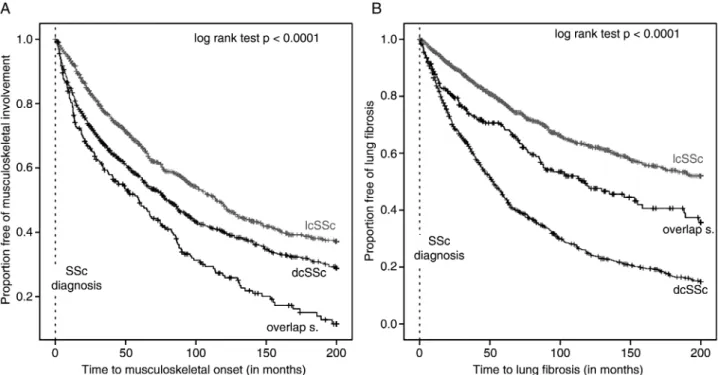
Patients with overlap syndrome developed significantly earlier and more often musculoskeletal involvement, followed by patients with diffuse and limited SSc (data shown in table 1 and figure 1A). Musculoskeletal manifestation included muscle weakness (36.9%), synovitis (22.8%), contractures (2.6%), muscle atrophy (19.7%) and elevated CK levels (17.8%).
Logistic regression analysis revealed, that overlap patients had threefold the risk of developing musculoskeletal involvement, compared to patients with lcSSc (OR 3.2; p<0.001; 95%-CI 2.5 to 4.2), and double the risk compared to dcSSc patients (OR 2.2; p<0.001; 95%-CI 1. 6 to 2.9).
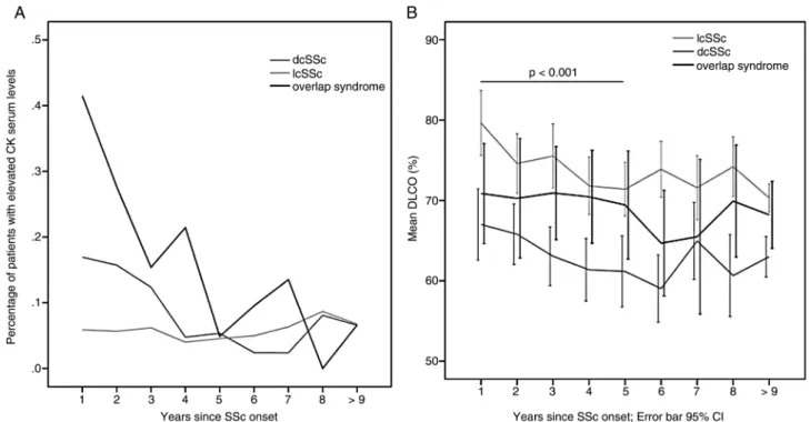
We also found that the course of initially elevated CK serum levels decreased substantially over time, especially in overlap and dcSSc patients (figure 3A).
In a subgroup of patients classified as overlap syndrome with myositis and CK elevation (44/325), we determined the fre- quency of gender distribution (70.5% women, 29.5% men), as well as clinical features; 59.1% had oesophageal involvement, 38.6% of patients had lung fibrosis, and 34.1% suffered from cardiac involvement, while less than 15.0% had kidney failure or signs for PH. Most of the patients selected for myositis and CK elevation were PmScl positive (22.7%) followed by 9.1%
anti-U1RNP antibodies, 6.8% ATA antibodies and 4.5% ACA, Ku- and Jo1-antibodies.
Cardiopulmonary involvement
Lung fibrosis and heart involvement was diagnosed earliest in patients with dcSSc, followed by SSc-overlap and lcSSc patients (log rank test p<0.0001); patients with SSc-overlap syndromes resulted in a clear intermediate position between patients suffer- ing from lcSSc and dcSSc (figures 1B and 2A).
Logistic regression analysis revealed that patients with overlap syndromes had a higher risk of developing lung fibrosis com- pared to lcSSc patients (OR 1.6; p<0.001; 95%-CI 1.2 to 2.1), and a reduced risk compared to dcSSc patients (OR 0.4;
p<0.001; 95%-CI 0.3 to 0.5). They also had a lower risk of developing lungfibrosis when they had a high DLCO-level (OR 0.9; p<0.001; 95%-CI 0.9 to 1.0). The analysis of DLCO within regular follow-up visits showed no significant interaction, for example, the decrease in DLCO over time does not differ between group memberships. However, there was an overall Table 2 Detailed autoantibody status of patients with overlap syndromes, compared to patients with lcSSc/dcSSc
SSc subsets p Values
Overlap
s. (n=325) lcSSc (n=1598) dcSSc (n=997) Overlap versus lcSSc Overlap versus dcSSc
Autoantibodies % md % md % md
ANAs positive 92.0 3.1 87.9 4.1 89.1 4.1 0.050 0.240
ACA positive 15.4 5.2 53.8 7.6 8.4 8.9 0.0001 0.001
ATA positive 13.2 4.9 16.0 7.9 52.4 8.0 0.155 0.0001
Other Abs 68.0 4.3 26.7 6.9 31.1 7.2 0.0001 0.0001
Other Abs than ACA & ATA, including (n=221) (n=426) (n=310)
PmScl 16.7 5.9 2.6 0.0001 0.0001
anti-U1RNP 33.0 5.4 3.9 0.0001 0.0001
Jo-1 4.1 0.5 1.9 0.001 0.183
Ku 3.6 0.5 2.3 0.006 0.601
Ro 24.4 23.5 27.7 0.770 0.484
La 10.9 10.6 14.2 0.894 0.294
md, missing data; missing data were less than 10%.
Abs, antibodies; ACA, anticentromer antibodies; ATA, antitopoisomerase antibodies; ANAs, antinuclear autoantibodies.
significant difference in DLCO between groups ( p<0.001) and between the years since SSc onset ( p<0.001). A posthoc test revealed additionally a significant difference between years 1 and 5, again the course of the curve of overlap patients was running between dcSSc and lcSSc patients (figure 3B).
Other organ manifestations
Disease progression, as determined by the onset of PH, oesophagus and kidney involvement of SSc-overlap patients, was similar to lcSSc patients. There was also no significant dif- ference in patients developing gastrointestinal involvement, Figure 1 Kaplan–Meier analysis of the onset of (A) musculoskeletal involvement and (B) lungfibrosis in lcSSc, dcSSc and SSc-overlap syndromes.
Events (defined as onset of musculoskeletal involvement or lungfibrosis) correspond to step-downs, while censored observations (defined as last follow up visit) are identified as ticks. The plot shows a significant difference ( p<0.0001) between the survival curves of lcSSc, dcSSc and overlap syndrome patients.
Figure 2 Kaplan–Meier analysis of the onset of (A) heart involvement and (B) PH in lcSSc, dcSSc and SSc-overlap syndromes. Events (defined as onset of heart involvement or PH) correspond to step-downs while censored observations (defined as last follow-up visit) are identified as ticks. The plot shows a significant difference ( p<0.0001) between the survival curves of lcSSc, dcSSc and overlap syndrome patients.
depending on their subset, gender and autoantibody status (table 1 and online supplementary table S1,figure 2B).
Autoantibody status
Detailed characterisation of overlap patients depending on their autoantibody status revealed that patients with anti-U1RNP-antibodies were significantly younger at RP onset, compared with PmScl-positive patients (36.2±1.8 years vs 44.6
±2.3 years; p<0.008) and tended to be younger at onset of skin manifestations (39.3±2.0 years vs 5.3±2.4 years). The interval between RP onset and skin onset was shortest for patients with Ku antibodies (0.7±0.7 years), followed by Jo1 (1.6±0.9 years), PmScl (2.0±0.6 years), ATA (2.1±0.8 years), Ro (5.0
±1.3 years), La (5.2±2.5 years), anti-U1RNP (6.7±1.4 years), followed by ACA-positive patients with 11.2±2.4 years.
DISCUSSION
SSc is a heterogeneous disease and includes subsets which are characterised by the extension of skin involvement and circulat- ing autoantibodies. For many years it has been observed, that not all SSc patients fit into the categories defined by LeRoy et al2A considerable number of patients present with symptoms and signs of SSc together with clinical features of other CTDs.5
Of the 3240 patients registered in the DNSS, 10.0% were categorised as overlap syndrome. This frequency is in agreement with other reported data, ranging between 10.0% and 38.0%.4 14 30–32Our study presents one of the largest studies, characterising patients with overlap syndromes in direct com- parison with the two main SSc subsets, and our data indicate that overlap patients clearly differ from lcSSc and dcSSc.
Totally, 62.5% of the overlap patients in this study had mus- culoskeletal manifestations, which confirms previously published data of Balbir–Gurmanet al, who reported myositis in 47.5%
of their 40 overlap patients, and Troyanovet al, who reported SSc-myositis overlaps in 42.6% of their cases.33
In this registry, patients with SSc/myositis, who suffered from muscle weakness together with elevated CK serum levels, 59.1%
had oesophageal involvement, 38.6% of patients had lungfibro- sis, 34.1% suffered from cardiac involvement, while less than 15.0% had kidney failure or signs for PH. These frequencies are lower than in the study of Balbir–Gurman who reported higher frequencies of occurrence, but the same trend in the order of frequencies; 84.2% of patients with SSc-myositis had gastro- intestinal involvement, 66.4% interstitial lung disease, and 26.3% cardiomyopathy or PH. Most of the patients showing myositis and CK elevation were PmScl positive (22.7%), fol- lowed by 9.1% anti-U1RNP antibodies, 7.0% ATA antibodies and 4.5% ACA, anti-Ku and -Jo1 antibodies. Compared to these data, other groups reported a higher frequency of ATA anti- bodies.4 The frequency of elevated CK serum levels decreased within the course of follow-up visits over a period of 9 years, which could indicate that myositis responds to treatment.34 As expected, significantly more SSc-overlap patients have been put on immunosuppressive treatment compared to lcSSc and/or dcSSc patients, confirming previously published data.26
We found significantly more overlap patients with joint involvement compared to lcSSc and dcSSc patients ( p<0.0001), but with no significant difference in the frequency of RF positiv- ity, although the presence of RF and the association with rheumatoid arthritis of SSc patients have been previously dis- cussed controversially.35
Regarding the onset of lung fibrosis and heart involvement, patients with SSc-overlap syndromes had an intermediate rate of disease progression in between lcSSc and dcSSc. This observa- tion is further supported by the DLCO levels following a similar course. Overlap patients, in general, had significantly less fre- quent PH and kidney involvement than dcSSc patients. The direct comparison between the three major SSc subsets, using Kaplan–Meier curves, visualised that the trend of the SSc-overlap curve was clearly between the curves standing for lcSSc and dcSSc patients (figures 1 and 2).
Figure 3 Course of (A) the percentage of patients with elevated CK serum levels and (B) mean DLCO levels across all follow-up visits per year for the three main SSc subsets.
Additionally, patients with SSc-overlap syndromes were sig- nificantly younger (48±1.2 years) than patients with lcSSc, con- firming the data of Garamaschi et al (48.5±13.3 years) and developed less frequently digital ulcers, resulting in significant differences, when compared with dcSSc patients.
Furthermore, the data of this study are in good agreement with Mierau et al and Hasegawa et al, who found that anti-U1RNP antibodies were associated with a younger age of disease onset when using a multiple regression analysis.27 36 However, Koschik et al reported that in their patient cohort, patients with PmScl-antibodies were significantly younger than those without PmScl-antibodies.32 These patients developed most frequently musculoskeletal involvement, including muscle weakness and synovitis, but in contrast with other studies, less lung fibrosis.37 Our study, however, did not allow analysing, whether symptoms and signs of other rheumatic diseases appeared prior or after first SSc features, but Caramaschiet al reported, that 40.5% of their patients were diagnosed with an additional autoimmune disease prior and 38.1% after SSc diagnosis.30
In summary, we could demonstrate that patients with overlap syndromes differ from lcSSc and dcSSc regarding lung fibrosis and heart involvement, and that musculoskeletal involvement is clearly the most frequent organ manifestation in overlap patients. Although these patients appear to have a milder course of the disease with a mean mRSS similar to lcSSc patients, but less lung fibrosis and heart involvement, when compared to dcSSc patients, they progressed more rapidly with earlier and more widespread significant organ involvement than patients with lcSSc. This study demonstrates that patients with SSc-overlap, on average, carry a higher disease burden than patients with the limited form.
Based on a large cohort, this study strongly supports the idea that patients with SSc-overlap syndromes should be regarded as a separate subset of patients with SSc.
Author affiliations
1Department of Dermatology, Cologne University Hospital, Cologne, Germany
2Department of Dermatology, Medical University of Graz, Graz, Germany
3Department of Rheumatology, Asklepios Clinic Altona, Hamburg, Germany
4Department of Internal Medicine, Division of Rheumatology, University of Heidelberg, Heidelberg, Germany
5Department of Rheumatology, University of Erlangen, Erlangen, Germany
6Department of Dermatology, University of Tuebingen, Aachen, Germany
7Department of Rheumatology, Clinic of Rheumatology of Aachen, Aachen, Germany
8Department of Dermatology, University-Hospital Carl Gustav Carus, Dresden, Germany
9Department of Dermatology, Munich University of Technology, Munich, Germany
10Department of Rheumatology, University of Tuebingen, Tuebingen, Germany
11Institute of Medical Statistics, Informatics and Epidemiology, University of Cologne, Cologne, Germany
12Department of Dermatology and Venereology, University of Muenster, Muenster, Germany
13Department of Internal Medicine and Nephrology (Centre for interdisciplinary Rheumatology), Robert-Bosch-Hospital, Stuttgart, Germany
14Department of Dermatology, Helios Clinic Oberhausen, Oberhausen, Germany
15Department of Haemato-Oncology and Rheumatology, University of Heidelberg, Heidelberg, Germany
16Department of Rheumatology and Clinical Immunology, Kerckhoff Clinic, Bad Nauheim, Germany
17Clinical Research Unit for Rheumatology, University Medical Center Freiburg, Freiburg, Germany
18Clinic for Dermatology, Hamburg Alstertal, Hamburg, Germany
19Department of Dermatology, Ulm University Hospital, Ulm, Germany
20Department of Rheumatology and Clinical Immunology, University of Berlin, Charité, Germany
21Department of Dermatology and Allergology, Ludwig Maximilian University, Munich, Germany
22Department of Dermatology and Venereology, Ruhr University Bochum, Bochum, Germany
23Department of Rheumatology and Clinical Immunology, University Medical Center Freiburg, Freiburg, Germany
24Department of Dermatology and Venerology, University of Berlin, Charité, Berlin, Germany
25Department of Rheumatology, Johanniter-Hospital, Treuenbrietzen, Germany Acknowledgements The work of Ebru Keser, Patricia Scholz and Gabriele Browne at the central office in keeping the Network going is gratefully acknowledged.
Collaborators Nil Mona Ahrazoglu, Department of Dermatology, Cologne University Hospital; Mike Oliver Becker, Department of Rheumatology and Clinical Immunology, University of Berlin, Charité; Rudolf Mierau, Department of Rheumatology, Clinic of Rheumatology Aachen; Gunther Neeck, Medical Center for Rheumatology Bad Doberan; Christoph Fiehn, Medical Center for Rheumatology, Baden-Baden; Aaron Juche, Department of Rheumatology, Immanuel Hospital Berlin-Buch; Andrea Rubbert, Department of Rheumatology, University of Cologne;
Manfred Weber, Department of Internal Medicine, Hospital Cologne Merheim;
Rebecca Fischer-Betz, Department of Nephrology and Rheumatology, University of Duesseldorf; Michael Sticherling, Department of Dermatology and Venereology, University of Erlangen; Harald Burkhardt, Michaela Koehm, Med. Clinic II, Department of Rheumatology, University of Frankfurt; Cornelia S Seitz, Claudia Herink, Department of Dermatology, Venereology and Allergology, University of Goettingen; Ivan Foeldvári, Department of Rheumatology, Schoen Clinic, Hamburg-Eilbek; Johannes Norgauer, Department of Dermatology and Venereology, University of Jena; Dieter Schoeffel, Clinic for Rheumatology, Mannheim; Regina Renner, Mirjana Ziemer, Department of Dermatology and Venereology, University of Leipzig; Kerstin Steinbrink, Department of Dermatology and Venereology, University of Mainz; Rudolf Stadler, Verena Müller, Department of Dermatology and Venereology, Johannes Wesling Hospital, Minden; Mahzad Ziai, Department of Dermatology, Munich University of Technology; Sigrid Karrer, Michael Landthaler, Department of Dermatology and Venereology, University of Regensburg; Rotraud Meyringer, Department of Internal Medicine, University of Regensburg; Michael Buslau, Clinic for Rehabilitation Rheinfelden (CH); Dirk Tomsitz, Hae- Hyuk Lee, Department of Dermatology and Venereology, University of Berlin, Charité; Percy Lehmann, Noemi Gaebelein-Wissing, Helios Clinic, Department of Dermatology, Wuppertal; Marieke Wagner, Department of Rheumatology, University of Tuebingen;
Joachim Ebel, Department of Rheumatology, Asklepios Clinic Altona; Christian Beyer, Department of Rheumatology, University of Erlangen; Rita Varga, Department of Dermatology and Allergology, Ludwig Maximilian University, Munich; Agnes Bretterklieber, Department of Dermatology, Medical University of Graz; Frank Reichenberger, Department of Pneumology, Asklepios Clinic Gauting,
Muenchen-Gauting; Andrea Himsel, Clinic for Internal Medicine and Rheumatology, GPR MVZ Ruesselsheim
Contributors PM, NH and TK have conceived and designed the study. All authors and coauthors (excluding LH and KK) were involved in data acquisition. LH and KK have made particular contribution to statistical analyses and interpretation of data.
All authors contributed to the interpretation of the data and jointly approved the final manuscript.
Funding This study was supported by a grant of the German Federal Ministry of Education and Research (BMBF) (01GM0310) and a grant of the Edith-Busch- Foundation. The work of Pia Moinzadeh was supported by a german‘Koeln Fortune’
and‘Deutsche Stiftung Sklerodermie’grant.
Competing interests None.
Patient consent Obtained.
Ethics approval Ethics Committee University of Cologne.
Provenance and peer reviewNot commissioned; externally peer reviewed.
Open Access This is an Open Access article distributed in accordance with the Creative Commons Attribution Non Commercial (CC BY-NC 4.0) license, which permits others to distribute, remix, adapt, build upon this work non-commercially, and license their derivative works on different terms, provided the original work is properly cited and the use is non-commercial. See: http://creativecommons.org/
licenses/by-nc/4.0/
REFERENCES
1 Preliminary criteria for the classification of systemic sclerosis (scleroderma).
Subcommittee for scleroderma criteria of the American Rheumatism Association Diagnostic and Therapeutic Criteria Committee.Arthritis Rheum1980;23:581–90.
2 LeRoy EC, Black C, Fleischmajer R,et al. Scleroderma (systemic sclerosis):
classification, subsets and pathogenesis.J Rheumatol1988;15:202–5.
3 Galluccio F, Walker UA, Nihtyanova S,et al. Registries in systemic sclerosis: a worldwide experience.Rheumatology (Oxford)2011;50:60–8.
4 Balbir-Gurman A, Braun-Moscovici Y. Scleroderma overlap syndrome.Isr Med Assoc J2011;13:14–20.
5 Elhai M, Avouac J, Kahan A,et al. Systemic sclerosis at the crossroad of polyautoimmunity.Autoimmun Rev2013.
6 Cappelli S, Bellando Randone S, Martinovic D,et al.“To be or not to be,”ten years after: evidence for mixed connective tissue disease as a distinct entity.Semin Arthritis Rheum2012;41:589–98.
7 Ziswiler HR, Urech R, Balmer J,et al. Clinical diagnosis compared to classification criteria in in a cohort of 54 patients with systemic sclerosis and associated disorders.Swiss Med Wkly2007;137:586–90.
8 Fiori G, Pignone A, Cerinic MM. Overlap syndromes.Reumatizam2002;49:12–15.
9 Jury EC, D’Cruz D, Morrow WJ. Autoantibodies and overlap syndromes in autoimmune rheumatic disease.J Clin Pathol2001;54:340–7.
10 Meier FM, Frommer KW, Dinser R,et al. Update on the profile of the EUSTAR cohort: an analysis of the EULAR Scleroderma Trials and Research group database.
Ann Rheum Dis2012;71:1355–60.
11 Jablonska S, Blaszczyk M. Scleroderma overlap syndromes.Adv Exp Med Biol 1999;455:85–92.
12 Pakozdi A, Nihtyanova S, Moinzadeh P,et al. Clinical and serological hallmarks of systemic sclerosis overlap syndromes.J Rheumatol2011;38:2406–9.
13 Hashimoto A, Endo H, Kondo H,et al. Clinical features of 405 Japanese patients with systemic sclerosis.Mod Rheumatol2012;22:272–9.
14 Iaccarino L, Gatto M, Bettio S,et al. Overlap connective tissue disease syndromes.
Autoimmun Rev2013;12:363–73.
15 Jablonska S, Blaszyk M. Scleromyositis (scleroderma/polimyositis overlap) is an entity.J Eur Acad Dermatol Venereol2004;18:265–6.
16 Szucs G, Szekanecz Z, Zilahi E,et al. Systemic sclerosis-rheumatoid arthritis overlap syndrome: a unique combination of features suggests a distinct genetic, serological and clinical entity.Rheumatology (Oxford)2007;46:989–93.
17 Ueda-Hayakawa I, Hasegawa M, Kumada S,et al. Usefulness of anti-cyclic citrullinated peptide antibody and rheumatoid factor to detect rheumatoid arthritis in patients with systemic sclerosis.Rheumatology (Oxford)2010;49:2135–9.
18 Ho KT, Reveille JD. The clinical relevance of autoantibodies in scleroderma.Arthritis Res Ther2003;5:80–93.
19 Maddison PJ. Overlap syndromes and mixed connective tissue disease.Curr Opin Rheumatol1991;3:995–1000.
20 Graf SW, Hakendorf P, Lester S,et al. South Australian Scleroderma Register:
autoantibodies as predictive biomarkers of phenotype and outcome.Int J Rheum Dis2012;15:102–9.
21 Steen VD. Autoantibodies in systemic sclerosis.Semin Arthritis Rheum2005;35:35–42.
22 Nihtyanova SI, Denton CP. Autoantibodies as predictive tools in systemic sclerosis.
Nat Rev Rheumatol2010;6:112–16.
23 Cepeda EJ, Reveille JD. Autoantibodies in systemic sclerosis andfibrosing syndromes:
clinical indications and relevance.Curr Opin Rheumatol2004;16:723–32.
24 Oddis CV, Okano Y, Rudert WA,et al. Serum autoantibody to the nucleolar antigen PM-Scl. Clinical and immunogenetic associations.Arthritis Rheum
1992;35:1211–17.
25 Hunzelmann N, Genth E, Krieg T,et al. The registry of the German Network for Systemic Scleroderma: frequency of disease subsets and patterns of organ involvement.Rheumatology (Oxford)2008;47:1185–92.
26 Hunzelmann N, Moinzadeh P, Genth E,et al. High frequency of corticosteroid and immunosuppressive therapy in patients with systemic sclerosis despite limited evidence for efficacy.Arthritis Res Ther2009;11:R30.
27 Mierau R, Moinzadeh P, Riemekasten G,et al. Frequency of disease-associated and other nuclear autoantibodies in patients of the German Network for Systemic Scleroderma:
correlation with characteristic clinical features.Arthritis Res Ther2011;13:R172.
28 Alarcon GS. Unclassified or undifferentiated connective tissue disease.Best Pract Res Clin Rheumatol2000;14:125–37.
29 Poormoghim H, Lucas M, Fertig N,et al. Systemic sclerosis sine scleroderma:
demographic, clinical, and serologic features and survival in forty-eight patients.
Arthritis Rheum2000;43:444–51.
30 Caramaschi P, Biasi D, Volpe A,et al. Coexistence of systemic sclerosis with other autoimmune diseases.Rheumatol Int2007;27:407–10.
31 Hudson M, Rojas-Villarraga A, Coral-Alvarado P,et al. Polyautoimmunity and familial autoimmunity in systemic sclerosis.J Autoimmun2008;31:156–9.
32 Koschik RW 2nd, Fertig N, Lucas MR,et al. Anti-PM-Scl antibody in patients with systemic sclerosis.Clin Exp Rheumatol2012;30:S12–16.
33 Troyanov Y, Targoff IN, Tremblay JL,et al. Novel classification of idiopathic inflammatory myopathies based on overlap syndrome features and autoantibodies:
analysis of 100 French Canadian patients.Medicine2005;84:231–49.
34 Burdt MAHR, Deutscher SL, Wang GS,et al. Long-term outcome in mixed connective tissue disease: longitudinal clinical and serologicfindings.Arthritis Rheum1999;42:899–909.
35 Schmeiser T, Pons-Kuhnemann J, Ozden F,et al. Arthritis in patients with systemic sclerosis.Eur J Intern Med2012;23:e25–9.
36 Hasegawa M, Hatta Y, Matsushita T,et al. Clinical and laboratory features dependent on age at onset in Japanese systemic sclerosis.Mod Rheumatol2012.
37 Skare TL, Fonseca AE, Luciano AC,et al. Autoantibodies in scleroderma and their association with the clinical profile of the disease. A study of 66 patients from southern Brazil.Anais brasileiros de dermatologia2011;86:1075–81.
systemic sclerosis
different from limited and diffuse cutaneous sclerosis-overlap syndrome is significantly Disease progression in systemic
Hunzelmann and and all participating DNSS centers
Peter Vaith, Margitta Worm, Gottfried Wozel, Gabriele Zeidler, Nicolas Marc Schmalzing, Cord Sunderkoetter, Laura Susok, Ingo H Tarner, Mueller-Ladner, Christiane Pfeiffer, Gabriela Riemekasten, Miklós Sárdy, Lorenz, Florian Meier, Inga Melchers, Hartwig Mensing, Ulf
Koetter, Alexander Kreuter, Thomas Krieg, Kathrin Kuhr, Hanns-Martin Guenther, Ruediger Hein, Joerg Henes, Lena Herich, Ilka Herrgott, Ina Joerg H W Distler, Gerhard Fierlbeck, Ekkehard Genth, Claudia
Pia Moinzadeh, Elisabeth Aberer, Keihan Ahmadi-Simab, Norbert Blank,
doi: 10.1136/annrheumdis-2013-204487
2014
2015 74: 730-737 originally published online January 3,
Ann Rheum Dishttp://ard.bmj.com/content/74/4/730
Updated information and services can be found at:
These include:
Material Supplementary
87.DC1
http://ard.bmj.com/content/suppl/2014/01/06/annrheumdis-2013-2044
Supplementary material can be found at:
References
#BIBL http://ard.bmj.com/content/74/4/730
This article cites 35 articles, 3 of which you can access for free at:
Open Access
http://creativecommons.org/licenses/by-nc/4.0/
non-commercial. See:
provided the original work is properly cited and the use is
non-commercially, and license their derivative works on different terms, permits others to distribute, remix, adapt, build upon this work
Commons Attribution Non Commercial (CC BY-NC 4.0) license, which This is an Open Access article distributed in accordance with the Creative
service Email alerting
box at the top right corner of the online article.
Receive free email alerts when new articles cite this article. Sign up in the
Collections
Topic Articles on similar topics can be found in the following collections
(145)
Interstitial lung disease(5144)
Immunology (including allergy)(4253)
Connective tissue disease(626)
Open accesshttp://group.bmj.com/group/rights-licensing/permissions
To request permissions go to:
http://journals.bmj.com/cgi/reprintform
To order reprints go to:
http://group.bmj.com/subscribe/
To subscribe to BMJ go to:
Notes
http://group.bmj.com/group/rights-licensing/permissions
To request permissions go to:
http://journals.bmj.com/cgi/reprintform
To order reprints go to:
http://group.bmj.com/subscribe/