An unusual presentation of macular telangiectasia type 2 with a large pigment deposit
Abstract
Macular telangiectasia type 2 (MacTel) is a bilateral retinal disease that seems to be limited to the juxtafoveal region of the macula. We herein
Sugandha Goel
1Purna Nangia
1report an unusual presentation of MacTel with a large pigment deposit
A. Joash Rijey
1at the macula. Fundus of the right eye showed a large pigment deposit
Kumar Saurabh
1at the macula and right-angled venule. The left eye fundus showed a grayish discoloration due to subretinal fibrosis, dark pigment clumps
Rupak Roy
1and right-angled venule in the macula. Lesions were highlighted on multicolor imaging and blue reflectance imaging. Spectral domain op-
tical coherence tomography (SD-OCT) of both eyes showed hyperreflectiv- 1 Department of Vitreo Retina, Aditya Birla Sankara ity on the inner aspect of the retina corresponding to the area of pigment
clumping. Nethralaya, Kolkata, West
Bengal, India Keywords:macular telangiectasia type 2, blue light reflectance, MacTel
Introduction
Macular telangiectasia type 2 (MacTel) is a bilateral ret- inal disease that seems to be limited to the juxtafoveal region of the macula [1]. It is usually diagnosed in the fifth or sixth decade of life. Clinical features range from the patient being asymptomatic to having minimal visual disturbances. Impaired reading ability is the most com- mon initial visual disturbance, or the patient may present with scotoma or metamorphopsia [2], [3]. We herein re- port an unusual presentation of MacTel with a large pig- ment deposit at the macula.
Case description
A 65-year-old female patient came to our hospital with the complaint of diminution of vision in both eyes. Best corrected visual acuity was counting fingers at 2 meters in the right eye, and 20/30 in the left eye. The patient gave a history of diabetes and hypertension for 10 years.
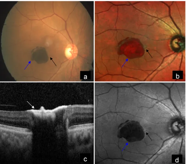
Fundus examination of the right eye showed a large pig- mented lesion and right-angled venule in the macula (Figure 1a). Multicolor imaging (MCI) showed the pigment- ed lesion as orange-red in color with well-demarcated borders as well as non-visibility of retinal blood vessels under the lesion, and highlighted the right-angled venule clearly (Figure 1b). Spectral domain optical coherence tomography (SD-OCT) at the level of the lesion showed inner layer hyperreflectivity, non-visibility of the outer retina, choroid and altered foveal contour (Figure 1c).
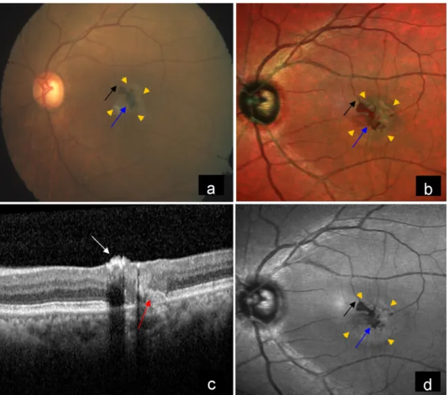
Blue reflectance (BR) showed decreased reflectance in the corresponding lesion and highlighted the right-angled venule (Figure 1d). Left eye fundus showed grayish dis- coloration due to subretinal fibrosis, dark pigment clumps
and right-angled venule in the macula (Figure 2a). MCI of the left eye showed orange-red pigments surrounded by grayish subretinal fibrosis and right-angled venule (Figure 2b). SD-OCT showed inner layer hyperreflectivity, altered foveal contour, retinal pigment epithelium (RPE) proliferation, as well as disruption of the ellipsoid zone and the interdigitation zone (Figure 2c). BR highlighted dark pigments and right-angled venule and picked up more areas of graying (Figure 2d).
Discussion
Reduced retinal transparency (“retinal graying”) in the parafoveolar area may be the first ophthalmoscopically visible change in patients with MacTel, which is seen as an area of increased reflectance on confocal BR imaging.
Paracentral vertically oriented slightly dilated right-angled venules are seen draining the telangiectatic area. These right-angled venules typically develop temporally. It has been suggested that a retinal Muller glial dysfunction or cell death may contribute to the formation of these venules [4]. Due to RPE migration into the retina along the course of the right-angled venules, retinal pigmented epithelial hyperplasia or clumps may be seen around the parafoveolar right-angled venules, and these pigments may extend into the inner retina to form irregular plaques enveloping these venules [5]. This pigmentary “sheath”
may also provide support for the abnormal blood vessels and increase the propensity of the tissue for fibrosis and tissue contraction [4]. Pigment clumping is initiated in the areas of outer retinal thinning and disruption, which suggests that atrophic changes in the photoreceptor layer may create a permissive environment for RPE transforma- tion. Pigment clumps increase in size over time, and in
1/4 GMS Ophthalmology Cases 2021, Vol. 11, ISSN 2193-1496
Case Report
OPEN ACCESS
Figure 1: a) Color fundus photograph (CFP) of the right eye showed a large pigment deposit (blue arrow) and right-angled venule (black arrow) in the macula. b) Multicolor image (MCI) of the right eye showed pigmented lesion as orange-red in color with well-demarcated borders (blue arrow) and highlighted the right-angled venule clearly (black arrow). c) Spectral domain optical coherence tomography (SD-OCT) showed inner layer hyperreflectivity (white arrow). d) Blue reflectance (BR) showed decreased
reflectance in the corresponding lesion (blue arrow) and highlighted right-angled venule (black arrow).
some cases, separate pigment clumps can coalesce and form a large contiguous pigment clump [6]. Similarly, in- traretinal migration of RPE can be noted in retinitis pig- mentosa and intermediate dry age-related macular de- generation. Bone spicule pigments in retinitis pigmentosa are formed in areas devoid of photoreceptors. It has been suggested that direct contact between inner retinal ves- sels and RPE appears to be a major trigger for migration of RPE cells [7], [8]. In dry age-related macular degener- ation, drusen may play physical and catalytic roles in fa- cilitating intraretinal RPE migration [9]. The laser channel of MCI that images deeper lesions is infrared, and the pseudocolor assigned to this channel is red. Thus, pig- ment is visualized as orange-red in color on MCI.
Optical coherence tomography angiography (OCTA) is a novel technique that visualizes vascular tissue using flow characteristics. Volume-rendered OCTA allows clear visualization of retinal vasculature at all depths. Subret- inal neovascularization can occur in MacTel. RPE abnor- malities may provide a conduit for abnormal vessels in the subretinal space to proliferate into the sub-RPE
compartment [10]. In OCT-A, right-angled vessels can be detected in early stages that can be tracked from super- ficial to outer retinal layers. They can form anastomoses in the outer retina with disease progression [11].
Extensive pigmentation can often be seen in patients treated with focal laser or photodynamic therapy. In our case, there was no previous history of laser treatment.
Interestingly we found an unusual large dark pigment deposit at the macula which has not yet been described in the literature. However, these pigmented lesions should be differentiated from tumors of the RPE that include solitary congenital hypertrophy of the RPE (CHRPE), con- genital hamartoma of the RPE, combined hamartoma of retina and RPE (CHRRPE) and adenoma of RPE. The dif- ferentiation between these conditions is primarily done clinically. CHRPE is a peripheral or mid-peripheral fundus lesion that is well-demarcated, flat or slightly elevated, and can either be black homogeneous with typical de- pigmented lacunae or completely depigmented [12]. Most solitary CHRPE lesions have a typical depigmented ring or “halo” around the margin [13]. Congenital hamartoma
2/4 GMS Ophthalmology Cases 2021, Vol. 11, ISSN 2193-1496
Goel et al.: An unusual presentation of macular telangiectasia ...
Figure 2: a) CFP of the left eye showed grayish discoloration at the macular area (yellow arrow heads) due to subretinal fibrosis, dark pigment clumps (blue arrow) and right-angled venule (black arrow). b) MCI of the left eye showed orange-red pigments (blue arrow) surrounded by grayish subretinal fibrosis (yellow arrow heads) and right-angled venule (black arrow). c) SD-OCT showed inner layer hyperreflectivity (white arrow), RPE proliferation and disruption of the ellipsoid zone and the interdigitation zone (red arrow). d) BR highlighted dark pigments (blue arrow), right-angled venule (black arrow) and picked up more areas of
graying (yellow arrow heads).
of the RPE is characterized by a small para-foveal, deeply pigmented nodular lesion of a size of 1 mm in diameter and 1 to 1.25 mm in thickness that protrudes through the sensory retina, sometimes into the vitreous cavity. It has well-defined margins, minimal feeder vessels and subtle retinal traction [14]. A CHRRPE is a rare benign, unilateral and solitary lesion located at the posterior pole.
It is characterized by an ill-defined gray retinal mass.
It varies widely in size, ranging from 1 mm to more than 10 mm in diameter. It shows tortuous or straightened retinal blood vessels, probably due to secondary retinal traction from excessive glial tissue on its surface [15].
RPE adenomas are rare and can be benign or malignant.
They are solitary, oval, unilateral, dark brown to black deep retinal tumors that invade over the sensory retina and acquire feeder vessels [13].
Conclusion
This case report highlights an unusual presentation of a large pigment clump in a case of MacTel which has not been previously reported.
Notes
Competing interests
The authors declare that they have no competing in- terests.
3/4 GMS Ophthalmology Cases 2021, Vol. 11, ISSN 2193-1496
Goel et al.: An unusual presentation of macular telangiectasia ...
References
1. Heeren TFC, Chew EY, Clemons T, Fruttiger M, Balaskas K, Schwartz R, Egan CA, Charbel Issa P; MacTel Study Group.
Macular Telangiectasia Type 2: Visual Acuity, Disease End Stage, and the MacTel Area: MacTel Project Report Number 8.
Ophthalmology. 2020 Nov;127(11):1539-48. DOI:
10.1016/j.ophtha.2020.03.040
2. Heeren TF, Holz FG, Charbel Issa P. First symptoms and their age of onset in macular telangiectasia type 2. Retina. 2014 May;34(5):916-9. DOI: 10.1097/IAE.0000000000000082 3. Finger RP, Charbel Issa P, Fimmers R, Holz FG, Rubin GS, Scholl
HP. Reading performance is reduced by parafoveal scotomas in patients with macular telangiectasia type 2. Invest Ophthalmol Vis Sci. 2009 Mar;50(3):1366-70. DOI: 10.1167/iovs.08-2032 4. Leung I, Sallo FB, Bonelli R, Clemons TE, Pauleikhoff D, Chew
EY, Bird AC, Peto T; MacTel Study Group. Characteristics of pigmented lesions in type 2 idiopathic macular telangiectasia.
Retina. 2018 Jan;38 Suppl 1(Suppl 1):S43-S50. DOI:
10.1097/IAE.0000000000001842
5. Charbel Issa P, Gillies MC, Chew EY, Bird AC, Heeren TF, Peto T, Holz FG, Scholl HP. Macular telangiectasia type 2. Prog Retin Eye Res. 2013 May;34:49-77. DOI:
10.1016/j.preteyeres.2012.11.002
6. Meleth AD, Toy BC, Nigam D, Agrón E, Murphy RP, Chew EY, Wong WT. Prevalence and progression of pigment clumping associated with idiopathic macular telangiectasia type 2. Retina. 2013 Apr;33(4):762-70. DOI: 10.1097/IAE.0b013e3182695bb3 7. Schuerch K, Marsiglia M, Lee W, Tsang SH, Sparrow JR.
Multimodal imaging of disease-associated pigmentary changes in retinitis pigmentosa. Retina. 2016 Dec;36 Suppl 1(Suppl 1):S147-S158. DOI: 10.1097/IAE.0000000000001256 8. Jaissle GB, May CA, van de Pavert SA, Wenzel A, Claes-May E,
Giessl A, Szurman P, Wolfrum U, Wijnholds J, Fischer MD, Humphries P, Seeliger MW. Bone spicule pigment formation in retinitis pigmentosa: insights from a mouse model. Graefes Arch Clin Exp Ophthalmol. 2010 Aug;248(8):1063-70. DOI:
10.1007/s00417-009-1253-9
9. Ho J, Witkin AJ, Liu J, Chen Y, Fujimoto JG, Schuman JS, Duker JS. Documentation of intraretinal retinal pigment epithelium migration via high-speed ultrahigh-resolution optical coherence tomography. Ophthalmology. 2011 Apr;118(4):687-93. DOI:
10.1016/j.ophtha.2010.08.010
10. Balaratnasingam C, Yannuzzi LA, Spaide RF. Possible choroidal neovascularization in macular telangiectasia type 2. Retina.
2015 Nov;35(11):2317-22. DOI:
10.1097/IAE.0000000000000887
11. Tzaridis S, Heeren T, Mai C, Thiele S, Holz FG, Charbel Issa P, Herrmann P. Right-angled vessels in macular telangiectasia type 2. Br J Ophthalmol. 2019 Feb 26. DOI: 10.1136/bjophthalmol- 2018-313364
12. Shields JA, Shields CL. Tumors and related lesions of the pigment epithelium. In: Shields JA, Shields CL, editors. Intraocular Tumors:
An Atlas and Textbook. 3rd ed. Philadelphia: Wolters Kluwer;
2015. p. 453-502.
13. Shields JA, Shields CL. Tumors and Related Lesions of the Pigmented Epithelium. Asia Pac J Ophthalmol. 2017 Mar- Apr;6(2):215-23. DOI: 10.22608/APO.201705
14. Gass JD. Focal congenital anomalies of the retinal pigment epithelium. Eye. 1989;3(Pt 1):1-18. DOI: 10.1038/eye.1989.2 15. Schachat AP, Shields JA, Fine SL, Sanborn GE, Weingeist TA,
Valenzuela RE, Brucker AJ. Combined hamartomas of the retina and retinal pigment epithelium. Ophthalmology. 1984 Dec;91(12):1609-15. DOI: 10.1016/s0161-6420(84)34094-5
Corresponding author:
Dr. Rupak Roy
Department of Vitreo Retina, Aditya Birla Sankara Nethralaya, 147 Mukundapur, E. M. Bypass, Kolkata 700099, West Bengal, India, Phone: +91 33 4401 3000 rayrupak@gmail.com
Please cite as
Goel S, Nangia P, Rijey AJ, Saurabh K, Roy R. An unusual presentation of macular telangiectasia type 2 with a large pigment deposit. GMS Ophthalmol Cases. 2021;11:Doc03.
DOI: 10.3205/oc000176, URN: urn:nbn:de:0183-oc0001762
This article is freely available from
https://www.egms.de/en/journals/oc/2021-11/oc000176.shtml Published:2021-01-28
Copyright
©2021 Goel et al. This is an Open Access article distributed under the terms of the Creative Commons Attribution 4.0 License. See license information at http://creativecommons.org/licenses/by/4.0/.
4/4 GMS Ophthalmology Cases 2021, Vol. 11, ISSN 2193-1496
Goel et al.: An unusual presentation of macular telangiectasia ...