Synthesis and Biological A ctivity of Functionalized Photochr omic Dithienylethens
Synthesis and Biological Activity of Functionalized
Photochromic Dithienylethens Natascha Kuzmanovic
2014
Fakultät für
Chemie und Pharmazie Universität Regensburg Universitätsstraße 31 93053 Regensburg
ISBN 978-3-86845-105-4
N. Kuzmanovic | 2
Fakultät für Chemie und Pharmazie
Natascha Kuzmanovic aus Hof, geb. am 16.03.1986
Studium: Chemie, Universität Regensburg, Abschluss Master of Science
Promotion: Prof. Dr. Burkhard König, Institut für Organische Chemie
Diese Dissertation umfasst neue Konzepte zur biologischen Anwen- dung von funktionalisierten photochromen Dithienylethenen, die als lichtabhängige Schalter für Enzymaktivität und Peptide agieren. Es wird die Entwicklung von Dithienylcyclopenten basierten Enzyminhi- bitoren berichtet, die die Aktivität des metabolischen Schlüsselen- zyms mtPriA reversibel durch Licht kontrollieren können. Weiterhin ist die Anwendung photochromer Dithienylcyclopentene als Zellsig- nalinhibitoren für die Kaskade der extrazellulär regulierten Kinase beschrieben. Um die Wasserlöslichkeit zu verbessern, die Hydrophilie zu erhöhen und eine leichtere Derivatisierung zu erreichen, wurden Dithienylmaleimid basierte Aminosäuren synthetisiert und in Peptide eingebaut. Darüber hinaus werden neue photochrome Dithienyl-Ma- leinsäurehydrazide vorgestellt. Die gewonnenen Erkenntnisse besit- zen besondere Relevanz im Hinblick auf die Entwicklung von neuarti- gen molekularen Hilfsmitteln um zelluläre Abläufe durch Licht zu kontrollieren.
Alumniverein Chemie der Universität Regensburg E.V.
alumniverein@chemie.uni-regensburg.de http://www.alumnichemie-uniregensburg.de
Synthesis and Biological Activity of Functionalized Photochromic Dithienylethens
Herausgegeben vom Alumniverein Chemie der Universität Regensburg e.V.
in Zusammenarbeit mit Prof. Dr. Burkhard König, Prof. Dr. Joachim Wegener, Prof. Dr. Arno Pfitzner und Prof. Dr. Werner Kunz.
Dissertationsreihe der Fakultät für Chemie und Pharmazie der Universität Regensburg, Band 1
Dithienylethenes
Dissertation zur Erlangung des Doktorgrades der Naturwissenschaften (Dr. rer. nat.)
der Naturwissenschaftlichen Fakultät IV - Chemie und Pharmazie der Universität Regensburg vorgelegt von
Natascha Kuzmanovic Regensburg
2014
Die Arbeit wurde von Prof. Dr. Burkhard König angeleitet.
Das Promotionsgesuch wurde am 21.01.2014 eingereicht.
Prüfungsausschuss: Vorsitzender: Prof. Dr. Joachim Wegener 1. Gutachter: Prof. Dr. Burkhard König 2. Gutachter: Prof. Dr. Kirsten Zeitler weiterer Prüfer: Prof. Dr. Reinhard Sterner
Natascha Kuzmanovic
of Funcionalized Photochromic
Dithienylethens
in der Deutschen Nationalbibliografie. Detailierte bibliografische Daten sind im Internet über http://dnb.ddb.de abrufbar.
1. Auflage 2014
© 2014 Universitätsverlag, Regensburg Leibnizstraße 13, 93055 Regensburg
Umschlagentwurf: Alumniverein Chemie der Universität Regensburg e.V.
Layout: Natascha Kuzmanovic Druck: Docupoint, Magdeburg ISBN: 978-3-86845-105-4
Alle Rechte vorbehalten. Ohne ausdrückliche Genehmigung des Verlags ist es nicht gestattet, dieses Buch oder Teile daraus auf fototechnischem oder elektronischem Weg zu vervielfältigen.
Weitere Informationen zum Verlagsprogramm erhalten Sie unter:
www.univerlag-regensburg.de
Now can come what want.
Uschi Disl, Biathletin
T ABLE OF C ONTENTS
1 SUMMARY ... 3
2 ZUSAMMENFASSUNG ... 5
3 EXPLOITING PROTEIN SYMMETRY TO DESIGN LIGHT-CONTROLLABLE ENZYME INHIBITORS ... 7
3.1 Introduction ... 9
3.2 Results and Discussion ... 11
3.2.1 Synthesis ... 11
3.2.2 Photochromism ... 12
3.2.3 Biological Test Results ... 13
3.2.4 Molecular Modeling ... 14
3.3 Conclusion ... 16
3.4 Supporting Information ... 17
3.4.1 Synthesis and Characterization of New Compounds ... 17
3.4.2 Photochromism of DTE-phosphates and DTE-phosphonates ... 24
3.4.3 Cloning, Heterologous Expression in E. coli, and Purification of mtPriA ... 26
3.4.4 Steady-state Enzyme Kinetics of mtPriA ... 26
3.4.5 Molecular Modeling ... 29
3.5 References ... 31
4 TOWARDS PHOTOSWITCHABLE KINASE INHIBITORS ... 35
4.1 Introduction ... 37
4.2 Results and Discussion ... 39
4.2.1 Molecular Docking ... 39
4.2.2 Synthesis ... 40
4.2.3 Photochromism ... 41
4.2.4 Biological Test Results ... 43
4.3 Conclusion ... 47
4.4 Experimental Materials and Methods ... 48
4.4.1 Synthesis and Characterization of New Compounds ... 48
4.4.2 Molecular Docking ... 51
4.4.3 Photochemical Investigations ... 52
4.4.4 Enzymatic Reisomerization with ERK2 ... 52
4.5 References ... 53
5 SYNTHESIS OF PHOTOCHROMIC DITHIENYLMALEIMIDE AMINO ACIDS ... 57
5.1 Introduction ... 59
5.2 Results and Discussion ... 61
5.2.1 Synthesis ... 61
5.2.2 Peptide Coupling ... 64
5.2.3 Photochromism ... 65
5.3 Conclusion ... 67
5.4 Experimental Section ... 68
5.4.1 General ... 68
5.4.2 New Compounds ... 69
5.4.3 Solid Phase Peptide Synthesis ... 74
5.4.4 Photochemical Investigations ... 75
5.5 References ... 76
6 NEW PHOTOCHROMIC DITHIENYL MALEIC HYDRAZIDES ... 79
6.1 Introduction ... 81
6.2 Initial Results and Discussion ... 82
6.3 Conclusion ... 86
6.4 Experimental Section ... 86
6.5 References ... 87
7 APPENDIX ... 89
7.1 Supplementary NMR spectra for Chapter 1 ... 91
7.2 Supporting Information for Chapter 2 ... 101
7.2.1 Supplementary Figures ... 101
7.2.2 Supplementary NMR spectra ... 103
7.3 Supporting Information for Chapter 3 ... 108
7.3.1 Supplementary Synthetic Data ... 108
7.3.2 Supplementary Figures ... 114
7.3.3 Supplementary NMR spectra ... 115
7.4 List of Abbreviations ... 127
7.5 Curriculum Vitae ... 131
7.6 Publications and Conference Contributions ... 132
7.7 Danksagung ... 133
1 S UMMARY
This thesis focuses on the design, synthesis and evaluation of novel functionalized photochromic dithienylethenes (DTEs) for applications in biology.
Chapter 1 deals with the creation of dithienylcyclopentene based enzyme inhibitors to reversibly control the activity of the metabolic branch-point enzyme PriA from Myco- bacterium tuberculosis (mtPriA) by light. The enzyme’s natural rotational symmetry encouraged us to design two-pronged DTEs with terminal phosphate or phosphonate functional groups. Switching from the flexible, ring-open to the rigid, ring-closed isomer reduces inhibition activity by one order of magnitude, whereby mtPriA’s performance can even be remote-controlled by light during catalysis. Molecular Dynamics simulations support our experiments showing that the open form is energetically more favorable while bound in the active site. Thus the concept of utilizing the enzyme structure for the inhibitor design has been proven.
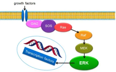
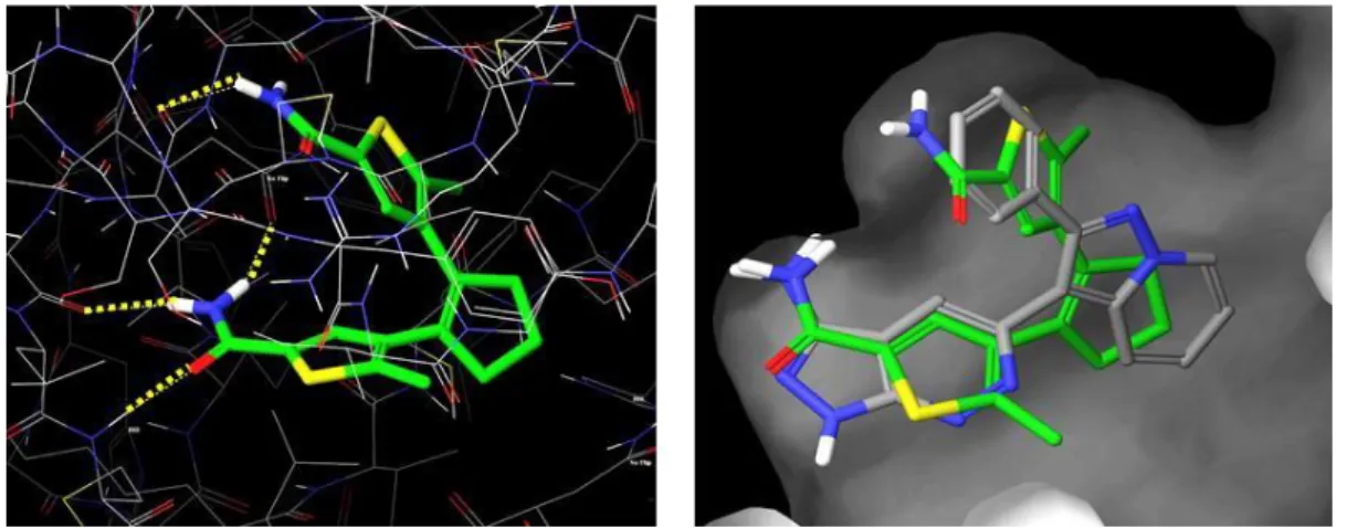
In chapter 2 the development of photochromic dithienylcyclopentenes as cell signal inhibitors for the extracellular-regulated kinase (ERK) pathway is reported. Through Molecular Docking we identified promising DTE based ERK inhibitors, which were subsequently synthesized and photochemically evaluated. The mediocre thermal stability of their ring-closed photoisomers requires that the test solutions are irradiated directly before use. Incubation of cells with the title compounds caused significant inhibition by the ring- open isomers, whereas the ring-closed forms’ activity seems reduced and cytotoxicity was observed. Further experiments with isolated ERK could not sufficiently substantiate these results though. This already gives a hint on the complexity of this topic as any other tier of the entire signal cascade might be affected. Nevertheless, interesting trends were found and new compounds could be identified, which allow for the light-dependent regulation of the ERK signal.
Aiming for better water-solubility, increased hydrophilicity and easier derivatization, dithienylmaleimide based amino acids were synthesized and incorporated in peptides, as described in chapter 3. We combined independently prepared N- and C-terminal thienyl components by Perkin condensation to form the maleimide core. Subsequent derivatization of the photoswitchable amino acid is easily achieved by standard solid phase peptide coupling. The resulting compounds show excellent photochromic performance in polar solvents and are thus suitable candidates for biological applications.
Moreover, novel photochromic dithienyl maleic hydrazides are introduced in chapter 4.
The reaction of dithienylmaleimides with hydrazine generates DTEs bearing maleic hydrazide as ethene unit. Initial NMR measurements indicate that it might occur predominantly as the monolactim tautomer. Concurrently, we observed photoswitchable solvatochromism of the obtained compound, but its repeated application is restricted by rapid photodegradation. Further investigations are necessary to thoroughly characterize the compounds and their behavior. These interesting pioneering observations could contribute to establish a new generation of DTE photoswitches.
In conclusion, this thesis presents new concepts for the biological application of functionalized photochromic dithienylethenes, which act as light-dependent switches for enzyme activity and peptides. The implications of this work are especially relevant for the development of novel molecular tools to remote-control cellular processes by light.
2 Z USAMMENFASSUNG
Diese Arbeit befasst sich mit der Gestaltung, der Synthese und der Untersuchung von neuen funktionalisierten photochromen Dithienylethenen (DTEs) für biologische Anwendungen.
Kapitel 1 handelt von der Entwicklung von Dithienylcyclopenten basierten Enzyminhibitoren, die die Aktivität des metabolischen Schlüsselenzyms PriA aus Mycobacterium tuberculosis (mtPriA) reversibel durch Licht kontrollieren können. Die natürliche Rotationssymmetrie des Enzyms hat uns dazu bewogen, symmetrische Inhibitoren mit terminalen Phosphat- oder Phosphonat-Ankern zu entwerfen. Durch das Schalten vom flexiblen, offenen Isomer zur starren, geschlossenen Form verringert sich die Inhibitionsaktivität um eine Größenordnung, wobei die Enzymsteuerung durch Licht auch während der Katalyse funktioniert. Moleküldynamik- Simulationen stützen die experimentellen Daten und zeigen, dass die Bindung der offenen Form im aktiven Zentrum energetisch bevorzugt ist. Damit wurde bewiesen, dass man abgeleitet von der Enzymstruktur Inhibitoren konstruieren kann.
In Kapitel 2 wird die Anwendung photochromer Dithienylcyclopentene als Zellsignalinhibitoren für die Kaskade der extrazellulär regulierten Kinase (ERK) berichtet. Mittels molekularen Dockings wurden potentielle, vom DTE-Gerüst abgeleitete, ERK-Inhibitorstrukturen bestimmt, welche anschließend synthetisiert und photochemisch untersucht wurden. Die mittelmäßige thermische Stabilität der geschlossenen Photoisomere erfordert, dass die Proben direkt vor ihrem Einsatz belichtet werden. Bei der Inkubation von Zellen mit den Zielsubstanzen können deren offene Isomere das ERK-Signal hemmen, wohingegen die Wirkung der geschlossenen Formen geringer erscheint und Zellschädigung festgestellt wurde. Weitere Experimente mit isolierter ERK konnten diese Ergebnisse jedoch nicht ausreichend untermauern und deuten auf die Komplexität dieser Thematik hin, da jede weitere Stelle der gesamten Signalkaskade betroffen sein könnte. Dennoch wurden interessante Tendenzen entdeckt und neue Substanzen gefunden, die das ERK-Signal abhängig von der Beleuchtung regulieren können.
Um die Wasserlöslichkeit zu verbessern, die Hydrophilie zu erhöhen und eine leichtere Derivatisierung zu erreichen, wurden Dithienylmaleimid basierte Aminosäuren synthetisiert und in Peptide eingebaut, was in Kapitel 3 beschrieben ist. Dabei verknüpften wir die separat hergestellten N- und C-terminalen Thienylkomponenten durch eine Perkin-Kondensation, wobei der Maleimidkern entstand. Eine anschließende Derivatisierung der photoschaltbaren Aminosäure kann durch gewöhnliche Festphasenpeptidsynthese per Standardprotokoll erfolgen. Die hergestellten Substanzen zeigen hervorragende photochrome Eigenschaften in polaren Lösungsmitteln und eignen sich daher für biologische Anwendungen.
Darüber hinaus werden neue photochrome Dithienyl-Maleinsäurehydrazide in Kapitel 4 vorgestellt. Die Reaktion von Dithienylmaleimiden mit Hydrazin erzeugt DTEs, die mit einem Maleinsäurehydrazid als Etheneinheit ausgestattet sind. Erste NMR-Messungen deuten darauf hin, dass es vermutlich als Monolactimtautomer vorliegt. Gleichzeitig beobachteten wir eine photoschaltbare Solvatochromie der hergestellten Substanz, jedoch ist eine wiederholte Anwendung durch den raschen Zerfall bei Beleuchtung eingeschränkt. Es sind weitere Untersuchungen nötig um eine fundierte Charakterisierung vorzunehmen. Diese interessanten wegbereitenden Beobachtungen können dazu beitragen eine neue Generation von DTE- Photoschaltern zu entwickeln.
Zusammenfassend zeigt diese Arbeit neue Konzepte zur biologischen Anwendung von funktionalisierten photochromen Dithienylethenen auf, welche als lichtabhängige Schalter für Enzymaktivität und Peptide agieren. Die gewonnenen Erkenntnisse besitzen besondere Relevanz im Hinblick auf die Entwicklung von neuartigen molekularen Hilfsmitteln um zelluläre Abläufe durch Licht zu kontrollieren.
C HAPTER 1
3 E XPLOITING P ROTEIN S YMMETRY TO D ESIGN L IGHT - C ONTROLLABLE E NZYME I NHIBITORS *
* This chapter was published as: B. Reisinger, N. Kuzmanovic, P. Löffler, R. Merkl, B. König and R. Sterner, Angew. Chem. 2014, 126, 606-609; Angew. Chem. Int. Ed. 2014, 53, 595-598. BR and NK contributed equally to this work. Design, calculations, synthesis, characterization and photophysical investigations of new compounds by NK. Protein cloning, expression, purification, steady-state enzyme kinetics and manuscript by BR (group of Prof. Dr. R. Sterner, University of Regensburg).
Molecular Modeling by PL (group of Prof. Dr. R. Merkl, University of Regensburg). BK and RS supervised the project and are the corresponding authors.
3.1 Introduction
The artificial control of biological processes by light is a rapidly emerging area of protein design.[1] Three basic strategies for the light-regulation of biomolecules have been reported:
Besides caging of key positions with photolabile protecting groups[2] and reprogramming of naturally occurring photoreceptors,[3-5] designed molecules that can be reversibly switched by light (photoswitches) have been used to direct protein or cellular function.[6] Recently, substantial progress has been made in the regulation of neuronal activity by designing light- inducible ligands for ion channels and receptors.[7-8] As the molecular recognition of specific ligand parts leads to a nonlinear signal response in neural systems, even small changes in the binding efficacy upon light irradiation significantly influence the cellular output.[9]
However, when aiming to reversibly control enzymatic activities, switching of a photoresponsive group must substantially affect the enzyme’s active site. Covalent incorporation of a molecular photoswitch near the catalytic center is able to fulfill this task.[6, 10] The required coupling step between protein and photoswitch can be circumvented by designing a light-controlled inhibitor.[6, 11-13]
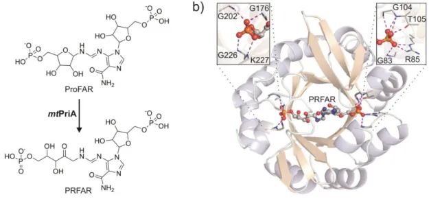
Figure 1. Reversible light-dependent control of mtPriA activity.
We aimed to design a light-controllable inhibitor for phosphoribosyl-isomerase A from Mycobacterium tuberculosis (mtPriA, Figure 1). mtPriA is a branch-point enzyme in amino acid biosynthesis as it catalyzes two chemically equivalent sugar isomerization reactions in tryptophan and histidine biosynthesis.[14] In the latter, the aminoaldose N′-[(5′- phosphoribosyl)-formimino]-5-aminoimidazole-4-carboxamide ribonucleotide (ProFAR) is converted to the corresponding aminoketose N´-[(5´-phosphoribulosyl)-formimino]-5- aminoimidazole-4-carboxamide-ribonucleotide (PRFAR) (Figure 2a). Since humans can neither synthesize histidine nor tryptophan, mtPriA is a potential target for anti- tuberculosis drugs.[15-16] Structurally, mtPriA belongs to the class of (βα)8-barrels, which is a frequently encountered and highly versatile fold among enzymes.[17-18] mtPriA exhibits a clear twofold symmetry (Figure 2b),[19-20] which indicates its evolution from a (βα)4-half- barrel precursor.[21] Consequently, two phosphate binding sites are found opposite each other to fix the substrate ProFAR and the product PRFAR (Figure 2b). We thus reasoned that
a C2-symmetric photoswitch with terminal phosphate-anchors would be an excellent foundation for building a light-controllable inhibitor of mtPriA.
Figure 2. Reaction and structure of mtPriA. a) mtPriA catalyzes the conversion of ProFAR to PRFAR within histidine biosynthesis. b) Ribbon diagram of the (βα)8-barrel structure of mtPriA with bound product PRFAR (PDB ID 3ZS4[19]). The view is along the two-fold symmetry axis of the protein. PRFAR is anchored by two opposite phosphate binding sites, which are enlarged in the insets. Formed hydrogen bonds are indicated by dashed lines (structure is slightly rotated for clarity in either case).
Two types of organic photochromic systems possess the desired twofold rotational symmetry: stilbene[22] or azobenzene switches[23] and the diarylethene scaffold.[24] Although azobenzene derivatives have been widely used in biological systems, they suffer from incomplete photoconversion and thermal reversibility.[1] In contrast, photoresponsive compounds based on 1,2-dithienylethene (DTE) generally feature switching rates of over 90% conversion with both photoisomers being thermally stable.[12, 24] Hence, we opted for DTE as core and provided it with different phosphate and phosphonate anchors (Scheme 1).
3.2 Results and Discussion 3.2.1 Synthesis
Starting with the bischlorodithienylethene 1, Suzuki coupling yielded either the aromatic hydroxides 2-4 or the aromatic bromides 8-9. The former were subsequently converted to ortho-, meta- and para-phosphates 5-7, while the latter were used to synthesize the phosphonic acid esters 10-11, which were finally hydrolyzed to afford meta- and para- phosphonates 12-13.
Scheme 1. Synthesis of DTE-phosphates and DTE-phosphonates.
3.2.2 Photochromism
The DTE molecular structure can reversibly be toggled between a ring-open and ring-closed photoisomer (Scheme 2), which significantly alters its overall conformational flexibility.[12]
Energy minimizations of the open and closed forms of all potential inhibitors confirmed for the phosphorous atoms distances between 15.8 and 19.6 Å (Scheme 2), which is in good accordance with the 16.9 Å observed for the corresponding atoms in the mtPriA structure with co-crystallized PRFAR (PDB ID 3ZS4[19]) (see Experimental Material and Methods for details). Only the open and closed isomers of ortho-phosphate 5 exhibit rather short P-P distances of 12.3 Å and 11.3 Å, respectively, in their energetically most favorable geometries; in addition, less populated, more extended conformers can be observed.
Scheme 2. Photochemical switching and corresponding calculated P-P distances in DTE- phosphates and DTE-phosphonates.
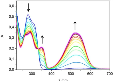
When ring-open forms of compounds 5-7, 12 and 13 are irradiated with 312 nm light, the absorption band at 280 nm immediately decreases. Simultaneously, new absorption maxima at 350 nm and 525 nm are formed turning the initially colorless solutions pink (see Figure 5). In each case, the spectral changes are completed after 30 s of irradiation and the corresponding photostationary states contain between 93% and 97% of the closed isomers, as judged by HPLC analyses (see Figure 6). The open forms can be recovered by irradiation with visible light (> 420 nm) and all switches are robust over several ring-closing/ring- opening cycles (see Figure 7).
3.2.3 Biological Test Results
The mtPriA activity can be monitored spectrophotometrically at 300 nm in a coupled enzyme assay (Scheme 3).[25] As all synthesized compounds were stable under assay conditions, their inhibitory effect could be investigated in steady-state enzyme kinetics. For this purpose, substrate saturation curves were measured in presence of different concentrations of compounds 5-7, 12 and 13 in their open and closed forms (curves are shown for compound 6 in Figure 8). Indeed, all investigated DTE-phosphates and DTE- phosphonates have the ability to inhibit the mtPriA reaction in both isomeric forms, thus proving the viability of the design concept. As expected for competitive inhibition, the turnover numbers kcat were identical in presence and absence of inhibitor (see experimental section, Table 2). The observed increase of the Michaelis constants caused by the inhibitors (Table 2) was used to calculate the inhibition constants Ki (Formula 1), which are given in Table 1.
Table 1. Inhibition constants Ki of compounds 5-7, 12 and 13 in their ring-open and ring-closed forms.
Inhibitor Ki / µM[a]
ring-open ring-closed
5 8.1 ± 2.1 7.0 ± 1.2
6 7 12 13
0.55 ± 0.12 3.5 ± 0.3 1.6 ± 0.1 6.8 ± 0.6
4.4 ± 0.3 3.7 ± 0.4 4.9 ± 0.3 22.7 ± 4.7
[a] The values of Ki were obtained from the data shown in Table 2 using Formula 1.
The determined inhibition constants are in the low micromolar range (Table 1) and therefore comparable or even better than the enzyme’s KM value for the natural substrate ProFAR (KMProFAR = 8.6 µM; see Table 2). In agreement with the well-suited distance of its phosphorous atoms (Scheme 2), the open isomer of phosphate 6 exhibits the highest binding affinity (Ki = 0.55 µM). However, when switched to its rigid, closed form, the inhibition activity is lowered by roughly one order of magnitude (Ki = 4.4 µM). A similar trend is observed for the respective phosphonate 12, whose binding affinity in the open form (Ki = 1.6 µM) is decreased about 3-fold upon ring-closure (Ki = 4.9 µM). In contrast, the inhibitory effects of phosphates 5 and 7 are nearly identical in their ring-open and ring- closed forms (Table 1). Interestingly, the introduction of a phosphonate moiety in para- position (compound 13) causes a 3-fold difference in the inhibition activity of the open (Ki = 6.8 µM) and closed photoisomer (K = 22.7 µM). The removal of the oxygen bridge between
the terminal anchor and the switchable core further reduces the overall flexibility, which seems to predominantly affect the already rigid, closed isomer.
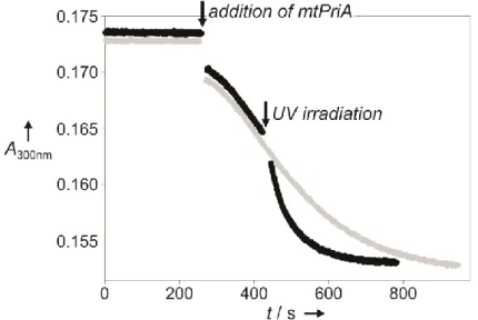
The performance of mtPriA can also be remote-controlled by light during catalysis. Starting up with compound 6 in its strongly inhibiting, open form, the reaction velocity is enhanced about 3-fold, when switching to the less active, ring-closed isomer (Figure 3).
Figure 3. Change in mtPriA activity upon ring-closure of compound 6. The turnover of 5 µM ProFAR was followed photometrically at 300 nm in the presence of 4 µM 6 in its open form under typical assay conditions (25 °C, 50 mM Tris/acetate pH 8.5, 100 mM ammonium acetate, 0.18 µM HisF and 0.15 µM mtPriA). After having reached its maximal velocity, the reaction mixture was either left in the photometer (grey) or removed to irradiate it with 312 nm light for 10 s (black). A reference solution without enzymes was used to correct the baseline shift resulting from different absorption values of the open and closed isomer at 300 nm.
3.2.4 Molecular Modeling
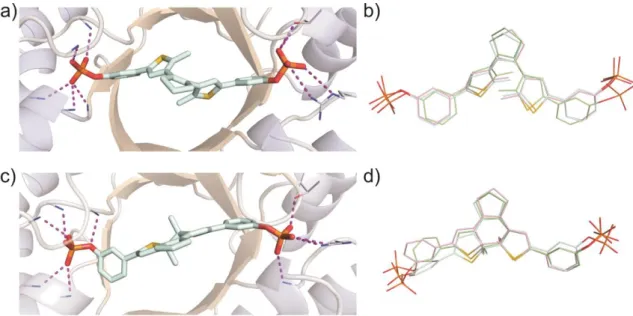
As pointed out before, the mechanistic principle, which induces the different binding affinities of the photoisomers, is based on a change in conformational flexibility.[12] Due to free rotation around the C-C bonds joining the thiophene heterocycles with the central cyclopentene ring and the terminal phenyl groups, DTE-phosphate 6 is able to adopt various geometries in its ring-open form. On the other hand, the closed isomer is completely conjugated and thus far more restricted in its motility. In order to get insights into the binding modes of the inhibitors, Molecular Dynamics (MD) simulations of the open and closed isomer of compound 6 bound to mtPriA were performed. Although both forms are clearly fixed at the phosphate binding sites (Figure 4a and 4c), obvious differences can be observed in their structural cores. While the open isomer converges to similar,
C2-symmetric conformers in three independent calculations (Figure 4b), more diverse binding modes are found for the ring-closed isomer (Figure 4d). Here, one terminal phenyl ring is twisted and adopts various geometries to facilitate proper coordination of the terminal phosphate group. The measured difference in inhibition activity is also reflected in the binding energies determined during the simulations, which consistently show that interaction with the open form is energetically more favorable (see Table 3). Taken together, the higher flexibility of the open isomer allows for better adaptation to the enzyme’s active site and apparently overcompensates the loss in entropy upon binding.
Figure 4. MD simulations of mtPriA and bound meta-phosphate 6. For each isomer, three independent calculations were performed and representative enzyme structures for the open (a) and closed (c) form are shown. A superposition of the energetically most favorable conformer of each simulation is depicted in the open conformation (b) and the closed conformation (d).
3.3 Conclusion
In summary, we have demonstrated that natural protein symmetry can be advantageously utilized to design light-controllable enzyme inhibitors. The two-pronged DTE switches can reversibly be toggled between a high- and low-affinity form, where both photoisomers are nearly quantitatively formed and thermally stable. Hence, the enzyme’s performance can alternately be enhanced and reduced by irradiation with UV and visible light, respectively.
The viability of a dual-anchored DTE inhibitor has been shown before[12] and the design concept can in principle be transferred to functionally quite different enzyme systems.
Phosphate is a frequently encountered element of metabolic substrates, and various other (βα)8-barrel enzymes such as pyridoxine 5’-phosphate synthase[26] or aldolases[27-28] possess two phosphate binding sites. For these enzymes, inhibitors may be designed with a similar approach, which would allow for an independent photocontrol of several metabolic processes in a spatiotemporal fashion.
3.4 Supporting Information
3.4.1 Synthesis and Characterization of New Compounds
General. Commercial reagents and starting materials were purchased from Acros Organics, Alpha-Aesar or Sigma Aldrich and used without further purification. Solvents were used in p.a. quality and dried according to common procedures, if necessary. Flash column chromatography was performed on a Biotage Isolera One automated flash purification system with UV/Vis detector using Sigma Aldrich MN silica gel 60 M (40-63 µm, 230-400 grain diameter) for normal phase or pre-packed Biotage SNAP cartridges (KP-C18-HS) for reversed phase chromatography. Reaction monitoring via TLC was performed on alumina plates coated with silica gel (Merck silica gel 60 F254, 0.2 mm). NMR spectra were recorded on a Bruker Avance 400 (1H 400.13 MHz, 13C 100.61 MHz, 31P 161.96 MHz, T = 300 K) instrument. The spectra are referenced against the NMR-solvent, chemical shifts δ are reported in ppm and coupling constants J are given in Hz. Resonance multiplicity is abbreviated as: s (singlet), d (doublet), t (triplet), m (multiplet) and b (broad). Carbon NMR signals are reported using DEPT 135 spectra with (+) for primary/tertiary, (−) for secondary and (q) for quaternary carbons. Mass spectra were recorded on Finnigan MAT95 (EI-MS), Agilent Q-TOF 6540 UHD (ESI-MS, APCI-MS), Finnigan MAT SSQ 710 A (EI-MS, CI- MS) or ThermoQuest Finnigan TSQ 7000 (ES-MS, APCI-MS) spectrometer. UV/Vis absorption spectroscopy was performed using a Varian Cary BIO 50 UV/Vis/NIR spectrometer.
Photochemistry. Standard hand-held lamps (Herolab, 312 nm, 6 W) were used for visualizing TLC plates and to carry out the ring-closure reactions at 312 nm. The ring- opening reactions were performed with the light of a 200 W tungsten light bulb which was passed through a 420 nm cut-off filter to eliminate higher energy light. The power of the light is given based on the specifications supplied by the company when the lamps were purchased. A light detector was not used to measure the intensity during the irradiation experiments.
General procedure A for Suzuki coupling. 1,2-Bis(5-chloro-2-methylthiophen-3- yl)cyclopent-1-ene 1[29] (1.0 eq.) was dissolved in dry THF under nitrogen atmosphere. After cooling to -78 °C, n-butyl lithium (2.2 eq.) was added dropwise via syringe and the resulting purple solution was stirred in the cold for 45 min. The reaction mixture was treated with trimethyl borate (4.0 eq.) and the yellow solution was stirred at -78 °C for 30 min.
Subsequently, the cold bath was removed and the reaction was stirred at r.t. for further 60 min to form the boronic ester intermediate. After quenching with aqueous sodium
carbonate (2 M) the two phase system was degassed for 15 min by nitrogen purge. Then the appropriate aryl bromide (2.2 or 4.0 eq.) and Pd(PPh3)2Cl2 (10 mol%) were added and the reaction mixture was heated to reflux overnight. After cooling to r.t., it was diluted with EtOAc and water and the phases were separated. The aqueous phase was extracted with EtOAc twice, the combined organic phases were dried over sodium sulfate and the solvents removed under reduced pressure. Purification of the crude product was performed by automated flash column chromatography.
General procedure B for phosphate ester formation. A crimp top vial was equipped with the desired bisphenol DTE (1.0 eq.) and a stirring bar and capped. It was set under nitrogen atmosphere and pyridine (20 eq.) in dry CH2Cl2 (1 mL) was added by syringe. After cooling to 0 °C a solution of phosphorous oxychloride (10 eq.) in dry THF (1 mL) was added dropwise via syringe over 5 min. The purple reaction mixture was stirred for 2 h at r.t.
under nitrogen atmosphere and subsequently quenched with acetone/water (1:1, 2 mL).
Volatiles were removed in vacuo and the crude product was purified by automated reversed phase flash column chromatography.
Synthesis of 2,2'-(4,4'-(cyclopent-1-ene-1,2-diyl)bis(5-methylthiophene-4,2-diyl)) diphenol (2). Compound 2 was prepared from 1,2-bis(5-chloro-2-methylthiophen-3- yl)cyclopent-1-ene 1 (1.50 g, 4.55 mmol), n-butyl lithium (1.3 M in hexane, 7.68 mL, 10.0 mmol), trimethyl borate (2.03 mL, 18.2 mmol), 2-bromophenol (1.16 mL, 10.0 mmol) and Pd(PPh3)2Cl2 (320 mg, 0.46 mmol) following the general procedure A for Suzuki coupling with 50 mL dry THF and 20 mL aq. Na2CO3 (2 M). Automated flash column chromatography (PE/EtOAc, 15-25% EtOAc) and subsequent reversed phase automated flash column chromatography (MeOH/water with 0.05% TFA, 3-100% MeOH) afforded 223 mg (0.50 mmol, 11%) of 2 as purple oil. 1H-NMR: δH(400 MHz, CDCl3): 2.10 (2H, dt, J = 7.4, 7.5 Hz, cyclopentene-CH2), 2.16 (6H, s, 2 CH3), 2.84 (4H, t, J = 7.4 Hz, 2 cyclopentene- CH2), 6.85 (2H, s, 2 thiophene-H), 6.89 − 6.97 (4H, m, 4 phenyl-H), 7.15 − 7.22 (2H, m, phenyl-H), 7.30 (2H, dd, J = 7.6, 1.6 Hz, phenyl-H); 13C-NMR: δC(101 MHz, CDCl3): 14.1 (+), 23.2 (−), 37.8 (−), 116.3 (+), 120.8 (+), 121.1 (q), 127.6 (+), 129.2 (+), 129.8 (+), 134.5 (q), 135.4 (q), 135.6 (q), 136.6 (q), 152.4 (q); ESI-MS: m/z (%): 443.1 (100) [M-H]-; calcd. for C27H24O2S2 443.1145; found 443.1147.
Synthesis of 3,3'-(4,4'-(cyclopent-1-ene-1,2-diyl)bis(5-methylthiophene-4,2-diyl)) diphenol (3). Compound 3 was prepared from 1,2-bis(5-chloro-2-methylthiophen-3- yl)cyclopent-1-ene 1 (1.84 g, 5.59 mmol), n-butyl lithium (1.6 M in hexane, 7.68 mL, 12.3 mmol), trimethyl borate (2.49 mL, 22.4 mmol), 3-bromophenol (2.13 g, 12.3 mmol) and Pd(PPh3)2Cl2 (393 mg, 0.56 mmol) following the general procedure A for Suzuki coupling with 100 mL dry THF and 20 mL aq. Na2CO3 (2 M). Automated flash column chromatography (PE/EtOAc, 15-30% EtOAc) and subsequent reversed phase automated flash column chromatography (MeOH/water with 0.05% TFA, 30-100% MeOH) afforded 1.04 g (2.35 mmol, 42%) of 3 as red oil. 1H-NMR: δH(400 MHz, CDCl3): 2.00 (6H, s, 2 CH3), 2.08 (2H, dt, J = 7.4 Hz, cyclopentene-CH2), 2.83 (4H, t, J = 7.4 Hz, 2 cyclopentene-CH2), 3.51 (2H, bs, 2 phenyl-OH), 6.71 (2H, dd, J = 1.9 Hz, 8.0 Hz, 2 phenyl-H), 6.95 − 6.99 (2H, m, 2 phenyl-H), 7.00 (2H, s, 2 thiophene-H), 7.07 (2H, J = 7.9 Hz, 2 phenyl-H), 7.19 (2H, t, J = 7.9 Hz, 2 phenyl-H); 13C-NMR: δC(101 MHz, CDCl3): 14.4 (+), 23.0 (−), 38.4 (−), 112.2 (+), 114.1 (+), 117.9 (+), 124.4 (+), 130.1 (+), 134.7 (q), 134.8 (q), 136.1 (q), 136.7 (q), 139.2 (q), 156.0 (q); EI-MS: m/z (%) 444.1 (100) [M]+, 429.0 (35) [M-CH3]+; calcd. for C27H24O2S2
443.1145; found 443.1156.
Synthesis of 4,4'-(4,4'-(cyclopent-1-ene-1,2-diyl)bis(5-methylthiophene-4,2-diyl)) diphenol (4). Compound 4 was prepared from 1,2-bis(5-chloro-2-methylthiophen-3- yl)cyclopent-1-ene 1 (209 mg, 0.64 mmol), n-butyl lithium (1.6 M in hexane, 0.88 mL, 1.41 mmol), trimethyl borate (0.29 mL, 2.56 mmol), 4-bromophenol (439 mg, 2.54 mmol) and Pd(PPh3)2Cl2 (42 mg, 0.06 mmol) following the general procedure A for Suzuki coupling with 50 mL dry THF and 10 mL aq. Na2CO3 (2 M). Automated flash column chromatography (PE/EtOAc, 15-25% EtOAc), subsequent reversed phase automated flash column chromatography (MeOH/water with 0.05% TFA, 30-100% MeOH) and lyophilization afforded 127 mg (0.29 mmol, 45%) of 4 as red oil. 1H-NMR: δH(400 MHz, CDCl3): 1.98 (6H, s, 2 CH3), 2.10 (2H, p, J = 7.7 Hz, cyclopentene-CH2), 2.83 (4H, t, J = 7.5 Hz, 2 cyclopentene-CH2), 4.88 (2H, bs, 2 phenyl-OH), 6.80 (4H, d, J = 8.7 Hz, 4 phenyl-H), 6.90 (2H, s, 2 thiophene-H), 7.37 (4H, d, J = 8.7 Hz, 4 phenyl-H); 13C-NMR: δC(101 MHz, CDCl3):
14.4 (+), 23.0 (−), 38.5 (−), 115.7 (+), 123.1 (+), 126.8 (+), 127.7 (q), 133.5 (q), 134.6 (q), 136.6 (q), 139.4 (q), 154.8 (q); EI-MS: m/z (%) 444.0 (100) [M]+, 429.0 (35) [M-CH3]+; calcd.
for C27H24O2S2 443.1145; found 443.1148.
Synthesis of (4,4'-(cyclopent-1-ene-1,2-diyl)bis(5-methylthiophene-4,2-diyl))bis(2,1- phenylene) bis(phosphate) (5). Compound 5 was prepared from 2 (65 mg, 0.15 mmol), pyridine (0.24 mL, 2.92 mmol) and phosphorous oxychloride (0.13 mL, 1.46 mmol) following the general procedure B for phosphate ester formation. Automated reversed phase flash column chromatography (MeOH/water with 0.5% NEt3, 20-100% MeOH) and subsequent lyophilization afforded 66 mg (0.09 mmol, 58%) of 5 as purple foam. 1H-NMR:
δH(400 MHz, MeOD): 1.21 (18H, t, J = 7.3 Hz, 2 H+N(CH2CH3)3), 1.99 (6H, s, 2 thiophene- CH3), 2.04 − 2.13 (2H, m, cyclopentene-CH2), 2.87 (4H, t, J = 7.5 Hz, 2 cyclopentene-CH2), 3.03 (12H, q, J = 7.3 Hz, 2 H+N(CH2CH3)3), 6.99 (2H, t, J = 7.6 Hz, 2 phenyl-H), 7.10 − 7.18 (2H, m, 2 phenyl-H), 7.40 (2H, s, thiophene-H), 7.51 (2H, d, J = 7.8 Hz, 2 phenyl-H), 7.63 (2H, d, J = 8.3 Hz, 2 phenyl-H); 13C-NMR: δC(101 MHz, MeOD): 9.1 (+), 14.4 (+), 24.0 (−), 39.6 (−), 47.5 (−), 121.4 (+), 123.8 (+), 126.6 (q), 128.2 (+), 128.4 (+), 128.7 (+), 135.8 (q), 136.1 (q), 136.5 (q), 137.4 (q), 150.8 (q); 31P-NMR: δP(162 MHz, MeOD): -3.40 (s); ESI-MS: m/z (%) 806.0 (100) [(M4-+3H)+2(N(Et)3)]-, 603.0 (44) [M4-+3H]-; calcd. for C27H25O8P2S2 603.0472;
found 603.0477.
Synthesis of (4,4'-(cyclopent-1-ene-1,2-diyl)bis(5-methylthiophene-4,2-diyl))bis(3,1- phenylene) bis(phosphate) (6). Compound 6 was prepared from 3 (26 mg, 0.06 mmol), pyridine (100 µL, 1.18 mmol) and phosphorous oxychloride (50 µL, 0.56 mmol) following the general procedure B for phosphate ester formation. Automated reversed phase flash column chromatography (MeOH/water with 0.5% NEt3, 20-100% MeOH) and subsequent lyophilization afforded 33 mg (0.03 mmol, 56%) of 6 as purple foam. 1H-NMR: δH(400 MHz, MeOD): 1.21 (36H, t, J = 7.3 Hz, 4 H+N(CH2CH3)3), 1.96 (6H, s, 2 thiophene-CH3), 2.11 (2H, dt, J = 7.5, 7.6 Hz, cyclopentene-CH2), 2.86 (4H, t, J = 7.4 Hz, 2 cyclopentene-CH2), 2.97 (24H, q, J = 7.3 Hz, 4 H+N(CH2CH3)3), 7.11 − 7.13 (4H, m, 4 phenyl-H), 7.15 (2H, s, 2 thiophene-H), 7.17 − 7.23 (2H, m, 2 phenyl-H), 7.50 (2H, s, 2 phenyl-H); 13C-NMR: δC(101 MHz, MeOD):
9.5 (+), 14.5 (+), 24.1 (−), 39.5 (−), 47.1 (−), 118.1 (+), 120.0 (+), 120.3 (+), 125.3 (+), 130.5 (+), 135.3 (q), 136.1 (q), 136.7 (q), 138.2 (q), 141.1 (q), 156.4 (q); 31P-NMR:
δP(162 MHz, MeOD): -0.78 (s); ESI-MS: m/z (%) 806.0 (100) [(M4-+3H)+2(N(Et)3)]-, 603.0 (21) [M4-+3H]-; calcd. for C27H25O8P2S2 603.0472; found 603.0478.
Synthesis of (4,4'-(cyclopent-1-ene-1,2-diyl)bis(5-methylthiophene-4,2-diyl))bis(4,1- phenylene) bis(phosphate) (7). Compound 7 was prepared from 4 (50 mg, 0.11 mmol), pyridine (191 µL, 2.36 mmol) and phosphorous oxychloride (103 µL, 1.12 mmol) following the general procedure B for phosphate ester formation. Automated reversed phase flash column chromatography (MeOH/water with 0.5% NEt3, 20-100% MeOH) and subsequent lyophilization afforded 73 mg (0.07 mmol, 64%) of 7 as purple oil. 1H-NMR: δH(400 MHz, MeOD): 1.26 (36H, t, J = 7.3 Hz, 4 H+N(CH2CH3)3), 1.99 (6H, s, 2 thiophene-CH3), 2.08 (2H, dt, J = 7.5, 7.6 Hz, cyclopentene-CH2), 2.84 (4H, t, J = 7.4 Hz, 2 cyclopentene-CH2), 3.09 (24H, q, J = 7.3 Hz, 4 H+N(CH2CH3)3), 6.97 (2H, s, 2 thiophene-H), 7.20 (4H, d, J = 8.7 Hz, 4 phenyl-H), 7.40 (4H, d, J = 8.6 Hz, 4 phenyl-H); 13C-NMR: δC(101 MHz, MeOD): 9.2 (+), 14.5 (+), 24.1 (−), 39.3 (−), 47.4 (−), 121.8 (+), 124.6 (+), 127.1 (+), 130.6 (q), 134.8 (q), 136.2 (q), 138.1 (q), 140.9 (q), 154.5 (q); 31P-NMR: δP(162 MHz, MeOD): -2.26 (s); ESI-MS: m/z (%) 806.0 (100) [(M4-+3H)+2(N(Et)3)]-, 603.0 (15) [M4-+3H]-; calcd. for C27H25O8P2S2 603.0472;
found 603.0465.
Synthesis of tetraethyl((4,4'-(cyclopent-1-ene-1,2-diyl)bis(5-methylthiophene-4,2- diyl))bis(3,1-phenylene)) bis(phosphonate) (10). A crimp top vial was loaded with compound 8[30] (277 mg, 0.49 mmol), palladium acetate (11 mg, 0.05 mmol), triphenylphosphine (39 mg, 0.15 mmol) and a stirring bar and capped. It was set under nitrogen atmosphere at which point diethyl phosphite (0.15 mL, 1.18 mmol), triethylamine (0.20 mL, 1.47 mmol) and ethanol (4 mL) were added via syringes and the purple solution was refluxed for 48 h. After cooling to r.t. the reaction mixture was diluted with ethanol (5 mL), transferred to a round bottom flask and the volatiles were removed at the rotary evaporator. The brown residue was purified by automated flash column chromatography (PE/EtOAc, 40-95% EtOAc) yielding 10 (128 mg, 0.19 mmol, 39%) as purple oil. 1H-NMR:
δH(400 MHz, CDCl3): 1.29 (12H, t, J = 7.1 Hz, 2 P(O)(OCH2CH3)2), 1.96 (6H, s, 2 thiophene- CH3), 1.99 − 2.13 (2H, m, cyclopentene-CH2), 2.82 (4H, t, J = 7.4 Hz, 2 cyclopentene-CH2), 3.91 − 4.24 (8H, m, 2 P(O)(OCH2CH3)2), 7.10 (2H, s, 2 thiophene-H), 7.39 (4H, td, J = 7.7, 4.4 Hz, 2 phenyl-H), 7.54 − 7.71 (4H, m, 4 phenyl-H), 7.93 (2H, dt, J = 14.1, 1.4 Hz, 2 phenyl-H); 13C-NMR: δC(101 MHz, CDCl3): 14.5 (+), 16.3 (+), 23.0 (−), 38.6 (−), 62.2 (−), 124.8 (+), 128.2 (+), 128.3 (q), 129.0 (+), 129.1 (+), 130.0 (+), 134.7 (q), 134.9 (q), 135.4 (q), 136.9 (q), 138.4 (q); 31P-NMR: δC(162 MHz, CDCl3): 19.0 (s); ESI-MS: m/z (%) 685.2 (100) [M+H]+, 1391.4 (65) [2M+Na]+; calcd. for C35H42O6P2S2 685.1971; found 685.1976.
Synthesis of tetraethyl ((4,4'-(cyclopent-1-ene-1,2-diyl)bis(5-methylthiophene-4,2- diyl))bis(4,1-phenylene)) bis(phosphonate) (11). A crimp top vial was loaded with compound 9[31] (248 mg, 0.44 mmol), palladium acetate (10 mg, 0.04 mmol), triphenylphosphine (34 mg, 0.14 mmol) and a stirring bar and capped. It was set under nitrogen atmosphere at which point diethyl phosphite (0.13 mL, 1.04 mmol), triethylamine (0.18 mL, 1.31 mmol) and ethanol (4 mL) were added via syringes and the purple solution was refluxed for 48 h. After cooling to r.t. the reaction mixture was diluted with ethanol (5 mL), transferred to a round bottom flask and the volatiles were removed at the rotary evaporator. The brown residue was purified by automated flash column chromatography (PE/EtOAc, 95% EtOAc) yielding 11 (102 mg, 0.15 mmol, 34%) as purple oil. 1H-NMR:
δH(400 MHz, CDCl3): 1.31 (12H, t, J = 7.1 Hz, 2 P(O)(OCH2CH3)2), 1.99 (6H, s, 2 thiophene- CH3), 2.02 − 2.14 (2H, m, cyclopentene-CH2), 2.83 (4H, t, J = 7.4 Hz, 2 cyclopentene-CH2), 3.92 − 4.29 (8H, m, 2 P(O)(OCH2CH3)2), 7.11 (2H, s, 2 thiophene-H), 7.55 (4H, dd, J = 8.3, 3.7 Hz, 4 phenyl-H), 7.75 (4H, dd, J = 12.9, 8.3 Hz, 4 phenyl-H); 13C-NMR: δC(101 MHz, CDCl3): 14.5 (+), 16.3 (+), 16.4 (+), 23.0 (−), 38.5 (−), 62.1 (−), 124.9 (+), 125.1 (+), 125.4 (+), 127.2 (q), 132.4 (+), 132.5 (+), 134.8 (q), 136.2 (q), 137.0 (q), 138.2 (q), 138.4 (q); 31P-NMR:
δC(162 MHz, CDCl3): 19.3 (s); ESI-MS: m/z (%) 685.2 (100) [M+H]+, 1391.4 (44) [2M+Na]+; calcd. for C35H42O6P2S2 685.1971; found 685.1977.
Synthesis of ((4,4'-(cyclopent-1-ene-1,2-diyl)bis(5-methylthiophene-4,2-diyl))bis(3,1- phenylene))diphosphonic acid (12). Compound 10 (118 mg, 0.17 mmol) was dissolved in
dry acetonitrile (15 mL) and treated with trimethylsilyl bromide (1.14 mL, 8.62 mmol). The reaction mixture was stirred at r.t. overnight. Volatiles were removed at the rotary evaporator. After purification by automated reversed phase flash column chromatography (MeOH/water with 0.5% NEt3, 3-100% MeOH) and lyophilization compound 12 (114 mg, 0.15 mmol, 87%) was obtained as dark purple oil. 1H-NMR: δH(400 MHz, MeOD): 1.23 (18H, t, J = 7.3 Hz, 2 H+N(CH2CH3)3), 1.98 (6H, s, 2 thiophene-CH3), 2.05 − 2.16 (2H, m, cyclopentene-CH2), 2.86 (4H, t, J = 7.4 Hz, 2 cyclopentene-CH2), 3.07 (12H, q, J = 7.3 Hz, 2 H+N(CH2CH3)3), 7.16 (2H, s, thiophene-H), 7.33 (2H, td, J = 7.7, 3.5 Hz, 2 phenyl-H), 7.44 − 7.59 (2H, m, 2 phenyl-H), 7.67 (2H, dd, J = 12.1, 7.5 Hz, 2 phenyl-H), 8.01 (2H, d, J = 13.1 Hz, 2 phenyl-H); 13C-NMR: δC(101 MHz, MeOD): 9.2 (+), 14.6 (+), 24.1 (−), 39.5 (−), 47.4 (−), 125.4 (+), 127.4 (+), 128.7 (+), 129.5 (+), 130.6 (+), 135.2 (q), 135.7 (q), 136.2 (q), 138.3 (q), 139.2 (q), 141.0 (q); 31P-NMR: δP(162 MHz, MeOD): 12.5 (s); ESI-MS: m/z (%) 571.1 (100) [M4-+3H]-; calcd. for C27H25O6P2S2 571.0573; found 571.0570.
Synthesis of ((4,4'-(cyclopent-1-ene-1,2-diyl)bis(5-methylthiophene-4,2-diyl))bis(4,1- phenylene))diphosphonic acid (13). Compound 11 (82 mg, 0.12 mmol) was dissolved in dry acetonitrile (5 mL) and treated with trimethylsilyl bromide (0.79 mL, 6.00 mmol). The reaction mixture was stirred at r.t. overnight. Volatiles were removed at the rotary evaporator. After purification by automated reversed phase flash column chromatography (MeOH/water with 0.5% NEt3,3-100% MeOH) and lyophilization compound 13 (76 mg, 0.10 mmol, 82%) was obtained as dark purple oil. 1H-NMR: δH(400 MHz, MeOD): 1.25 (18H, t, J = 7.3 Hz, 2 H+N(CH2CH3)3), 1.99 (6H, s, 2 thiophene-CH3), 2.09 (2H, dt, J = 7.5, 7.6 Hz, cyclopentene-CH2), 2.85 (4H, t, J = 7.4 Hz, 2 cyclopentene-CH2), 3.09 (12H, q, J = 7.3 Hz, 2 H+N(CH2CH3)3), 7.17 (2H, s, thiophene-H), 7.50 (4H, dd, J = 8.4, 2.8 Hz, 4 phenyl-H), 7.73 (4H, dd, J = 12.1, 8.3 Hz, 4 phenyl-H); 13C-NMR: δC(101 MHz, MeOD): 9.2 (+), 14.7 (+), 24.1 (−), 39.4 (−), 47.4 (−), 125.4 (+), 125.8 (q), 132.6 (+), 136.2 (q), 136.7 (q), 136.9 (q), 138.4 (q), 138.5 (q), 140.7 (q); 31P-NMR: δP(162 MHz, MeOD): 12.9 (s); ESI-MS: m/z (%) 571.1 (100) [M4-+3H]-; calcd. for C27H25O6P2S2 571.0573; found 571.0579.
3.4.2 Photochromism of DTE-phosphates and DTE-phosphonates
Photochemical syntheses of ring-closed isomers. Solutions of compounds 5-7, 12 and 13 (12.5 µM) in 50 mM Tris/acetate pH 8.5 were irradiated for 30 s with a 312 nm lamp yielding pink solutions containing the ring-closed isomers. The changes in the UV/Vis absorption spectra are representatively shown for compound 6 in Figure 5. The photostationary states contain between 93 and 97% of the ring-closed isomers according to HPLC analyses. The respective HPLC chromatograms of compound 6 in its open and closed form are depicted in Figure 6.
Figure 5. UV/Vis absorption spectra evolution of compound 6 upon irradiation with 312 nm light.
Arrows indicate the changes of the absorption maxima of 12.5 µM 6 in 50 mM Tris/acetate pH 8.5 with irradiation intervals of 2 s from 0 to 30 s, which is illustrated by the color change from blue (open isomer) to purple (closed isomer).
Photochemical cycling studies. In order to test the robustness of the photochromic systems, photochemical cycling studies were recorded for all switches (Figure 7). In each case, a solution of 12.5 µM inhibitor in 50 mM Tris/acetate pH 8.5 was alternately irradiated with 312 nm light for 30 s and with visible light for 15 min over various cycles and the absorption change at 525 nm was monitored.
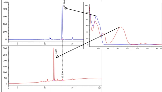
Figure 6. Representative HPLC chromatograms for the determination of the photostationary state of compound 6 (100 µM in 50 mM Tris/acetate pH 8.5). The UV/Vis signal was detected at 288 nm. The chromatograms before (upper) and after (lower) irradiation with 312 nm light for 30 s indicate that 97% of the open isomer (blue curve) were converted to the closed isomer (red curve). The absorption spectra of both forms are shown in the right panel.
Figure 7. Cycle performance of compounds 5-7, 12 and 13. Changes in absorption at 525 nm were measured during an alternated irradiation of each inhibitor solution (12.5 µM in 50 mM Tris/acetate pH 8.5) with 312 nm light for 30 s and greater than 420 nm light for 15 min.
5 10 15
mAU
0 100 200 300
400 13.166
0 5 10 15 min
0 50 100 150 200 250 300
11.663 13.230
300 400 500 600 700 800 900 nm
mAU
0 100 200 300 400 500
3.4.3 Cloning, Heterologous Expression in E. coli, and Purification of mtPriA
The gene coding for mtPriA was amplified by PCR using pETM-11-mtPriA[19] as a template and cloned into the vector pET24a(+) (Stratagene) via the terminal restriction sites for NdeI and XhoI. mtPriA was heterologously expressed in E. coli BL21-Gold cells (Stratagene) transformed with pET(24a)(+)-mtpriA. To this end, five liters of Luria Broth (LB) medium supplemented with 75 μg/mL kanamycin were inoculated with a pre-culture and incubated at 37 °C. After an OD600of 0.6 was reached, the temperature was lowered to 20 °C.
Expression was induced by adding 0.5 mM IPTG, and growth was continued overnight. Cells were harvested by centrifugation (Sorvall/RC5B, GS3, 15 min, 4000 rpm, 4 °C) and suspended in 50 mM Tris/HCl pH 8.0, 300 mM NaCl, 10 mM imidazole. After lysing by sonication (Branson Sonifier W-250D, 2 x 2 min in 15 sec intervals, 45% pulse, 0 °C), cells were centrifuged again (Sorvall/RC5B, SS34, 30 min, 13.000 rpm, 4 °C) to separate the soluble from the insoluble fraction of the cell extract. As a first purification step, mtPriA was subjected to a Ni2+ affinity chromatography using its C-terminal hexa-histidine tag. To this end, the soluble supernatant was loaded onto a HisTrapFF crude column (5 mL; GE Healthcare), which had been equilibrated with 50 mM Tris/HCl pH 8.0, 300 mM NaCl, 10 mM imidazole. After washing the column with the equilibration buffer, a linear gradient of 10-500 mMimidazole was applied and mtPriA eluted between 40 and 100 mM imidazole.
Main fractions were pooled and further purified via size exclusion chromatography. For this purpose, a Superdex200 column (HiLoad 26/60, 320 mL, GE Healthcare) was equilibrated and operated with 50 mM Tris/HCl pH 8.0, 200 mM sodium chloride, 2 mM dithiothreitol at 4 °C. Fractions with pure protein were pooled and dialyzed twice against 50 mM Tris/HCl pH 8.0. According to SDS-PAGE (12.5% acrylamide), mtPriA was more than 95% pure.
About 1 mg of protein was obtained per liter of culture.
3.4.4 Steady-state Enzyme Kinetics of mtPriA
Steady-state enzyme kinetics of the mtPriA reaction were measured spectrophotometrically at 300 nm as described.[25] In a coupled enzyme assay the substrate ProFAR was initially converted to PRFAR, which was further processed by the enzyme HisF from Thermotoga maritima (tmHisF) to yield imidazole glycerol phosphate (ImGP) and aminoimidazole carboxamide ribonucleotide (AICAR) (Scheme 3). As ProFAR and PRFAR exhibit identical absorption spectra, the second step is indispensible in order to monitor the reaction progress at 300 nm (Δε300(ProFAR-AICAR) = 5.637 mM-1cm-1). It furthermore prevents product inhibition of the mtPriA reaction.[32]
Scheme 3. Coupled enzyme assay for photometric determination of mtPriA activity.
The steady-state catalytic parameters of mtPriA in the absence of inhibitor were determined in triplicate at 25 °C in 50 mM Tris/acetate pH 8.5 in the presence of 100 mM ammonium acetate, 2-73 µM ProFAR, and 0.2 µM tmHisF (Table 2). Reactions were initiated with 0.2 µM mtPriA, when the baseline signal was constant. To determine the inhibition constants of compounds 5-7, 12 and 13 in their open and closed forms, three saturation curves were measured in presence of different inhibitor concentrations in each case. Reaction mixtures contained 100 mM ammonium acetate, 3-190 µM ProFAR, 0.4-20 µM inhibitor (closed inhibitor forms were synthesized photochemically prior to addition to the reaction mixture), and 0.04-0.2 µM tmHisF in 50 mM Tris/acetate pH 8.5. Substrate turnover was initiated with 0.04-0.2 µM mtPriA. The molar concentration of mtPriA and tmHisF did not exceed a tenth of the respective inhibitor concentration. Additionally, to rule out artifacts, the photostability of inhibitor 6 was representatively investigated under assay conditions:
15 µM of 6 in both isomeric forms were irradiated for 30 minutes at 300 nm in the spectrophotometer and no change in the absorption spectra was observed. Moreover, it was assured that the performance of mtPriA is rate-limiting in each kinetic setup, as doubling of its concentration resulted in a doubled initial velocity. Finally, the inhibition constant Ki of each DTE compound could be calculated from Formula 1, where Km is the Michaelis constant of mtPriA and Kmapp is the apparent Km value determined in the presence of a distinct inhibitor concentration c(I). All kinetic parameters are given in Table 2 and steady-state enzyme kinetics of 6 in its open and closed form are illustrated in Figure 8.
m ( )
i app
m m
K c I K K K
Formula 1. Determination of inhibition constant Ki.
Figure 8. Substrate saturation curves of mtPriA in the presence of inhibitor 6 in its open (upper curves) and closed (lower curves) form. See Table 2 for deduced steady-state kinetic parameters.
Table 2. Steady-state kinetic constants for the ProFAR isomerization activity of mtPriA in absence and presence of compounds 5-7, 12 and 13 in their ring-open and ring-closed forms.
Inhibitor c(I) / µM kcat / s-1 Km / µM Ki / µM open / closed
- 0.49
0.49 0.57
8.6 9.2 8.1
5 5.0 / 5.0
10.5 / 10.5 18.0 / 18.0
0.47 / 0.58 0.59 / 0.55 0.55 / 0.60
12.7 / 13.9 20.9 / 21.5 32.5 / 35.2
10.5 / 8.1 7.3 / 7.0 6.5 / 5.8
6 0.4 / 2.0
0.6 / 3.5 0.8 / 8.0
0.52 / 0.56 0.59 / 0.60 0.55 / 0.61
16.2 / 12.4 18.7 / 15.9 18.5 / 23.7
0.45 / 4.5 0.51 / 4.1 0.69 / 4.6
7 2.0 / 3.5
3.5 / 7.0 5.0 / 10.5
0.47 / 0.58 0.47 / 0.55 0.47 / 0.53
13.1 / 16.2 17.7 / 24.8 21.7 / 35.6
3.8 / 4.0 3.3 / 3.7 3.3 / 3.3
12 1.5 / 4.0
2.5 / 10.0 4.0 / 15.0
0.57 / 0.55 0.58 / 0.57 0.60 / 0.55
16.3 / 15.2 22.3 / 27.1 29.4 / 34.9
1.7 / 5.2 1.6 / 4.6 1.7 / 4.9
13 5.0 / 15.0
10.0 / 15.0 15.0 / 20.0
0.60 / 0.58 0.60 / 0.57 0.59 / 0.57
15.7 / 15.1 20.7 / 15.0 26.6 / 14.7
6.1 / 19.8 7.1 / 20.2 7.2 / 28.2
Three substrate saturation curves were recorded at different inhibitor concentrations c(I) for each form (open/closed). The deduced kcat and Km values are shown. The shown Ki values were calculated with Formula 1 from the increase of the apparent Km value with respect to the Km value in absence of inhibitor. The mean values and standard deviations of Ki for each inhibitor are given in Table 1.
3.4.5 Molecular Modeling
Energy minimization of the open and closed isomers of compounds 5-7, 12 and 13.
The conformer distribution of compounds 5, 6, 7, 12 and 13 in their open and closed isomers was calculated by Wavefunction Spartan’10[33] using molecular mechanics in water (MMFFaq) in the ground state with 20.000 conformers examined, a total charge of -2 for phosphonates or -4 for phosphates, singlet multiplicity, subject to symmetry and global calculations. Sorted by energy minimum the five best results for each calculation were averaged and are shown in Scheme 2.
Molecular Dynamics (MD) simulations of mtPriA with compound 6 bound in its open and closed form. Modeling and MD simulations were performed with YASARA Structure Version 13.4.21 employing the YAMBER3 force field.[34] Simulations were run at 298 K under periodic boundary conditions and with explicit water, using a multiple time step of 1 fs for intramolecular and 2 fs for intermolecular forces. Lennard-Jones forces and long- range electrostatic interactions were treated with a 7.86 Å cutoff, the latter were calculated using the Particle Mesh Ewald method.[35] Temperature was adjusted using a Berendsen thermostat based on the time-averaged temperature and simulations were carried out at constant pressure. The parameterization of the open and closed forms of compound 6 was performed using the AM1BCC protocol[36], that assigns atomic charges by applying additive bond charge corrections (BCCs) to semi-empirical AM1 atomic charge calculations. Based on the crystal structure of mtPriA (PDB ID 3ZS4), the original ligand PRFAR was removed and manually replaced by compound 6 in the open and closed form in order to fit the phosphate binding pockets. The simulation cell was defined as 5 Å larger than the protein along each axis (cell dimensions 52 × 47 × 44 Å3), filled with water to a density of 0.997 g/mL, and counterions were added to a final concentration of 0.9% NaCl. The protonation states of protein side chains were assigned according to Krieger et al.[37] In order to remove conformational stress, two phases of equilibration were conducted: After a 100 ps equilibration with frozen protein coordinates, the whole system was equilibrated for 1 ns.
The two equilibrated models of the open and closed form were subsequently used for the six