protein-protein interactions in three enzyme superfamilies
D ISSERTATION
ZUR
E
RLANGUNG DESD
OKTORGRADES DERN
ATURWISSENSCHAFTEN(D
R.
RER.
NAT.)
DERF
AKULTÄT FÜRB
IOLOGIE UND VORKLINISCHEM
EDIZIN DERU
NIVERSITÄTR
EGENSBURGvorgelegt von Maximilian Plach
aus Bad Kötzting
Januar 2017
Die Arbeit wurde angeleitet von Prof. Dr. Reinhard Sterner.
Unterschrift:
Superfamilies are a classification system to combine proteins that are related through a common evolutionary origin, share similar sequences, structures, and core reaction mech- anisms, but exert different functions. Today, for most superfamilies tens of thousands of sequences and hundreds of structures are known and most of the different functions of their members have been elucidated. Superfamilies thus provide a formal and biologically sensible framework to study evolutionary relationships between proteins. In the present work, the frameworks of three enzyme superfamilies were utilized to get insights into several important aspects of enzyme evolution.
The first part of this work addresses the question how enzymatic mono- and bi-functio- nality have evolved in the superfamily of ribose-binding (βα)8-barrel sugar isomerases.
This superfamily contains the homologous enzymes HisA and TrpF, which catalyze similar reactions in histidine and tryptophan biosynthesis, as well as the bi-functional enzyme PriA, which catalyzes both the HisA and TrpF isomerization reactions. HisA and TrpF are ubiquitous in Archaea and Bacteria, whereas PriA is only found in certain Actinobacteria.
These species have lost the dedicated TrpF enzyme and PriA is consequently part of both tryptophan and histidine biosynthesis. Much has been speculated on the evolutionary relationship of these enzymes and whether the bi-functionality of PriA is a remnant from ancient evolutionary times or a more recent development in Actinobacteria. Using ances- tral sequence reconstruction it was demonstrated in this work that evolutionary ancestors of modern HisA enzymes display bi-functionality, reminiscent of PriA. A detailed enzy- matic characterization of three reconstructed HisA ancestors showed that they catalyze not only the HisA but also the TrpF reaction with comparable catalytic efficienciesin vitro.
Metabolic complementation experiments withhisA andtrpF deficientEscherichia coli strains furthermore demonstrated that the bi-functional HisA ancestors could support both histidine and tryptophan biosynthesisin vivo. By a combination of sequence- and network-basedin silicomethods, several modern HisA enzymes were subsequently iden- tified that possess sequence motifs typical for bi-functional PriA enzymes. The enzymatic characterization of three such modern HisA representatives revealed that they are also
bi-functional, albeit to a lesser extent, although the respective organisms possess dedi- cated TrpF enzymes. Thus, the ancestral bi-functionality has pertained for billions of years in HisA enzymes, without any obvious selective pressure. Consequently, a new model for the evolution of HisA, TrpF, and PriA was proposed: The bi-functionality of ancient HisA variants may have played an important role in maintaining early metabolism by supporting both histidine and tryptophan biosynthesis. After the emergence of dedicated TrpF enzymes the bi-functionality of the ancestors became expendable and diminished to the level observed in modern HisA enzymes. However, the inherent bi-functionality of HisA contributed to the robustness of microbial metabolism and made possible to compensate the loss of a dedicatedtrpF gene in some Actinobacteria. In these organisms, the available bi-functionality of HisA was exploited, selected for, and enhanced, which eventually led to the modern PriA enzymes.
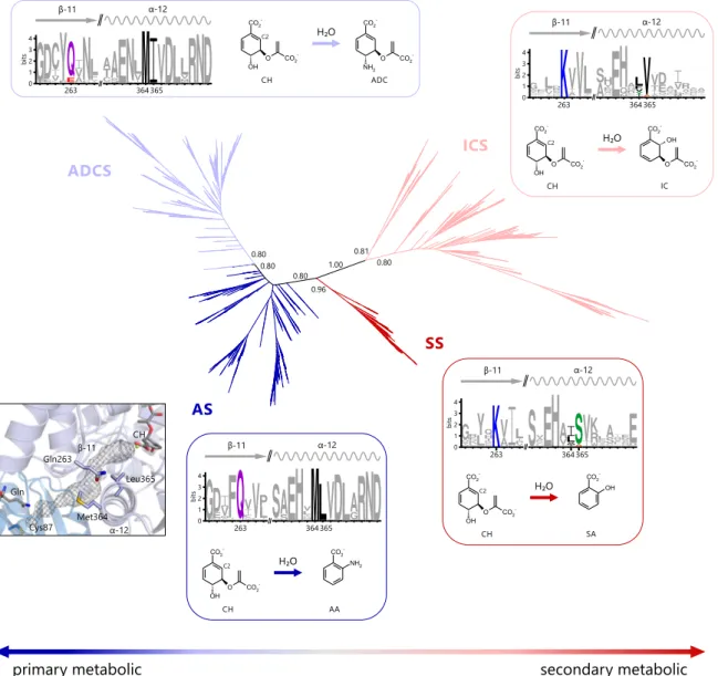
The second part of this work deals with the evolution of substrate specificity and secondary metabolic enzymes in a superfamily of chorismate-utilizing enzymes, named MST-superfamily. Chorismate is a central metabolic node molecule and the starting point for the biosynthesis of various important metabolites, including aromatic amino acids, folate, or iron-chelating siderophores. The MST-enzymes catalyze the committed steps of these biosynthetic pathways and are highly similar in sequence, structure, and reaction mechanism. However, the MST-enzymes that are part of primary metabolic pathways employ exclusively ammonia as a nucleophile to aminate chorismate, whereas those that are part of secondary metabolic pathways exclusively employ water as a nucleophile to hydroxylate chorismate. Based on the notion that secondary metabolic enzymes are descendants of primary metabolic ones, it was investigated in this part of this work by which mechanism the transition from primary metabolic to secondary metabolic MST- enzymes went along with a change in nucleophile-specificity from ammonia to water.
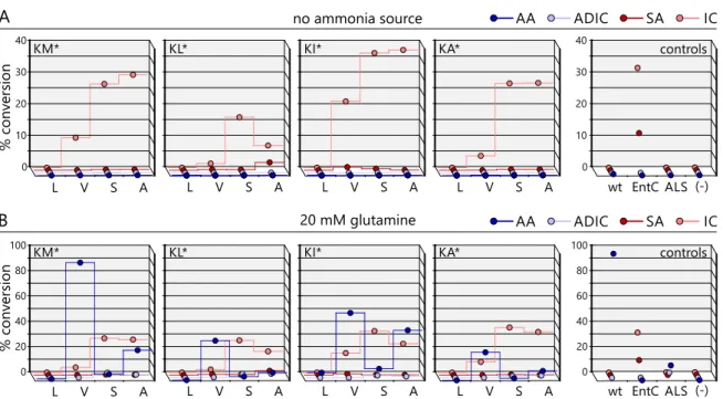
Initially, network-based, phylogenetic, and structure-basedin silicomethods were applied to identify two key amino acids in the nucleophile access channel of the active site that dis- tinguish primary-metabolic/ammonia-utilizing and secondary-metabolic/water-utilizing MST-enzymes. The importance of these key positions was subsequently examined by rationally designing sixteen variants of the MST-enzyme anthranilate synthase, which normally employs ammonia as a nucleophile. The enzymatic characterization of these variants by HPLC-MS showed that the right combination of amino acids at the two key positions indeed resulted in a broadening of nucleophile specificity to also include water.
These anthranilate synthase variants hydroxylated chorismate and formed isochorismate with efficiencies comparable to native secondary-metabolic/water-utilizing isochorismate synthases. Moreover, these variants were still able to employ ammonia as a nucleophile
switch from ammonia to water. Moreover, the observed bi-functionality of the anthrani- late synthase variants argues that the evolution of secondary metabolic MST-enzymes may have proceeded through bi-functional intermediates. Such metabolic generalists may have allowed for the formation of novel metabolites (isochorismate) while maintaining the formation of important primary metabolic metabolites (anthranilate). This scenario con- sequently does nota priorirequire gene duplication events and thus precludes negative metabolic effects linked to retaining redundant gene copies.
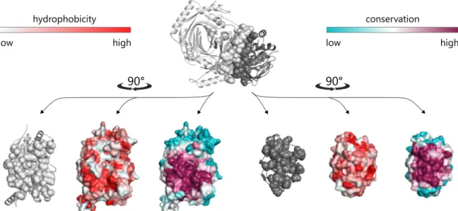
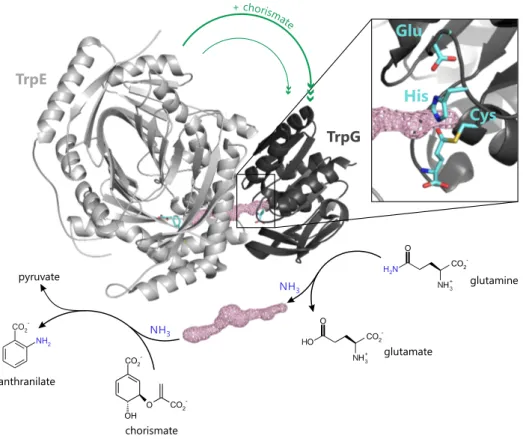
The third part of this work pursues the question how protein-protein interaction specificity is assured in superfamilies of structurally related protein complexes and how the determinants of interaction specificity have evolved. Specific interactions between proteins are vital for almost all cellular functions. This specificity is usually achieved by shape and electrostatic complementarity of protein interfaces. However, the number of different protein folds and interface geometries found in Nature is limited, due to the constraints imposed by efficiently packing hydrogen-bonded secondary structure elements. It is thus a challenging question how interaction specificity is achieved despite structural limitations and how the formation of non-physiological complexes is avoided when several possible interaction partners with similar interface geometries are avail- able. In order to address this problem, initially a comprehensive computational survey of the interface geometries of over 300 bacterial, heteromeric protein complexes and all their homologs of respective superfamilies was performed. This survey revealed that in about 10% of the superfamilies interface geometries vary significantly between related complexes that share homologous subunits. In these cases interfaces were extended by so- called interface add-ons, which typically comprise 10-20 amino acids, form well-defined secondary structure elements, and significantly contribute to complex stability. These characteristics suggested that interface add-ons differentiate between structurally related protein complexes and contribute to interaction specificity through negative design. In order to back this assumption, the case of the interface add-on found in a superfamily of glutamine amidotransferase complexes involved in tryptophan and folate biosynthesis was subsequently analyzed in detail. These complexes comprise synthase and glutami- nase subunits that interact to transfer ammonia from glutamine to an acceptor substrate.
A subset of synthase subunits exclusively involved in tryptophan biosynthesis contains the interface add-on, whereas it is absent in all other homologous synthase subunits, includ- ing those exclusively involved in folate biosynthesis. The comprehensive experimental characterization of 54 combinations of different synthase and glutaminase subunits by
chromatographic methods, light scattering, mass spectrometry, and enzyme kinetics demonstrated that the presence or absence of the interface add-on determines interaction specificity. Anin silicogenetic profiling of over 15 000 archaeal and bacterial genomes together within vivogrowth assays showed that the interface add-on found in complexes of tryptophan biosynthesis is biologically relevant for preventing cross-interactions with the homologous complexes of folate biosynthesis, which would lead to harmful metabolic cross-talk that negatively affects cellular fitness. It was finally shown by protein design that the evolution of the interface add-on in these complexes most likely proceeded via intermediary complexes with relaxed interaction specificity. In conclusion, this part of this work demonstrates that interface add-ons are evolutionary tools to facilitate interaction specificity in superfamilies of homologous proteins or in cases where a protein has to discriminate between several potential interaction partners that share similar interface geometries.
In summary, the presented work leads to an improved understanding of the mech- anisms behind the evolution of enzymatic mono- and bi-functionality, emphasizes the importance of generalist, bi- or multi-functional enzymes for the evolution of secondary metabolic pathways, and finally describes a so far overlooked structural tool for the evolu- tionary specification of protein-protein interactions.
Superfamilien stellen ein Klassifikationsschema zur Gruppierung von Proteinen dar, die auf einen gemeinsamen evolutionären Ursprung zurück gehen und Ähnlichkeiten in Sequenz, Struktur und Reaktionsmechanismus besitzen, jedoch unterschiedliche Funk- tionen ausüben. Die meisten Superfamilien sind heute umfassend charakterisiert und häufig sind für eine Superfamilie mehr als zehntausend Sequenzen, mehrere hundert Strukturen sowie die zentralen Funktionen bekannt. Superfamilien erlauben daher als formales und gleichzeitig biologisch sinnvolles Rahmenwerk die Untersuchung der evolu- tionären Zusammenhänge zwischen Proteinen. Für die vorliegende Arbeit wurden drei Enzym-Superfamilien herangezogen, um Einblicke in mehrere wichtige Fragestellungen der Evolution von Enzymen zu gewinnen.
Der erste Teil dieser Arbeit befasst sich mit der Evolution enzymatischer Mono- und Bifunktionalität in der Superfamilie der Ribose-bindenden Zuckerisomerasen mit (βα8)- Fass Struktur. Diese Superfamilie enthält unter anderem die verwandten Enzyme HisA und TrpF, die ähnliche Reaktionen in der Biosynthese der Aminosäuren Histidin und Tryp- tophan katalysieren. Zusätzlich enthält die Familie auch das bifunktionale Enzym PriA, welches sowohl die HisA, als auch die TrpF Isomerisierungsreaktion katalysiert. Während HisA und TrpF in den meisten Archaeen und Bakterien vorkommen, ist PriA ausschließlich in bestimmten Actinobakterien zu finden. Diesen Spezies fehlt ein dediziertes TrpF En- zym und PriA ist daher essentiell für die Biosynthese sowohl von Histidin als auch von Tryptophan. Es wurden bisher mehrere Hypothesen über die genauen evolutionären Zusammenhänge dieser drei Enzyme aufgestellt; so auch beispielsweise ob die in PriA beobachtete Bifunktionalität ein Überrest von evolutionsgeschichtlich alten Enzymen ist oder ob sie sich erst in neuerer Zeit speziell in Actinobakterien entwickelt hat. Anhand der Rekonstruktion anzestraler Proteinsequenzen wurde in dieser Arbeit gezeigt, dass die evolutionären Vorfahren moderner HisA Enzyme eine zu PriA ähnliche Bifunktionalität aufweisen. Die genaue enzymatische Charakterisierung dreier HisA-Vorläufer ergab, dass diese nicht nur die HisA-Reaktion, sondern auch die TrpF-Reaktionin vitromit vergleich- barer Effizienz katalyiseren. Mittels metabolischer Komplementationsexperimente wurde
weiterhin gezeigt, dass die bifunktionalen HisA-Vorläuferin vivodie Aufrechterhaltung von Histidin- und Tryptophanbiosynthese ermöglichen. Darauf aufbauend wurden mittels einer Kombination aus sequenz- und netzwerkbasiertenin silicoMethoden mehrere mod- erne HisA Enzyme identifiziert, die Sequenzmotive enthalten, die für bifunktionale PriA Enzyme typisch sind. Die enzymatische Charakterisierung dreier solcher HisA Vertreter zeigte, dass diese ebenfalls einen gewissen Grad an Bifunktionalität aufweisen. Interessan- terweise besitzen die zugehörigen bakteriellen Spezies jedoch eigenständige TrpF Enzyme.
Die ursprüngliche Bifunktionalität von HisA hat sich folglich über mehrere Milliarden Jahre erhalten; offenbar ohne direkten Selektionsdruck oder evolutionären Vorteil. Auf- bauend auf diesen Ergebnissen wurde abschließend ein neues Modell zur Erklärung der Evolution von HisA, TrpF und PriA vorgeschlagen: In der evolutionären Frühzeit waren bifunktionale HisA Varianten vermutlich wichtig für die Aufrechterhaltung einer primi- tiven Art von Metabolismus, da sie sowohl die entscheidende Isomerisierungsreaktion in der Histidinbiosynthese, als auch die analoge Reaktion in der Tryptophanbiosynthese katalysieren konnten. Nachdem im Laufe der Evolution allerdings für letztere Reaktion dedizierte TrpF Enzyme entstanden waren, führte ein geringerer Selektionsdruck auf HisA zu einer Abnahme der Bifunktionalität auf das Niveau moderner HisA Vertreter. Dennoch spielte die den HisA Enzymen eigene Bifunktionalität eine wichtige evolutionäre Rolle, da sie die Anpassungsfähigkeit des bakteriellen Metabolismus vergrößerte. So ermöglichten bifunktionale HisA Varianten beispielsweise den Verlust eines dedizierten TrpF Enzyms in Actinobakterien auszugleichen. In diesen Organismen wurde die HisA Bifunktionalität in Folge durch Selektion verstärkt, woraus letztendlich die modernen, bifunktionalen PriA Enzyme hervorgingen.
Im zweiten Teil dieser Arbeit wurde die Evolution von Substratspezifität und sekundär- metabolischen Enzymen am Beispiel einer Superfamilie von Chorismat-bindenden En- zymen, der MST-Superfamilie, untersucht. Chorismat ist ein zentraler metabolischer Knotenpunkt und der Ausgangspunkt für die Biosynthese zahlreicher wichtiger Metabo- lite wie der aromatischen Aminosäuren, Folsäure, oder eisenchelatierender Siderophore.
Die initialen Reaktionen dieser Biosynthesewege werden ausgehend von Chorismat von Enzymen der MST-Superfamilie katalysiert, die hohe Ähnlichkeiten in Sequenz, Struktur und Reaktionsmechanismus aufweisen. Dennoch nutzen diejenigen MST-Enzyme, die Teil von primärmetabolischen Stoffwechselwegen sind, ausschließlich Ammoniak als Nukleophil für die Bildung von aminosubstituierten Chorismatderivativen, wogegen die MST-Enzyme, die Teil von sekundärmetabolischen Wegen sind, ausschließlich Wasser als Nukleophil für die Bildung von hydroxysubstituierten Chorismatderivativen nutzen.
Gemäß der Vorstellung, dass sich sekundärmetabolische Enzyme aus den evolutionär
MST-Enzymen mit dem Übergang von Ammoniak auf Wasser als Nukleophil zusammen- hängt. Dazu wurden zunächst mit netzwerkbasierten, phylogenetischen und struktur- basiertenin silicoMethoden zwei entscheidende Aminosäuren in einem Zugangskanal des aktiven Zentrums identifiziert, die sich signifikant zwischen den primärmetabolis- chen, Ammoniak-umsetztenden und den sekundärmetabolischen, Wasser-umsetztenden MST-Enzymen unterscheiden. Der Beitrag dieser beiden Aminosäuren zur Substrat- bzw. Nukleophilspezifität wurde mittels rationalem Design von sechzehn Varianten des primärmetabolischen MST-Enzyms Anthranilatsynthase überprüft. HPLC-MS Experi- mente zeigten, dass verschiedene Kombinationen von zueinander passenden Aminosäuren an den beiden identifizierten Positionen zu einer Verbreiterung der Nukleophilspezifität von Ammoniak auf Wasser führen. Die aktiven Varianten waren in der Lage, Chorismat unter Verwendung von Wasser als Nukleophil zu Isochorismat umzuwandeln; deren Effizienz war dabei mit der von nativen Isochorismatsynthasen vergleichbar. Diese Vari- anten konnten ebenso Ammoniak als Nukleophil für die Bildung von Anthranilat zu nutzen, waren also bifunktional. Zusammenfassend wurde durch diese Experimente gezeigt, dass die Nukleophilspezifität von MST-Enzymen ohne große Hürden von Am- moniak auf Wasser ausgedehnt werden kann. Die beobachtete Bifunktionalität der An- thranilatsynthasevarianten lässt vermuten, dass die Evolution der sekundärmetabolischen MST-Enzyme über vergleichbare, bifunktionale Enzymintermediate verlaufen ist. Solche, oft als metabolische Generalisten bezeichnete, Enzyme könnten die Bildung von neuar- tigen Sekundärmetaboliten (Isochorismat) unter gleichzeitiger Aufrechterhaltung der Biosynthese wichtiger primärer Metaboliten (Anthranilat) erlaubt haben. Ein solches evo- lutionäres Szenario setzt nicht zwingenderweise eine initiale Genduplikation voraus und vermeidet folglich negative metabolische Effekte, die beispielsweise aus der Beibehaltung von duplizierten, redundanten Genkopien resultieren.
Der dritte Teil dieser Arbeit befasst sich mit der Problematik, wie spezifische Protein- Protein Interaktionen in Superfamilien von strukturell ähnlichen Proteinkomplexen sicher- gestellt werden und wie die für Interaktionsspezifität entscheidenden Faktoren evolviert sind. Praktisch alle zellulären Prozesse sind essentiell abhängig von spezifischen Interak- tionen zwischen Proteinen. Die nötige Spezifität wird generell durch das Zusammenspiel von räumlich und elektrostatisch komplementären Proteinoberflächen, den so genannten interfaces, erreicht. Allerdings ist die Zahl der in der Natur vorkommenden Proteinstruk- turen und folglich auch die Zahl der Proteininterfacesbegrenzt; dieser Umstand ist bedingt durch den Zwang bei der Proteinfaltung die 2D-Strukturelemente räumlich möglichst
effizient zu packen. Zentrale Fragen sind daher, wie trotz der strukturellen Limitationen ausreichende Interaktionsspezifität sichergestellt und wie die Bildug unphysiologischer Komplexe vermieden wird, wenn mehrere Interaktionspartner mit strukturell ähnlichen interfacesvorhanden sind. Um sich diesen Problemen zu nähern, wurden in diesem Teil der Arbeit zunächst dieinterfaceGeometrien von über 300 bakteriellen, heteromeren Proteinkomplexen im Vergleich zu ihren homologen Verwandten aus den entsprechen- den Superfamilien bioinformatisch ausgewertet. Diese Untersuchung zeigte, dass in 10% aller Komplexe, die strukturell ähnliche Untereinheiten besitzen, deutliche Varia- tionen in deninterfaceGeometrien auftreten. In diesen Fällen sind dieinterfacesdurch zusätzliche, definierte Sekundärstrukturelemente, die so genannteninterface add-ons, erweitert, die aus ca. 10-20 Aminosäuren bestehen und signifikante Beiträge zur Stabil- ität der Proteinkomplexe leisten. Aufgrund dieser Eigenschaften lässt sich folgern, dass interface add-onsals eine Art negatives Designelement wirken, dadurch strukturell ähn- liche Proteinkomplexe voneinander differenzieren und somit zur Interaktionsspezifität von Proteinkomplexen beitragen. Um diese Vermutung experimentell zu untersuchen, wurde anschließend eine Superfamilie von Glutaminamidotransferasekomplexen, die an der Biosynthese von Tryptophan und Folsäure beteiligt sind, genauer untersucht: Diese heteromeren Komplexe bestehen aus Synthase- und Glutaminaseuntereinheiten, welche miteinander interagieren, um Ammoniak von Glutamin auf ein Akzeptorsubstrat zu über- tragen. Ein Teil der Glutaminamidotransferasen dieser Superfamilie – ausschließlich solche, die an der Biosynthese von Tryptophan beteiligt sind – trägt in der Synthaseun- tereinheit eininterface add-on. Alle anderen homologen Synthaseuntereinheiten der Superfamilie – auch diejenigen, die an der Biosynthese von Folsäure beteiligt sind – besitzen diesesinterface add-onnicht. Die umfassende Charakterisierung von insge- samt 54 Kombinationen verschiedener Synthase- und Glutaminaseuntereinheiten mittels chromatografischer, biophysikalischer, massenspektrometrischer und enzymkinetischer Methoden zeigte eindeutig, dass die An- bzw. Abwesenheit desinterface add-onsin der Synthaseuntereinheit die Spezifität für die Interaktionen mit den Glutaminaseunterein- heiten bestimmt.In silicoerstellte Profile von über 15 000 archaeellen und bakteriellen Spezies sowiein vivoWachstumsexperimente machten weiterhin deutlich, dass dasinter- face add-onder Komplexe aus der Tryptophanbiosynthese biologische Relevanz besitzt:
Dasinterface add-onverhindert Kreuzinteraktionen mit den Untereinheiten des homolo- gen Komplexes aus der Folsäurebiosynthese, welche sich ansonsten negativ auf die zel- luläre Fitness auswirken und das Zellwachstum behindern. Abschließend wurde anhand von rational designten Glutaminaseuntereinheiten aufgezeigt, dass in der Evolution des interface add-onsdieser Superfamilie vermutlich intermediäre Komplexe mit breiterer
zu etablieren, wenn aufgrund von ähnlicheninterfaceGeometrien zwischen mehreren potentiellen Interaktionspartnern unterschieden werden muss.
Zusammenfassend vertieft die vorliegende Arbeit das Verständnis der Mechanismen, die der Evolution von enzymatischer Mono- und Bifunktionalität zu Grunde liegen, unter- streicht die Bedeutung von multifunktionellen, generalistischen Enzymen für die Evolu- tion des Sekundärmetabolismus und beschreibt ein bisher nicht erkanntes strukturelles Element zur evolutionären Spezifizierung von Protein-Protein Interaktionen.
This cumulative dissertation is composed of the following published or submitted manuscripts:
A Plach, M.G., Reisinger, B., Sterner, R., and Merkl, R. (2016). Long-term persistence of bi-functionality contributes to the robustness of microbial life through exapta- tion.PLoS Genetics12:e1005836
B Plach, M.G., Löffler, P., Merkl, R., and Sterner, R. (2015). Conversion of anthranilate synthase into isochorismate synthase: Implications for the evolution of chorismate- utilizing enzymes.Angewandte Chemie International Edition54:11270-11274 C Plach, M.G., Semmelmann, F., Busch, F., Busch, M., Heizinger, L., Wysocki, V.H.,
Merkl, R., and Sterner, R. (2017). Evolutionary diversification of protein-protein interactions by interface add-ons.Submitted for Publication
In the course of this work, I contributed to further publications, which are not part of the dissertation:
D Veprinskiy, V., Heizinger, L.,Plach, M.G., and Merkl, R. (2017). Assessingin silico the recruitment and functional spectrum of bacterial enzymes from secondary metabolism.BMC Evolutionary Biology. Accepted.
E Kandlinger, F.,Plach, M.G., and Merkl, R. (2017). AGeNNT: annotation of enzyme families by means of refined neighborhood networks.Submitted for publication
Publication A
The research was designed by myself, Bernd Reisinger, Reinhard Sterner, and Rainer Merkl.
The experiments were performed by myself and Bernd Reisinger in equal parts. Ancestral sequence reconstruction was done by Rainer Merkl. The work was supervised by Reinhard Sterner and Rainer Merkl, and the publication was written by myself, Reinhard Sterner, and Rainer Merkl.
Publication B
The research was designed and the experimental was work performed by myself. Patrick Löffler performed molecular dynamics simulations. The work was supervised by Rainer Merkl and Reinhard Sterner, and the publication was written by myself, Patrick Löffler, Rainer Merkl, and Reinhard Sterner.
Publication C
The research was designed by myself, Florian Semmelmann, Rainer Merkl, and Rein- hard Sterner. Cloning, expression, purification, and experimental characterization of glutaminases and synthases were done by myself and Florian Semmelmann. Florian Busch performed mass spectrometry experiments and was supervised by Vicky Wysocki.
All other experiments were performed by myself. Computational tools and scripts were provided by myself, Markus Busch, and Leonhard Heizinger. Rainer Merkl and Reinhard Sterner supervised the work. The publication was written by myself, Florian Semmelmann, Rainer Merkl, and Reinhard Sterner.
Abstract ix
Kurzfassung der Arbeit xiv
List of Publications xvi
Personal Contributions xviii
1 General Introduction 1
1.1 A short history of evolution . . . . 1
1.2 The driving forces of evolution . . . . 3
1.3 The concept of homology . . . . 4
1.4 Protein superfamilies . . . . 7
1.5 Aim and scope of this work . . . . 10
1.6 Guide to the following chapters . . . . 11
2 Long-term persistence of bi-functionality contributes to the robustness of microbial life through exaptation (Synopsis of publication A) 13 2.1 Introduction . . . . 13
2.1.1 The superfamily of ribulose-phosphate binding (βα)8-barrels . . . 13
2.1.2 Promiscuity and bi-functionality . . . . 15
2.1.3 Ancestral sequence reconstruction . . . . 16
2.2 Summary and Discussion . . . . 19
2.2.1 Ancient HisA precursors are bi-functional, PriA-like enzymes . . . . 19
2.2.2 Ancient bi-functionality persists in modern HisA enzymes . . . . . 23
3 Conversion of anthranilate synthase into isochorismate synthase: Implications for the evolution of chorismate-utilizing enzymes (Synopsis of Publication B) 27 3.1 Introduction . . . . 27
3.1.1 Chorismate-utilizing enzymes and the MST superfamily . . . . 27
3.1.2 Primary and secondary metabolism . . . . 34
3.2 Summary and Discussion . . . . 38
3.2.1 Establishing isochorismate-synthase activity on an anthranilate- synthase scaffold . . . . 38
3.2.2 Secondary-metabolic MST-enzymes might have evolved via bi- functional intermediates . . . . 42
4 Evolutionary diversification of protein-protein interactions by interface add-ons (Synopsis of Publication C) 47 4.1 Introduction . . . . 47
4.1.1 Protein-protein interactions . . . . 47
4.1.2 Protein interfaces . . . . 51
4.1.3 Glutamine amidotransferases . . . . 54
4.2 Summary and Discussion . . . . 56
4.2.1 The role of interface add-ons for protein-protein interaction specificity 56 4.2.2 Biological relevance of interface add-ons . . . . 64
4.2.3 Evolution of protein-protein interactions in AS and ADCS com- plexes: The role of the interface add-on . . . . 66
5 Abbreviations 71 6 References 75 7 Publications 107 7.1 Publication A . . . . 107
7.2 Publication B . . . . 133
7.3 Publication C . . . . 163
List of figures 233
List of tables 235
Acknowledgements 237
1.1 A short history of evolution
Life in its entire spectrum formally bursts with complexity. One, for instance, might think of the remarkable biosynthetic capabilities of microorganisms, the specialization and connectivity of individual cells in multi-cellular plants and animals, or the highly sophisticated cognitive abilities of the human brain. These developments did, of course, not come overnight but rather from a painstakingly slow but gradual adaptation process.
This process, referred to asevolution, was first formulated as a scientific theory in the early 19th century (Hoyle and Wickramasinghe, 2000). At that time scholars recognized that the driving forces behind the differentiation of life into the countless species were constant adaptation to ecological niches and competition for resources (Darwin, 1859).
But what did evolution begin with? One longstanding theory suggests that life itself started in the primordial oceans. During a time-period with a reducing atmosphere (Laz- cano and Miller, 1996; Oparin and Morgulis, 2003) complex organic compounds formed from carbon dioxide or methane under the influence of ultraviolet light or electricity (Bada, 2013; Miller, 1953). Despite some disagreements (Bernhardt, 2012), it is widely acknowledged that RNA was among these first compounds (de Farias et al., 2016; Gilbert, 1986). This "RNA world" concept suggests that RNA molecules could catalyze simple biochemical reactions and also store the information needed for their replication. The concept is supported by the observation that some modern biosynthetic machineries like ribosomes contain catalytic RNA molecules and by the simple enzymatic reduction of ribose nucleotides that yields deoxyribose nucleotides as they appear in the later emerged DNA (Higgs and Lehman, 2015).
Evolution really kicked off when the information stored in RNA and DNA became meaningful in early forms of genes. These served as blueprints for small primordial peptides that were able to interact with RNA and to support RNA-based catalysis (Alva et al., 2015). These small peptides are thought to have gradually fused, multiplied, and recombined to eventually give rise to the relatively small set of not more than a thousand
different modern protein geometries or folds (Chothia, 1992). Recent experimental dis- coveries identified sets of primordial peptides (Farías-Rico et al., 2014), showed that they can self-assemble and form larger, biochemically active proteins (Smock et al., 2016), and highlighted that duplication and fusion can lead to symmetric proteins structurally similar to modern ones (Longo and Blaber, 2014; Park et al., 2015; Richter et al., 2010).
The inclusion of such primitive proteins together with RNA, DNA, and other organic molecules in cell-like bubbles made of amphipathic fatty molecules was the basis for first simple self-replicating units. These units eventually formed the last universal common ancestor (LUCA) of all living things, which existed approximately between 3.8 and 4.5 bil- lion years ago (Nisbet and Sleep, 2001). It is assumed that this enigmatic single entity was rather simple (Di Giulio, 2011; Koonin, 2003; Woese, 1998), yet its true nature and physiol- ogy is still widely debated (Kim and Caetano-Anollés, 2011; Weiss et al., 2016). For instance, analyses of gene content, co-evolution, and proteomes favor a more sophisticated LUCA physiology (Doolittle, 2000; Ouzounis et al., 2006; Tuller et al., 2010). Originating from the LUCA, formation and diversification of species ("macro-evolution") took on, first yielding uni-cellular microorganisms like archaea and bacteria and eventually the evolutionary young multi-cellular plants and animals. Given some remarkable similarities between different animal species or between animals and plants, this common origin has already been noticed in the 17th century (Hoyle and Wickramasinghe, 2000). Indeed, modern methods of genetic analyses allow one to reconstruct the evolutionary relationship of all known species back to their common root in a so-called tree-of-life (Figure 1).
Finally, there is one more point to stress out: Evolution is no completed process that has begun sometime in the past and that has led to all the species and organisms we know today. On the contrary, evolution is highly dynamic and new species, new organisms, and new functions constantly emerge. This is best seen from the rapid development of microbial drug resistance against common antibiotics that have only been in use for nearly a decade (Barlow and Hall, 2002; Boyanova et al., 2015; Hall, 2004) or the appearance of microbial enzymes that are capable of degrading chemicals that have not been in the ecosystem for longer than 50 years (Copley, 2009; Hartley et al., 2006; Janssen et al., 2005;
Seffernick and Wackett, 2016). The herbicide atrazine, for example, was invented in 1958 and has been applied in millions of tons since then. Already 40 years later, bacterial strains were identified that are able to degrade atrazine completely and metabolize its degradation products (Boundy-Mills et al., 1997; de Souza et al., 1998b). The atrazine-hydrolyzing enzymes produced by these bacteria are assumed to have emerged very recently from amidohydrolases like melamine deaminase in soil-living Pseudomonas species (de Souza et al., 1998a; Seffernick et al., 2001; Shapir et al., 2007).
Figure 1. Tree-of-life.
Phylogenetic tree, as constructed from the comparison of 31 concatenated genes that oc- cur in 191 species with fully sequenced genomes (Ciccarelli et al., 2006). In this illustration the root of the tree is in the middle and the branches radiate outwards, with each new species descending from a common ancestor resulting in a new branch. Note that branch lengths are not to scale. Bacterial and archaeal species are spanned by blue and magenta arcs, respectively. Eukaryotic species are marked by a green arc.
1.2 The driving forces of evolution
The common ancestry of all species and the relationship between them makes clear how new species emerge: By segregation from already existing ones. Such speciation events take place when two populations of the same species get separated by some kind of barrier, for instance an entrapment in an ecological niche, and adapt to their respective conditions. Over time the populations will genetically drift apart, up to a point where genetic exchange between the daughter and the parental population is no longer possible (Safran and Nosil, 2012). Evolution is thus driven by constant adaptation processes to changing environments through the emergence of novel functionalities, which are eventually just novel genes that lead to novel proteins with novel capacities.
But how do novel genes arise? In fact, this is simply a matter of chance. DNA is not just an information carrier that is filed somewhere inside a cell but is constantly transcribed into RNA blueprints for proteins and replicated for genetic reproduction.
Although a highly complex and sophisticated catalytic machinery has developed for these
essential tasks, errors occasionally occur. For instance, sometimes the wrong nucleotide is incorporated into replicated DNA thus changing the genetic information (Kunkel and Bebenek, 2000). Additionally, response systems can fail to repair DNA damage coming from the exposure to UV light or oxidizing compounds and different parts of a genome can be recombined due to viruses or erroneous DNA ligation (Friedberg, 2003; Hakem, 2008).
These changes in the genetic information are known as mutations, which can be classified into insertions, deletions, and substitutions, depending on whether additional nucleotides were inserted into the sequence, a stretch of nucleotides was lost, or a single nucleotide was changed to another one. Although the effects of mutations in protein coding genes are buffered by a redundancy in coding nucleotide triplets, many mutations are deleterious. For instance, a mutation may lead to a loss of function by substituting a hydrophobic residue in the protein core with a polar one, which could cause the protein to misfold. Likewise, substituting a catalytic residue in an enzyme might render it inactive.
For uni-cellular organisms like bacteria such mutations can have lethal consequences, clearing the corresponding mutants from the population and the gene pool. However, by chance, some mutations will occur at just the right spot; they may lead to a more stable protein by filling a hole in its structure with a bulky amino acid, may allow the defense against antibiotic agents by modifying the catalytic capacity of a hydrolase enzyme, or increase the catalytic efficiency of an enzyme by introducing an amino acid that aids in binding its substrate. Such mutations lead to novel functionalities that can give the mutant a lead in competition and adaptation, eventually allowing it to prevail in the population and conserve the new functionality.
1.3 The concept of homology
A great caveat of mutational adaptation is, as mentioned above, that most mutations are deleterious and severely impair the viability of organisms. Today, evolution is thus no longer viewed as a strictly linear, gradual adaptation process but more as a drift through a landscape of neutral mutations, which are neither beneficial nor harmful, with random fixation of mutations (Kimura, 1983). Moreover, it has been realized that other processes of greater genetic variation must exist that enable the accumulation of mutations while buffering their negative effects. Ohno postulated in his seminal workEvolution by Gene Duplication(Ohno, 1970) that the duplication of a gene or even larger genomic segments creates redundant copies that are free from selective pressure, which allows for the accu- mulation of otherwise detrimental or lethal mutations.
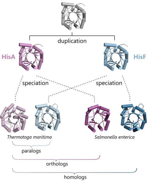
Genes that are related through such duplication events and that have diverged in function are formally referred to as paralogs(Figure 2). The frequency of paralogs in bacterial genomes is quite high; already in a genome comprising 2000 genes about one fourth of the genes are paralogs (Hooper and Berg, 2003). Although the term paralog, in its original definition referred to genes (Fitch, 1970), it is commonly used to describe the relationship of proteins. In this sense, paralogous proteins share many properties like their overall three-dimensional structure but often differ in crucial features like enzymatic function or stability. For example, the enzyme 1-(5-phosphoribosyl)-5-[(5-phosphoribosylamino)- methylideneamino]-imidazole-4-carboxamide isomerase (HisA) and the cyclase subunit of the imidazole glycerol phosphate synthase (HisF) share the same fold and are similar with respect to their amino acid sequences, but have evolved quite different catalytic activities for the biosynthesis of the amino acid histidine.
In contrast to paralogs,orthologsare genes that are related through speciation events and that have retained their original function in the descendant species (Fitch, 1970), because they are still subject to the same selective pressure as in the paternal species (Figure 2). Following the example from above, the HisA enzymes fromThermotoga mar- itimaandSalmonella entericaare orthologs, because they both have descended from their common HisA ancestor and they both still catalyze the fourth step of histidine biosynthesis.
In a generalization of these two concepts, genes and their products are calledhomologs, if they are related to each other by descent from a common ancestor through a series of duplication and speciation events (Figure 2). However, it is complicated to infer homol- ogy for genes or proteins that have diverged from a common ancestor billions of years ago, because their sequences often show hardly any similarities. Thus, a more practical definition of homology is applied today: Two genes (or two proteins) are homologs, if they are simply similar enough that it is unlikely that this similarity arose just by coincidence.
The most common lower threshold for inferring homology of proteins is a minimum of 20-30% identical amino acids across the length of their aligned sequences, as this value correlates with an almost certain structure similarity (Orengo and Thornton, 2005).
duplication
speciation
Thermotoga maritima Salmonella enterica
HisA HisF
speciation
paralogs
orthologs
homologs
Figure 2. Illustration of the concepts of paralogs, orthologs, and homologs.
The enzymes HisA (magenta) and HisF (blue) have emerged from a common ancestor by gene duplication but have evolved different functions in the biosynthesis of the amino acid histidine. Their relation is apparent from structural similarity and the similar amino acid sequences of modern HisA and HisF enzymes (41% and 42% for the pairs fromS. enterica andT. maritima, respectively, determined with EMBOSS Needle (Rice et al., 2000)). HisA and HisF are paralogs, because they are related through a duplication event and have diverged in their respective functions. In contrast, orthologs are genes or proteins that are related through a speciation event and that have retained the same function. In general, homologs are genes or proteins that are related through descent from a common ancestor by a series of duplication and speciation events. The homology between HisA fromT. maritimaand HisF fromS. entericais evident from their structural similarity and the high degree of sequence similarity (43%, determined with EMBOSS Needle).
1.4 Protein superfamilies
Homology is no completely precise concept: For instance, closely related homologs may share almost identical structures and perform very similar functions, like the catalysis of the same reaction with just a slightly different substrate. On the other hand, distantly related proteins that may have little in common – their structures may be rather strong variations of the same core fold and they may catalyze completely different chemistries – are counted as homologs as long as their sequences are similar enough. Thus a more fine-grained classification of homology in the context of proteins is necessary. Such is provided by combining homologs that have highly similar sequences (e.g. >40% identity) and that also share common functional properties intofamilies. In turn, several families can be further grouped together to larger entities whose members have less similar se- quences (generally <30% identity) and have fewer traits in common, for instance only the overall three-dimensional structure or a core reaction chemistry. Margaret Dayhoff first coined the termsuperfamilyfor these groups to reflect their super-ordinate character and broader evolutionary relationship compared to families (Dayhoff, 1965). Superfamilies are frequently functionally diverse (Almonacid and Babbitt, 2011); only about 60% of the pro- teins from the same superfamily also have the same function (Hegyi and Gerstein, 2001) and this variability can even span all six Enzyme Commission classes in some enzyme superfamilies (Baier et al., 2016). To summarize, the term superfamily shall be defined within the scope of this thesis as follows: A superfamily is a group of homologous proteins that (i) are derived from a common evolutionary ancestor, (ii) hence share similarities in sequence, structure, and – for enzymes – core reaction mechanism, but (iii) differ in their specific functions.
The first comprehensive implementation of the family-superfamily concept was achieved in theStructural Classification of Proteinsdatabase (SCOP) (Murzin et al., 1995).
This database provides a hierarchical classification of proteins into families and superfam- ilies, based on evolutionary relationship, and into folds and classes, based on structural features and similarities (Figure 3). For example, different glucosidases and glycosyltrans- ferases that all act onα-linked oligosaccharides are summarized in theα-amylase family.
This family is, in turn, grouped together with other families like theβ-galactosidases, -glucanases, and -amylases to the superfamily of glycosyltransferases. While families are collections of related but very similar enzymes, superfamilies provide an overview of the different functions that have been established on a common protein scaffold and the evolutionary relationship between these different functions.
SCOP
α β α/β α+β
Rossman fold Flavodoxin-like α/β-barrel
TIM Trp biosynthesis Glycosyltransferases RuBisCo
β-Galactosidases β-Glucanase α-Amylase β-Amylase
Acid α-Amylase Cyclodextrin Glycosyltransferase Oligo-1,6-glucosidase
Escherichia coli Bacillus subtilis Bacillus macerans Bacillus cereus
Class
Fold
Family
Protein Superfamily
structure based
evolution based Species
Figure 3. Hierarchical illustration of the SCOP database.
The hierarchical classification of proteins in the SCOP database comprises six levels. At the bottomspeciesrepresent distinct protein sequences, which are grouped together with similar proteins of essentially the same function on theproteinlevel. Thefamily level summarizes proteins with highly similar sequences and similar functions. Thesuperfamily level combines individual families with similar structural features, a common evolutionary origin, but different functions. Above this levelfoldandclassgroup superfamilies based on structural features and do not imply homology. The figure is adapted from the SCOP version 1.75 documentation (Andreeva et al., 2008).
A look at frequently used databases that classify proteins into families and superfamilies points out, however, that the definition of a superfamily is not general and that it is of- ten not clear where to draw the line between a family and a superfamily. For example, the latest release of the SCOP database (version 1.75, June 2009) lists 1962 superfamilies and 3902 families. More recent and more sophisticated databases like InterPro (ver- sion 60.0, November 2016) and Pfam (version 30.0, June 2016) yet list 1749/19 851 and 595/16 306 corresponding entries, respectively. These databases have been created us- ing high-performance and high-sensitive algorithms that accurately assign new protein sequences to the correct families and superfamilies (Finn et al., 2016; Mitchell et al., 2014).
An equally challenging task is to put the inherent information of protein superfamilies on evolutionary relationship and functional diversity to use. Traditional phylogenetic approaches to dissect and sub-divide a superfamily are often problematic, because the data-sets frequently contain tens of thousands of sequences and the amino acid simi- larities between iso-functional sub-families can be very low (Punta et al., 2012). A more
E = 1·10-114
60% seq-id
E = 1·10-101
44% seq-id
E = 1·10-260
73% seq-id
E = 1·10-97
40% seq-id
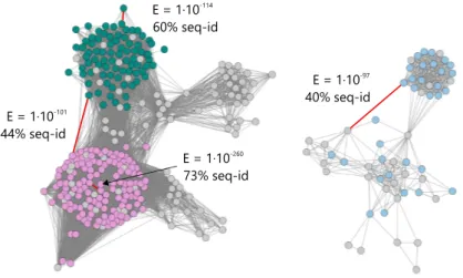
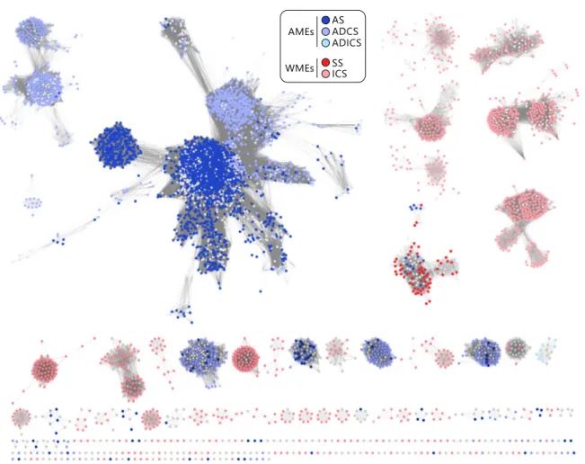
Figure 4. Visualization of pairwise sequence similarity relationships between mem- bers of a protein superfamily in a sequence similarity network.
The SSN shows the relationship between selected members of the glycosyl transferase superfamily (InterPro entry IPR000312), which comprises structurally similar but func- tionally distinct AMP-phosphorylases (green), thymidine phosphorylases (magenta), and anthranilate phosphoribosyltransferases (blue). The network was generated at a similarity cut-off of 10−97(BLAST E-value9 and is shown in a force-directed layout generated with Cytoscape 3.4 (Shannon et al., 2003). Selected edges are marked in red and annotated with their respective E-value and the corresponding sequence identity (seq-id) of the connected sequences.
robust and computationally less demanding alternative nowadays widely used for pro- cessing superfamilies are sequence similarity networks (SSNs), developed by Babbitt and co-workers (Atkinson et al., 2009) and made available by theEnzyme Function Initiative (Gerlt et al., 2015). SSNs rely on the pre-defined protein family and superfamily data-sets from InterPro or Pfam and are constructed from an all-by-all sequence comparison be- tween all members of a superfamily using the well-established BLAST algorithm (Altschul et al., 1990). The key of SSNs is that these inter-relationships can be visualized as a net- work graph (Figure 4). In such networks, nodes represent individual members of the superfamily (protein sequences) and connecting edges represent the pairwise sequence relation (sequence similarity score or BLAST E-value), if the corresponding proteins are more similar than a pre-defined threshold.
For most thresholds closely related members of a superfamily will be inter-connected more densely than distantly related members. With the threshold chosen appropriately for a specific superfamily, the network can reveal iso-functional groups within a superfamily.
For example, an exemplary SSN of the glycosyltransferase superfamily (Figure 4) shows that AMP phosphorylases and thymidine phosphorylases are more closely related to each
other than to the anthranilate phosphoribosyltransferases. This simple visual analysis tells much about the evolution of this superfamily and the functional relationship between its members. It is thus hardly surprising that superfamily data-sets together with SSNs have been used frequently with great success to get insight about enzyme evolution (Brown and Babbitt, 2014), to discover and predict enzymatic activities of uncharacterized proteins (Baier et al., 2016; Chow et al., 2015; Gerlt et al., 2015; Zhao et al., 2014), and to analyze the sequence-structure-function relationship, the fundamental axiom of modern molecular biology (Mashiyama et al., 2014).
1.5 Aim and scope of this work
Although the number of protein and whole genome sequences as well as the number of elucidated protein structures and functions have soared in the last decade, the evolu- tionary relationship between proteins and the paths that evolution took from the ancient ancestral entities to the modern ones is still enigmatic in all but few cases. And yet the lessons learned from unraveling, for example, how a specific enzymatic function is dis- tributed across the members of the respective superfamily and how this function might have been established during evolution are the most important; first and foremost for a better understanding of the principles of biology and evolution. But these lessons will also tell us which enzyme scaffolds might be used to alter their catalytic capabilities to a desired function and how the transition to the new function might be achieved in the laboratory or via computer-aided design. Eventually, these lessons will help us to better understand the sequence-structure-function relationship, which in turn could tell what functional aspects or structural features of a protein superfamily might be targeted by drugs that could aid human health.
To study the evolution of protein traits and characteristics, one heavily relies on ex- ploiting homology relationships between proteins. Superfamilies are of great value in this context, because they provide a formal but also biologically sensible framework to sub-divide and dissect groups of related proteins and – with the right tools in hand – to draw far-reaching conclusions and gain valuable insights. In this thesis, these concepts are used together with computational, biochemical, and biophysical techniques to investigate the evolution of enzymatic mono- and bi-functionality, the determinants of substrate specificity in enzyme superfamilies, the evolution of secondary metabolic enzymes, as well as the structural determinants of protein-protein interactions, their evolutionary history, and their physiological significance.
1.6 Guide to the following chapters
The following three chapters each deal with one of the three first-author publications (A-C) that constitute this thesis. These chapters are not intended to repeat the publications one-to-one. They are rather formulated as synopses that give the reader a profound introduction into the individual concepts and ideas important for the respective topics, highlight the key findings of each publication, and discuss these with attention to the corresponding literature.
In the chapterLong-term persistence of bi-functionality contributes to the robustness of microbial life through exaptationSSNs and ancestral sequence reconstruction (ASR) are used to demonstrate that the evolution of modern sugar isomerases with different catalytic functions originated from a bi-functional common ancestor. The chapter further describes that this bi-functionality has persisted to a certain degree in modern members of the corresponding sugar isomerase superfamily.
In the following chapterConversion of anthranilate synthase into isochorismate synthase:
Implications for the evolution of chorismate-utilizing enzymesthe functional divergence within a superfamily of enzymes is examined and key catalytic residues that determine substrate specificity of these enzymes are identified. It is further demonstrated that this information can be used to modify the function of enzymes from this superfamily and finally the implications of this functional change for the evolution of secondary metabolic enzymes from primary metabolic enzymes are highlighted.
In the final chapterEvolutionary diversification of protein-protein interactions by interface add-onsstructural determinants of protein-protein interaction specificity are identified from an extensive analysis of heteromeric protein complex structures. A superfamily of glutamine amidotransferases is then used to demonstrate that these structural features prevent the formation of non-physiological complexes and an evolutionary scenario for the emergence of these structural elements is provided.
nality contributes to the robustness of microbial life through exaptation (Synopsis of Publication A)
2.1 Introduction
2.1.1 The superfamily of ribulose-phosphate binding (βα)8-barrels
Theribulose-phosphate binding barrelsuperfamily (SCOP 51366) comprises, among oth- ers, several iso-functional families of (βα)8-barrel enzymes from histidine and tryptophan biosynthesis. One of its members is the enzyme HisA, which catalyzes the isomerization of the aminoaldose N’-[(5’-phosphoribosyl)-formimino]-5-aminoimidazole-4-carboxamide- ribonucleotide (ProFAR) to the aminoketose N’-[(5’-phosphoribulosyl)-formimino]-5- aminoimidazole-4-carboxamide-ribonucleotide (PRFAR) (Henn-Sax et al., 2002) for the fourth step of the biosynthesis of the essential amino acid histidine (Figure 5A). Another prominent member of this superfamily is the enzyme TrpF, which catalyzes a similar iso- merization reaction involving the aminoaldose N-(5’-phosphoribosyl)anthranilate (PRA) and the aminoketose 1-(o-carboxyphenylamino)-1-deoxyribulose-5-phosphate (CdRP) in the biosynthesis of tryptophan (Hommel et al., 1995). The common evolutionary history of HisA and TrpF is not only evident from their highly similar structures and their high degree of sequence similarity (List et al., 2011) but is also supported by establishment of PRA isomerase activity on HisA scaffolds through both random mutagenesis (Jürgens et al., 2000) and spontaneous mutations under selective pressure (Näsvall et al., 2012).
The InterPro database associates HisA with the phosphoribosylformimino-5-amino- imidazole-carboxamide-ribotide isomerase superfamily (InterPro entry IPR023016). In the current InterPro release (version 60.0, November 2016) this entry comprises 9565
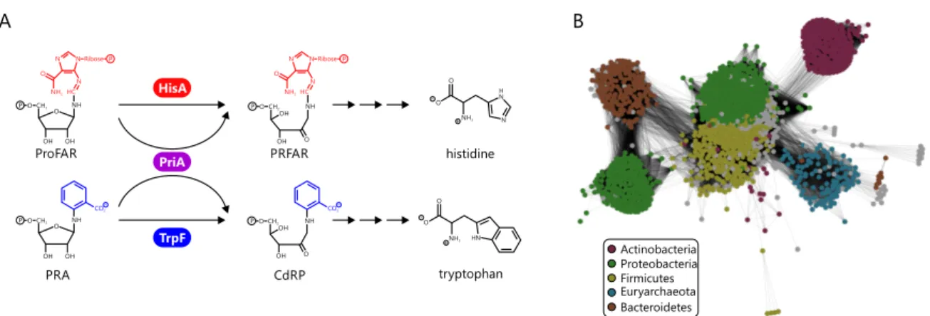
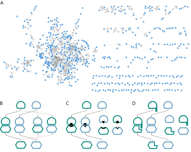
Figure 5. Reactions catalyzed by the homologous enzymes HisA, PriA, and TrpF and a SSN representation of the HisA InterPro family.
(A) HisA and TrpF catalyze analogous Amadori rearrangements of the aminoaldoses ProFAR and PRA to the corresponding aminoketoses PRFAR and CdRP in histidine and tryptophan biosynthesis, respectively. The bi-functional enzyme PriA catalyzes both reac- tions. (B) SSN of the phosphoribosylformimino-5-aminoimidazole-carboxamide-ribotide isomerase superfamily. Nodes are colored by the five main bacterial phyla contributing to the superfamily. The figure is adapted from publication A.
members, which mainly correspond to HisA enzymes from bacterial and archaeal species.
An SSN shows that the superfamily is basically iso-functional, with a certain sub-cluster structure as a result of phylum specific sequence variations (Figure 5B). The most uniform and most separated sub-cluster is that of Actinobacteria. Most Actinobacteria do not possess HisA enzymes, but rely for the biosynthesis of histidine and tryptophan on a homolog of HisA called phosphoribosyl isomerase A (PriA). PriA is a bi-substrate spe- cific enzyme processing ProFAR as well as PRA with equal efficiency (Barona-Gómez and Hodgson, 2003). Actinobacteria that possess apriA gene lack a dedicatedtrpF gene, and consequently PriA is part of both histidine and tryptophan biosynthesis. The genomic neighborhoods ofhisA andpriA, i.e. the genes upstream and downstream, are highly simi- lar, further indicating that PriA is a homolog of HisA (Publication E). Structural comparison with HisA makes clear that PriA utilizes the same two phosphate binding sites formed by βα-loops three and four as well asβα-loops seven and eight to bind ProFAR (Due et al., 2011; Wright et al., 2008). The C-terminal site used for binding PRA is also identical to the single phosphate binding site of its superfamily relative TrpF (Due et al., 2011; Henn-Sax et al., 2002). Its quite unusual characteristics have made PriA a well-examined member of this superfamily with respect to structure and catalytic mechanism (Due et al., 2011;
Küper et al., 2005; Wright et al., 2004) as well as its evolutionary relationship with HisA and TrpF (Noda-García et al., 2013, 2015; Verduzco-Castro et al., 2016).