RESEARCH PAPER
Thermodynamic, Spatial and Methodological Considerations for the Manufacturing of Therapeutic Polymer Nanoparticles
Sara Maslanka Figueroa1&Daniel Fleischmann1&Sebastian Beck1&Achim Goepferich1
Received: 9 January 2020 / Accepted: 11 February 2020
#The Author(s) 2020
ABSTRACT
Purpose
Evaluate fundamental parameters that dictate the effectiveness of drug loading.
Methods
A model water-soluble drug lacking ionizable groups, pirfenidone (PFD), was encapsulated through nanoprecipitation in poly(ethylene glycol)-poly(lactic acid) (PEG-PLA)-poly(lactic-co-glycolic acid) (PLGA) NPs.
Firstly, the thermodynamic parameters predicting drug- polymer miscibility were determined to assess the system’s suitability. Then, the encapsulation was evaluated experi- mentally by two different techniques, bulk and microfluidic (MF) nanoprecipitation. Additionally, the number of mol- ecules that fit in a particle core were calculated and the loading determined experimentally for different core sizes.
Lastly, the effect of co-encapsulation of
α-lipoic acid (LA),a drug with complementary therapeutic effects and en- hanced lipophilicity, was evaluated.
Results
The thermodynamic miscibility parameters predicted a good suitability of the selected system. MF manufacturing enhanced the encapsulation efficiency by 60–90% and achieved a 2-fold higher NP cellular uptake. Considering spa- tial constrictions for drug encapsulation and increasing the size of the PLGA core the number of PFD molecules per NP was raised from under 500 to up to 2000. More so, the co-encapsulation of LA increased the number of drug mole- cules per particle by 96%, with no interference with the re- lease profile.
Conclusions
Thermodynamic, spatial and methodological parameters should be considered to optimize drug encapsulation.
KEYWORDS
encapsulation . microfluidics . nanoprecipitation . pirfenidone . polymeric nanoparticlesABBREVIATIONS
χsp
Flory-Huggins interaction parameter
FRRFlow rate ratio
LA α-lipoic acid PEG
Poly(ethylene glycol)
PFDPirfenidone
PLA
Poly(lactic acid)
PLGA
Poly(lactic-co-glycolic acid)
MFMicrofluidic
ΔHMT
Mixing enthalpy obtained from the total solubility parameters
ΔHM
Mixing enthalpy obtained from the partial solubility parameters
NPs
Nanoparticles
δSolubility parameter
TFRTotal flow rate
INTRODUCTION
Nanoprecipitation (1), or solvent displacement, is a frequently used technique for the preparation of therapeutic polymer nanoparticles (NPs), as it is a simple, clean, and versatile ap- proach (2). More so, it enables drug encapsulation without requiring additional steps, such as covalently coupling the drug to structural particle components.
Electronic supplementary material The online version of this article (https://doi.org/10.1007/s11095-020-2783-4) contains supplementary material, which is available to authorized users.
* Achim Goepferich achim.goepferich@ur.de
1 Department of Pharmaceutical Technology, University of Regensburg Universitaetsstrasse 31, 93053 Regensburg, Germany
https://doi.org/10.1007/s11095-020-2783-4
There are several elements that to some extent determine the success of a nanoprecipitation-mediated drug encapsula- tion, such as the physicochemical characteristics of the selected drug. As this technique involves the addition of a small volume of organic polymer phase into a large volume of aqueous phase, it has mostly been exploited for the encapsulation of lipophilic drugs that have little to no water solubility. Its application to water-soluble drugs is often inefficient, but can be improved to some extent by modulating the pH value of the water phase (3–5) or promoting electrostatic interaction with excipients (4).
Alternatively, some authors have resulted to modify the drug molecule itself (6), or released it from its salt form (2), for which doxorubicin is a common example (7). However, these approaches are not universally applicable. For the former the presence of ionizable groups is a mandatory requirement and the latter may not be a feasible option due to the additional regulatory burdens associated with changing a drug’s structure.
More so, there is a lack of research regarding the encapsulation through nanoprecipitation of water-soluble molecules that lack the aforementioned criteria.
An additional element that has an impact on drug encap- sulation is the thermodynamic compatibility, i.e. miscibility, of the drug and particle-forming polymers. Therefore, thermo- dynamic parameters such as the Flory-Huggins interaction parameter (χ
sp), or the solubility parameters (δ) are frequently used to assess drug-polymer miscibility (8). However, there are other elements that can determine the successful encapsula- tion of a drug in a selected particle system, such as dimensional restrictions, the used nanoprecipitation method or the pres- ence of a co-encapsulated drug molecule.
Therefore, the goal of this study was to encapsulate through nanoprecipitation a partially water-soluble drug lack- ing ionizable groups and evaluate different parameters dictat- ing the drug loading.
As a drug model for our purposes we selected pirfenidone (PFD) (Fig.
1). PFD is a small molecule drug, with a logP= 2.14. Therefore, it would be initially considered a good can- didate for encapsulation through nanoprecipitation.
However, it is soluble in water up to 2 mg/mL, which is exceedingly high for this technique, due to the large volumes of water phase being used.
PFD is an antifibrotic agent, approved for the treatment of idiopathic pulmonary fibrosis (9). Additionally, recent studies showed its potential for the treatment of diabetic kidney dis- ease (10,11) and glomerulosclerosis (12). However, it has a considerable plasma protein binding (50%) and fast elimina- tion half-life (2.4 h), which requires a large oral dose intake (>
2 g/day) to achieve therapeutic effects (13). This elevated daily intake prompts considerable gastrointestinal side effects which contribute to therapy discontinuation by a large number of patients (11,14). Therefore, it is an excellent candidate that would benefit from incorporation in a nanoparticulate system.
Over the past years PFD has been encapsulated in PLGA NPs by the emulsion solvent evaporation method for the treatment of pulmonary fibrosis (15) and corneal wounds (16), in chitosan-alginate nanocarriers through the pre-gelation meth- od for transdermal delivery (17) and in liquid crystalline nano- particles (18). However, it has never been encapsulated before by means of nanoprecipitation, which would considerably ease NP preparation and allow for simple cost-effective upscaling and reproducible results (19).
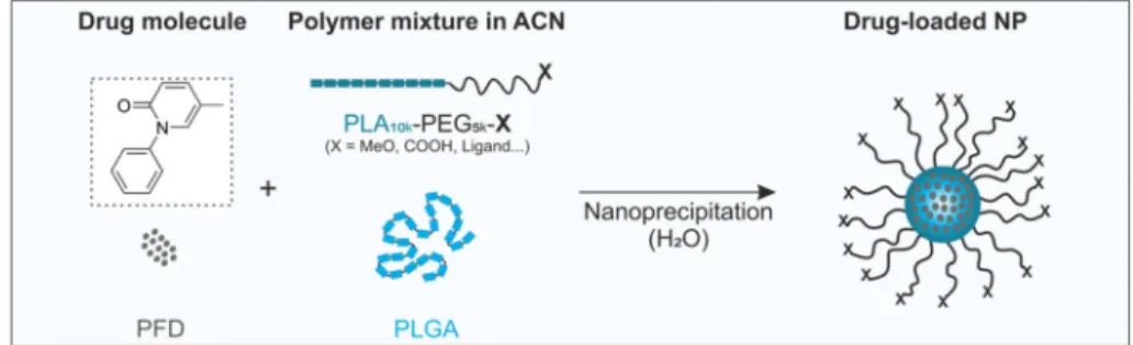
For our systematic investigation of the PFD encapsulation we relied on well-established poly(ethylene glycol)-poly(lactic acid) (PEG-PLA) block copolymer NPs (20) with a poly(lactic- coglycolic acid) (PLGA)-stabilized core (Fig.
1). Such particlesare known for their good biocompatibility and highly tunable composition. Additionally, by modifying the PEG end-groups with ligands the particles can be targeted to receptors on the cellular membrane to increase their specificity (21). To evalu- ate the suitability of our particle system for the encapsulation of PFD we firstly estimated the compatibility of PFD with the particle-forming polymers through the calculation of thermo- dynamic miscibility parameters. Then, we prepared the NPs through two different nanoprecipitation techniques, bulk and microfluidic (MF) manufacturing, and assessed the drug load- ing. More so, we investigated the influence of the two different approaches on the NP characteristics and their interaction at cellular level. Additionally, we evaluated the spatial constrain on drug loading by calculating the number of molecules that can fit in a single NP core and experimentally determined the influence of this parameter by preparing particles with larger cores. Finally, we investigated the effect of introducing an
Fig. 1 Encapsulation of PFD in block copolymer NPs. Particles are prepared through nanoprecipitation of organic mixtures of PEG-PLA, PLGA and the drug in aqueous medium.
additional drug molecule in the formulation,
α-lipoic acid(LA), on PFD encapsulation.
MATERIALS AND METHODS Materials
Methoxy and carboxylic acid end-functionalized PEG (MeO-PEG
5k-OH and COOH-PEG
5k-OH) with a molec- ular weight of 5000 Da were purchased from JenKem Technology USA Inc. (Allen, TX, USA). PFD was obtained from MedChem Express (Sollentuna, Sweden).
Dulbecco’s phosphate-buffered saline (DPBS) was pur- chased from Thermo Fisher Scientific (Waltham, MA, USA). LA, PLGA with a molecular weight of 13.4 kDa, and all other reagents and chemicals in analytical grade were purchased from Sigma Aldrich (Taufkirchen, Germany). Ultrapure water for particle preparation was obtained from a Millipore Milli-Q water purification sys- tem (Billerica, MA, USA).
Compatibility of Drug and Particle-Assembling Polymers
To predict the miscibility of particle-forming polymers with either PFD or LA, different thermodynamic param- eters were calculated. To that end, an ideal particle poly- mer distribution was assumed where PEG conforms the shell, PLGA the core, and PLA is situated at the core-shell interphase. The interference of concurring NP polymers in the miscibility was not taken into consideration. The total solubility parameters (δ) were obtained from litera- ture values for the polymers (PEG (24.0 MPa
1/2) (22), PLA (22.0 MPa
1/2) (22) and PLGA (22.3 MPa
1/2) (23)), and calculated for PFD and LA using the partial solubility parameters after Krevelen and Nijenhuis (24) determined by the group contributions methods using Eq.
1–3.δd,δp,
δhare the partial solubility parameters associated with the dispersion forces, the polar interactions and the hydrogen bonding components, respectively.
δd ¼∑Fdi
V ð1Þ
δp¼∑F2pi1=2
V ð2Þ
δh¼ ∑Ehi
V 1=2
ð3Þ
The estimation of the molecular volume of PFD (V = 119 cm
3) and LA (V = 144 cm
3) was done by the group
contribution methods after Fedors (25). The total solubility parameters were determined using Eq.
4(24).
δ2¼δ2d þδ2pþδ2h ð4Þ
The differences in solubility parameters (Δδ,
Δδd,Δδp,
Δδh) were calculated by subtracting the polymer’s from the drug’s solubility parameter. The mixing enthalpy cal- culated from the total- or partial solubility parameters (ΔH
MTand
ΔHM, respectively) was determined according to Eq.
5(26) and 6 (27), where
Φ1and
Φ2and
δ1and
δ2represent the volume fractions and the solubility parame- ters of the drug and polymer, respectively.
ΔHMT ¼ϕ1ϕ2 δdrug−δpolymer
2
ð5Þ ΔHM ¼ϕ1ϕ2 ðδd1−δd2Þ2þ δp1−δp2
2
h þðδh1−δh2Þ2i ð6Þ
The Flory-Huggins interaction parameter (χ
sp) was calcu- lated with the Hildebrand-Scatchard equation (Eq.
7) usingthe obtained total solubility parameters.
χsp¼ðδ1−δ2Þ2Vdrug
RT ð7Þ
Polymer Synthesis
Block copolymers (PEG
5k-PLA
10k) with different PEG-end functionalization (MeO, or COOH) were synthesized after Qian
et al.(28) with slight modifications as previously de- scribed by our group (29). In brief, for the ring opening poly- merization of cyclic 3,6-dimethyl-1,4-dioxane-2,5-dione (lac- tide), MeO-PEG
5k-OH and COOH-PEG
5k-OH were used as macroinitiators using 1,8-diazabicyclo[5.4.0]undec-7-ene (DBU) as a catalyst. As products, MeO-PEG
5k-PLA
10and COOH-PEG
5k-PLA
10kwere obtained, for non-targeted and targeted NP preparation, respectively (please refer to supple- mentary methods for ligand modification of the polymers).
Particle Preparation Via Bulk or Microfluidic Nanoprecipitation
Block copolymer NPs were prepared through nanoprecipita-
tion of PEG
5k-PLA
10kand PLGA
13.4kpolymer mixtures. To
that end, and if not indicated otherwise, PEG-PLA and PLGA
were mixed at a 70:30 mass ratio in acetonitrile (ACN) to a
final polymer concentration of 10 mg/mL (1 mL). For drug-
loaded particles, different amounts of PFD ranging from
25
μg to 10 mg (1–600-fold molar excess to PLGA) wereadded to the polymer mixture. Afterwards, for bulk nanopre-
cipitation, they were pipetted in stirring sterile filtrated
Millipore water (5 mL) at a 1:5 organic to aqueous phase ratio
to a polymer concentration of 2 mg/mL.
For MF manufacturing, the particles were prepared using the NanoAssemblr™ Benchtop (Precision NanoSystems Inc., Vancouver, Canada). Process parameters were controlled us- ing the NanoAssemblr™ controller software (v1.09). If not noted otherwise the particles were prepared at a total flow rate (TFR) of 2 mL/min and a flow rate ratio (FRR) of 1:5 organic to water phase. To investigate the effect on drug load- ing of the different microfluidic parameters, the TFR (2–
12 mL/min) and the FRR (1:1–1:10) were varied.
For both preparation techniques, immediately after prep- aration, particles were diluted to 20 mL with Millipore water and purified and concentrated through ultracentrifugation with a 30-kDa molecular weight cutoff Microsep advance centrifugal device (Pall corporation, NY, USA) at 959 x
gfor 20 min. Purification from free or adsorbed drug was per- formed through thoroughly washing the NPs with Millipore water and ultracentrifugation, as described above (2×).
Particle Characterization
The particle size was determined through dynamic light scat- tering (DLS) using a ZetaSizer Nano ZS. The device was equipped with a 633 nm He-Ne laser at a backscatter angle of 173° (Malvern Instruments GmbG, Lappersdorf, Germany).
Measurements were performed at 25°C using Millipore water as dispersing medium and a NP concentration of 1 mg/mL.
The measurement position was set at 4.65 mm and the data was recorded with Malvern Zetasizer software 7.11 (Malvern Instruments, Worcestershire, United Kingdom).
The particle PEG content was determined using a colori- metric iodine complexing assay (30) as previously described by our group (29).
The NP mass concentration was determined gravimetrical- ly after lyophilization and the PFD content was measured photometrically after particle disruption in ACN at 300 nm using a FLUOStar Omega microplate reader (BMG Labtech, Ortenberg, Germany). The NP molar concentration (PNC) was calculated using Eq.
8where
mis the NP mass determined gravimetrically after lyophilization,
ρNPis the density of the particles (1.25 g/cm
3) (31),
dhis the hydrody- namic diameter of the NPs determined through DLS,
NAis the Avogadro number, and
Vthe volume of the samples.
PNC¼ m
ρNP 4 3 π dh
2
3
x
1NAV ð8Þ
The encapsulation efficiency (EE) was determined using Eq.
9,
where
mEis the quantified mass of entrapped drug, and
mTis the total mass of drug added initially to the formulation.
EEð Þ ¼% mE
mT
x100 ð9Þ
The loading capacity (LC) was determined through Eq.
10,where M
Tis the total mass of the particle formulation.
LCð Þ ¼% mE
MT
x100 ð10Þ
The number of drug molecules per NP was calculated by dividing the molar concentration of the drug by the molar concentration of the NPs determined as described above.
Determination of the Spatial Restriction of PFD Loading
To determine the number of molecules that fit inside a NP, the volume of the particle core was estimated. To that end, the mean NP size was used as a starting point. For the calculation it was considered that the PLA is located at the core-shell interface and that the PLGA and PEG form the core and shell, respectively. To estimate the core size, first the confor- mation of the PEG on the particle surface was investigated using the Flory radius (R
F) (32). When the distance between polymer chains on a particle surface is smaller than the R
F, they assume an extended brush conformation. Otherwise, they take a folded mushroom configuration. The R
Fcan be calculated using Eq.
11, whereαis the length of a PEG mono- mer (0.278–0.358 nm) (33) and
Nis the number of monomers in one molecule (each monomer has the molecular weight of 44 g/mol).
RF ¼αN35 ð11Þ
The surface (S) that is taken by the polymers on the particle surface can be calculated after Gref
et al.(34) using Eq.
12.MPEG
represents the molecular weight of the PEG, and
fis the mass fraction of PEG in the PEG-PLA blends. The
Scan be used to determine the distance between polymers (D) on the particle surface using Eq.
13(34).
S¼ 6MPEG
dh NA f ρNP ð12Þ
D¼2 S π
1
2 ð13Þ
The length in nm of a PEG brush can be calculated by multiplying the monomer length
α= 0.35 nm by
N. This valuewas subtracted twice from the
dhof the NP to obtain the diameter of the particle core (d
c). The volume of the core (V
core) was calculated assuming a spherical shape after Eq.
14,where
rcis the calculated radius of the particle core (d
c/2).
Vcore¼4
3πr3c ð14Þ
The estimation of the molecular volume of PFD (V
drug= 0.2 nm3) was conducted using the group contribution methods
after Fedors (25). The number of drug molecules that are able to fit inside a single particle core were calculated through Eq.
15,
assuming the maximal packing efficiency of poly-sized spheres (90%) (35), a spherical molecule shape and an even drug distribution among all the particles.
Drug molecules per NP¼ Vcore
Vdrug
x0:9 ð15Þ
In order to prepare particles with increasing PLGA core sizes, the PLGA content of the formulations was increased.
Particles with a PEG-PLA to PLGA mass ratio of 70:30, 60:40, 50:50 and 40:60 were prepared to a total final polymer concentration of 10 mg/mL, as described above. NPs were prepared through bulk nanoprecipitation and an initial PFD mass of 25
μg, as described above.Co-Encapsulation of LA
For the co-encapsulation of LA and PFD, the NPs were pre- pared as described above. LA and PFD were simultaneously added to the organic polymer mixture prior to particle prep- aration. The PFD to LA molar ratio was varied to determine the ideal particle composition. Additionally, the initial PFD amount was set to 2 mg and LA in a molar excess (ranging from 0.1 to 3.5) was added to the different formulations. As a control, NPs only encapsulating LA at the same concentra- tions added to the PFD-containing formulations were pre- pared. As an additional control the molar excess of LA added was replaced by the same molar excess of PFD. The PFD or LA content in the formulations was determined photometri- cally after particle lyophilization and disruption in ACN at 300 or 334 nm, respectively, using a FLUOStar Omega microplate reader (BMG Labtech, Ortenberg, Germany).
The EE and number of drug molecules per particle was de- termined as described above.
In Vitro
Release Studies
The
in vitrorelease of PFD was assessed through the dialysis bag method. To that end, NPs containing PFD (NP-PFD), or PFD and LA (NP-PFD/LA) were prepared at a 70:30 PEG- PLA to PLGA mass ratio and purified as described above. An initial PFD and LA mass of 2 mg and 4.5 mg, respectively, were used. The samples were adjusted to a final volume of 2 mL (60 mg/mL NPs) and placed in a 3.5-5 kDa molecular weight cut off Spectrum™ Spectra/Por™ 3 RC Dialysis Membrane (Spectrum Laboratories, Inc., Rancho Dominguez, CA, USA). The dialysis membrane was intro- duced in a 35 mL glass vial containing 27 mL of DPBS (pH 7.3) under sink conditions. Vials were kept in a 37°C shaking water bath and 0.5 mL samples were taken at differ- ent time points and replaced with fresh pre-warmed buffer. As
a control, particles with no encapsulated drug (NPMeO), and free drug (PFD, 2.2 mg) were used. The PFD concentration was quantified at 300 nm using a FLUOStar Omega micro- plate reader (BMG Labtech, Ortenberg, Germany).
Statistical Analysis
Statistical analysis was performed using GraphPad Prism Software 6.0. Student’s t test (Fig.
3and Fig.
5a) was employedto evaluate statistical significance. Levels of statistical signifi- cance and
“n”numbers for each experiment are indicated in the text and figure legends.
RESULTS AND DISCUSSION
Thermodynamic Considerations for PFD Encapsulation
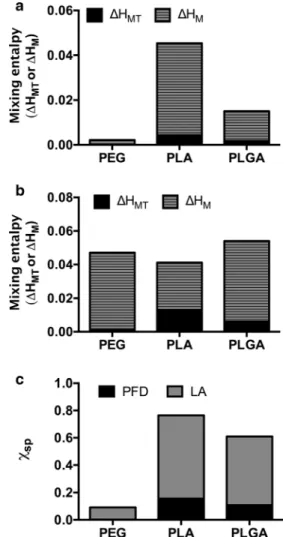
A frequent approach to estimate the compatibility of a drug in a polymer is the calculation of their thermodynamic interac- tions. This approach has been used by several authors to select the most suitable polymers for encapsulating a certain drug or retroactively explain experimental outcomes (26,27,36–38).
Here, we assessed the thermodynamic miscibility of PFD and LA with either PEG, PLA or PLGA to determine the suitability of our particle system for the selected drug(s). As depicted in Eq.
16, the thermodynamic miscibility of two sub-stances is given by the free energy of mixing (ΔG
M), where
ΔHMTand
ΔSMare the enthalpy and entropy of mixing, respectively. If the
ΔGMis negative, the two substances are considered mutually soluble.
ΔGM ¼ΔHMT−TΔSM ð16Þ
The mixing enthalpy (ΔH
M) per volume unit for each polymer-drug can be used as an indicator for the miscibility of two substances. It is given by Eq.
5after Hildebrand (26).
For its determination the volume fractions and the solubility parameters of the drug and polymer,
Φ1and
Φ2and
δ1and
δ2respectively, are considered.
The solubility parameters for some common drugs and polymers are reported in the literature (22,23). Otherwise they can be calculated after van Krevelen and Nijenhuis (24) using Eq.
4by combining the contribution of the partial solubility parameters associated with the dispersion forces (δ
d), the polar interactions (δ
p) and the hydrogen bonding components (δ
h).
The total- and partial solubility parameters of PFD, LA and the particle-forming polymers are depicted in Table
I.The miscibility of two components is promoted when their solubility parameters have similar values and thus,
ΔHMTtends to 0. Therefore, the differences among solubility param-
eters of drug and polymer (Δδ
,Δδd,Δδp, and
Δδh) can also be
used to predict miscibility (37) (Table
II).However, as Liu
et al.pointed out, Eq.
5does only take into consideration dispersion forces (27). As an alternative, they proposed the determination of
ΔHMtaking into account all forces of the interaction by using the partial solubility param- eters, as depicted in Eq.
6.The most frequently used indicator to predict the miscibil- ity of drug molecules with different polymers is the Flory- Huggins interaction parameter (χ
sp) (8). It is given by the Hildebrand-Scatchard equation (Eq.
7), which takes into ac-count the total solubility parameters (δ) of the drug and poly- mer in combination with the molar volume of the drug (V
drug).
A complete miscibility is achieved when
χspis <0.5 (24) which requires a very near match of both solubility parameters of drug and polymer. The closer the
χspis to zero, the more favorable interactions between the two components, and thus the better compatibility of drug and polymer.
With the determined solubility parameters (δ
,δd,δp, and
δh) (Table
I) and their differences (Δδ,Δδd,Δδp, and
Δδh) (Table
II), we calculated the mixing enthalpy (ΔHMTor
ΔHM), and
χspin order to predict the miscibility of our drugs with the particle system (Fig.
2).Comparing the total solubility parameters (δ) (Table
I), it isevident that all substances (drugs and polymers alike) have very similar values ranging between 22 and 25 MPa
1/2. This indicates a very good match for the selected drug and poly- mers, which is confirmed by the values of
ΔHMTand
ΔHMof the PFD- or LA-polymer mixtures (Fig.
2). As pointed out byLiu
et al.(27), the absolute values for
ΔHMTand
ΔHMdiffered.
Furthermore, for the case of LA, a complete opposite tenden- cy was seen (Fig.
2b). TheΔHMTpredicted a higher miscibility
for PEG > PLGA > PLA, whereas the
ΔHManticipated a better miscibility with PLA > PEG > PLGA. Nevertheless, the enthalpy values for all mixtures were very close to zero pre- dicting good miscibility from a thermodynamic point of view.
As the most common used parameter to predict drug- polymer compatibility is the
χsp, we also compared these val- ues for the polymers composing our NPs. As depicted in Fig.
2c, theχsp
values predict total miscibility of PFD with all the polymers forming the particle with a predilection to PEG >
PLGA > PLA. However, for LA it indicates low miscibility with PLGA and PLA, as
χsp< 0.5 only for PEG, disagreeing with the other estimated parameters. Overall, as suggested previously by other authors (27), the different parameters pre- dicting drug-polymer miscibility varied in terms of the poly- mer which showed the highest miscibility. Nevertheless, ex- cept the
χspfor the LA-PLA or LA-PLGA mixture, all the obtained values indicated thermodynamically favorable mis- cibility. In general, the highest miscibility for PFD is predicted to be with PEG, followed by PLGA and PLA (Fig.
S1Aand
Table I Partial and Total Solubility Parameters for the Drugs (PFD and LA)and the Particle-Assembling Polymers
Component δd δp δh δ
PFD* 20.5 9.4 7.7 23.8
LA* 22.9 2.9 10.2 25.2
PEG#,(22) 20.3 9.6 6.0 24.0
PLA#,(22) 19.8 4.0 6.7 22.0
PLGA#,(23) 17.4 9.1 10.5 22.3
Obtained from literature#, calculated*
Table II Calculated Differences Between the Solubility Parameters or Polarity of PFD or LA and the Particle-Forming Polymers
Particle component PFD LA
Δδd Δδp Δδh Δδt Δδd Δδp Δδh Δδt
PEG 0.2 - 0.2 1.7 - 0.2 2.6 - 6.7 4,2 1.2
PLA 0.7 5.4 1.0 1.8 3.1 - 1.1 3.5 3.2
PLGA 3.1 0.3 - 2.8 1.5 5.5 - 6.2 - 0.3 2.9
Fig. 2 Prediction of the drug-polymer miscibility. Mixing enthalpy of the particle-assembling polymers and (a) PFD or (b) LA. (c) Flory-Huggins inter- action parameter.
Table
SI). For LA the same trend in miscibility is to beexpected (Fig.
S1Band Table
SI).As all determined parameters unanimously predict that PFD is very compatible with all the particle-forming polymers, we determined that the block copolymer PEG-PLA/PLGA particle system is an appropriate candidate for its encapsulation.
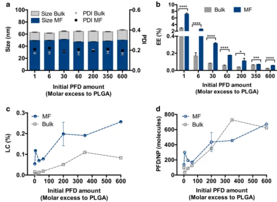
Influence of the Nanoprecipitation Technique on PFD Loading
In order to experimentally investigate the encapsulation of PFD in PEG-PLA/PLGA block copolymer NPs, we evaluat- ed two different manufacturing techniques, bulk or MF nano- precipitation (Fig.
3). To that end, PFD-polymer mixtures inACN were precipitated into a water phase, either manually or with the assistance of a microfluidic device. The initial PFD addition was varied to determine its influence on the drug loading. The resulting particles were homogenous in terms of size with narrow polydispersity indexes (PDI) (Fig.
3a) inde-pendent of the amount of drug added to the formulation. As frequently observed, MF manufacturing rendered particles about 15 nm smaller (~ 50 nm) compared to bulk nanopreci- pitated particles (~ 65 nm). This is due to the fact that the MF mixing is faster than the time the particles need to nucleate, which does not occur during bulk nanoprecipitation (39).
For both methods an increase in initial drug concentrations reduced the EE (Fig.
3b), which can be partially explained by anincrease in mixing enthalpy with increasing volume fractions of
drug (Fig.
S2). Interestingly, MF manufacturing achieved signif-icantly higher EE, a phenomenon also described by other authors (40,41). As an additional advantage, MF preparation allows for a precise control over the manufacturing parameters, such as the TFR (Fig.
S3) and the FRR (Fig.S4) which, in turn,permit drug loading optimization and a decrease in batch to batch variations. Furthermore, the resulting particles have the ideal size (~ 50 nm) for optimal NP-cell interaction (42) (Fig.
S5).Despite the decrease in efficiency of the process with higher PFD amounts, the LC (Fig.
3c) and number of drug moleculesper particle (Fig.
3d) increased with larger initial PFD addi-tions. This is frequent for particles entrapping the drug during NP preparation (39), and is probably promoted in this partic- ular case by the water solubility of PFD (~ 2 mg/mL).
Interestingly, both methods achieved a comparable number of PFD molecules per particle, which is probably due to a larger number of smaller particles being produced by MF compared to bulk nanoprecipitation.
However, the low number of drug molecules that were being incorporated into the particle system (under 1000 molecules per NP) was quite surprising, considering the high predicted miscibility of PFD with the particle- forming polymers (Fig.
2). By those calculations, PFD dis-played a very high compatibility with PEG. Therefore, part of the drug may end up in the particle’s shell.
However, during NP purification, this non-core-bound drug is washed out through the several purification steps, promoted by the water solubility of PFD, causing a lower than expected EE.
Fig. 3 PFD encapsulation in block copolymer NPs through bulk and MF nanoprecipitation. Particle characterization in terms of (a) size and PDI, (b) Encapsulation Efficiency (EE) (c) Loading Capacity (LC) and (d) number of PFD mol- ecules per particle. Results are shown as mean ± SD of at least n= 3 measurements. Levels of statistical significance are indicated as
*p≤0.005, ***p≤0.001, and
****p≤0.0001.
Overall, we could demonstrate that PFD encapsulation in block copolymer NPs is feasible by nanoprecipitation.
Comparing both manufacturing techniques, it is apparent that MF is superior. Not only did it lead to a significantly higher EE than bulk nanoprecipitation, but this could be fur- ther enhanced up to 40% by adjusting the manufacturing parameters (Fig.
S3and
S4). More so, a higher cellular uptakewas achieved by MF-nanoprecipitated particles (Fig.
S5)which is highly relevant for
in vitroand
in vivoapplications.
Spatial Limits on the Drug Encapsulation
Due to the low number of PFD molecules being incorporated in our particles (Fig.
3), despite the high predicted miscibilitywith the selected polymers, we decided to estimate the number of drug molecules that would actually fit inside a single par- ticle’s core (Fig.
4). For our determination we considered aparticle structure as depicted in Fig.
4a, where the PEG com-poses the shell, the PLGA forms the particle core, and the PLA is located at the core-shell interface. Given that after NP man- ufacturing, the particles are purified from drug that is free or entangled in the PEG shell through various washing and cen- trifugation steps, we considered that the particle-bound drug would mostly be located in the PLGA core. We speculated that the spatial constriction given by the core size would de- termine the number of PFD molecules that can be fitted inside the particle.
To determine the size of the core, the length of the PEG brush was calculated and subtracted from the overall particle diameter (~ 65 nm). To that end, we firstly determined the conformation of the PEG shell on the particle corona. For our NPs prepared at a 70:30 PEG-PLA to PLGA mass ratio, the PEG assumes an extended brush conformation. This corre- sponds to a length of approximatively 30 nm (for a PEG with a molecular weight of ~ 4800 Da). This would leave a particle core of about 5 nm in diameter, which would allow for the encapsulation of about 200 PFD molecules per NP, assuming spherical shapes and 90% packing efficiency. It needs to be noted that this calculation does not take into consideration the space occupied by the core-forming PLGA itself. However, the value is in the same size-range of that was experimentally determined (Fig.
3).Therefore, we hypothesized that the number of encapsu- lated molecules per particle could be improved by increasing the size of the PLGA core. To confirm these assumptions, particles with an increase in PLGA mass ratio were prepared.
From the initial PEG-PLA to PLGA 70:30 mass ratio, we raised the PLGA fraction to 60:40, 50:50 and 60:60. As expected, the NPs increase in size in a linear manner from 60 to 120 nm (Fig.
4b). However, for all the formulations thePEG assumed a brush conformation (Table
SII), indicatingthat the size increase was core-derived (Fig.
S6). More impor-tantly, the number of PFD molecules per particle increased
exponentially (Fig.
4c), therefore confirming our theory thatthere is a spatial restriction for drug loading in a particle.
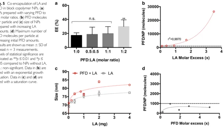
Co-Encapsulation of LA and PFD in Block Copolymer NPs
Even though increasing the size of the PLGA core dramati- cally enhanced the number of PFD molecules per NP, for certain applications this may not be a feasible option. More so, the number of PFD molecules per NP did not reach the extent that would be expected from the linear size increase (Fig.
S7). This can be explained, on the one hand, by thevolume that the PLGA takes up in the core by itself and which
Fig. 4 Core volume-dependent PFD encapsulation. (a) structure of a block copolymer NP. d(NP): particle diameter; r(PEG): PEG-brush radius; d(core):particle core diameter. (b) NP size and (c) PFD molecules per NP with increasing PLGA mass fraction. Results are shown as mean ± SD of at least n = 3 measurements. Data in (b) and (c) are fitted with a linear and an exponential growth equation, respectively.
was not considered for the calculations. On the other hand, the high affinity of PFD to PEG may limit its core localization and promote a partial distribution among the NP shell. This PEG-associated PFD is removed during particle purification due to water solubility of the drug (2 mg/mL). However, a PEG shell is highly necessary for the
in vivoapplications of NPs, as it reduces protein adsorption (29,34) and increases their blood residence (43).
As an alternative approach, we hypothesized that inclusion of an additional drug molecule, also compatible with PEG but high- ly water insoluble, would increase the number of PFD molecules per NP by limiting the PFD diffusion to the aqueous phase.
As a candidate we selected LA. It has a very poor water solubility (0.2 mg/mL), which is 10-times lower than for PFD (2 mg/mL). Additionally, as determined by the miscibility prediction (Fig.
2), it is compatible with the particle formingpolymers, especially with PEG. More so, at a therapeutic level it may show complementary effects to PFD as they both dem- onstrate positive antioxidative effects in doxorubicin-induced cardiac and renal toxicity (44) and oxidative liver damage (45).
To test our hypothesis, we prepared NPs encapsulating both PFD and LA (Fig.
5). Initially we combined different PFD andLA ratios and examined the EE. As depicted in Fig.
5a, a LAmolar excess significantly increased the EE from 20% to 40%.
We then maintained a constant PFD concentration and varied the formulation’s LA content. Interestingly, an exponential increase/decrease in PFD molecules per NP was observed as LA content was increased/lowered (up to 25,000 PFD mole- cules/NP) (Fig.
5b). When comparing the size of particles onlyincorporating LA with the ones encapsulating LA and PFD, there was only a 10 nm size difference (Fig.
5c). Furthermore,the number of PFD molecules per NP reached with increasing LA addition could not be achieved by just increasing the initial PFD added to the formulations (Fig.
5d), which reached a max-imum of about 1000 molecules per particle (dashed line in Fig.
5b
and
d). This rising number of PFD molecules per NP can beexplained by a decrease in mixing enthalpy with increasing amounts of LA, which may promote the miscibility of PFD with the particle-forming polymers (Fig.
S8). Additionally, the lowerwater solubility of LA and its interaction with PFD may promote a stronger particle binding of the latter. This is reinforced by the fact that the single encapsulation of LA, reaches a higher number of molecules per NP (Fig.
S9) compared to the individual PFDencapsulation.
Fig. 5 Co-encapsulation of LA and PFD in block copolymer NPs. (a) NPs prepared with varying PFD to LA molar ratios. (b) PFD molecules per particle and (c) size of NPs prepared with increasing LA amounts. (d) Maximum number of PFD molecules per particle at increasing initial PFD amounts.
Results are shown as mean ± SD of at least n = 3 measurements.
Levels of statistical significance are indicated as **p≤0.01 and *p≤ 0.05 compared to NPs without LA, n.s.: non-significant. Data in (b) are fitted with an exponential growth equation. Data in (c) and (d) are fitted with a saturation curve.
Fig. 6 Release profile of PFD from block copolymer NPs. PFD: Free drug;
NP-PFD: PFD-loaded NPs; NP-PFD/LA: PFD- and LA-loaded NPs;
NPMeO: control, drug-free NPs. Results are shown as mean ± SD of at least n= 3 measurements.
However, the co-encapsulation of an additional drug mole- cule can raise concerns about its interference with the release profile of the original drug. Therefore, we investigated the PFD
in vitrorelease from the different NP formulations using the dial- ysis bag method. To that end, NPs containing PFD (NP-PFD) or PFD and LA (NP-PFD/LA) were compared. As controls, the release profiles under the same conditions of drug-free particles (NPMeO) and free drug (PFD) were evaluated. As depicted in Fig.
6, after 1 h all of the free-drug was released from the dialysismembrane. At that time point, about 70% of the PFD was released from the NPs. The remaining 30% of the particle- bound drug was released very slowly over the course of the next 5 h. This fast release is probably due to the large surface area to volume ratio resulting from the small particle size. Additionally, the water-solubility of PFD may boost this effect. However, these results are in line with the release of other hydrophilic drugs prepared through nanoprecipitation, such as procaine hydro- chloride from PLGA NPs (4). Comparing the release profiles of NP-PFD and NP-PFD/LA it becomes apparent that both par- ticle formulations show the same release curve. Therefore, we can conclude that the LA incorporation has no detrimental effects on the PFD release (Fig.
6).CONCLUSION
In this study we systematically examined the encapsulation of PFD, a water-soluble drug lacking ionizable groups, in block copolymer NPs through nanoprecipitation. We determined dif- ferent thermodynamic parameters which predicted a good mis- cibility of the drug with the particle-forming polymers (PEG, PLA and PLGA) (Fig.
2). We showed that the calculation ofdifferent parameters yielded slight differences among their pre- dictions (Fig.
S1), which is in agreement with previous studies.More so, even though useful for selecting appropriate polymers for the drug encapsulation, they cannot be exclusively consid- ered. We demonstrate that spatial constrictions regarding the particle’s core size can also limit the number of molecules that can be encapsulated in polymer NPs (Fig.
4) and by modulatingthem the number of encapsulated molecules can be increased.
We experimentally assessed the EE, LC and number of encap- sulated drug molecules per particle using two common nano- precipitation techniques (Fig.
3). Among them, we were able todemonstrate that MF manufacturing achieves significantly higher drug loading with the additional benefit of precise ad- justment of the process parameters (Fig.
S3and
S4) and en-hanced NP-cell interactions (Fig.
S5).Lastly, we demonstrated how the encapsulation of an ad- ditional drug, LA, with similar polymer miscibility but lower water solubility is able to exponentially increase the number of PFD molecules per NP (Fig.
5) without affecting the drug’srelease profile (Fig.
6).Overall our study demonstrates that the encapsulation of a water-soluble drug without ionizable groups is feasible when taking into consideration and adjusting the limits due to size constrictions and appropriately selecting the manufacturing method (MF vs bulk nanoprecipitation). More so, by consid- ering the co-encapsulation of an additional appropriate mol- ecule an additional increase in drug loading can be achieved.
ACKNOWLEDGMENTS AND DISCLOSURES
We thank Viktoria Eismann for her excellent technical assistance.
Financial support from the German Research Foundation (DFG), Grant GO 565/17-3, is gratefully acknowledged.
FUNDING INFORMATION
Open Access funding provided by Projekt DEAL.
Open Access This article is licensed under a Creative Commons Attribution 4.0 International License, which per- mits use, sharing, adaptation, distribution and reproduction in any medium or format, as long as you give appropriate credit to the original author(s) and the source, provide a link to the Creative Commons licence, and indicate if changes were made. The images or other third party material in this article are included in the article's Creative Commons licence, unless indicated otherwise in a credit line to the material. If material is not included in the article's Creative Commons licence and your intended use is not permitted by statutory regulation or exceeds the permitted use, you will need to obtain permission directly from the copyright holder. To view a copy of this licence, visit
http://creativecommons.org/licenses/by/4.0/.REFERENCES
1. Fessi H, Puisieux F, Devissaguet JP, Ammoury N, Benita S.
Nanocapsule formation by interfacial polymer deposition following solvent displacement. Int J Pharm. 1989;55:1–4.
2. Almoustafa HA, Alshawsh MA, Chik Z. Technical aspects of prepar- ing PEG-PLGA nanoparticles as carrier for chemotherapeutic agents by nanoprecipitation method. Int J Pharm. 2017;533:275–84.
3. Barichello JM, Morishita M, Takayama K, Nagai T. Encapsulation of hydrophilic and lipophilic drugs in PLGA nanoparticles by the Nanoprecipitation method. Drug Dev Ind Pharm. 1999;25:471–6.
4. Govender T, Stolnik S, Garnett MC, Illum L, Davis SS. PLGA nanoparticles prepared by nanoprecipitation: drug loading and re- lease studies of a water soluble drug. J Control Release. 1999;57:
171–85.
5. Peltonen L, Aitta J, Hyvönen S, Karjalainen M, Hirvonen J.
Improved entrapment efficiency of hydrophilic drug substance dur- ing Nanoprecipitation of poly(l)lactide nanoparticles. AAPS PharmSciTech. 2004;5.
6. Wang H, Xie H, Wu J, Wei X, Zhou L, Xu X,et al.Structure-based rational Design of Prodrugs to enable their combination with poly- meric nanoparticle delivery platforms for enhanced antitumor effi- cacy. Angew Chem Int Ed. 2014;53:11532–7.
7. Yang Q, Tan L, He C, Liu B, Xu Y, Zhu Z,et al.Redox-responsive micelles self-assembled from dynamic covalent block copolymers for intracellular drug delivery. Acta Biomater. 2015;17:193–200.
8. Thakral S, Thakral NK. Prediction of drug-polymer miscibility through the use of solubility parameter based Flory-Huggins inter- action parameter and the experimental validation: PEG as model polymer. J Pharm Sci. 2013;101:2271–80.
9. Kim ES, Keating GM. Pirfenidone: a review of its use in idiopathic pulmonary fibrosis. Drugs. 2015;75:219–30.
10. RamachandraRao SP, Zhu Y, Ravasi T, McGowan TA, Toh I, Dunn SR,et al.Pirfenidone is Renoprotective in diabetic kidney disease. J Am Soc Nephrol. 2009;20:1765–75.
11. Sharma K, Ix JH, Mathew AV, Cho M, Pflueger A, Dunn SR,et al.
Pirfenidone for diabetic nephropathy. J Am Soc Nephrol. 2011;22:
1144–51.
12. Cho ME, Smith DC, Branton MH, Penzak SR, Kopp JB.
Pirfenidone slows renal function decline in patients with focal seg- mental glomerulosclerosis. Clin J Am Soc Nephrol. 2007;2:906–13.
13. George PM, Wells AU. Pirfenidone for the treatment of idiopathic pulmonary fibrosis. Expert Rev Clin Pharmacol. 2017;10:483–91.
14. Bonella F, Wessendorf T, Costabel U. Clinical experience with pirfenidone for the treatment of idiopathic pulmonary fibrosis.
Eur Respir J. 2013;42.
15. Trivedi R, Redente EF, Thakur A, Riches DWH, Kompella UB.
Local delivery of biodegradable pirfenidone nanoparticles amelio- rate s ble om ycin-induced pulmonary fibros is in mice.
Nanotechnology. 2012;23:505101.
16. Chowdhury S, Guha R, Trivedi R, Kompella UB, Konar A, Hazra S. Pirfenidone nanoparticles improve corneal wound healing and prevent scarring following alkali burn. PLoS One. 2013;8:e70528.
17. Abnoos M, Mohseni M, Mousavi SAJ, Ashtari K, Ilka R, Mehravi B. Chitosan-alginate nano-carrier for transdermal delivery of pirfe- nidone in idiopathic pulmonary fibrosis. Int J Biol Macromol.
2018;118:1319–25.
18. Silva RO, da Costa BL, da Silva FR, da Silva CN, de Paiva MB, Dourado LFN,et al.Treatment for chemical burning using liquid crystalline nanoparticles as an ophthalmic delivery system for pirfe- nidone. Int J Pharm. 2019;568:118466.
19. Vauthier C, Bouchemal K. Processing and scale-up of polymeric nanoparticles. Pharm Res. 2008;36:1025–58.
20. Xiao RZ, Zeng ZW, Zhou GL, Wang JJ, Li FZ, Wang AM. Recent advances in PEG-PLA block copolymer nanoparticles. Int J Nanomedicine. 2010;5:1057–65.
21. Maslanka Figueroa S, Veser A, Abstiens K, Fleischmann D, Beck S, Goepferich A. Influenza a virus mimetic nanoparticles trigger se- lective cell uptake. Proc Natl Acad Sci. 2019;116:9831–6.
22. Adamska K, Voelkel A, Berlińska A. The solubility parameter for biomedical polymers—application of inverse gas chromatography.
J Pharm Biomed Anal. 2016;127:202–6.
23. Schenderlein S, Lück M, Müller BW. Partial solubility parameters of poly(D,L-lactide-co-glycolide). Int J Pharm. 2004;286:19–26.
24. van Krevelen DW, te Nijenhuis K. Properties of polymers. Their correlation with chemical structure; their numerical estimation and prediction from additive group contributions. Endeavour. 2009.
https://doi.org/10.1016/0160-9327(92)90023-i.
25. Fedors RF. A method for estimating both the solubility parameters and molar volumes of liquids. Polym Eng Sci. 1974;14:147–54.
26. Li Y, Taulier N, Rauth AM, Wu XY. Screening of lipid carriers and characterization of drug-polymer-lipid interactions for the rational Design of Polymer-Lipid Hybrid Nanoparticles (PLN).
Pharm Res. 2006;23:1877–87.
27. Liu J, Xiao Y, Allen C. Polymer–drug compatibility: a guide to the development of delivery systems for the anticancer agent, ellipti- cine. J Pharm Sci. 2004;93:132–43.
28. Qian H, Wohl AR, Crow JT, Macosko CW, Hoye TR. A strategy for control of“random”copolymerization of Lactide and Glycolide: appli- cation to synthesis of PEG- b -PLGA block polymers having narrow Dispersity. Macromolecules. 2011;44:7132–40.
29. Abstiens K, Maslanka Figueroa S, Gregoritza M, Goepferich AM.
Interaction of functionalized nanoparticles with serum proteins and its impact on colloidal stability and cargo leaching. Soft Matter.
2019;15:709–20.
30. Childs CE. The determination of polyethylene glycol in gamma globulin solutions. Microchem J. 1975;20:190–2.
31. Rabanel J-M, Faivre J, Tehrani SF, Lalloz A, Hildgen P, Banquy X. Effect of the polymer architecture on the structural and biophys- ical properties of PEG–PLA nanoparticles. ACS Appl Mater Interfaces. 2015;7:10374–85.
32. de Gennes PG. Polymers at an interface; a simplified view. Adv Colloid Interf Sci. 1987;27:189–209.
33. Kreuzer HJ, Wang RLC, Grunze M. Single molecule force spec- troscopy by AFM indicates helical structure of poly(ethylene-glycol) in water related content effect of stretching on the molecular con- formation of oligo (ethylene oxide): a theoretical study stretching of single polymer strands. New J Phys. 1999;1:6.1–6.11.
34. Gref R, Lück M, Quellec P, Marchand M, Dellacherie E, Harnisch S,et al.‘Stealth’corona-core nanoparticles surface modified by polyethylene glycol (PEG): influences of the corona (PEG chain length and surface density) and of the core composition on phago- cytic uptake and plasma protein adsorption. Colloids Surf B:
Biointerfaces. 2000;18:301–13.
35. Kansal AR, Torquato S, Stillinger FH. Computer generation of dense polydisperse sphere packings. 2002;https://doi.org/10.
1063/1.1511510.
36. Hancock BC, York P, Rowe RC. The use of solubility parameters in pharmaceutical dosage form design. Int J Pharm. 1997;148:1–21.
37. Greenhalgh DJ, Williams AC, Timmins P, York P. Solubility parameters as predictors of miscibility in solid dispersions. J Pharm Sci. 1999;88:1182–90.
38. Latere Dwan’Isa JP, Rouxhet L, Préat V, Brewster ME, Ariën A.
Prediction of drug solubility in amphiphilic di-block copolymer micelles:
the role of polymer-drug compatibility. Pharmazie. 2007;62:499–504.
39. Kamaly N, Xiao Z, Valencia PM, Radovic-Moreno AF, Farokhzad OC. Targeted polymeric therapeutic nanoparticles: design, devel- opment and clinical translation. Chem Soc Rev. 2012;41:2971.
40. Liu Z, Fontana F, Python A, Hirvonen JT, Santos HA.
Microfluidics for production of particles: mechanism, methodolo- gy, and applications. Small. 2019;1904673.
41. Karnik R, Gu F, Basto P, Cannizzaro C, Dean L, Kyei-Manu W,et al.
Microfluidic platform for controlled synthesis of polymeric nanopar- ticles. Nano Lett. 2008.https://doi.org/10.1021/nl801736q.
42. Jiang W, Kim BYS, Rutka JT, Chan WCW. Nanoparticle- mediated cellular response is size-dependent. Nat Nanotechnol.
2008;3:145–50.
43. Klibanov AL, Maruyama K, Torchilin VP, Huang L. Amphipathic polyethyleneglycols effectively prolong the circulation time of lip- osomes. FEBS Lett. 1990;268:235–7.
44. Giri SN, Ali Al-Bayati M, Du X, Schelegle E, Mohr FC, Margolin SB.
Amelioration of doxorubicin-induced cardiac and renal toxicity by pirfenidone in rats. Cancer Chemother Pharmacol. 2004;53:141–50.
45. Macias-Barragan J, Caligiuri A, García-Banuelos J, Parola M, Pinzani M, Armendariz-Borunda J. Effects of alpha lipoic acid and pirfenidone on liver cells antioxidant modulation against oxi- dative damage. Rev Med Chil. 2014;142:1553–64.
Publisher’s Note