Elucidating the molecular function of CLUH in mammalian system
Inaugural-Dissertation
zur
Erlangung des Doktorgrades
der Mathematisch-Naturwissenschaftlichen Fakultät der Universität zu Köln
vorgelegt von Jie (Jane) Gao
aus China
Köln 2015
Berichterstatter
Prof. Dr. Elena Rugarli
Prof. Dr. Aleksandra Trifunovic
Tag der Mündlichen Prüfung: 04 May 2015
To my dear father, mother and husband
致我亲爱的父母与丈夫Table of Contents
Abstract ... 7
1 Introduction ... 8
1.1 Mitochondria ... 9
1.1.1 Origin of mitochondria ... 9
1.1.2 Mitochondrial genome ... 9
1.1.3 Brief introduction of the mitochondrial function ... 11
1.2 Biogenesis of nuclear encoded mitochondrial proteins ... 15
1.2.1 Coordination between nucleus and mitochondria to ensure appropriate protein expression ... 15
1.2.2 Regulation of nuclear encoded mitochondrial proteins ... 17
1.2.3 Mitochondrial post-translational versus co-translational protein targeting and import. ... 17
1.2.4 The targeting and sorting sequence of mitochondrial proteins ... 19
1.2.5 TOM and the sorting machinery ... 21
1.3 mRNA localisation ... 22
1.3.1 mRNA localisation is an ubiquitous phenomenon ... 22
1.3.2 mRNA localisation to mitochondria... 25
1.3.3 PUF3p ... 26
1.4 CLUH ... 28
1.4.1 Clu mutant leads to mitochondrial clustering and abnormal mitochondrial morphology ... 28
1.4.2 Clu mutants of multicellular organism display growth defects ... 30
1.4.3. Primary structure and the molecular function of Clu ... 32
2 Aim of the Thesis ... 36
3 Material and Method ... 37
3.1 Cell biology ... 38
3.1.2 Cell culture ... 38
3.1.3 Long-term galactose experiment ... 38
3.1.4 Short-term galactose and acetoacetate treatment ... 39
3.1.5 Bezafibrate treatment ... 39
3.1.6 RNA interference ... 40
3.1.7 Immunofluorescence and imaging ... 40
3.2 Molecular biology ... 42
3.2.1 Polymerase chain reaction (PCR) and agarose gel electrophoresis ... 42
3.2.2 Gel extraction, DNA extraction, DNA digestion and ligation ... 43
3.2.3 Bacterial transformation and colony selection ... 43
3.2.4 Generation of CLUH-FLAG and CLUH deletion HEK 293T cell lines ... 44
3.2.5 Crosslinking immunoprecipitation (CLIP) and RNA immunoprecipitation ... 45
3.2.6 RNA extraction ... 46
3.2.7 Reverse transcription and quantitative real-time PCR ... 46
3.2.8 RNA sequencing and analysis ... 48
3.3 Biochemistry ... 49
3.3.1 Protein extraction for western blot analysis ... 49
3.3.2 Protein quantification and preparation ... 49
3.3.3 SDS-PAGE, western blot analysis and quantification ... 50
3.3.4 Cell fractionation ... 51
3.3.5 Sucrose gradient and polysome profiling ... 52
3.3.6 Metabolic labeling and biotinylation of the newly synthesised proteins... 53
3.4 Obtaining mouse tissues ... 53
3.5 Statistical analysis ... 54
4 Results ... 55
4.1 CLUH is coregulated with genes encoding mitochondrial proteins and involved in translation ... 56
4.2 CLUH is an RNA binding protein ... 57
4.3 CLUH preferentially binds to mRNAs encoded in the nucleus for mitochondrial proteins ... 59
4.4 Global analysis of the mRNAs bounds by CLUH ... 61
4.5 The mRNA of the cytosolic protein astrin is bound by CLUH ... 71
4.6 CLUH affects the protein level of its mRNA targets ... 71
4.7 CLUH overexpression upregulates the protein level of some of its targets ... 73
4.8 Global cytosolic translation is unaffected in the absence of CLUH ... 74
4.9 Translation of CLUH targets is affected ... 77
4.10 Localisation of CLUH ... 79
4.11 CLUH level in response to galactose and acetoacetate treatment ... 83
4.12 CLUH protein level is reduced in PGC-1α deficient MEFs ... 88
4.13 CLUH levels in mouse liver during development and aging ... 90
4.14 Identification of the function of CLUH protein domains ... 92
5 Discussion ... 100
5.1 Elucidation of the molecular function of CLUH unravels the cause of Clu mutant phenotypes. ... 101
5.2 Identification of the molecular function of CLUH could be a support for co-translational import ... 106
5.3 CLUH affects the levels of protein encoded by target mRNAs ... 107
5.4 Regulation of CLUH ... 109
5.4.1 Effect of galactose treatment on CLUH ... 109
5.4.2 CLUH level during acetoacetate ... 112
5.4.3 CLUHs level during different life stages in mouse liver ... 113
5.5 Conclusions ... 114
Zusammenfassung ... 115
Reference ... 117
Appendix ... 126
List of Abbreviations ... 138
Acknowledgement ... 139
Eidesstattliche Erklärung... 141
Curriculum Vitae ... 142
Abstract
The mitochondrial proteome is composed of both mitochondrial and nuclear encoded proteins. Restructuration of mitochondrial proteome is often required upon on change in nutrient availabilities and stress conditions. To facilitate this process, the nuclear and mitochondrial genome must coordinate their protein expression in a well-concerted manner. Additional factors are present to mediate the coordination. These factors are diverse in function ranging from energy sensing to translation of the specific nuclear encoded mitochondrial proteins.
Clu is a well-conserved cytosolic protein, however its molecular function remains unknown. A small fraction of Clu is observed outside of mitochondria in Drosophila.
Clu mutant of multicellular organisms display phenotypes of growth defect, failure to respond to stress and early death. However these phenotypes are absent from single cellular organisms. Nevertheless, all Clu mutants have clustered mitochondria and this leads to the speculation that CLUH and mitochondria are functionally closely related.
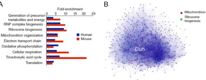
In this study, we analysed the genes that coregulate with human and murine
CLUH/Cluh, respectively, and identified two main clusters of genes: nuclear encoded
mitochondrial genes and cytosolic translation related genes. After this, we identified the
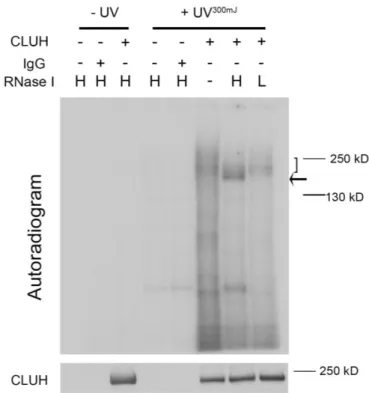
molecular function of human CLUH as an mRNA binding protein by using crosslinking
immunoprecipitation (CLIP). By comparing the mRNAs enriched by CLUH to control
IgG, we discovered that CLUH binds specifically to mRNAs of nuclear encoded
mitochondrial proteins (NEMP). Among the mRNAs bound by CLUH, we found
enrichments for genes involved in branched-chain amino acid degradation,
mitochondrial dysfunction, TCA cycle, oxidative phosphorylation and fatty acid α-
oxidation. Subsequently, we showed that CLUH is a positive translation regulator of its
target mRNAs, because in the absence of CLUH, translation of its target mRNAs
appeared to be reduced. However, general translation was not affected in the absence of
CLUH. Furthermore, we found a reduction of CLUH in PGC-1α null mouse embryonic
fibroblasts (MEFs), which indicates that CLUH expression might be under the
transcriptional control of PGC-1α. Together, we conclude that CLUH might be
important for the biogenesis of its target proteins under influence of cellular metabolism.
I n t r o d u c t i o n | 8
1 Introduction
I n t r o d u c t i o n | 9
1.1 Mitochondria
1.1.1 Origin of mitochondria
Mitochondria were first observed and described with various names as early as the 1800s, when the German histologist Richard Altmann proposed that this cellular granule to be autonomous and forming colonies in the cytoplasm in his book “Die Elementarorganismen”. It took another seven decades to discover the existence of DNA in mitochondria (mtDNA) (Nass and Nass 1963) and another two decades to find the evidence that mtDNA is not originated from the host genome via invagination during evolution (Farrelly and Butow 1983). Altogether, these evidences strongly support the endosymbiotic theory, which explains that mitochondria arose as a result of either a singular or multiple events of fusion of α-proteobacterium and eukaryotic progenitor (Macklin 1927). To many respects, these original endosymbiotic events mark the rise of the eukaryotes since a nucleus-bearing living eukaryotes completely devoid of mitochondria has yet to be discovered (Williams, Hirt et al. 2002; Chan, Slotboom et al.
2005; van der Giezen and Tovar 2005). This cooperation allowed the expansion of eukaryotes due to the acquisition of the super energy-producing “plant”. Along the course of evolution, mitochondria have lost and transferred most of their genes to the nucleus (Adams and Palmer 2003) and mitochondria only retain a skeletal genome, relatively small compared to the bacterial ancestor and encoding only components of the mitochondrial translation machinery and of the oxidative phosphorylation (OXPHOS).
1.1.2 Mitochondrial genome
In human, the circular mtDNA is composed of only 16,569 base pairs. Despite the
comparatively small size of this genome (only 1/3000th of the size of our smallest
chromosome 21), mutation, deletion and depletion of mtDNA can lead to severe
diseases. Human mitochondrial genome encodes for 37 genes including 22 tRNAs, two
rRNAs and 13 polypeptides. The mtDNA is a double stranded molecule with cytosine-
rich light strand and a guanine-rich heavy strand (Anderson, Bankier et al. 1981).
I n t r o d u c t i o n | 10
Unlike the nuclear genome, multiple copies of mtDNAs can be found per mitochondrion as a result of regulated replication and this condition is referred as polyploidy. Despite the existence of DNA repair systems in plant and mammalian mitochondria (Boesch, Ibrahim et al. 2009; Boesch, Ibrahim et al. 2010), high rate of mutations still occurs and leads to heteroplasmy (Dianov, Souza-Pinto et al. 2001). In such case, mitochondria will host a heterogeneous mix of healthy and mutated genomes, the specific threshold at which mutated genome exceeds the wildtype genome can determine the onset of mitochondrial dysfunction (Wong 2007). Since mtDNAs are maternally inherited, this condition can be prevented through three parents in vitro fertilisation or mitochondrial replacement therapy, in which the resulting embryo contains the nuclear genetic material from two parents and mitochondrial genetic material from a healthy female donor (Klitzman, Toynbee et al. 2015).
mtDNA is not naked, instead, multiple copies are packed by proteins and form nucleoid structures. This is in contrast to the nucleosome, where DNA wraps around the histone complex (Table 1) (Taylor and Turnbull 2005). Nucleoids are not free floating in the matrix but associate with the inner mitochondrial membrane (Wang and Bogenhagen 2006; Holt, He et al. 2007). It is estimated that one nucleoid is capable of packing 5-7 copies of the mtDNA (Iborra, Kimura et al. 2004). The exact composition of the nucleoids as well as the very nature of the molecule or complex tethering the mtDNA to the mitochondrial inner membrane (IM) is still debated today (Kukat and Larsson ; Holt, He et al. 2007). The mtDNA copy number is subjected to tight regulation and failures to maintain the desired copy numbers lead to mtDNA depletion syndromes (MDS) (Macmillan and Shoubridge 1996; Choi, Kim et al. 2001; Blokhin, Vyshkina et al. 2008; Xing, Chen et al. 2008). MDS can be caused by mutations in both nuclear and mitochondrial genomes.
Mitochondria are not only involved in the aforementioned diseases but also have a
possible role in the aging process. Indeed, the original Harman theory of aging
postulated that the accumulation of free radical damage over time ultimately led to
cellular aging (Harman 1956). While this theory became widely accepted, it has been
challenged by work done on the mutator mice, in this model, aging and mutation load
were studied in mice deficient in mtDNA repair mechanisms but free radicals did not
I n t r o d u c t i o n | 11
correlate with accumulation of mutations and aging (Trifunovic, Wredenberg et al.
2004).
Table 1: comparison between the human nuclear and mitochondrial genome (Taylor and Turnbull 2005)
1.1.3 Brief introduction of the mitochondrial function
Mitochondria are present in virtually all eukaryotic cells, with the notable exception of the erythrocytes, which are also devoid of nuclei, Golgi and endoplasmic reticulum in mammals. They constantly undergo fusion and fission events to assist in content exchanges and protect against stresses (Chan 2006). Mitochondria are compartmentalised by two membranes composed of phospholipid bilayers (Figure 1):
the two membranes, namely the outer membrane (OM) and inner membrane are
separated by the intermembrane space (IMS) while the compartment enclosed by IM is
referred as the matrix. IM infolds into the matrix and forms cristae. Cristae structure
alters according to the respiratory state of mitochondria and apoptosis (Hackenbrock
1966; Scorrano, Ashiya et al. 2002; Frezza, Cipolat et al. 2006; Yamaguchi, Lartigue et
I n t r o d u c t i o n | 12
al. 2008). The OM is not considered as a potent membrane barrier due to the presence of a large, pore forming complex, the porin or voltage dependant anion channel (VDAC) which allows for the passive diffusion of molecules under 5000 kDa (Scheffler 2007).
In contrast, the IM harbors many carriers and uniporters that selectively transport molecules such as ADP/ATP, Pi, NAD
+/NADH, H
+, Ca
2+,Fe
2+, Cu
+, Zn
2+, pyruvate, amino acids and many more (Kunji 2004). It is estimated that 39 carriers with different functions are present in the IM. Moreover, it also provides sites for the complexes of OXPHOS for ATP production.
The function of mitochondrial proteins are extremely diverse, including ATP production, type II fatty acid synthesis, fatty acids and amino acids oxidation, pyrimidine biosynthesis, calcium homeostasis, iron-sulfur cluster synthesis, urea cycle, ROS and heat production, apoptosis and viral defense response. Here I will only focus on pathways involved in direct ATP production (Scheffler 2007).
Figure 1: Mitochondrial compartment and proteome distribution (A) Schematic illustration of a mitochondrion. (B) Estimation of protein distribution across the compartments of mitochondrion, data extracted from Distler, 2008).
TCA cycle
Tricarboxylic acid cycle or TCA cycle is central for metabolism. It is in this pathway that the substrates derived from carbohydrates, fats and proteins are interconnected.
TCA cycle utilises acetyl-CoA to form diverse metabolic chemicals, such as NADH,
FADH
2, ATP (GTP), ubiquinone and other precursors. NADH and FADH
2are then
conveyed to the OXPHOS to create a proton gradient that generates ATP. The cycle
I n t r o d u c t i o n | 13
itself is generally controlled by the end product of each reaction. All protein components of the TCA cycle are encoded by the nuclear genome.
The TCA cycle provides many metabolites and precursors for the cellular activities.
Oxaloacetate and α-ketoglutarate can be taken from the cycle by cataplerotic reaction to form amino acids, or they can be put back directly into the cycle by anaplerotic reactions. Glutamine, when provided externally, is transported into the matrix and converted to glutamate and subsequently α-ketoglutarate, and enters the TCA cycle. At the end of TCA cycle, oxaloacetate can be taken to yield aspartate and asparagine or alternatively, oxaloacetate is redirected to gluconeogenesis. Acetyl-CoA is essential to form fatty acid, ketone bodies, and cholesterol, while acetyl-CoA can be replenished back from these metabolites. The cycle is also important in providing acetyl-group required for post-translational modification. Succinyl-CoA from TCA cycle can combine with glycine to form δ-aminolevulinic acid (ALA). This is the initial reaction for heme synthesis and is catalysed by the mitochondrial ALA synthase.
Oxidative phosphorylation
OXPHOS is a metabolic process where ATP is produced from ADP and P
iby
transfering electrons from NADH and FADH
2to O
2. This process requires the
participation of the electron transport chain (ETC) and ATP Synthase (complex V), both
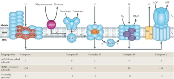
are made up by nuclear and mitochondrial encoded proteins (Figure 2). The ETC is
composed of four complexes, collectively known as oxidoreductases, which couple
electron transport with translocation of protons across the IM to IMS (apart from
complex II, which does not possess proton pumping activity).
I n t r o d u c t i o n | 14
Figure 2: Distribution of nuclear encoded (nDNA, blue, except for DHOD) and mitochondrial encoded protein (other colours) in the OXPHOS in human (Schon, DiMauro et al. 2012)
Complex I, also known as NADH-ubiquinone oxidoreductase is the largest in size with 45 core subunits and an enormous amount of assembly factors, it forms an L shape complex in the IM (Diaz, Kotarsky et al. 2011). In this step, the electron carrier NADH generated from TCA cycle is oxidized to NAD
+, generating four protons across the membrane. It is also the major source of reactive oxygen species inside mitochondria (Murphy 2009).
Complex II (succinate:ubiquinoneoxidoreductase, or succinate dehydrogenase) is the simplest complex of the ETC with only four subunits which are exclusively nuclear encoded in mammals. The two largest subunits SDHA and SDHB locate on the peripheral of the IM. Complex II also takes part in TCA cycle to facilitate conversion of succinate to fumarate. The resulting electrons are then transferred directly through complex II to ubiquinone in the ETC.
Complex III or ubiquinone-cytochrome c oxidoreductase, also known as cytochrome c reductase is another major source of reactive oxygen species production in mitochondria.
Mammalian complex III consists of 11 subunits, however only three are functionally important since they are the only components of the bacterial counterparts, they are:
cytochrome b (mtDNA encoded) cytochrome C
1and the Rieske iron-sulfur protein.
I n t r o d u c t i o n | 15
Complex IV, the cytochrome c oxidase, catalyses the final electron transfers to H
2O with O
2as the electron acceptor. It contains 13 subunits, three of the largest subunits are encoded by the mitochondrial genome (COX I, II and III) however around 30 factors are required for the assembly of this complex.
Complex V or ATP synthase utilises the proton gradient generated in the IMS to catalyse the phosphorylation of ADP to ATP. Complex V is a lollipop-like structure with a “head” facing the matrix and its “neck”inserted in the IM. The “head” is named F
1-ATP synthase and is made up of nine subunits: 3α, 3β, 1γ, 1δ, and 1ε. The neck is F
0- synthase and consists of a subunit c-ring and a, b, d, F6 and the oligomycin sensitivity- conferring protein (OSCP). There are also other subunits associated with F
0: e, f, g and A6L. Two of the F
0subunits are encoded by mtDNA: ATP6 (a) and ATP8 (A6L) (Anderson, Bankier et al. 1981) (Jonckheere, Smeitink et al. 2012)
Complexes I, III and IV are found to form specific respirasome (complex I + III + IV) and/or supercomplexes (complex I + III, complex III + IV) (Dudkina, Sunderhaus et al.
2008; Lapuente-Brun, Moreno-Loshuertos et al. 2013) while ATP synthase can dimerise and form long oligomeric chains. Moreover because of such formation, assembly of one complex can affect the other: for instance, complex I disassembles in the absence of complex III and IV (Scheffler 2007).
1.2 Biogenesis of nuclear encoded mitochondrial proteins
1.2.1 Coordination between nucleus and mitochondria to ensure appropriate protein expression
Because of the coexistence of nuclear and mitochondrial genomes, concerted
coordination is required for the expression of nuclear and mitochondrial encoded
proteins. For example, the OXPHOS houses nearly 80 subunits with only 13 being
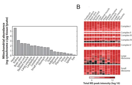
encoded in the mitochondria (Figure 2A). It is estimated that about 1500 proteins are
present in a given mitochondrion depending on the sample type, purification method
and detection sensitivity (Calvo and Mootha 2010). Moreover, mitochondrial protein
expression pattern is tightly controlled in specific cell types depending on the metabolic
I n t r o d u c t i o n | 16
demand (Figure 3A and B) (Calvo and Mootha 2010) Therefore, mitochondria and nucleus need to constantly communicate with each other to monitor protein expression.
Figure 3: Mitochondrial proteome. (A) Mitochondrial abundance in tissues (B) Expression profile of the subunit of protein complexes in different tissues. Black indicates lack of detection whereas red is the level of abundance (Calvo and Mootha 2010).
There is an enormous amount of factors involved in the cross-talk processes, residing or shuttling in between mitochondria, cytosol and the nucleus. If we take the example of ATP production, mitochondrial function is monitored in terms of metabolites (pyruvate, NADH and ATP) derived from glycolysis, TCA cycle and the OXPHOS, respectively.
During fasting, in the liver, the increase of pyruvate is coupled with an increased ratio
of NAD
+: NADH, this subsequently leads to post-translational upregulation of Sirt1 (a
NAD
+dependent deacetylase) in the nucleus (Rodgers, Lerin et al. 2005). In the
meantime the ratio of AMP: ATP or ADP levels are monitored by AMP-acivated
protein kinase (AMPK) in the cytosol. When AMP:ATP ratio increases, AMPK is
phosphorylated and induces a cascade of changes (Mihaylova and Shaw 2011). AMPK
phosphorylation and Sirt1 activation can lead to the upregulation of the master
I n t r o d u c t i o n | 17
transcriptional coactivator: peroxisome proliferators-activated receptor γ coactivator 1- alpha (PGC-1α). Consequently, this triggers the transcription induction of NEMPs such as OXPHOS components (Puigserver, Wu et al. 1998; Nunnari and Suomalainen 2012).
1.2.2 Regulation of nuclear encoded mitochondrial proteins
PGC-1 was first identified as a cold inducible activator of uncoupling protein 1 (UCP-1), which is important for energy dissipation in the brown fat cells. Under normal condition, PGC-1 is expressed in the heart, kidney, brown fat and brain but at very low level in lung, muscle, liver, white fat, testis and spleen (Puigserver, Wu et al. 1998). PGC-1 expression can be induced in the liver under fasting or diabetic condition (Yoon, Puigserver et al. 2001) or cold shock in brown fat and slow-twitch skeletal muscles (Puigserver, Wu et al. 1998). It interacts with various transcription factor partners, to name a few: peroxisome proliferator-activated receptor γ (PPARγ), thyroid hormone receptor (TR), retinoic acid receptor (RA), estrogen receptors (Puigserver, Wu et al.
1998), PPARα (Vega, Huss et al. 2000), PPARβ (Wang, Lee et al. 2003), nuclear respiratory factor 1 and 2 (Wu, Puigserver et al. 1999) and estrogen-related receptors α (ERRα) (Huss, Kopp et al. 2002; Schreiber, Knutti et al. 2003). Depending on the combination of the complexes, different set of genes can be activated: ERRα, PPARα PPARγ and NRF-1, which regulates the transcription of genes involved in mitochondrial fatty acid beta-oxidation and oxidative phosphorylation and mtDNA transactions (Scarpulla 2002). All nuclear encoded proteins are translated in the cytosol, however whether they are localised to mitochondria before or after translation is still under debate, this is especially true for the NEMPs.
1.2.3 Mitochondrial post-translational versus co-translational protein targeting and import.
Two main mitochondrial protein targeting/import models exist (Figure 4): post-
translational (Neupert and Herrmann 2007) and co-translational (MacKenzie and Payne
2007). In the former, the NEMPs are translated in the cytosol, unfolded by chaperons,
I n t r o d u c t i o n | 18
imported via the translocases of the outer membrane (TOM complex) and sorted to their respective destination. This mode of protein localisation and import to mitochondria relies on the presence of mitochondrial targeting signals which is also a determinant for mitochondrial protein sorting after passing TOM (Chacinska, Koehler et al. 2009). The mRNA, in this case, does not participate in the event of protein targeting to mitochondria. Evidences supporting post-translational import include: (1) Many precursor proteins can be made and imported into isolated mitochondria by in vitro protein import assays (Harmey, Hallermayer et al. 1977). (2) Not all nuclear encoded mRNAs are associated with mitochondria, many are present in the cytosol and associate with free polysomes (Marc, Margeot et al. 2002). (3) The order of protein translation dictates that all proteins have to be translocated from the N-terminal in co-translational model. This is challenged by protein with a non-canonical C-terminal targeting and sorting sequence.
On the other hand, the co-translational model suggests that localisation of mRNAs to the vicinity of mitochondria prior to their translation. After translation, the peptide emerges from the ribosome exit tunnel of the ribosome and is “captured” and “pulled”
through the TOM complex. Therefore, in this case, mRNA targeting to mitochondria precedes protein localisation and is important for mitochondrial biogenesis. This model is supported by increasing evidence of (1) A fraction of NEMP mRNAs localise in the vicinity of mitochondria (Marc, Margeot et al. 2002). (2) Mitochondrial targeting sequence is not necessary to be a determinant for mitochondrial localisation of some proteins (Mukhopadhyay, Ni et al. 2004). In fact mRNA localisation is not only observed in mitochondria but also in peroxisomes and ER (Marc, Margeot et al. 2002;
Pyhtila, Zheng et al. 2008; Zipor, Haim-Vilmovsky et al. 2009). When translation
elongation is inhibited, ribosomes accumulate on the surface of mitochondria. Moreover
the signal peptides are believed to be translated during mRNA transport and acting as a
guide for path finding to the proper destination. Tom20, part of the TOM complex can
mediate localisation of some mRNAs (Eliyahu, Pnueli et al. 2010; Kilchert and Spang
2011). It is, of course, possible that in nature, the two models coexist. Localisation of
mRNAs and localisation of mRNA to mitochondria will be discussed further in the next
chapter.
I n t r o d u c t i o n | 19
Figure 4: Protein targeting models. In post-translational model, protein targeting relies on the presence of mitochondrial targeting signals whereas co-translational model requires the targeting of mRNAs to the vicinity of mitochondria followed by localised translation.
1.2.4 The targeting and sorting sequence of mitochondrial proteins
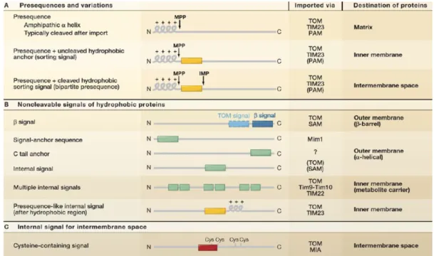
The function of a protein dictates its conformation and location. Newly synthesised proteins must be sorted and folded correctly by the virtue of the other proteins as well as the intrinsic targeting signal that the new proteins possess (Table 2) (Chacinska, Koehler et al. 2009).
The best-known mitochondrial targeting sequence (MTS) is the cleavable amphipathic α-helix presequence at the N-terminal of a protein. This sequence can be up to 100 amino acids and is cleaved off by the matrix processing peptidase (MPP) after import.
Therefore, it is used by most of matrix proteins. For some IM proteins, a hydrophobic segment is present after the amphipathic α-helix. This segment can halt further translocation of the preprotein and remains as a part of the mature protein. In contrast to some IMS proteins, the mature protein is formed after translocating through the IM, followed by cleavage by MPP in the matrix as well as cleavage of the hydrophobic IM insertion by inner membrane peptidase (Table 2 A).
None of the OM proteins possesses any of the aforementioned features mainly because
they do not translocate the IM and reach the matrix, however large number of IM
I n t r o d u c t i o n | 20
proteins and IMS proteins do not have any cleavable targeting sequence. The targeting sequence of OM proteins can be found at the N-terminus, internal or the C-terminus (Table 2B) based on the topology of the proteins: α-helices with N-terminal, internal domain or C-terminal insertion into the membrane or β-barrel conformation.The IM contains many multi-spanning transmembrane metabolite carriers that harbor multiple intrinsic α-helix targeting signals. A number of IM proteins contain a presequence like structure with positively charged α-helices after hydrophobic stretch (Table 2B). IMS proteins embody mitochondrial intermembrane space signal (MISS) with a cysteine residue that can be recognised and sorted (Table 2C) (Chacinska, Koehler et al. 2009).
Table 2: Targeting and sorting signals present in nuclear encoded mitochondrial proteins
(Chacinska, Koehler et al. 2009).
I n t r o d u c t i o n | 21
1.2.5 TOM and the sorting machinery
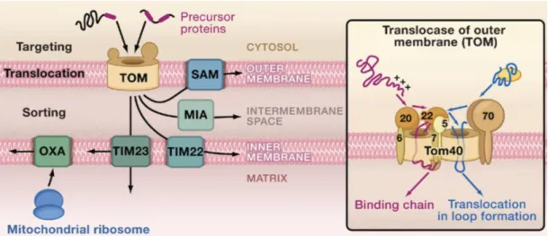
TOM complex is the entry gate of almost all nuclear encoded mitochondrial proteins.
TOM is composed of 7 subunits: two receptors TOM70 and TOM20 and the general import core TOM40 (forms oligomeric β-barrel), TOM22, TOM7, TOM6 and TOM5 (Figure5 ) (Model, Meisinger et al. 2008). TOM20 and TOM70 are the initial contact sites for preprotein with presequence and IM proteins, respectively. They are also important for the localisation of mRNAs in co-translational import model together with TOM7 (Eliyahu, Pnueli et al. 2010; Gadir, Haim-Vilmovsky et al. 2011). After the initial contact, the preproteins are then transferred to TOM22, TOM5, translocated through TOM40 to bind to the IMS domain of TOM22. From this point onward, preproteins are sorted to OM via the SAM (sorting and assembly machinery), to the IMS via MIA (mitochondrial intermembrane space assembly), to the IM via TIM 22 (transloase of the inner mitochondrial membrane) and to the matrix via TIM23 (Figure 5).
Figure 5: Mitochondrial protein sorting pathways. After entry through TOM, the precursor
proteins are sorted into different compartments. OXA is the insertase of mitochondrial encoded
IM protein(Chacinska, Koehler et al. 2009).
I n t r o d u c t i o n | 22
1.3 mRNA localisation
1.3.1 mRNA localisation is an ubiquitous phenomenon
mRNA localisation in the cell is evolutionarily conserved from single cell organism to multicellular systems and serves as a mean of stimuli-controlled local translation. This regulation ensures that the right amount of proteins can be made at the right time, in the right place with right conformation and post-translational modifications. In yeasts, to ensure that the daughter cells cannot switch mating types, ASH 1 mRNA is transported to the daughter bud via myosin and actin prior to fission (Long, Singer et al. 1997).
Tightly controlled compartmentalisation of mRNAs is observed during early Drosophila embryogenesis (Lecuyer, Yoshida et al. 2007). The mRNA of cytoskeletal
elements: actin, vimentin and tubulin have been shown to localise to cell extremities, perinuclear and peripheral cytoplasm, respectively in chicken embryonic myoblasts and fibroblasts (Lawrence and Singer 1986). In highly polarised and specialised cell types such as neurons, many different types of mRNAs are found to be localised to direct the growth of axons (Zivraj, Tung et al. 2010) and dendrites (Kleiman, Banker et al. 1990).
Normally, functionally related mRNAs are bundled and translated coordinately and therefore can be efficiently controlled (Gerber, Herschlag et al. 2004; Keene 2007). This precise local translational control is favored over directly transporting protein, because (1) One mRNA can be translated to the exact desired amount of proteins according to demand. (2) Local translation provides rapid response to local demand when compared to protein localisation. (3) During protein transport, cellular stress could impose undesired damage to the protein. (4) For some hydrophobic membrane proteins, it can be energetically favorable to insert the protein while being translated to avoid misfolding and aggregation.
mRNA localisation is different from transport and it can be seen as a final step
following the transportation of mRNA. mRNA protein complexes (mRNPs) are
composed of mRNA, mRNA binding proteins or other associating proteins and proteins
that connect the mRNP complex to cytoskeleton via motor proteins (Gagnon and
Mowry 2011). As soon as the mRNAs are transcribed and spliced, they are sorted
according to their destination with different protein compositions (i.e. RNP granules).
I n t r o d u c t i o n | 23
These granules are distinct from both stress or P granules in composition and function even though, general translation is halted in all cases (Anderson and Kedersha 2006;
Anderson and Kedersha 2009).
Transport of mRNAs to their final destination is a result from two factors: the cis-acting RNA sequence and the trans-acting proteins (RNA binding proteins or RBP) that bind to the mRNAs.
Cis-acting RNA sequences, also known as "zipcodes", lie within the mRNAs and dictate the direction of transport, they could be in the 3’-UTR (for example, ASH 1 and β-actin) (Bassell, Zhang et al. 1998; Chartrand, Meng et al. 1999), 5’-UTR (kor, mRNA of kappa opioid receptor) (Tsai, Bi et al. 2007), and even within the protein coding sequence (mRNA target of FMRP, loss of function mutation causes fragile X syndrome) (Darnell, Van Driesche et al. 2011). The coding message can be hidden either in a short or long stretch of nucleotides or multiple units scattered through the sequence. It can also be coded in its secondary structure. Different signals may even be employed at different stages of mRNA transport and localisation (Hervé, Mickleburgh et al. 2004).
One representative of such complexity is the localisation of the mRNA of myelin basic protein (MBP) to myelin: it is firstly transported to the oligodendrocytes via a 21- nucleotide sequence in the 3´-UTR (RNA transport signal), followed by localisation to myelin compartment via a long stretch of nucleotides in 3´-UTR that forms a stable secondary structure (the RNA localisation region) (Ainger, Avossa et al. 1997). Another example is that the bicoid mRNA localisation to the anterior pole in Drosophila embryo requires two different cis-acting elements for early and late localisation and formation of double stranded helical structure to achieve the anchorage in the anterior cytoplasm (Ferrandon, Koch et al. 1997).
Trans-acting proteins are the other factor that dictates the transport and localisation of mRNAs. They bind to the cis-acting elements of the mRNAs, their binding or association depends on the life cycle of an mRNA: transport, localisation or anchorage.
In cultured HeLa cells, approximate 860 proteins have been identified as RNA binding
proteins, the actual number, however might be even more due to the stringent threshold
of the analysis (Castello, Fischer et al. 2012). To complicate the matters further, one
RBP is able to bind to hundreds of different mRNAs (Gerber, Herschlag et al. 2004) and
I n t r o d u c t i o n | 24
one RNA can be bound by numerous RBPs. For example, Staufen recognises the loop structure formed by two strands of different RNAs in the zipcode, its target mRNAs include prospero and bicoid in flies. The association between Staufen and its target mRNAs is location specific: anterior to the anchoring of bicoid, and posterior to the localisation of oskar (St Johnston, Beuchle et al. 1991; Ferrandon, Koch et al. 1997).
Therefore, it is reasonable to extrapolate that the RNA granules are extremly dynamic structures that change in time, space and cellular stresses. It is believed that during transport, mRNA translation is inhibited and mRNA is stabilised/protected and only become activated when reaching its destination (Abaza and Gebauer 2008; Besse and Ephrussi 2008).
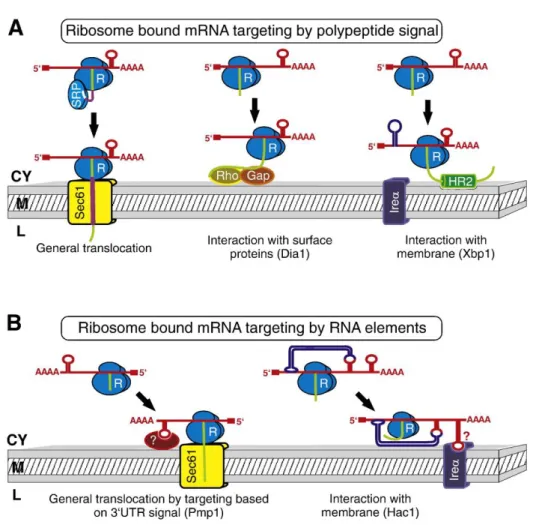
Proper localisation of mRNAs in either polarised or less polarised cells is an essential
feature to determine the morphogenesis and development. However, mRNAs are also
localised to different organelles (Weis, Schleiff et al. 2013). For example, the mRNAs
for secretory, integral proteins and some cytosolic proteins are localised to the ER prior
to complete translation, the signal recognition particle (SRP) pathway. After the release
of mRNP into the cytosol, the complex is bound by ribosomes and a small length of
polypeptide (signal peptide) is translated. Together this forms ribosome mRNA-nascent
polypeptide complexes (RNCs) that can be recognised by the SRP. At the ER, SRP
receptor recognises RNC-SRP, RNC stalks at the translocation channel and continues
the translation while the nascent chain is “pulled” inside of the ER (Figure 6A)
(Nicchitta, Lerner et al. 2005). However this classical co-translational model is also
challenged by other models where the cis-acting element is important for localisation to
the surface of ER instead of the SRP (Figure 6B).
I n t r o d u c t i o n | 25
Figure 6: mRNA localisation to the ER. (A) Three forms of mRNA localisation mediated by the signal peptide. (B) mRNA localisation imposed by the cis-acting element of the mRNA together with ribosomes (Weis, Schleiff et al. 2013).
1.3.2 mRNA localisation to mitochondria
This brings us to the question of whether a similar mRNA localisation pathway also occurs in the mitochondria. The vast majority of the work on mRNA localisation to mitochondria was carried out in yeast owing to ease in manipulation, however this is also observed in mammalian cells (Matsumoto, Uchiumi et al. 2012; Gehrke, Wu et al.
2015). First of all, ribosomes are found to locate on the surface of mitochondria (Kellems and Butow 1972; Keyhani 1973). Secondly, about 500 mRNAs were estimated to reside close to mitochondria. Furthermore, the authors show that the localised mRNAs are likely to be ancient with bacterial origins (Marc, Margeot et al.
2002; Saint-Georges, Garcia et al. 2008). The association of mRNA in the vicinity of
mitochondria requires the presence of protein import machinery, the cis-acting
I n t r o d u c t i o n | 26
sequences and the RBP (Eliyahu, Pnueli et al. 2010; Gadir, Haim-Vilmovsky et al.
2011). The best-studied mRNA binding protein involved in localisation of NEMP mRNA is the yeast PUF3p.
1.3.3 PUF3p
The yeast PUF3p is one of the six PUFs (Pumilio and FBF) with known RNA binding domains. Members of this family are well conserved from yeast to human and can recognise the specific sequence at the 3´-UTR in order to regulate mRNA localisation, translation and stability (Wickens, Bernstein et al. 2002). Yeasts contain five PUFs, Drosophila, one, mammals, two. The RNA binding motif of RBP and the RNA signature for binding of the targets are also well conserved (Wickens, Bernstein et al.
2002). The RNA binding domain of PUF proteins are PUM-HD-type which forms an arc-like shape that can interact with both RNA and protein (Zamore, Williamson et al.
1997; Zhang, Gallegos et al. 1997). Members of this protein family display different functions, they can either suppress or activate translation or involved in targeting mRNA to subcellular localisation/organelle (Murata and Wharton 1995; Sonoda and Wharton 1999). In Drosophila, PUMILIO can repress the translation of hunchback during early embryogenesis. Yeast Puf6p can regulate both localisation and translation of ASH1 for mating type switching while Puf5p can affect the localisation of PEX14 to peroxisomes (Wreden, Verrotti et al. 1997; Zipor, Haim-Vilmovsky et al. 2009).
Puf3p binds preferentially to approximately 162 mRNAs of NEMPs in yeast to facilitate
localisation and co-translational import. The protein products of these mRNAs are
involved in mitochondrial protein synthesis, structural components of the ribosome,
tRNA ligases, OXPHOS and translational regulators (Huh, Falvo et al. 2003; Gerber,
Herschlag et al. 2004). Puf3p is also found to be a component of P-body under stress
(Lee, Dudley et al. 2009; Eliyahu, Pnueli et al. 2010). In the absence of Puf3p, the target
mRNAs mislocate (Olivas and Parker 2000; Saint-Georges, Garcia et al. 2008; Eliyahu,
Pnueli et al. 2010). Puf3p associate with mitochondria via binding to Mdm12, the
component of ER-mitochondria encounter structure (Garcia-Rodriguez, Gay et al. 2007).
I n t r o d u c t i o n | 27
One would expect that Puf3p could be important for mitochondrial biogenesis regarding its molecular function, however in reality, it is more complicated. Mitochondrial biogenesis and Puf3p reduction occur at the same time, and both overexpression and deletion lead to impairment in respiratory growth (Gerber, Herschlag et al. 2004;
Garcia-Rodriguez, Gay et al. 2007; Saint-Georges, Garcia et al. 2008; Eliyahu, Pnueli et al. 2010). Puf3p activity is dependent on the carbon source without alteration in transcript or protein level. (Gerber, Herschlag et al. 2004; Foat, Houshmandi et al.
2005). Furthermore, Puf3p is essential for the growth of TOM20 mutant yeast in glycerol. In the absence of TOM20, puf3 mRNA increases and so is its protein level, however Puf3p associates less with the mitochondria (Eliyahu, Pnueli et al. 2010).
Moreover mitochondria in Δpuf3 are fragmented and aggregated with impaired anterograde movement (Garcia-Rodriguez, Gay et al. 2007). Based on these observations, it is suggested that Puf3p exists as two pools: mitochondrial and non- mitochondrial. Puf3p on mitochondria localises and tethers the mRNAs to mitochondria.
Non-mitochondrial Puf3p locates in the cytoplasm and may inhibit translation when mitochondrial biogenesis is not required (Garcia-Rodriguez, Gay et al. 2007; Quenault, Lithgow et al. 2011).
It is worth remembering that Puf1p also localises to mitochondria and facilitates the association of mitochondria to Arp2/3 complex of actin in yeast (Moreau, Madania et al.
1996).There are two Pumilio homolog (PUM1 and PUM2) in human (Spassov and Jurecic 2002; Galgano, Forrer et al. 2008). PUM2 knockout mice displays smaller testis but without obvious effect on fertility (Xu, Chang et al. 2007). The mRNA targets of PUM1 shares little similarity to Puf3p or Pumilio and is not enriched for NEMP (Gerber, Herschlag et al. 2004; Gerber, Luschnig et al. 2006; Morris, Mukherjee et al. 2008) despite the conservation of the protein binding domain and RNA motif. There is no protein with similar role to Puf3p that has been identified so far in mammalian system.
Furthermore, it is unclear if mRNA localisation to mitochondria occurs at all in the
mammalian system.
I n t r o d u c t i o n | 28
1.4 CLUH
1.4.1 Clu mutant leads to mitochondrial clustering and abnormal mitochondrial morphology
Clu stands for clustered mitochondria and is well conserved from amoeba to human (see the gene and protein annotations in Table 3). For the sake of simplicity, all homologs mentioned below will be referred as Clu when general function is introduced. Its name originated from the phenotype of mitochondrial clustering after clu is deleted. CluA was first identified in 1992 as an unconventional myosin in Dictyostelium because of its cross-reactivity with the antiserum specific for myosin-IC (Zhu and Clarke 1992).
However it neither possesses any myosin like activity nor shares significant homology to myosin (Zhu, Hulen et al. 1997).
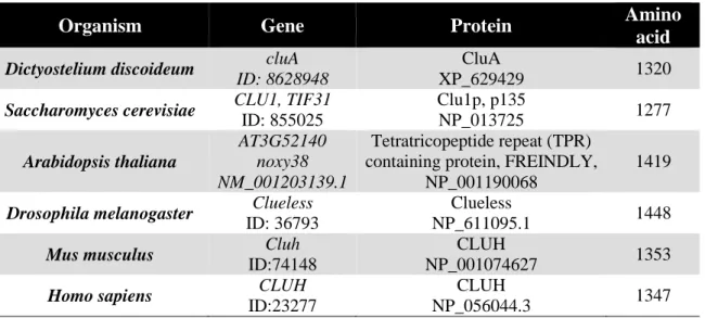
Table 3: Annotation of clu genes and proteins in different organisms.
Organism Gene Protein Amino
acid Dictyostelium discoideum cluA
ID: 8628948
CluA
XP_629429 1320
Saccharomyces cerevisiae CLU1, TIF31 ID: 855025
Clu1p, p135
NP_013725 1277
Arabidopsis thaliana
AT3G52140 noxy38 NM_001203139.1
Tetratricopeptide repeat (TPR) containing protein, FREINDLY,
NP_001190068
1419 Drosophila melanogaster Clueless
ID: 36793
Clueless
NP_611095.1 1448
Mus musculus Cluh
ID:74148
CLUH
NP_001074627 1353
Homo sapiens CLUH
ID:23277
CLUH
NP_056044.3 1347
In Dictyostelium, when cluA is deleted, mitochondria cluster at the cell center while
peroxisomes and ER remain morphologically unchaged (Zhu and Clarke 1992; Fields,
Arana et al. 2002). Disruption of microtubules or actin filaments, does not affect the
distribution of existing mitochondria (i.e. mitochondria that were already transported to
the positions) (Fields, Arana et al. 2002). Mitochondria within the cluster are free to
undergo deformation and rearrangement, however, individual mitochondrion appears to
I n t r o d u c t i o n | 29
have constrained movement. Half of the CluA mutants are multinucleated, indicating a failure in cytokinesis.
Yeast mitochondria in clu1Δ cells were condensed and accumulated on one side of the cell. However, the inheritance of functional mitochondria to daughter was not affected.
No multinucleated cells were observed in yeast mutants. Interestingly, yeast Clu1p (sharing only 50 % similarity in primary structure to CluA) can correct the cytokinetic defect in Dictyostelium cluA
-cells to wildtype level. However it can only partially rescue the mitochondrial clustering phenotype in cluA
-cells: Clu1p complemented mitochondria were dispersed in the cytoplasm but in the form of small aggregates (Fields, Conrad et al. 1998). The Arabidopsis homolog friendly mitochondria (FMT, also known as noxy 38 or friendly) also gave rise to clustered mitochondria (Logan, Scott et al. 2003; Vellosillo, Aguilera et al. 2013; El Zawily, Schwarzlander et al. 2014).
However only a portion of total mitochondria were clustered, others resemble mitochondria in the wildtype cells. Consistent with the observation in Dictyostelium, other organelles in Arabidopsis mutant are not affected morphologically (El Zawily, Schwarzlander et al. 2014). Importantly, two fmt mutations were studied and both led to aggregated (clustered) mitochondria. Fmt mutant identified by Logan and colleague contains G-to-A mutation of the first base of the second intron and this result in almost complete absence of the respective transcript. In contrast, Vellosio and colleague´s noxy38 mutant contains G-to-A transition in the last nucleotide of intron 22. This transcript is translated into a truncated protein (1053 amino acids) without the TPR domains (see more information of this domain later).
In Drosophila clueless mutants, mitochondria clustered in the anterior region of germline stem cells, mitotic germline stem cells, old germ cells, stage 5 nurse cells, somatic follicle cells and neuroblast cells (Cox and Spradling 2009; Sen, Damm et al.
2013). Mitochondrial clusters were present in microtubule plus end (Cox and Spradling
2009). Moreover, the electron microscope (EM) data showed swollen mitochondria
with loss of cristae in the 3-days old flight muscles and ovaries. Sperm from sterile male
mutants was immobile and the spermatide mitochondria derivative was irregular and
swollen. Most interestingly, the fly mutant with double heterozygous park (gene
associates with Parkinson´s) and clu leads to clustered mitochondria suggesting that
I n t r o d u c t i o n | 30
these two proteins genetically interact. The clustering phenotype of mitochondria was also observed in mouse, monkey and human cells (Gao, Schatton et al. 2014).
Analyses were conducted to understand mitochondrial clusters. The EM images of mitochondria from cluA
-cells displayed interconnection between neighboring mitochondria. This observation suggests a failure of mitochondrial fission at a very late stage (Fields, Arana et al. 2002). However, interconnected mitochondria were not observed in the other organisms. The clustered mitochondria in Arabidopsis did not have interconnected OM between mitochondria (Logan, Scott et al. 2003; El Zawily, Schwarzlander et al. 2014). However, electron dense regions were observed between individual mitochondrion within a cluster of the mutant.
The cytoskeleton in all Clu mutants appeared to be intact and functional (Zhu and Clarke 1992; Fields, Arana et al. 2002; Cox and Spradling 2009). Moreover mitochondrial transport was unaffected (Cox and Spradling 2009; El Zawily, Schwarzlander et al. 2014). Deletion of Clu did not affect the transport and accumulation of mitochondria to Balbiani bodies (Balbiani body is a collection of organelles, mRNA and inclusions present in the early oocyte of all animals, it is normally close to the nucleus.). In friendly mutant, small mitochondrial aggregates as well as individual mitochondrion could be transported on actin. Altogether, these evidences indicate that mitochondrial clustering phenotype is independent of cytoskeletal alteration and defects in mitochondrial transport. It is postulated that the clustering phenotype might be caused by prolonged association between mitochondria (El Zawily, Schwarzlander et al. 2014).
1.4.2 Clu mutants of multicellular organism display growth defects
Clu function is not essential for single cellular organisms. Dictyostelium cluA
-mutant
displayed a moderate reduction for growth that could be caused by unequal distribution
of mitochondria after cell division (Zhu, Hulen et al. 1997). Yeast clu1Δ mutants
showed no defect in growth in non-fermentable media (glycerol, lactate and ethanol),
glucose media, heat and osmotic stress (Fields, Conrad et al. 1998). No signs of failure
in cell wall synthesis or mitochondrial membrane potential were observed in yeast. It
I n t r o d u c t i o n | 31
was not even considered to be an essential gene in yeast, since TIF31 mutant could still grow at temperatures higher or lower than 30
oC (Vornlocher, Hanachi et al. 1999).
Arabidopsis Friendly mutant leaves were smaller, shorter and more rounded than the wildtype (El Zawily, Schwarzlander et al. 2014). The length and width of root were significantly reduced and this might due to increased cell death. Along with this, the mutant root displayed increased number and size of acid vacuoles (El Zawily, Schwarzlander et al. 2014). Interestingly, the noxy38 mutant was more susceptible to pathogen invasions (Vellosillo, Aguilera et al. 2013).
Despite the clustering phenotype during development, the fly clueless mutants strived to adulthood. However, there was a prolonged larval development. At the end, only 40 % of the pupae can manage to eclose (Sen, Damm et al. 2013). These flies then developed phenotypes similar to park and pink1 mutants: short lifespan, small size, un- coordination, sterility, flight muscle defects (wings up and down) and problems with utilising larval fat body in the new adults (Cox and Spradling 2009) (Goh, Zhou et al.
2013). Clu was highly expressed in neuroblasts but functionally dispensable (Goh, Zhou et al. 2013; Sen, Damm et al. 2013). In Clu mutant neuroblasts, mitochondria were not evenly segregated into the daughter cells. However, this did not have any impact on cell development or mitochondrial numbers.
Little is known regarding the function of clu1 in C. elegans, however genomic RNAi
studies revealed that clu-1 mutant is embryonic-lethal (6 %, 24 hours recovery after
soaking the worms with RNAi) (Maeda, Kohara et al. 2001). Interestingly,
mitochondria are not clustered in C. elegans (Logan, Scott et al. 2003). However, it is
not clear if the above RNAi data on C. elegans can be correlated.
I n t r o d u c t i o n | 32
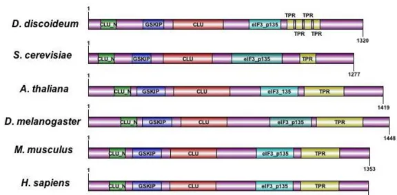
1.4.3. Primary structure and the molecular function of Clu
The structure and molecular functions of Clu proteins are unknown, however provisional bioinformatics studies show that all Clu homologs share similar domains arrangement: CLU_N domain also known as TIF31-like, GSKIP domain, CLU domain, eIF_ p135 (or Clu_central) domain and finally the TRP domain (Figure 7).
The nomenclature of TIF31 and eIF_p135 domain was based on the finding of Vornlocher et al. that TIF31 co-purified with eukaryotic translation initiation factor eIF3 (Vornlocher, Hanachi et al. 1999). However, deletion of TIF31 affected neither the function of eIF3 nor the global cytosolic translation. Moreover, TIF31 is neither always associated with eIF3 complex nor identified in eIF3 complex in human or plant (Browning, Gallie et al.). Consequently, Clu is not considered as part of the core eIF-3 complex despite its interaction with p33 (or TIF35, eIF3G, an essential eIF3 subunits) in yeast. There is no evidence so far that Clu plays any roles in translation.
The TPR motif contains degenerate sequences and proteins with these repeats act as scaffolds for the assembly of multicomplexes or protein-protein interactions. As mentioned before, truncated FMT protein without the TPR domains can lead to mitochondrial clustering phenotype. Moreover, FRIENDLY protein is subjected to post- translational modification, it contains two acetylation sites Lys-1022 and Lys-1029 which is located in between eIF_p135 and TPR domains (Finkemeier, Laxa et al. 2011;
El Zawily, Schwarzlander et al. 2014). The acetylation status of FRIENDLY might be
involved in mitochondrial clustering and other functions.
I n t r o d u c t i o n | 33
Figure 7: Illustration of the predicted protein domain of CLUH. CLU_N domain is also known as TIF31-like, eIF_p135 domain is also known as CLU-central. Protein domain information obtained from NCBI Conserved domains and InterPro from EMBL-EBI (for GSKIP domain). Protein domains are annotated with DOG (Ren, Wen et al. 2009).
It is also interesting to note that the mRNA level of cluA is elevated from the vegetative stage to the developing stage of the cell (Zhu, Hulen et al. 1997).
Clu locates in the cytoplasm as discrete puncta / granule (Fields, Conrad et al. 1998;
Fields, Arana et al. 2002; Cox and Spradling 2009; Gao, Schatton et al. 2014). Some Clueless particles were observed outside of mitochondria, while some other associated with microtubules. However it is not clear if Clueless particles, mitochondria and microtubules colocalise (Cox and Spradling 2009). The ovary microtubule network in clueless mutants remained undisrupted. Deletion of Clu in flies and mammalian cells led to reductions of a subset of mitochondrial proteins (Cox and Spradling 2009; Gao, Schatton et al. 2014).
Transcriptome studies of Clu mutants indicated mitochondrial dysfunction. Gene
expression study of the mild clueless mutant (with low expression level of Clu) showed
downregulation of genes involved in reactive oxygen species protecting agents, DNA
repair, apoptosis regulator and fatty acid production regulator. Some candidate
metabolic genes involved in amino acid/fatty acid transporter, putative choline
transporter and putative aminoadipate-semialdehyde were upregulated (Cox and
Spradling 2009). Transcriptome analysis in Arabidopsis indicates alteration in genes
I n t r o d u c t i o n | 34
involved in protein synthesis, degradation, photosynthesis, photosynthetic light reaction, abiotic stress, redox and TCA cycle (El Zawily, Schwarzlander et al. 2014).
Biochemical analysis of the mutants in different organisms also suggests defects in mitochondria. In Arabidopsis, mutation induced the upregulation of the alternative oxidase pathway without affecting the average oxygen uptake (Vellosillo, Aguilera et al.
2013; El Zawily, Schwarzlander et al. 2014). AOX directs reduction of oxygen with the electrons donated from ubiquinol. Therefore, it reduced the total electron flow to complex III and the eventual production of ATP. Similarly, clueless adult mutants displayed severely reduced level of ATP as well as aconitase activity. However both phenotypes were absent in the mutant larvae.
Investigation of the neuroblasts of Clueless mutant showed that Clu was important for the asymmetric cell division of neuroblasts. Neuroblast asymmetric cell division generates one big daughter neuroblast cell and another small ganglion mother cell.
These two cells inherit different materials including cell fate determinants Numb and Miranda. Numb and Miranda are positioned to the basal cortex of neuroblasts before mitosis. After cell division, they are inherited by ganglion mother cell. Positioning of the cell fate determinants is achieved by phosphorylation conducted by the Par complex.
Par complex is composed of atypical protein kinase C (aPKC), Defective 6 (Par6) and Bazooka. In interphase, Par6 and aPKC are associated with lethal giant larvae (Lgl). It is proposed that aPKC/Baz/Par6 (active) complex is in equilibrium with aPKC/Lgl/Par 6 (inactive) complex in the apical cortex to direct the phosphorylation of cell fate determinents Miranda and Numb. Phosphorylation of these proteins results in exclusion of their apical position. Interestingly, a small portion of neuroblast mutant cells displayed mislocalised Miranda during metaphase. Clu was mainly if not exclusively present in the neuroblasts throughout asymmetric cell division (Goh, Zhou et al. 2013;
Sen, Damm et al. 2013). In addition, Clu coimmunoprecipitated with aPKC and Baz but
not Lgl. Lgl mutant also led to mislocalisation of Miranda and Numb to a large extent
and mislocalisation could be rescued by depleting clu or park. The author concluded
that the function of park and clu is likely to be independent from mitochondrial function
during neurogenesis (Goh, Zhou et al. 2013). However according to Sen and colleagues,
the number of neuroblats in clu mutant were comparable to the wildtype and neurons
I n t r o d u c t i o n | 35
can differentiate properly. Therefore it is still unclear why neuroblasts require high expression level of Clu.
In spite of mitochondrial clustering phenotype, Clu is a cytosolic protein and might be
located in close proximity to mitochondria (Cox and Spradling 2009). The relationship
between clu and mitochondria remains to be elucidated.
A i m o f t h e T h e s i s | 36
2 Aim of the Thesis
Mitochondria are indispensible organelles to support the normal function of cells in all eukaryotic systems, including mammals. The status of mitochondria in response to metabolic alteration and signaling is vital for cell survival. It is well established that mitochondrial biogenesis requires concerted coordination between the nuclear and mitochondrial genomes. This process demands the participation of armies of factors:
transcriptional regulators, transcriptional factors, mRNA export of nucleus, mRNA transport and localisation, translation, post-translational modification, protein folding, and protein import to mitochondria.
Accumulated evidences in different organisms indicate the important relationship between Clu and mitochondria. Clu is a well-conserved cytosolic protein with a similarly conserved peculiar mitochondrial clustering phenotype, yet its function remains to be elucidated. Despite being a cytosolic protein, Clu is proposed to be involved in regulating mitochondrial position in response to physiological changes. A small portion of Clu is observed in close proximity to mitochondria. It can affect the protein levels of OXPHOS while genetically interacting with Park in Drosophila. In yeast, Clu copurifies with eIF3, however it is not an essential components of the cytosolic translation machinery.
The aim of this project is to investigate the molecular function of CLUH in mammalian
system in order to address the following questions: (1) what is the molecular function of
CLUH? (2) What is the relationship between CLUH and mitochondria? (3) How is
CLUH regulated transcriptionally and in respect to cellular alterations?
M a t e r i a l a n d M e t h o d s | 37
3 Material and Method
M a t e r i a l a n d M e t h o d s | 38
All chemicals used in this study were purchased from Merck, Sigma-Aldrich, Serva or Roth unless stated otherwise.
3.1 Cell biology
3.1.2 Cell culture
Mouse embryonic fibroblasts (MEFs) and HeLa cells were cultured in DMEM (Life Techologies, 11960085), supplemented with 10 % FCIII (HyClone, FetalCloneIII, SH30109.03, Life Techologies), 2 % penicillin-streptomycin (Life Technologies, 151401232), and 2 mM L-glutamine (Life Technologies, 25030024). NSC34 cells (Cashman, Durham et al. 1992) were cultured in DMEM, supplemented with 10 % defined FBS (Life Techologies, SH30070.03), 2 % penicillin-streptomycin, and 2 mM L-glutamine.
HEK 293T cells were maintained in media containing DMEM, 7.5 % tetracycline-free fetal bovine serum (Biochrom AG, S0115), 1 mM sodium pyruvate (Life Technologies, 1136- 070), 1x non-essential amino acids (Life Technologies, 1140-035), 2 mM L-glutamine and 1.5 mg/ml of hygromycin (Invivogen, ant-hm-5) and 150 µg/ml of blasticidin (Invivogen, ant-bl-5b). 1 g/ml of tetracycline (dissolved in ethanol) was used to induce protein expression.
PGC-1α MEFs were kindly provided from Dr. Tina Wenz. The embryos were collected at E12.5, and immortalized using the classic 3T3 protocol. PGC-1α knockout mice were obtained by breeding PGC-1α floxed (http://jaxmice.jax.org/strain/009666.html) with a full body Cre-deletor mouse line.
Cluh knockout MEFs were prepared by Dr. Henriette Hansen.
3.1.3 Long-term galactose experiment
The scheme of the galactose experiment is provided below (Figure 8). In general, HeLa
cells were cultured in galactose media containing DMEM (glucose free, Life Technologies,
M a t e r i a l a n d M e t h o d s | 39
11966-025), 10 % dialysed serum (Life Technologies, 26400-036) 1 mM sodium pyruvate;
6 mM galactose and 2 % penicillin-streptomycin). Galactose media were changed every 24 hours.
Figure 8: Experimental scheme of long-term galactose treatment.
3.1.4 Short-term galactose and acetoacetate treatment
The treatment was performed by Dr. David Pla-Martin. Briefly, 400,000 of MEF cells were plated on P60 plates and incubated in complete DMEM for overnight. The media was replaced with media containing galactose or acetoacetate (glucose free DMEM, 10%
dialysed FBS, 100 mM acetoacetate (Sigma-Aldrich, 164100-50G) and 2 % penicillin- streptomycin).
3.1.5 Bezafibrate treatment
The treatment was conducted in MEFs as illustrated below (Figure 9). Bezafibrate solution (B7273, sigma) was prepared by dissolving 0.199 g of bezafibrate in 2 ml of DMSO (100%) for a final concentration of 275 mM. MEFs were treated with a final concentration
0 24 48 72 96 120 144 168
168
144
120
96
72
48
24 Splitting cells
Change medium to galactose
Split into 2 plates, one for protein, one for RNA 20%
Treatment time
20%
10%
20%
10%
20 %
Cell time point 10%