Severe acute syphilitic posterior placoid chorioretinitis with complete spontaneous resolution: The natural course
Abstract
Purpose: We report on a case of unilateral acute syphilitic posterior placoid chorioretinitis (ASPPC) with spontaneous resolution of the le-
Mónica Franco
1Vanda Nogueira
1sions, and discuss the role of an altered versus adequate immune re- sponse as the major pathogenic factor.
Methods:We describe a case of acute loss of visual acuity (VA) in the left eye (LE) in a 55-year-old healthy man.
1 Instituto de Oftalmologia Dr.
Gama Pinto, Lisboa, Portugal Results:The patient presented with VA of 20/20 in the right eye (RE)
and hand movements in the LE. Fundoscopy revealed a large yellowish placoid macular lesion with subretinal fluid in the LE, with no abnormal- ities detected in the RE. Fluorescein angiography showed early hypo- fluorescence with late staining in the affected area. The clinical findings progressed fast during the first week, with extension of the initial lesion outside the temporal retinal vascular arcades and the appearance of new lesions in the same eye. The patient abandoned the clinic for two weeks with no treatment. When observed again, VA of the LE had re- covered to 20/20 and the lesions had completely resolved. Venereal disease research laboratory (VDRL) and fluorescent treponemal antibody absorption (FTA-ABS) tests results were positive and HIV antibody test titers negative. The diagnosis of ASPPC in the left eye was made. The patient accepted treatment with penicillin G only 45 days after the initial presentation. AV remained stable at 20/20 both eyes and no relapses of the lesions were observed during this period without therapy. The patient was followed for 3 months after treatment. He remained asymptomatic and the ophthalmic examination was unremarkable.
Conclusions:The pathogenesis of ASPPC is still not understood. Our case showed a sequential pattern of the chorioretinal lesions, with initial aggravation and complete posterior spontaneous resolution, showing the natural course of the disease. These findings suggest the presence of an adequate ocular immune response in patients with ASPPC, not supporting the initially proposed hypothesis of the importance of a modified immune response as the major pathogenic factor.
Keywords:syphilis, acute syphilitic posterior placoid chorioretinitis, immune response
Introduction
Syphilis is a sexually transmitted, systemic infection caused by the spirochete bacteriumTreponema pallidum [1], with increasing incidence in the USA and Europe [2], [3], [4]. Ocular involvement may be silent or present as anterior uveitis, choroiditis, interstitial keratitis, retinal vasculitis, retinitis, optic neuritis, dacryoadenitis, or scleritis [5], [6], [7]. Acute syphilitic posterior placoid chorioretinitis (ASPPC) is a rare manifestation of syphilis, characterized by large, yellow-white geographic lesions involving the macula at the level of the outer retina/retinal pigment epithelium (RPE) [8]. It can present in immuno- competent and immunodepressed patients and the pathogenesis still remains unknown. Penicillin is the mainstay of treatment and is usually given early after
serologic diagnosis of syphilis, so the natural course of the ocular lesions in not known. We present an untreated patient with complete spontaneous resolution of ASPPC.
Purpose
To report on a case of unilateral acute syphilitic posterior placoid chorioretinitis in an immunocompetent patient with spontaneous resolution of the lesions and discuss the role of an altered versus adequate immune response as the major pathogenic factor.
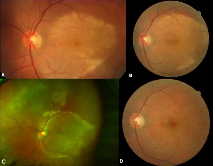
Figure 1: A) Colour photograph of the left eye shows a large yellow macular placoid lesion with a curvilinear edge. B) Colour photograph of the left eye, three days after the initial presentation, the placoid lesion had extended. C) Colour photograph of the left eye, one week after the initial presentation, placoid lesion had increased outside the temporal retinal vascular arcades and there are new multiple yellow lesions and hemorrhages in the retinal superior nasal area. D) Colour photograph of the left
eye three weeks after presentation and before treatment shows the lesion’s complete resolution.
Case report
A 55-year-old man presented with acute visual loss in the left eye (LE). The patient denied other ocular or systemic symptoms. He was not taking any medications and his medical, social and family history was unremarkable.
Visual acuity (VA) was 20/20 in the right eye (RE) and hand movements in the LE. Slit-lamp examination findings of the anterior segment were normal in both eyes (OU) and intra-ocular pressure was 12 mmHg bilaterally.
Dilated fundoscopy of the LE revealed a few vitreous cells and a large yellow macular placoid lesion with a curvilin- ear edge (Figure 1A). Fluorescein angiography (FA) showed early hypofluorescence with late staining in the affected area (Figure 2). Optical coherence tomography (OCT) in the LE demonstrated subretinal fluid overlying the macular lesion (Figure 3). No abnormalities were de- tected in the RE. Screening blood tests, including syphilis serology, were requested and the patient was monitored without treatment.
Three days after the placoid lesion had extended (Figure 1B). The patient did not follow our recommenda- tion for serologic work-up and the tests were ordered
again. One week after the initial presentation, VA re- mained stable but the placoid lesion had increased out- side the temporal retinal vascular arcades (Figure 1C).
New multiple yellow lesions and hemorrhages were ob- served in the retinal superior nasal area. Indocyanine green angiography (ICGA) revealed persistent leakage of the choriocapillaris and hypofluorescence areas in the early and late phases (Figure 4). The patient continued not to follow our recommendation for the blood tests and they were ordered for the third time. He was informed again about his clinical situation and the importance of the work-up was underlined.
The patient abandoned the clinic for two weeks and when observed again, VA had improved to 20/20 in the LE and the lesions showed complete spontaneous resolution (Figure 1D). RE examination remained normal. Finally, we had access to the serologic tests results that revealed a positive VDRL (1/128) and FTA-ABS. HIV titers were negative. All other tests were normal. The diagnosis of acute syphilitic posterior placoid chorioretinitis in the LE was made. The patient was referred to the Infectious Disease Department. He had no other syphilitic manifest- ations and no abnormalities were detected after com-
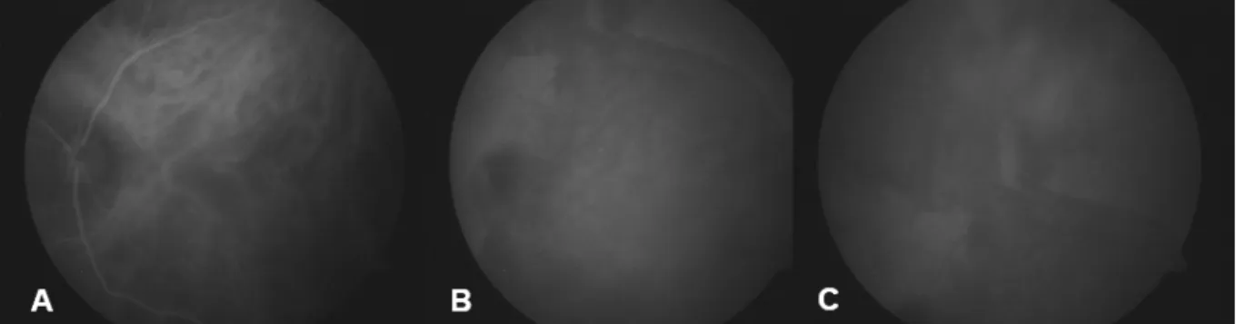
Figure 2: A) The early phase FA – corresponding to Figure 1A – shows hypofluorescence in the affected area. B) Late-phase FA shows progressive staining in the area of the lesion.
Figure 3: The ICGA – corresponding to Figure 1C – shows persistent leakage of the choriocapillaris and hypofluorescence areas in the early (A) and late phases (B, C).
Figure 4: A) OCT of the left eye at presentation reveals subretinal fluid overlying the macular lesion. B) OCT image three weeks after presentation and before treatment demonstrates complete anatomical resolution.
puted tomography of the cranium and orbits. A lumbar puncture revealed a positive VDRL and CSF-TPHA in the cerebrospinal fluid (CSF). The patient accepted to start treatment only 45 days after the initial presentation.
During this period no relapse of the ocular lesions oc- curred and no systemic manifestation was observed. The patient was treated with intravenous aqueous penicillin G (four million units given every 4 h for 2 weeks). Three months after treatment, VA was 20/20 and fundoscopy remained normal both eyes. Follow-up serologic testing revealed a decrease of VDRL to 1/32.
Discussion
Acute syphilitic posterior placoid chorioretinitis is a variant of syphilitic chorioretinitis characterized by a solitary large placoid lesion localized in the macular or juxtapapillary areas, first described by de Souza et al. [9] and sub- sequently by Gass et al. [8], who coined the term. Thought to occur primarily in immunocompromised patients, was also described in immunocompetent individuals [10], [11], [12], [13]. Although the fundoscopic and angiograph- ic features of ASPPC are quite distinctive and suggestive of primary inflammation at the level of the choriocapillaris- retinal pigment epithelium complex, the histopathology and pathogenesis of this entity remain unknown.
immune status in modulating the clinical signs by report- ing the cause-and-effect relationship between oral pred- nisolone and ASPPC.
Nevertheless, Yoo et al. [11] questioned this relationship by reporting a case of bilateral ASPPC in which the antibi- otics were started late in the course of the disease.
Chorioretinitis presented at different stages in both eyes and, regardless of the treatment with prednisone, the same clinical course was observed: onset-aggravation- resolution. The authors considered this an atypical case but suggested ASPPC could have its own clinical course, independent of steroid use.
Our immunocompetent patient presented with severe ASPCC. Since the diagnosis was delayed, no treatment was started. Over one week we observed deterioration of the clinical features with enlargement of the lesion outside the macular limits and to the nasal quadrant.
After this period complete spontaneous recovery was noticed and the patient was free of any sign of inflamma- tion for 2 months, until he accepted to receive antimicro- bial therapy to treat the systemic disease. To our know- ledge this is the first report of a non-treated patient with complete anatomical and functional resolution of ASPPC.
This shows the natural course of the disease not yet re- ported, since patients usually receive prompt antimicro- bial treatment after simple serologic investigation. Eandi et al. [10] recently reported a case series of sixteen pa- tients, all of them treated. We found no reports of un- treated patients. Yoo et al. [11] and Meira-Freitas [19]
described two patients initially not treated that showed a sequential pattern similar to our case. Nevertheless, even if the inflammation showed signs of spontaneous improvement, it subsided completely only after antimicro- bial treatment. In addiction, both patients received early systemic steroids, a confounding factor to the disease’s natural course.
We believe that this sequential pattern with complete spontaneous resolution of inflammation reflects an ad- equate response of the immune system to this infection, being able to control it locally. The presence of spiro- chetes in the eye is probable, since they are wide dissem- inated in secondary syphilis and present, for example, in the mucocutaneous lesions that accompanies 44% of the patients with ASPPC [10]. So the ocular sequential
Gass et al. [8].
By better understanding the response to infectious agents, we could learn about our immune system, his defense mechanisms and the pathophysiology of infec- tious diseases.
To prevent damage in the eye and to avoid other mani- festations of a systemic disease frequently undiagnosed, adequate antimicrobial treatment should always be pre- scribed to patients with ASPPC.
Notes
Competing interests
The authors declare that they have no competing interests and that no financial support was received for this sub- mission.
Patient’s consent
The authors obtained written consent from the patient for the publication of her anonymised clinical data.
References
1. Lutchman C, Weisbrod DJ, Schwartz CE. Diagnosis and management of syphilis after unique ocular presentation. Can Fam Physician. 2011 Aug;57(8):896-9.
2. Foti C, Carnimeo L, Delvecchio S, Guerriero S, Cassano N, Vena GA. Secondary syphilis with progressive ocular involvement in an immunocompetent patient. Eur J Dermatol. 2009 May- Jun;19(3):288. DOI: 10.1684/ejd.2009.0660
3. Ghanem KG. Evaluation and Management of Syphilis in the HIV- Infected Patient. Curr Infect Dis Rep. 2010 Mar;12(2):140-6.
DOI: 10.1007/s11908-010-0083-6
4. Puech C, Gennai S, Pavese P, Pelloux I, Maurin M, Romanet JP, Chiquet C. Ocular manifestations of syphilis: recent cases over a 2.5-year period. Graefes Arch Clin Exp Ophthalmol. 2010 Nov;248(11):1623-9. DOI: 10.1007/s00417-010-1481-z 5. Tamesis RR, Foster CS. Ocular syphilis. Ophthalmology. 1990
Oct;97(10):1281-7.
6. Lukehart SA, Hook EW 3rd, Baker-Zander SA, Collier AC, Critchlow CW, Handsfield HH. Invasion of the central nervous system by Treponema pallidum: implications for diagnosis and treatment.
Ann Intern Med. 1988 Dec 1;109(11):855-62.
7. Margo CE, Hamed LM. Ocular syphilis. Surv Ophthalmol. 1992 Nov-Dec;37(3):203-20.
8. Gass JD, Braunstein RA, Chenoweth RG. Acute syphilitic posterior placoid chorioretinitis. Ophthalmology. 1990 Oct;97(10):1288- 97. DOI: 10.1016/S0161-6420(90)32418-1
9. de Souza EC, Jalkh AE, Trempe CL, Cunha S, Schepens CL.
Unusual central chorioretinitis as the first manifestation of early secondary syphilis. Am J Ophthalmol. 1988 Mar 15;105(3):271- 6. DOI: 10.1016/0002-9394(88)90009-8
10. Eandi CM, Neri P, Adelman RA, Yannuzzi LA, Cunningham ET Jr;
International Syphilis Study Group. Acute syphilitic posterior placoid chorioretinitis: report of a case series and comprehensive review of the literature. Retina (Philadelphia, Pa). 2012 Oct;32(9):1915-41. DOI: 10.1097/IAE.0b013e31825f3851 11. Yoo C, Kim SK, Huh K, Oh J. Atypical acute syphilitic posterior
placoid chorioretinitis. Korean J Ophthalmol. 2009 Jun;23(2):108- 11. DOI: 10.3341/kjo.2009.23.2.108
12. Joseph A, Rogers S, Browning A, Hall N, Barber C, Lotery A, Foley E, Amoaku WM. Syphilitic acute posterior placoid chorioretinitis in nonimmuno-compromised patients. Eye (Lond). 2007 Aug;21(8):1114-9. DOI: 10.1038/sj.eye.6702504
13. Chen J, Lee L. Posterior placoid chorioretinitis: An unusual ocular manifestation of syphilis. Clin Ophthalmol. 2008 Sep;2(3):669- 73. DOI: 10.2147/OPTH.S2743
14. Tran TH, Cassoux N, Bodaghi B, Fardeau C, Caumes E, Lehoang P. Syphilitic uveitis in patients infected with human
immunodeficiency virus. Graefes Arch Clin Exp Ophthalmol. 2005 Sep;243(9):863-9. DOI: 10.1007/s00417-005-1137-6 15. Erol N, Topbas S. Acute syphilitic posterior placoid chorioretinitis
after an intravitreal triamcinolone acetonide injection. Acta Ophthalmol Scand. 2006 Jun;84(3):435. DOI: 10.1111/j.1600- 0420.2005.00641.x
16. Song JH, Hong YT, Kwon OW. Acute syphilitic posterior placoid chorioretinitis following intravitreal triamcinolone acetonide injection. Graefes Arch Clin Exp Ophthalmol. 2008 Dec;246(12):1775-8. DOI: 10.1007/s00417-008-0928-y
17. Brito P, Penas S, Carneiro A, Palmares J, Reis FF. Spectral-domain optical coherence tomography features of acute syphilitic posterior placoid chorioretinitis: the role of autoimmune response in pathogenesis. Case Rep Ophthalmol. 2011;2(1):39-44. DOI:
10.1159/000324086
18. Zamani M, Garfinkel RA. Corticosteroid-induced modulation of acute syphilitic posterior placoid chorioretinitis. Am J Ophthalmol.
2003 Jun;135(6):891-4. DOI: 10.1016/S0002-9394(02)02160- 8
19. Meira-Freitas D, Farah ME, Höfling-Lima AL, Aggio FB. Optical coherence tomography and indocyanine green angiography findings in acute syphilitic posterior placoid choroidopathy: case report. Arq Bras Oftalmol. 2009 Nov-Dec;72(6):832-5. DOI:
10.1590/S0004-27492009000600019
Corresponding author:
Mónica Franco, MD
Instituto de Oftalmologia Dr. Gama Pinto, Travessa Larga, nº 2, 1169-019 Lisboa, Portugal, Phone: +351
213553069
monicasofiafranco@gmail.com
Please cite as
Franco M, Nogueira V. Severe acute syphilitic posterior placoid chorioretinitis with complete spontaneous resolution: The natural course. GMS Ophthalmol Cases. 2016;6:Doc02.
DOI: 10.3205/oc000039, URN: urn:nbn:de:0183-oc0000398
This article is freely available from
http://www.egms.de/en/journals/oc/2016-6/oc000039.shtml Published:2016-02-16
Copyright
©2016 Franco et al. This is an Open Access article distributed under the terms of the Creative Commons Attribution 4.0 License. See license information at http://creativecommons.org/licenses/by/4.0/.