Borque et al.: Automated turbidimetry of rheumatoid factor 521 Eur. J. Clin. Chem. Clin. Biochem.
Vol. 29, 1991, pp. 521-527
© 1991 Walter de Gruyter & Co.
Berlin - New York
Automated Turbidimetry of Rheumatoid Factor without Heat Inactivation of Serum
By L· Borque, A. Rus and R. Ruiz
Laboratorio de Bioquimica, Hospital San Mülän, Logrono, Spam
(Received January 7/May 27, 1991)
Summary: We describe an improved turbidimetric procedure for rheumatoid factor measurement in human serum. Heat-aggregated human IgG is used äs antigen. Interference produced by the complement component Clq, which required a previous heat pretreatment of sera, is avoided by the addition of the chemical inhibitor, poly(vinyl sulphonic acid). This inhibitor eliminates Clq interference without affecting the attachment of rheumatoid factor to the Fc pari of IgG, thus permitting füll automation of the assay.
After studying the reactivity of different heat-aggregated IgG preparations, we optimized the procedure for preparing the antigen in order to attain maximal reactivity: IgG was heated at 63 °C until the absorbance at 340 nm of a 1/10 dilution of the antigen lay between 0.65 and 0.95 absorbance units. The study of antigen stability showed a maximum aggregation two weeks after preparation. The antigen was then stable and could be üsed in the assay.
The Standard curve extends from 30 to 500 l O3 IU/1. Intra- and inter-assay CV were less than 5.5%.
Relative analytical sensitivity and specificity were 98.3% and 95.2%, respectively. The results agreed well with those obtained by the non-improved turbidimetric procedure (r = 0.996) and with nephelometry values on the Behring nephelometer analyser (r = 0.964). The correlation with an enzyme immunoassay was also good (r = 0.923).
. To initiate activation of the classical pathway of com- Introducdon plement, globular head regions of Clq are involved Rheumatoid factors are antibodies directed against in ligand binding to the Fc parts of IgG of immune the Fc portion of the IgG molecule. Rheumatoid complexes (6), äs well äs a variety of non-antibody factor can be detected in the serum of the majority ligands, such äs polyanions (7 — 8), polynucleotides of patients with rheumatoid arthritis (1). Rheumatoid (9) and several bacterial components (10). Therefore, faetor measurement is important for the diagnosis these non-antibody agents could be used to eliminate and prognosis of rheumatoid arthritis, äs thöse pa- the harmful effect of Clq in rheumatoid factor assays.
tients with high concentrations tend to suffer a more ^ ^ shows how ^ effect Qf lement is
severe form of illness and to develop extra-jomt com- ^^^ fey adding a chemical inhibkor In this wayj
phcations more often (2). a previousiy pubiished turbidimetric method (l 1) was Several methods for rheumatoid factor measurement improved, making it comparable in simplicity and (3, 4) haye the disadvantage of requiring prior heating analytical quality to the new automated nephelome- of serum at 56 °C for 30 minutes in order to prevent tric tests (12). The improved method was extensively the binding of Clq to the Fc part of IgG, which evaluated and the results compared with those ob- would cause a positive interference (5). tained with three other quantitative techniques.
Eur. J. Clin. Chem. Clin. Biochem. / Vol. 29,1991 / No. 8
522 Borque et al.: Automated turbidimetry of rheumatoid faetor
Materials and Methods Samples
Samples from our clinical laboratory and from the rheumatol- ogy unit of our hospital were used. Sera that were not measured immediately were stored in plastic Containers at — 30 °C until analysis. For method comparison studies, only one freeze/thaw cycle was used. Lipaemic samples were delipidated by the "Li- poclean" treatraent.
Apparatus
A Cobas-Bio centrifugal analyser (Hoffmann-La Röche, Basel CH-4002, Svvitzerland) equipped with a DENS program to adjust non-linear standardization curves was used.
Rheumatoid faetor calibrator
The rheumatoid factor-positive calibrator used throughout this study was pooled from sera with concentrations of rheumatoid faetor higher than 300 l O3 IÜ/1 äs determined by nephelo- metry. The rheumatoid faetor value, following the procedure described below, was established with reference to the Standard rheumatoid arthritis serum of t|ie WHO (Ist. International Reference Preparation. Centraal Laboratorium V. D. Bloed- transfusiedienst. Amsterdam. The Netherlands). Rheumatoid faetor concentration was adjusted to 500 l O3 IU/1, and the calibrator was stored in aliquots at —30 °C. A calibration curve was prepared prior to analysis by serial two-foid dilutions of the rheumatoid factor caiibrator using äs diluent a heat^inac- tivated negative rheumatoid factor pool sera. The following points were obtained: 31.3, 62.5, 125, 250 and 500 3 IU/1.
Reagents
Polyethylene glycol (PEG; Mr 5000-7000) was purchased from E. Merck, Darmstadt, W-6100, Germany; Poly(vinyi sulphonic acid, sodium salt), 317 g/l in water was from Aldrich Chemical Company, Milwaukee, WIS 53233, USA, Ref. 27,842,4; Hep- arin (calcium salt) and dextran sulphate (sodium salt) were from Sigma Chemical Co., St. Louis, MO 63178, USA, Refs.
H-9768 and D-6001 respectively. Bilirubin was from Sigma, Ref. B-4126; Intralipid 20% was from Kabivitrum, Stockholm, Sweden; and "Lipoclean" Clearing agent from Behring Institute (Behringwerke AG, Marburg, Germany). Purified human Clq was from Chemicon International, Los Angeles, USA. All other reagents were analytical grade.
Reagent l
Borate buffer (50 mmol/1, pH 7.6) containing 0.2 mol of NaCl, 15 g of polyethylene glycol, and 15 mmol of NaN3 per litre, with the pH adjusted to 7.6 with a 0.2 mol/1 solution of NaOH.
One part of polyvinyl sulphonate (PVS) is added to 9 parts of this buffer. Reagent l prepared in this way contains 31.7g polyvinyl sulphonate per litre and was stable at least up to 8 months if kept at 4 °C.
Turbidimetric method in the Cobas-Bio (11)
The analysis is based upon the turbidity prodüced by the reaction between antigen (solüble heat-aggregated IgG) and the rheumatoid factor antibody in the sera. The fonnation of im- mune complexes is measured spectfophotometrically at 340 nm.
To perform the assay, 30 of rheumatoid factor calibratprs, controls or patient' sera is ädded automatically to the appro- priate wells of the rotor, along with 100 of reagent l and 10 of water diluent. The contents of cuvettes ön the rotor are mixed by centrifugation and incubated for 10s at 25 °C, after which the first absprbance reading at 340 nm is taken ("sample blank")· The mstrüment then adds 50 of Start reagent and 20 of water diluent and monitors the reaction via the absorbance at 340 nm. The final reading is made 300 s after mixing.
Each nm includes a cuvette containing only the reagents. The analyser calculates the increase in absorbance, corrects for the reagent blank and estimates unknown concentrations (in l O3 IU/1) by comparison with the concutrently analysed rheumatoid factor calibrators. The calibration curve is adjusted after a logit transformation by the program for the adjustment of non- linear curves from the Cöbas-rBio. Samples were not heat- treated at 56 °C for 30 min when the chemical inhibitor was used. " ···
Antigen diluent
Borate buffer (50 mmol/1, pH 7.6), with 150 mmol of NaCl and 15 mmol of NaN3 per litre,. pH adjusted äs above.
Preparation of heat-aggregated IgG
Human IgG was isolated from a large pool of normal human sera by precipitation with 45% saturated ammonium sulphate solution, followed by elution from diethylaminoethyl-cellulose with sodium phosphate buffer (25 mmol/1, pH 8.0). The IgG so collected was concentrated by precipitation with 140 g/l sodium sulphate, dialysed extensively against saline until the dialysates were sulphate-free, and stored in aliquots at -70 °C.
Preparations of IgG were tested for purity by imraunoelectro- phoresis against antisera to whole human serum; only a single precipitation line was formed. To IgG in saline (50 g/l) was added bovine albumin to give a final concentration of 5 g/l.
This mixture was heated at 63 ± l °C until the absorbance at 340 nm of a ten-fold dilution in antigen diluent lay between 0.65 and 0.95 absorbance units.
Start reagent
Working antigen reagent was prepared just before use by di- luting the heat-aggregated IgG 25-fold with antigen diluent.
Nephelometry
Serum samples were analysed in the Behring nephelometer analyser (BNA) (Behringwerke AG, Marburg, Germany) fol- lowing the instructions of the manufacturer. The Behring NA- Latex-RF Reagent kit (Ref OUUA 10/11) for rheumatoid factor measurement was used.
Enzyme immunoassay
IgM-rheumatoid factor activity of sera was measured using the Rheumatoid Factor Microassay kit (Ref 783^22, Diamodix Corp. Miami, FL, USA).
Results
Elimination of tbe complement iiiterference The usefülness of several polyanions such äs dextran sulphate or heparin äs Clq Inhibitors in the rheu^
mätoid factor test was evaluated. Although böth of them inhibited the binding of Clq to IgG, unspecific precipitations were observed^in the samples (results
Eur. J. Clin. Chem. Clin. Biochem. / Vol. 29,1991 / No. 8
Borque et al.: Automated turbidimetry of rheuraatoid factor 523 not shown). We investigated polyvinyl sulphonate s
Inhibitor, in view of its chemical similarity with the above mentioned polyanions.
We checked complement interference directly by spik- ing heat-inactivated negative and positive sera with purified human Clq. A dilution series, up to a spiked Clq concentration of 250 mg/1, was measured for rheumatoid factor concentration both before and after heat inactivation, and with or without the ad- dition of the chemical Clq inhibitor polyvinyl sul- phonate, using the turbidimetric method. Also, we tested the Clq interference in the BNA analyser. Re- sults are expressed in figure l. We found that, in the absence of polyvinyl sulphonate, there was an increase in rheumatoid factor values proportional to the Clq added, in both positive and negative sera. However, there was no significant Variation of rheumatoid fac- tor values after heat inactivation of sera or when the Clq inhibitor was in the reaction medium, indicating that polyvinyl sulphonate eliminates the effect of Clq in the turbidimetric test. In addition, we did not find any effect of Clq in the BNA System, using the latex nephelometric immunoassay for rheumatoid factor quantitation.
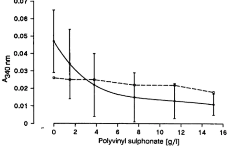
In order to select the Optimum polyvinyl sulphonate concentration, twenty-two rheumatoid factor nephe- lometry-negative sera were analysed, using increased quantities of polyvinyl sulphonate in reagent l. Sera were not heat inactivated. Figure 2 shpws the mean
300 n
100 150
C1q spiked [rrig/l]
200 250
Fig. l Effect of added Clq on different rheumatoid factor assays. Heat-ihactivated positive and negative rheuma- tpid factor samples were spiked with purified Clq from 0 to 250 mg/1 and analysed by:
Δ — Δ ο —ο old turbidimetric method without heat- inactivation;
A — A ·—· old turbidimetric method with heat*in- activation;
Bi_ n Q —ία improved turbidimetric method;
*—* nephelometric method.
0.07 - 0.06 - 0.05 - 0.04 - 0.03 - 0.02- 0.01 - 0 -
4 6 8 10 12
Polyvinyl sulphonate [g/l] 14 16
Fig. 2. Inhibition of the complement effect.
Reactivity (mean ± 2 S. D.) of 22 rheumatoid factor- negative sera versus the inhibitor concentration in the reaction cuvette.
Reactivity of Standard 1: D-D (31.3 χ ΙΟ3 IU/1).
± 2 Standard deviations of the absorbance of these sera measured with the turbidimetric rheumatoid fac- tor assay versus the polyvinyl sulphonate concentra- tion in the reaction cuvette. Complement interference in negative rheumatoid factor samples decreased with the increase in the quantity of polyvinyl sulphonate.
We chose a polyvinyl sulphonate concentration of 15.1 g/l in the reaction cuvette. This concentration of polyvinyl sulphonate produced no decrease in rheu- matoid factor reactivity, and at this polyvinyl sul- phonate concentration all normal serum samples tested gave rheumatoid factor values lower than the cut-off level of the assay. Moreover, unspecific pre- cipitations were not observed in 60 serum samples in the turbidimetric rheumatoid factor test to which no heat aggregated IgG had been added (sample blank).
Reactivity of heat aggregated IgG of differ- ent degrees of aggregation
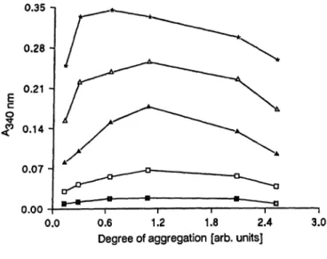
The aggregation process of IgG was studied by heat- treatment at 63 °C of seven identical aliquots of a solution containing 50 g/l IgG and 5 g/l bovine serum albumin in saline, using heating times between 5 and 45 minutes. The degree of aggregation is expressed s the absorbance at 340 nm of a 1/10 dilution in antigen diluent of the different heat aggregated IgG prepa- rations. Figure 3 shows the reactivity of these heat aggregated IgG preparations. This experiment was carried out 2 weeks after heating. Maximum reactivity was obtained when the degree of aggregation was between 0.9 and 1.5 absorbance units, which corre- sponds to approximately 20 minutes of thermal treat- ment.
Eur. J. Clin. Chem. Clin. Biochem.,/ Vol. 29,1991 / No. 8
524 Borque et al.: Automated turbidimetry of rhcumatoid factor
0.35 -i
0.28-
|0.21 -
0.14-
0.07 -
0.00
0.0 0.6 1.2 1.8 2.4 Degree of aggregation [arb. units]
3.0 Fig. 3, Reactivity of Standards with different degrees of antigen
aggregation.
Degree of aggregation is expressed s the absorbance at 340 nm of 1/10 dilutions in antigen diluent of the different aggregated-IgG preparations.
rheumatoid factor, l O3 IU/1:
D-D 31.3; α-α 62.5; Δ - A 125; Δ - Δ 250;
*-* 500.
Stability of the aggregation in time
The stability of heat aggregated IgG preparations obtained in the previous experiment was studied. The antigens were kept at 4 °C and their absorbance at 340 nm were measured at different times after the initial heating (15 min, 24 h, 7 days, 14 days, 21 days, 42 days and 4 months). Figure 4 shows that, after 2 weeks, no increase in the degree of aggregation of the IgG was observed. However, the heat aggregated IgG
which was kept 45 min at 63 °C became unstable, resulting in precipitation after 48 hours.
Heat aggregated IgG used afterwards had a degree of aggregation of 0.7, s measured immediately after heating, and 1.1 absorbance units after 2 weeks of stabilization. ·f
Analytical performances
Calibration curve: Figure 5 shows the mean ± 2 Standard deviations of 10 calibration curves obtained on 10 different days. The increase in absorbance from 30-500 χ 103 IU/1 was higher than 0.3 absorbance units.
0.400 η
0.300 -
0.200 -
0.100 -
100 200 300 400 Rheumatoid factor [103 IU/I]
500
Fig. 5. Rheumatoid factor calibration curve for the improved method. Absorbance (mean ± 2 S. D. of 10 calibrations) versus Standard concentrations.
3.0 Ί
7/Γ
1η 2.4 Η Α Ϊ Ι - β Η toσ>
Sg 1-2-1 'S0)
£ 0.6-
0.0 1—
14 d —l—
15 min 24 h 7 d 14 d 21 d 42 d 421 d Time after antigen preparation m Fig. 4. Stability of aggregated-IgG preparations in time.
The increase of aggregation in time is expressed s the
"degree of aggregation" (absorbance at 340 nm of 1/10 dilutions in antigen diluent of the different antigens).
The different aggregated-IgG preparations are ex- pressed by the heating times (in minutes) employed in their preparation.
Δ - Δ 5;n-a 10; V - Υ 15; G-o20;*-*25;
o-o 30; ©-045.
Precision: Three pools of sera with different rheu- matoid factor values were obtained. Intra-assay pre- cision was determined with a single rotor charged with 12 aliquots of each pool. Inter-assay precision was evaluated by analysing one aliquot of each con- centration in 12 different r ns with a new calibration curve each time. Precision data are given in table 1.
For interference studies we.follpwed the procedure of Glick (13). We assessed the effect of bilirubin and haemoglobin by adding known amo nts of these s b- stancestoab selineserumpoolcontaining.165 χ l O3
IU/1 of rheumatoid factor. interference from lipaemia was assessed by adding, various amo nts of a 20%
Intralipid solution to the same pool. Figure 6 shows interferences assoeiated with addition of haemoglo- bin, bilirubin and Intralipid. No effects were seen with levels up to 340 μπιοΐ/ΐ for bilirubin and 2300 mg/1 for haemoglobin. A negative interference was found when Intralipid was added. After treatment of the Intralipid spiked samples with "Lipoclean" Clearing agent, the rheumatoid factor activity was completely recovered (x = 101.1 ± 1.8%*'a = 4).
Eur. J. Clin. Chein. Clin. Biochem. / Vol. 29,1991 / No. 8
Borque et al.: Automated turbidimetry of rheumatoid factor 525 Tab. 1. Precision data
Rheumatoid factor (l O3 1U/1)
Mean S. D.
12 n = 12
CV (%) Intra-assay*
Sample I Sample II Sample III Inter-assayb
Sample I Sample II Sample III
12350 225 12754 223
2.51.6 4.5 2.94.6 6.7
3.12.0 2.0 5.33.6 3.0
0 0.20 0.40 0.60 0.80 1.00 Concentration of interfering substance, fraction of highest level Fig. 6. Interference studies in the improved turbidimetric test.
On the abscissa are represented the cöncentrations of interfering substances, expressed äs % of highest level:
V — V Intralipid 0.5%; A — A haemoglobin 2300 mg/1;
D—D bilirubin 340 >1/1; ·—· Intralipid-spiked series treated with "Lippclean" Clearing agent.
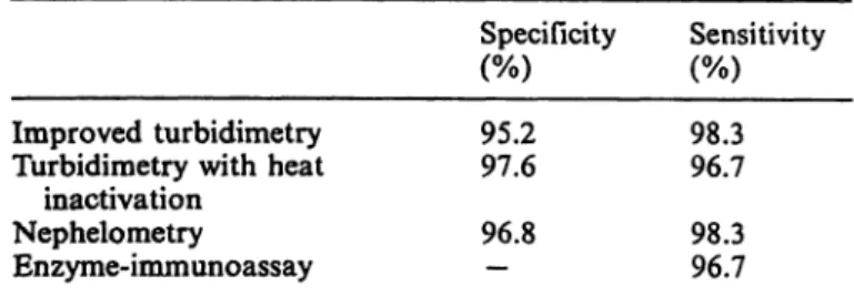
Tab. 2. Relative sensitivities and specificities of the several rheumatoid factor methods
Specificity Sensitivity Improved turbidimetry
Turbidimetry with heat inactivation
Nephelometry Enzyme-immunoassay
95.297.6 96.8—
98.396.7 98.396.7
Tab. 3. Method comparison (correlation coefficients)
Enzyme-immunoassay Nephelometry Turbidimetry with
heat inactivation
Improved turbidi- metry
0.923 0.964 0.996
Turbidi- metry with heat inacti- vation 0.917 0.962
Nephelo- metry
0.926
Correlations between all the assays are shown in table 3. It can be seen that the coefficients of correlation between methods were all higher than 0.90. In figure 7, the serum values obtained with the improved method are plotted against the three other tests.
Values obtained from the previous comparison studies were used to calculate relative analytical sensitivities.
A serum was considered rheumatoid factor-positive or rheumatoid factor-negative when it was, respec- tively, positive or negative in at least 3 out of 4 tests.
The analytical relative sensitivity (tab. 2) is defined äs the ratio of true positives to the sum of true positives and false negatives for each method.
Relative analytical specificity of the im- proved rheumatoid factor test
Rheumatoid factqr äctivity of 130 routine samples from our laboratory was analysed by the nephelo^
metric and by the improved and old turbidimetric rnethöds. A serum was considered rheumatoid factor- positive or rheumatoid factor-negative whenever at least two out of three test results were, f espectively, positive or negative. The relative specificities ob- tained, defined äs the ratio of true negatives to the sum of true negatives and false positives for eaeh method, äfe shöwn in table 2.
Method comparison and relative analytical sensitivity ,
A wide ränge of rheumatoid factor cöncentrations in sera was measured by the turbidimetric method with chemical inhibitor, and by the other three methods.
Discussion
Several authors have described the binding of poly- anions to the Clq fraction of complement (15 — 17).
Although dextran sulphate and heparin inhibited Clq binding to IgG, unspecific precipitations occurred in the samples, disqualifying the use of these inhibitors in the turbidimetric assay of rheumatoid factor. We researched the possibility of using polyvinyl sulphon- ate äs Clq inhibitor, in view of its chemical similarity with the abpve mentioned polyanions.
Complement interference, which required previous in- activation of samples at 56 °C for 30 minutes, was avoided by adding polyvinyl sulphonate to the reac- tion medium. Unspecific precipitations were not ob- served with this polyanion. The harmful effect of heat- pretreatment of samples on endogenous rheumatoid factor has been described (5, 18). Elimination of this step, therefore enabled us to avoid this inconvenience
Eur. J. Clin. Chem. Clin. Biochera. / Vol. 29,1991 / No. 8
526 Borque et al.: Automated turbidimetry of rheuraatoid fafctor oo
in(M
oo
S
_ οοin
oo
μ o
-§
U)oo inCM
oo oCM
oo in
oo o
oo in
fi/ni eoiJ
OO O
Oo om
8*5 "n
ooinCM oo oOJ
oo
o o
[|/niecH]
g
jopej pioieujnaijy"
ι
•S
l
2α
ε
£ «
s 2 ,
£> °^
ε<=>
s* s n
<2S
-o ·«·*
l'
.ss
3α S
C g II
•822 H*>
2 J§' - -S-r 8G Λ 7J
O w G O cd in
II o
•S 1 n
[Ι/ΠΙ gOI.]
(poiflauj paAOjduuj '
u ^|3 Ebb
and achieve complete automation of rheumatoid fac- tor turbidimetry.
By determining reactivity for different degrees of ag- gregation, it was possible to standardize the prepa- ration of heat-aggregated IgG- For this purpose, the 30 minute heat treatment at 63 °C (19) is replaced by more homogene us and reprod cible final conditions, giving maximal reactivity in the measurement of rheu- matoid factor. The heat-aggregated IgG so obtained, stored at 4 °C, continues to aggregate for approxi- mately 2 weeks, and cannot be used until then. It then remains stable for at least 4 months, in contrast to the aggregated-IgG obtained by other investigators (20).
This improved method shows intra- and inter^assay CVs similar to those of the original procedure (11).
Thus, even for low rheumatoid factor concentrations, the CVs were less than 5.5%, values comparable or better than those of othef quantitative techniques (3, 21 —23). The test has excellent relative analytical spec- ificity and sensitivity compared with other assays (24, 25). The correlation coefficient between the two tur- bidimetric procedures was exceptional (r = 0.996), probably due tp the fact that both use the same antigen and the same apparatus. On the other hand, the discrepancy in bias might be explained by the effect of heat inactivation (5). Also, the improved turbidimetry procedure correlates well with the ne- phelometric assay (r = 0.964), but not so well with an enzyme-immunoassay (r = 0.923). Correlation of the enzyme-immunoassay values with those of the rest of the procedures was in every case worse than the other comparisons (tab. 3), a result already obtained by other authors (26, 27). Proportional differences between the present method and the nephelometric and enzyme-immunoassay tests were considerable.
This indicates possible differences in the standardiza- tion of the procedures, although the calibrations in all c ses were in accordance with the WHO Standard;
similar observations have been reported elsewhere (27).
In conclusion, addition of polyvinyl s lphonate to-the reagent buffer of a turbidimetric procedure for rheu- matoid factor quantification eliminates the time-con- suming sample heat-inactivation and removes the er- ror assoeiated with this treatment on rheumatoid fac- tor values (5). This improved method correlates well with other quantitative assays, showing the advan- tages of complete automation, simple Operation and feduced costs, The analytical quality similar to that of the other methodologies. In addition, the procedure
"can be easily automated in other analytical Systems.
Eur. J. Clin. Chern. Clin. Biochem. / Vol. 29 1991 / No, 8
Borque et al: Automated turbidimetry of rheumatoid factor 527 Acknowledgement
We thank Dr. H. Dubais (Boehringer Mannheim GmbH, Mann- heim, Germany) for providing the isolated human Clq prepa-
ration. This work was supported in part by the Fondo de Inyestigaciones Sanitarias de la Seguridad Social (Grant No 90/0330).
References
1. Zrein, M., De Marcillac, G. & Van Regenmortel, M. H. V.
(1986) Quantitation of rheumatoid factor by enzyme im- munoassay using biotinylated human IgG. J. Immunol.
Methods 87, 229-237.
2. Masi, A. T., Maldonado-Cocco, J. A., Kaplan, S. B., Fri- genbaum, S. L. & Chandler, R. W. (1976) Prospective study of the early course of rheumatoid arthritis in young adults:
comparison of patients with and without rheumatoid factor positivity at entry and identification of variables correlating with outcome. Semin. Arthritis Rheum. 5, 299—326.
3. Painter, P. C, Lyon, J. M., Evans, J., Powers, W. W., Whittaker, R. L. & Decker, M. J. (1982) Performance of a new rate-nephelometric assay for rheumatoid factor, and its correlation with tube-titer results for human sera and synovial fluid. Clin. Chem. 28, 2214—2218.
4. Melamies, L. M., Ruutsalo, H. M. & Nissilä, H. (1986) Evaluation of a quantitative immunoturbidimetric assay for rheumatoid factor. Clin. Chem. 32, 1890-1894.
5. Borque, L., Ruiz, R. & Rus, A. (1990) Effect of serum heat treatment on rheumatoid factor measurement (Abstract).
Quim. Clin. 9, 242.
6. Renata, A., Rodrick, M., Knobel, H. R. & Isliker, H. (1979) Inhibition of the interaction between the complement com- ponent Clq and immune complexes. Int. Axchs. Allergy Appl. Immun. 58, 140-149.
7. Hughes-Jones, N. C. & Gadner, B. (1978) The reaction between the complement subcomponent Clq, IgG com- plexes and polyionic molecules. Immunology 34,459—463.
8. Rent, R., Ertel, N., Eisenstein, R. & Gewürz, H. (1975) Complement activation by interaction of polyanions and polycations. I. Heparin protamine induced consumption of complement. J. Immunol. 114, 120—124.
9. Yachin, S., Rosenblum, D. & Chatman, D. (1964) Biologie properties of polynucleotides. V. Studies on the Inhibition of the flrst component of complement by polyinosinic acid:
the interaction with Clq. J. Immunol. 93, 542—548.
10. Cooper, N. R. & Morrison, D. C. (1978) Binding and activation of the first component of human complement by the lipid A region of lipopolysaccharide. J. Immunol.
120, 1862-1868.
11. Borque, L., Yago, M., Mär, C. & Rodriguez, C (1986) Turbidimetry of rheumatoid factor in serum with a centrif- ugal analyzer. Clin. Chem. 32, 124-129.
12. Collins, R. J., Neu, J. C. & Wilson, R. J. (1990) Rate nephelometric determinatipns of rheumatoid factor: com- parison between Kallestad QM-300 and Beckman ICS-II (RF) methods. J. Clin, Pathol. 43, 243-245.
13. Glick, M. R., Ryder, K. W. & Jackson, S. A. (1988) Graph- ical comparisons of interferences in clinical chemistry In- strumentation. Clin. Chem. 32, 470—475.
14. Passing, H. & Bablok, W. (1983) A new biometrical pro- cedure for testing the equality of measurements from two different analytical methods. J. Clin. Chem. Clin. Biochem.
27, 709-720.
15. Loos, M. (1982) Antibody-independent activation of Cl.
The first component of complement. Ann. Immunol. (Inst.
Pasteur) 133C, 165-179.
16. Zeipeli, G., Hanson, H-S. & Stedingk, L-V. (1977) Purifi- cation from euglobulin of the first component (Cl) of complement and its subcomponents by heparin-sepharose chromatography. Acta Pathol. Microbiol. Scand. 85, 123 — 17. Takada, A. & Takada, Y. (1980) Interaction of Cl130. s and Cl
inactivator in the presence of heparin, dextran sulfate and protamine sulfate. Thromb. Res. 18, 847-859.
18. Soltis, D., Hasz, D., Morris, M. J. & Wilson, I. D. (1979) The effect of heat inactivation of serum on aggregation of immunoglobulins. Immunology 35, 37—45.
19. Yago, M., Borque, L., Mär, C. & Rodriguez, C. (1985) A stable heat-aggregated IgG preparation suitable for rheu- matoid factor determination using fluid-phase precipitin techniques. Rev. Diag. Biol. 34, 375-377.
20. McCarthy, D. A., Field, M., Mumford, R, Moore, S. R., Holborow, E. J. & Maini, R. N. (1985) Soluble IgG aggre- gates produced by heating remain stable on freeze-drying.
J. Immunol. Methods 82, 155-160.
21. Borque, L., Cambiaso, C. L. & Masson, P. L. (1987) Im- munoassay of rheumatoid factor by latex particle counting.
Clin. Chem. 33, 704-707.
22. Winkles, J. W., Lunec, J. & Gray, L. (1989) Automated enhanced latex agglutination assay for rheumatoid factor in serum. Clin. Chem. 35, 303-307.
23. Desjerlais, F. & Daigneault, R. (1985) Rheumatoid factors measured in serum with a fully automated laser nephelo- meter, and correlation with agglutination tube titers. Clin.
Chem. 31, 1077-1078.
24. Moxley, G. (1989) Heightened sensitivity of quantitative ELISA for IgM rheumatoid factor with the use of the Biotin-Streptavidin System. Am. J. Clin. Pathol. 92, 630- 25. Carpenter, A. B. & Bartkowiak, C. D. (1989) Rheumatoid636.
factor determined by fluorescence immunoassay: compari- son with qualitative and quantitative methods. Clin. Chem.
35, 464-466.
26. Lu, A. C., Pierce, C. S., Batholomew, W. R. & Kalinka, C.
B. (1985) Discordant rheumatoid factor values for reference controls. Clin. Chem. 31, 1913-1914.
27. Jaspers, J. P. M. M., Van Oers, R. J. M. & Leerkes, B.
(1988) Nine rheumatoid factor assays compared. J. Clin.
Chem. Clin. Biochem. 26, 863 — 871.
Dr. Luis Borque de Larrea Laboratorio Bioquimica Hospital San Millan c/Autonomia de la Rioja, 3 E-26004 Logrono-La Rioja Spain
Eur. J. Clin. Chem. Clin. Biochem. / Vol. 29,1991 / No. 8