Screening for components involved in NLR-mediated immune signalling
I n a u g u r a l – D i s s e r t a t i o n
zur
Erlangung des Doktorgrades
der Mathematisch-Naturwissenschaftlichen Fakultät der Universität zu Köln
vorgelegt von
Harald Frank Bielig
aus Neuss
Köln 2012
Berichterstatter: Prof. Dr. Jonathan Howard Prof. Dr. Mats Paulsson
Tag der mündlichen Prüfung: 13.04.2012
Table of contents
Table of contents I
Abbreviations III
Symbols for amino acids VIII
1 Introduction 1
1.1 Innate immunity and pattern recognition receptors 1
1.2 NLRs 3
1.3 NOD1 and NOD2 5
1.3.1 Signalling pathways and regulation 5 1.3.2 Bacterial sensing and physiological relevance of NOD1 and NOD2 8
1.4 Apoptosis: Programmed cell death 13
1.5 The BIRC-family: Functions beyond inhibition of apoptosis 14 1.6 RNAi and systematic screening approaches 16
1.7 Aim of the study 18
2 Materials and Methods 19
2.1 Materials 19
2.1.1 Cell lines and bacteria 19
2.1.2 Chemicals, reagents and enzymes 21
2.1.3 Kits 22
2.1.4 Plasmids 22
2.1.5 Primer 23
2.1.6 siRNAs 24
2.1.7 Antibodies 25
2.1.8 Instruments 25
2.1.9 Software 26
2.2 Methods 27
2.2.1 Cell biological methods 27
2.2.2 Screening protocols 31
2.2.3 Molecular biological methods 34
2.2.4 Biochemical methods 38
3 Results 41
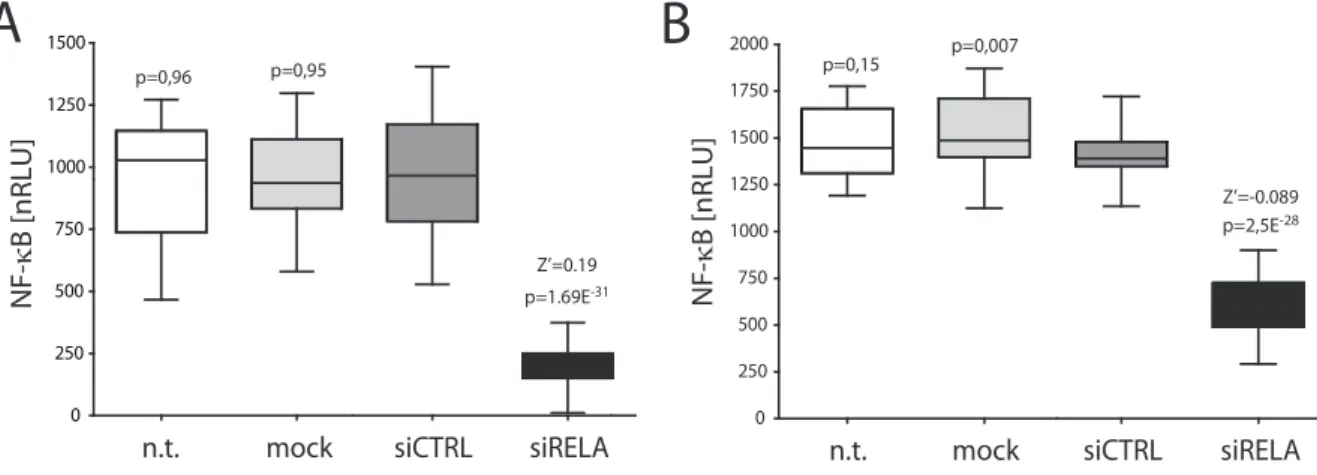
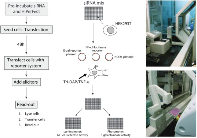
3.1 High-throughput siRNA screen to identify factors involved in NOD1 signalling 41 3.1.1 Establishment of the assay system in HEK293T cells 42 3.1.1.1 Establishment of the reporter system 42 3.1.1.2 Establishment of the siRNA knock-down conditions 45 3.1.1.3 Proof of principle for the differential read-out 46 3.1.1.4 Validation of the assay principle 47 3.1.1.5 Downscaling and automation of the protocol 48 3.1.2 Pilot screen with an apoptosis library 49
3.1.3 Druggable genome screen 52
3.1.3.1 Primary druggable genome screen 52 3.1.3.2 Validation and TNF-α counter-screen 53
3.1.4 THP1-Blue validation screen 55
3.1.4.1 Assay setup, downscaling and automation 55
3.1.4.2 THP1 screen and results 59
3.1.5 Final hitlist generation 60
3.1.6 Hit validation: XIAP 62
3.1.7 Hit validation: BMPR-2 65
3.1.8 Analysis of the roles of BIRC proteins in NOD1 signalling 68 3.2 Bacterial outer-membrane vesicles trigger NOD-mediated inflammatory responses
in a quorum sensing-dependent manner 70
3.2.1 Activation of NOD1 and NOD2 by extracellular bacteria 70 3.2.2 The role of quorum sensing in subverting host-detection by NODs 73
4 Discussion 77
4.1 Screen results 77
4.1.1 Pilot screen 77
4.1.2 Druggable genome screen 78
4.1.3 Comparison with published screens 81 4.1.4 XIAP and BMPR-2 are involved in NOD1 signalling 84 4.2 Roles for BIRC proteins in NOD1 signalling 88 4.3 V. cholerae OMVs trigger NOD signalling in a quorum sensing-dependent manner 90
5 References 93
6 Abstract 108
7 Zusammenfassung 110
Danksagung 112 Erklärung 113 Kollaborationserklärung 114
Lebenslauf 115
Abbreviations
°C degree Celsius
μg microgramme
μl microlitre
A desoxyadenosine
A20 TNFAIP3, tumor necrosis factor, alpha-induced protein 3 AAMP angio-associated migratory cell protein
ADP adenosine diphosphate
AIM2 absent in melanoma 2
AMP anti-microbial peptide
AP-1 activator protein 1
APAF1 apoptotic protease-activating factor 1
APS ammonium persulfate
ASC apoptosis-associated speck-like protein containing a CARD ATG16L1 ATG16 autophagy related 16-like 1 (S. cerevisiae)
ATP adenosine triphosphate
BCL-2 B cell lymphoma 2
BID BH3 interacting domain death agonist BIR baculovirus inhibitory repeat
BIRC baculoviral IAP repeat containing
BISC BMP-induced signalling complex
BMDMs bone marrow-derived macrophages
BMP bone morphogenetic protein
BMPR bone morphogenetic protein receptor bp basepairs
Bruce also BIRC6, Appollon
BS Blau syndrome
BSA bovine serum albumin
C desoxycytidine CARD caspase activating and recruitment domain caspase cysteine-dependent aspartate-directed proteases
CC coiled-coil
CD Crohn’s disease
CDH1 cadherin 1, type 1; also E-cadherin
cDNA complementary DNA
CENTB1 Centaurin beta 1
CHUK conserved helix-loop-helix ubiquitous kinase; also IKKα
CHX cycloheximide
cIAP cellular inhibitor of apoptosis CLR C-type lectin receptor
C-terminus carboxyl-terminus
CTNNB1 -catenin
CUL1 cullin 1
CYLD cylindromatosis (turban tumor syndrome) DAMP danger-associated molecular pattern
DAP diaminopimelic acid
DIAP2 Drosophila IAP 2
DISC death-induced signalling complex
DMEM Dulbecco’s modified Eagle’s medium
DMSO dimethylsulfoxide
DNA desoxyribonucleic acid
dNTP desoxynucleotide triphosphate
ds double stranded
EDTA ethylenediaminetetraacetic acid ELISA enzyme-linked immunosorbent assay
EM electron microscopy
Erbin Erbb2 interacting protein
ERK extracellular-signal-regulated kinase; also p44/42 et al. et alii; and others
FBS fetal bovine serum
g gravitational force
g gramme
G desoxyguanosine GAPDH glycerinaldehyd-3-phosphat-dehydrogenase GDF-5 growth and differentiation factor 5
GEF-H1 guanine nucleotide exchange factor H1
GO gene ontology
GPR17 G protein-coupled receptor
GRIM-19 gene associated with retinoid-interferon-induced mortality 19
GTP guanosine triphosphate
h hour
HapR hemagglutinin/protease regulatory protein (V. cholerae) HEK human embryonic kidney cells
HIN200 hematopoietic interferon-inducible nuclear antigens with 200 amino acid repeats HtrA2 High temperature requirement protein A2
HTS high troughput screening
HUVSMCs human umbilical vein smoth muscle cells
HRP horseradish peroxidase
i.e. id est; that is
ie-DAP γ- D -glutamyl-meso-diaminopimelic acid IBD inflammatory bowel disease
IBM IAP-binding motif
IFI16 interferon--inducible protein 16
IFN interferon
Ig immunoglobulin
IKK IB kinase
IL interleukin ILP-1 IAP-like protein 1, also BIRC4, XIAP ILP-2 IAP-like protein 2, also BIRC8
imd Immune deficiency pathway (Drosophila)
IRF IFN regulatory factor
ITCH itchy E3 ubiquitin ligase homolog (mouse) IBα NF-B inhibitor, alpha; also NFKBIA
JNK c-Jun N-terminal kinase
kb kilobasepairs
kDa kilodalton
LGP2 laboratory of genetics and physiology 2 LPS lipopolysaccharide
LRR leucine-rich repeat
M molar MAPK mitogen-activated protein kinase mc monoclonal
MCL Markov Cluster Algorithm
MDA5 melanoma differentiation associated gene 5 ML-IAP melanoma-IAP, also BIRC7, Livin
MDP muramyl dipeptide
MEF mouse embryonic fibroblast
MEKK4 mitogen-activated protein kinase kinase kinase 4
mg milligram
min minute
ml millilitre
mM millimolar MOI multiplicity of infection
MOMP Mitochondrial outer-membrane permeabilisation
mRNA messenger RNA
MyD88 myeloid differentiation primary response gene 88 NACHT domain present in NAIP, CIITA, HET-E and TP1 NAIP neuronal apoptosis inhibitory protein; also BIRC1
NBD nucleotide-binding domain
NBS-LRRs nucleotide-binding site leucine-rich repeat NEMO NF-B essential modulator; also IKK
NF-AT nuclear factor of activated T-cells
NF-B Nuclear factor -light-chain-enhancer of activated B cells ng nanogram
NIK NF-B-inducing kinase
NLR nucleotide-binding domain and leucine-rich repeat containing protein NOD Nucleotide-binding oligomerization domain
NOS3 nitric oxide synthase 3 nRLU normalised relative light unit N-terminus amino-terminus
OD optical density
OMP Outer membrane protein (V. cholerae)
OMVs outer-membrane vesicles
ONPG o-Nitrophenyl--D-galactopyranosid PAGE polyacrylamid gelelectrophorese
p50 NFKB1, nuclear factor of kappa light polypeptide gene enhancer in B-cells 1
PBS phosphate buffered saline
pc polyclonal
PCR polymerase chain reaction
PepT human peptide transporter
PFC Pre-formed complex
PGN peptidoglycan
PLK polo-like kinase
PMA phorbol 12-myristate 13-acetate
PRF1 perforin 1
PRR pattern-recognition receptor
PYD pyrin domain
PYHIN pyrin and HIN domain-containing protein qRT-PCR quantitative real-time PCR
RAC1 ras-related C3 botulinum toxin substrate 1
RELA v-rel reticuloendotheliosis viral oncogene homolog A; also p65 R protein resistance protein
RIG-I retinoic acid-inducible gene I RING really interesting new gene
RIP2 receptor-interacting serine/threonine-protein kinase 2; also RIPK2, RICK RISC RNA-induced silencing complex
RLR RIG-I-like receptor
RNA ribonucleic acid
RNAi RNA interference
RNF31 ring finger protein 31,; also HOIP
RNP ribonucleoprotein complex
ROS reactive oxygen species
rpm rounds per minute
RpoS RNA polymerase subunit sigma S
RPS-4 TIR-NBS-LRR class disease resistance protein
RT room temperature
RT-PCR reverse transcription PCR
SCF Skp, Cullin, F-box containing complex
SD standard deviation
SDS sodium dodecyl sulphate
SEAP secreted alkaline phosphatase sec seconds
shRNA short hairpin RNA
siRNA small interfering RNA
SMAC second mitochondria-derived activator of caspase SNAI1 snail homolog 1 (Drosophila)
SOP standard operating procedure
ss single stranded
SSH1 slingshot-homolog 1 (Drosophila)
ssRNA single-stranded RNA
Survivin also BIRC5
T desoxythymidine
TAB TAK1-binding protein
TAK1 transforming growth factor (TGF)-beta activated kinase; also MAP3K7 TR1 TGF-beta type I receptor
TBE TRIS-Borate-EDTA
TBK TANK-binding kinase
TBS TRIS buffered saline
TEMED N,N,N’,N’-Tetramethylethylendiamin Tetra-DAP L -Ala- D -Glu-meso-DAP- D -Ala (Tetra-DAP) TGF- transforming growth factor (TGF)-beta Th1, Th2 type I helper T cell, type II helper T cell
TIR Toll-IL-1 receptor
TNFR1 TNF receptor 1, also TNFRSF1A TNF-α tumor necrosis factor-alpha TRAF TNF receptor-associated factor Tri-DAP L -Ala- D --Glu-mDAP
TRIF Toll/IL-1R domain containing adaptor inducing IFN-β TRIS Tris(hydroxymethyl)-aminomethan Triton X-100 polyethylene glycol p-(1,1,3,3-tetramethylbutyl)-phenyl ether Tween 20 polyoxyethylen-(20)-sorbitan monolaureate
UV ultraviolet
V volt
WT wild type
XIAP X-linked inhibitor of apoptosis; also BIRC4, ILP-1 XLP X-linked lymphoproliferative syndrome
XPS X-ray photoelectron spectroscopy
Symbols for amino acids
Amino acid Three letter code One letter code
Alanine Ala A
Arginine Arg R
Asparagine Asn N
Aspartate Asp D
Cysteine Cys C
Glutamate Glu E
Glutamine Gln Q
Glycine Gly G
Histidine His H
Isoleucine Ile I
Leucine Leu L
Lysine Lys K
Methionine Met M
Phenylalanine Phe F
Proline Pro P
Serine Ser S
Threonine Thr T
Tryptophane Trp W
Tyrosine Tyr Y
Valine Val V
1 Introduction
1.1 Innate immunity and pattern recognition receptors
All multicellular organisms are permanently challenged by pathogens, such as bacteria, viruses, fungi and protozoans. Therefore it is of crucial importance for the organism to be able to detect harmful agents and to launch a potent defence response for its own protection. All animals and plants possess an innate immune system that relies on germline encoded invariant pathogen receptors and is characterised by an instantaneous reaction. Vertebrates and jawed fish have additionally evolved an adaptive (or acquired) immune system that relies on somatic recombina- tion of genes and clonal expansion of B- and T-lymphocytes. The adaptive immune system provides an unlimited number of receptors against all possible types of pathogens and is char- acterised by a delayed response.
The receptors of the evolutionary older innate immune system are invariant; they detect a limited number of conserved and mostly invariant patterns of the pathogens that are not prone to structural changes provoked by mutations, referred to as P ATHOGEN -A SSOCIATED
M OLECULAR P ATTERNS (PAMPs) (reviewed in Medzhitov and Janeway, 1997). These patterns, also referred to as “microbial non-self ”, such as bacterial cell wall components like peptidogly- can, flagellin, lipopolysaccharide (LPS) or nucleic acids, are recognised by specialised P ATTERN
R ECOGNITION R ECEPTORS (PRRs) (reviewed in Akira et al., 2006) that are situated on the surface of or within the host cell. Additionally, the innate immune system surveys the integrity of cells and tissues, as it also reacts to D ANGER -A SSOCIATED M OLECULAR P ATTERNS (DAMPs). These are endogenous substances that are normally spatially confined from PRRs, such as cellular DNA, ATP or uric acid, or substances that are produced upon injury, such as reactive oxygen species (ROS). Upon cellular rupture, DAMPs are released and cause an innate immune reaction mediated by PRRs (Medzhitov, 2007).
PRRs are highly expressed in innate immune cells like macrophages and dendritic cells (DCs), but they are also abundant in a variety of non-professional immune cells like epithelial cells or fibroblasts and mediate the onset of immune responses (reviewed in Takeuchi and Akira, 2010).
PRRs are compartmentalised; there are specialised receptors that monitor the extracellular milieu, the vesicular compartment and the intracellular milieu. Transmembrane receptors detect extracellular pathogens and pathogens that enter the vesicular compartment. There are also PRRs that are secreted into the extracelluar milieu (reviewed in Litvack and Palaniyar, 2010).
Invasive pathogens that avoid detection by surface receptors can be detected by intracellular
PRRs that monitor the cytoplasm.
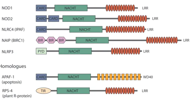
PRRs are evolutionary conserved. Toll-like receptors (TLRs), for example, are found in many organisms, including insects, fish and humans (reviewed in Aderem and Ulevitch, 2000). An- other striking example of the conservation of PRRs among different organisms are the resis- tance proteins (R-proteins) of the nucleotide-binding site leucine-rich repeat (NBS-LRR) class found in plants that share high structural similarities with mammalian nucleotide binding and oligomerisation domain (NOD)-like receptors (NLRs) (reviewed in Maekawa et al., 2011) (Fig. 1) . In most cases, activated PRRs trigger signalling cascades that culminate in inflammatory re- sponses, mainly mediated by activation of the transcription factor nuclear factor B (NF-B) and of mitogen-activated protein kinases (MAPKs). That in turn leads to the onset of antim- icrobial responses, such as phagocytosis, autophagy, production of reactive oxygen species (ROS), degranulation or secretion of antimicrobial peptides, which have direct antimicrobial effects. Furthermore, activation of PRRs leads to secretion of inflammatory chemokines and cytokines that mediate inflammation and recruit immune cells, such as macrophages or neutro- phils, to the site of infection. Some PRRs also induce antiviral responses by triggering type I interferon (IFN) expression (reviewed in Takeuchi and Akira, 2010). Additionally, this first de- fence reaction mediated by the innate immune system is essential to trigger and to direct a full adaptive immune response (Fritz et al., 2007; Dabbagh and Lewis, 2003).
There are at least four classes of PRRs: TLRs and the C-type lectin receptors (CLRs), which are membrane-bound and RIG-I-like receptors (RLRs) and NLRs, which are localised in the cytoplasm. The most extensively studied PRRs are the TLRs. Receptors of that family are exclu- sively membrane-bound and either face the extracellular milieu or the vesicular compartment.
Pattern recognition is mediated by leucine-rich repeats (LRRs) pointing to the extracellular mi- lieu, whereas the intracellular Toll/IL-1R homology (TIR) domain serves intracellular down- stream signalling. In humans, there are 10 TLRs known so far, conferring protection against a wide variety of bacteria and viruses. They display a very broad substrate specificity reaching from cell wall components, such as lipopolysaccharide (LPS), lipoproteins or flagellin to nucleic acids, such as CpG DNA, double-stranded RNA (dsRNA) and single stranded RNA (ssRNA).
Upon activation, TLRs trigger NF-B- and MAPK-mediated expression of pro-inflammatory cytokines and interferon response factor (IRF) 3/IRF7-mediated expression of type I IFNs via the adaptor proteins MyD88 (myeloid differentiation primary response gene 88) and TRIF (Toll/IL-1R domain containing adaptor inducing IFN-β), respectively (reviewed in Akira and Takeda, 2004).
Similar to the TLRs, CLRs are also membrane-bound. They sense carbohydrates present on
fungi, viruses and bacteria through a carbohydrate-binding domain and trigger MAPK-, NF-B-
and NF-AT (nuclear factor of activated T-cells)-mediated expression of pro-inflammatory cyto-
kines upon activation. CLRs also seem to be involved in the regulation of TLR-mediated re- sponses (reviewed in Geijtenbeek and Gringhuis, 2009).
RLRs, in contrast, are localised in the cytoplasm. They contain one or two N-terminal caspase activation and recruitment (CARD) domains, a central DEAD box helicase/ATPase domain and a C-terminal regulatory domain. Thus far, there are three known members: RIG-I (retinoic acid inducible gene I), MDA5 (melanoma-differentiation associated gene 5) and LGP2 (labora- tory of genetics and physiology 2). RIG-I and MDA5 confer protection to viruses through rec- ognition of viral nucleic acids via their C-terminal domains. LGP2, in contrast, might not rec- ognise nucleic acids directly, but was shown to negatively regulate RIG-I and MDA5 signalling.
Activation of RIG-I and MDA5 triggers IRF3/IRF7-dependent type I IFN production and expression of inflammatory cytokines (reviewed in Takeuchi and Akira, 2010).
Recently, a novel class of PRRs has been discovered. The pyrin and HIN200 (hematopoietic interferon-inducible nuclear antigens with 200 amino acid repeats) domain containing proteins absent in melanoma 2 (AIM-2) and interferon--inducible protein 16 (IFI-16) have been shown to be sensors for cytoplasmic foreign DNA. Engagement of these receptors triggers inflamma- tory responses, such as pro-IL-1 processing and IFN signalling through formation of multi- protein complexes termed I NFLAMMASOMES (Unterholzner et al., 2010; Burckstummer et al., 2009).
1.2 NLRs
In humans, there are 23 NLR members identified so far; many of these act as cytoplasmic PRRs.
They are expressed in various cell types, including myeloid and epithelial cells. Characteristic for NLRs is a tripartite domain structure similar to the structure of the apoptotic protease- activating factor 1 (APAF-1) (Fig. 1) . The C-termini of NLRs consist of leucine-rich-repeats (LRRs), whereas APAF-1 contains C-terminal WD40 repeats. The LRRs are thought to mediate recognition of elicitors. Furthermore, all members contain a central oligomerisation domain referred to as present in NAIP, CIITA, HET-E and TP-1 (NACHT). NLRs are divided into sub-groups based on their N-terminal domains that consist of pyrin (PYD), baculovirus inhibi- tory repeat (BIR) or CARD domains (reviewed in Wilmanski et al., 2008) (Fig. 1) . These motifs have in common that they mediate protein-protein interactions.
NLRs can be roughly divided into two functional groups: I NFLAMMASOME and N ODOSOME
NLRs. Some PYD-containing NLRs, such as NLRP3 and NLRP1, but also BIR-containing
NLRs, such as NLRC4, are referred to as inflammasome NLRs. Upon activation, these proteins
interact with the adaptor protein ASC (apoptosis-associated speck-like protein containing a
CARD), leading to formation of large multimeric protein complexes that contain NLR proteins, ASC and pro-caspase-1. The induced proximity leads to auto-activation of caspase-1, which in turn mediates processing and secretion of the pro-inflammatory cytokines IL-1β and IL-18.
This inflammatory response consists of a two-step process: Expression of pro-caspase-1, pro- IL-1β and pro-IL-18 is triggered by detection of microbial substances by TLRs, but a second signal, provided by the inflammasome, is required for maturation and secretion of the cytokines.
Inflammasome NLRs display very broad substrate specificities. They can be activated by nu- merous bacteria and bacterial toxins and by a variety of DAMPs, but also by crystalline aggre- gates, such as asbestos, urea crystals or cholesterol crystals (reviewed in Schroder and Tschopp, 2010). For most cases, there is no proof for direct interactions of NLRs with their elicitors available, suggesting that their activation occurs via intermediate factors, and may not rely on direct ligand binding, analogous to the example of TOLL in Drosophila that recognises the ligand Spaetzle, which is proteolytically cleaved by upstream cascades upon PAMP recognition (reviewed in Valanne et al., 2011).
NOD1 NOD2 NLRC4 (IPAF) NAIP (BIRC1) NLRP3
APAF-1 (apoptosis)
NACHT CARD
CARD CARD NACHT
CARD NACHT
NACHT NACHT
NACHT CARD
BIR PYD
BIR BIR
LRR LRR LRR
LRR LRR
WD40
NACHT LRR
RPS-4 TIR
(plant R-protein) NLR proteins
Homologues
Figure 1. Domain architecture of several NLR proteins and homologues. The NLR family is characterised by a tripartite domain composition similar to that of the pro-apoptotic factor APAF-1. The C-termini consist of lecine-rich repeats (LRRs) involved in detection of pathogens, the central parts contain so-called present in NAIP, CIITA, HET-E and TP1 (NACHT) domains that mediate oligomerisation. The N-termini consist of effec- tor domains, such as caspase activation and recruitment (CARD), pyrin (PYD) or baculovirus inhibitory re- peat (BIR) domains. NLR proteins share close homology with plant NBS-LRR R-proteins, such as RPS-4 (based on Fritz et al., 2006; Meylan et al., 2006).
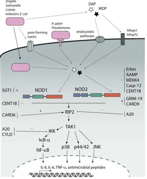
A sub-group of CARD domain containing NLRs, namely NOD1 and NOD2, are referred to as nodosome NLRs. NOD1 and NOD2 detect bacterial peptidoglycan (PGN) fragments with different substrate specificities, upon activation they trigger signalling cascades that culminate in activation of NF-B and MAPKs. This mediates expression of pro-inflammatory cytokines, chemokines and anti-microbial peptides (Fig. 2) (reviewed in Kufer, 2008).
1.3 NOD1 and NOD2
1.3.1 Signalling pathways and regulation
Human NOD1 is encoded by the CARD4 gene (Bertin et al., 1999), NOD2 by the CARD15 gene (Ogura et al., 2001). NOD1 and NOD2 share the typical tripartite domain structure com- mon to all NLR proteins consisting of C-terminal LRRs, a central NACHT domain and N- terminal effector domains consisting of one or two CARD domains, respectively (Fig. 1) .
NOD1 and NOD2 show a predominantly cytoplasmic localisation, however, recent reports indicate that they also localise to the plasma membrane upon activation (Zurek et al., 2012; Tra- vassos et al., 2010; Kufer et al., 2008; Barnich et al., 2005a; McDonald et al., 2005). The exact role of this membrane-association still remains elusive, however, it seems to be dependent on actin and actin remodelling enzymes, such as RAC1 (ras-related C3 botulinum toxin substrate 1) (Eitel et al., 2008; Legrand-Poels et al., 2007).
NOD1 and NOD2 were the first NLR proteins that were described to detect microbial PAMPs. In recent years, a multitude of publications elucidated the main signalling cascades em- ployed by these proteins. Recognition of PGN by NOD1 and NOD2 is thought to occur through the LRRs, as mutants lacking this domain proved to be incapable of PGN-sensing (Girardin et al., 2005; Tanabe et al., 2004). However, there is no solid evidence for a direct inter- action of the LRRs with PGN so far. There is also the possibility that NOD1 and NOD2 might act as downstream adaptors of so far unidentified PRRs, as this is the case for NLRC4 that acts downstream of the actual PRR NAIP (Kofoed and Vance, 2011; Zhao et al., 2011). A similar mechanism has been proposed for NLRP3, as there is evidence that NLRP3-mediated re- sponses to PGN require upstream activation of NOD2 (Pan et al., 2007). However, this impor- tant issue still needs clarification.
Under normal conditions, NOD1 and NOD2 are thought to be kept in an inactive state. Fol-
lowing recognition of PGN, these molecules likely undergo conformational changes that render
them active by exposing the NACHT-domains that mediate homo-oligomerisation of the mole-
cules. The exact nature of such conformational changes has not been worked out so far; the
presumed model is based on structural information on the activation of APAF-1 (reviewed in Riedl and Salvesen, 2007), which is closely related to NLRs (Fig. 1) . Oligomerisation leads to re- cruitment and activation of the serine-threonine kinase RIP2 (Receptor interacting protein 2) via homophilic CARD-CARD interactions (Inohara et al., 2000). This creates a molecular signal- ling platform. RIP2 thereby acts as a molecular scaffold for the recruitment of downstream components. The kinase activity of RIP2 itself is not essential for this function, but it is re- quired to stabilise the protein (Hasegawa et al., 2008; Windheim et al., 2007). NOD1- as well as NOD2-mediated recruitment of RIP2 promotes conjugation of RIP2 with K63-linked ubiq- uitin within the kinase domain at lysine 209. In contrast to K48-linked ubiquitination, K63- linked ubiquitination does not target proteins for proteasomal degradation, but serves as a regu- latory mechanism, especially in NF-B signalling (reviewed in Chen, 2005). K63-linked ubiquitin chains on RIP2, possibly attached by the E3 ligases TRAF2, TRAF5 (TNF receptor associated factor) and ITCH (itchy E3 ubiquitin ligase homolog (mouse)) (Tao et al., 2009; Hasegawa et al., 2008), serve as docking sites for TAK1 (transforming growth factor- activated kinase 1) (Hasegawa et al., 2008). Recruitment of TAK1 is mediated by the ubiquitin-binding proteins TAB1 (TAK binding protein) and TAB2 that form a complex with TAK1 (Hasegawa et al., 2008; Abbott et al., 2007). Polyubiquitinated RIP2 also serves as a docking site for the IKK- complex by direct interaction with the regulatory subunit IKK (NEMO, NF-B essential modifier). In NOD2 signalling, IKK itself gets conjugated with K63-linked ubiquitin chains at lysine 399 in a RIP2-dependent manner (Abbott et al., 2004). The interactions of RIP2 with IKK and TAK1 establish a close proximity between the IKK complex and the TAB/TAK complex, thus mediating TAK1-dependent activation of IKKα and IKK. The activated IKK complex phosphorylates the NF-B inhibitory protein inhibitor of B (IB) that sequesters NF-B in the cytoplasm. Phosphorylation of IB triggers its proteasomal degradation, causing NF-B to translocate to the nucleus and activate its target genes (reviewed in Hayden and Ghosh, 2008). Additionally, there is evidence that NOD2 can also activate the alternative NF-
B pathway by interaction with the NF-B-inducing kinase (NIK) in a RIP2-independent man- ner (Pan et al., 2006).
Interestingly, the TLR pathways leading to NF-B activation utilise partly the same ubiquitina-
tion mechanisms as the NOD1/2 pathway, for example, ubiquitination of IKK at the same site
is essentially involved (reviewed in Akira and Takeda, 2004). NOD1- and NOD2-dependent
activation of TAK1 via RIP2 also induces c-Jun N-terminal kinase (JNK) and p38 MAPK-
pathways (da Silva Correia et al., 2007; Windheim et al., 2007; Opitz et al., 2006; Girardin et al.,
stood, but there is evidence that the protein CARD9 is essentially involved at least in NOD2- mediated p38 activation (Hsu et al., 2007) (Fig. 2) .
Erbin AAMP MEKK4 Casp-12 CENT1B GRIM-19 CARD9
SGT1 NOD1 NOD2
RIP2
TAK1
p38 p44/42 JNK NF- κ B
IKK I κ B- α
hPept1 hPepT2 MDP
Shigella DAP
Salmonella Listeria enteroinv. E. coli
H. pylori Pseudomonas
endocytotic pathways
+
+ -
IL-8, IL-6, TNF- α , antimicrobial peptides
A20
A20 CYLD
-
- CENT1B -
CARD6
pore-forming toxins
+
Figure 2. Uptake of PGN triggers NOD1- and NOD2-dependent signalling cascades. Bacterial PGN can gain access to the cytoplasm by invasion of bacteria, by injection via bacterial secretion systems, by pH- dependent endocytotic events and by the transmembrane translocators hPepT1 and hPepT2. Intracellular PGN fragments trigger NOD1 and NOD2 signalling cascades that involve RIP2 and TAK1, leading to expres- sion of pro-inflammatory cytokines, chemokines and antimicrobial peptides. Factors that are known to regulate NOD signalling are indicated. For further information please refer to the main text (based on Le Bourhis et al., 2007).
Overwhelming inflammatory responses can be detrimental to the host, therefore the NOD1
and NOD2 signalling cascades have to be tightly controlled. Even though NOD1 and NOD2
share the same core signalling cascade, they appear to be partly differentially regulated by asse-
sory proteins (Fig. 2) .
A common factor that dampens NF-B activation in NOD1 and NOD2 signalling, but also in TLR signalling, is the de-ubiquitinating enzyme A20 (also known as TNFAIP3; tumor necrosis factor, alpha-induced protein 3), which reverses K63-linked ubiquitination of RIP2 (Hasegawa et al., 2008; Hitotsumatsu et al., 2008). NOD2-specific K63-linked ubiquitination of IKK can also be reversed by A20 and the deubiquitinating enzyme CYLD (cylindromatosis (turban tumor syndrome)) (Zhang et al., 2006; Boone et al., 2004). There are more examples of factors that uniquely regulate NOD2 signalling. For instance, it has been shown that MEKK-4 (mitogen- activated protein kinase kinase kinase 4) and caspase-12 down-regulate NOD2-mediated re- sponses by interfering with the NOD2/RIP2 interaction (Clark et al., 2008; LeBlanc et al., 2008).
Furthermore, it was reported that the protein Erbin (erbb2 interacting protein) binds NOD2 and exerts a negative regulatory function on NOD2 signalling (Kufer et al., 2006; McDonald et al., 2005). Recently, we discovered that the angio-associated migratory cell protein (AAMP) also interacts with NOD2 and negatively influences NOD2-mediated NF-B activation (Bielig et al., 2009). GRIM19 (gene associated with retinoid-IFN-induced mortality-19), in contrast, has been described to positively contribute to NOD2-mediated NF-B activation (Barnich et al., 2005b).
The regulation and fine-tuning of NOD1 signalling, in contrast, is less well characterised.
Thus far, it has only been shown that the GTPase-activating protein centaurin- 1 (CENTB1) negatively regulates NOD1 and also NOD2 signalling. (Yamamoto-Furusho et al., 2006). Fur- thermore, CARD6 exerts positive effects on NOD1-mediated NF-B activation by interaction with RIP2. However, these effects are not very strong and CARD6 also seems to act on NF-B pathways triggered by other stimuli (Dufner et al., 2006).
The regulatory networks that control positive- and negative-regulation of NOD1 and NOD2 are still very poorly defined. For Erbin and CENT1B there is evidence that they serve as feed- back inhibitors, as their expression is up-regulated following NOD1 and NOD2 stimulation (Kufer et al., 2006; Yamamoto-Furusho et al., 2006). Given the potentially dangerous effects of overwhelming inflammation, it can be assumed that NOD signalling is tightly regulated, as this is the case for TLR signalling (reviewed in Akira and Takeda, 2004). However, to date it is still not known how detection of elicitors occurs, or how NOD-mediated inflammatory responses are terminated, for example.
1.3.2 Bacterial sensing and physiological relevance of NOD1 and NOD2
Peptidoglycan (PGN) is present in the cell wall of nearly all bacteria and is essential for con-
structing the cell shape. PGN is constantly remodeled during cell-growth and -division (re-
viewed in Park and Uehara, 2008). There are recycling mechanisms, but at least some PGN and degradation products are shed by growing bacteria (reviewed in Boneca, 2005).
NOD1 specifically recognises diaminopimelic acid (DAP)-containing PGN fragments that are produced as breakdown-products during growth of Gram-negative bacteria. The minimal struc- tural motif required for NOD1-activation was identified to be γ- D -glutamyl-meso-diaminopimelic acid (ie-DAP), a dipeptide that serves to crosslink the carbohydrate-backbones of Gram- negative PGN (Chamaillard et al., 2003; Girardin et al., 2003c). Human NOD1 preferentially recognises L -Ala- D -Glu-meso-DAP (Tri-DAP) containing PGNs. Of note, murine NOD1 has a different substrate specificity as it is activated better by L -Ala- D -Glu-meso-DAP- D -Ala (Tetra- DAP) than by ie-DAP (Girardin et al., 2003a). Thus NOD1 should provide protection primarily against Gram-negative bacteria. Consistently, it has been shown that NOD1 detects the invasive Gram-negative pathogens Shigella flexneri (Girardin et al., 2001), enteroinvasive Escherichia coli (Kim et al., 2004) and Chlamydia spp. (Welter-Stahl et al., 2006; Opitz et al., 2005), among others. It has also been shown that NOD1-deficient mice have a higher bacterial burden after infection with the Gram-negative pathogen H. pylori compared to WT mice (Viala et al., 2004). In addition, NOD1 also recognises some Gram-positive bacteria that contain DAP-type PGNs, such as Ba- cillus spp. or Listeria monocytogenes (Hasegawa et al., 2006; Opitz et al., 2006).
NOD2, in contrast, recognises the PGN-moiety MurNac- L -Ala- D -isoGln (MDP, for muramyl dipeptide), the minimal motif common to all PGNs that is conserved in Gram-negative as well as in Gram-positive bacteria (Girardin et al., 2003b; Inohara et al., 2003). Due to its ability to sense MDP, NOD2 should be able to detect a very broad spectrum of bacteria. Indeed, it has been shown that NOD2 detects a variety of Gram-positive and Gram-negative bacteria, such as Streptococcus pneumoniae, Salmonella typhimurium and Mycobacterium tuberculosis (Ferwerda et al., 2005;
Opitz et al., 2004; Hisamatsu et al., 2003). NOD2 also detects a huge variety of PGNs of heat killed bacteria in vitro (Hasegawa et al., 2006). Furthermore, it has been shown that NOD2 is essentially involved in protection against L. monocytogenes in the intestine of mice (Kobayashi et al., 2005).
Bacterial pathogens can be roughly divided into two groups: Extracellular bacteria, which evade phagocytic uptake and/or subsequent lysosomal degradation and invasive bacteria, which ac- tively enter host cells. The latter either reside freely within the cytosol or stay entrapped and replicate within altered compartments of the host’s endocytic pathway (so-called “pathogen- containing vacuoles”) (reviewed in Raupach and Kaufmann, 2001).
Due to their cytoplasmic localisation, NOD1 and NOD2 are able to detect invasive bacteria,
such as S. flexneri or L. monocytogenes that invade host cells and reside freely within the cytosol. It
has been shown that PGN fragments derived from invasive Shigella activate NOD1-signalling in epithelial cells (Nigro et al., 2008). Furthermore, MDP can potentially be generated by secreted bacterial autolysins in cells infected with L. monocytogenes (Lenz et al., 2003). There is also evi- dence that NOD1 and NOD2 detect bacteria that normally reside within intracellular vacuoles, such as Legionella pneumophila (Berrington et al., 2010; Frutuoso et al., 2010), S. typhimurium (Le Bourhis et al., 2009) or Mycobacterium tuberculosis (Brooks et al., 2011; Pandey et al., 2009). However, it is not well understood how in general PGN is translocated from the vesicular compartment to the cytosol where it is sensed by NOD1 and NOD2, although translocation via pore-forming toxins and peptide transporters is involved in some cases (see below).
Initially, it has been presumed that only bacteria colonising intracellular niches are recognised by intracellular PRRs and that extracellular bacteria are detected by cell surface receptors, for example by TLRs. Nowadays there is increasing evidence that extracellular bacteria can also be detected by intracellular PRRs. This raises the question how the hydrophilic PGN fragments shed by extracellular bacteria are delivered into the host cells. One possibility is delivery via bac- terial secretion systems, as it has been shown that H. pylori can deliver PGN fragments into the cytosol of host cells via its type IV secretion system encoded by the cag pathogenicity island (Viala et al., 2004). It is also possible that pore-forming toxins secreted by some bacteria facili- tate entry of PGN to host cells, as it has been observed that NOD1 activation by PGN derived from Haemophilus influenzae required the pore-forming toxin pneumolysin from S. pneumoniae.
Similar effects were observed for pore-forming toxins from Bacillus anthracis and Staphylococcus aureus (Ratner et al., 2007).
Host cell factors might contribute to uptake of PGN as well, as it has been shown that clathrin- and dynamin-dependent pathways mediate an active uptake of PGN to the cytosol in a pH-dependent manner (Lee et al., 2009; Marina-Garcia et al., 2009). Moreover, the NOD2- elicitor MDP can specifically be taken up into the cytosol via the plasma membrane-located peptide transporter hPepT1 (human peptide transporter 1), whereas NOD1-elicitors can be taken up by hPepT2 (Swaan et al., 2008; Ismair et al., 2006; Vavricka et al., 2004) (Fig. 2) . However, these mechanisms are still not fully worked out and there are very likely further mechanisms that contribute to PGN uptake.
Whereas NOD1 is widely expressed in many tissues of the stromal and haematopoetic com-
partments, NOD2 shows the highest expression in immune cells like macrophages and dendritic
cells and in paneth cells in the intestinal crypts, but it is also expressed in epithelial cells of the
intestine and the lung (Hitotsumatsu et al., 2008; Uehara et al., 2007; Tada et al., 2005; Ogura et al.,
2003; Ogura et al., 2001; Inohara et al., 1999). NOD1 and NOD2 are indispensable for intestinal
homeostasis and pathogen recognition. As there are myriads of commensals in the gut, expres- sion of TLRs in the adult is restricted in intestinal epithelial cells to prevent constant inflamma- tion (Abreu et al., 2001). Moreover, TLR signalling can be rendered anergic due to constant ex- posure to PAMPs (Shahin et al., 1987). This prevents harmful overwhelming immune reactions to commensals. Pathogens rupturing the epithelial barrier or invading the host cells are subse- quently recognised by cytoplamic NLRs. There is a close cross-talk between NLRs and TLRs ensuring a proper distinction between commensals and pathogens. NOD1 and NOD2 are es- sential to trigger anti-microbial responses in cells that are anergic for TLR signalling in vivo (Kim et al., 2008). Furthermore, there are strong synergistic effects between NOD1 and NOD2 elici- tors and agonists of various TLRs (Fritz et al., 2005; Uehara et al., 2005; van Heel et al., 2005;
Traub et al., 2004).
Activation of NOD1 and NOD2 in epithelial and endothelial cells by bacteria induces expres- sion of pro-inflammatory cytokines and chemokines, such as IL-8 (CXCL-8), a chemokine that mediates the recruitment of neutrophils (Opitz et al., 2006; Kim et al., 2004; Viala et al., 2004).
Furthermore, activation of NOD1 and NOD2 in macrophages and dendritic cells induces the release of IL-8 and the cytokines IL-6, IL-1β and TNF-α (reviewed in Fritz et al., 2006). The important chemo-attractant RANTES (regulated upon activation, normal T-cell expressed and secreted/CCL5) is secreted by primary murine macrophages in vivo following stimulation with NOD agonists, underlining the importance of NOD signalling in co-ordinating innate immune responses (Werts et al., 2007). Importantly, stimulation of NOD1 and NOD2 additionally leads to secretion of anti-microbial peptides (AMPs) in the gut. AMPs are secreted short peptides that kill bacteria by forming pores in bacterial cell membranes; they are the key effectors in cell autonomous immunity, i.e. defence mechanisms that are executed by the attacked cell itself that do not rely on the recruitment of immune cells. They are not only produced by professional immune cells, but also by epithelial cells. Secretion of AMPs is important to control the num- bers of commensal bacteria and is thus essential for maintaining intestinal homeostasis (re- viewed in Auvynet and Rosenstein, 2009; Selsted and Ouellette, 2005). Especially in intestinal crypts, a compartment that is likely kept sterile, NOD2 seems to play a pivotal role in the pro- tection against pathogens. NOD2 is highly expressed in the AMP-secreting paneth cells in the crypts and it triggers secretion of AMPs upon pathogen intrusion (Petnicki-Ocwieja et al., 2009;
Kobayashi et al., 2005). Furthermore, NOD2 controls the expression of the AMP human - defensin in epithelial cells (Voss et al., 2006).
NOD1 as well is crucial for the regulation of intestinal homeostasis. It is required for produc- tion of human and murine -defensins in response to H. pylori infection (Boughan et al., 2006;
Hamanaka et al., 2001) and has been shown to mediate lymphoid tissue genesis induced by com-
mensals in the intestine of mice. Formation of intestinal B-cell containing isolated lymphoid follicles (ILFs) is essentially regulated by recognition of Gram-negative bacteria by NOD1 (Bouskra et al., 2008).
Of note, NOD1 and NOD2 have also been shown to contribute to autophagy by recruiting ATG16L1 (ATG16 autophagy related 16-like 1 (S. cerevisiae)) to the plasma membrane. Auto- phagy is a process that serves to maintain cellular homeostasis and mediates degradation of intracellular pathogens (reviewed in Ramjeet et al., 2010).
Within the last years, it became clear that NOD1 and NOD2 also contribute to the induction of adaptive immune responses. NOD1 and NOD2 elicitors, in particular in synergy with TLR agonists, have been shown to induce Th1- and Th2-type immune responses (Fritz et al., 2007;
Kobayashi et al., 2005; Tada et al., 2005). Furthermore, it has been demonstrated that administra- tion of NOD1 elicitors restored neutrophil function in mice depleted in intestinal microbiota.
Consistently, NOD1 deficient mice proved to be more susceptible to early pneumococcal sepsis due to unefficient neutrophil activation (Clarke et al., 2010).
Taken together, innate immune mechanisms involving NOD1 and NOD2 counter-act bacte- rial infections by the release of anti-microbial factors, recruitment of phagocytes and by mediat- ing the onset of adaptive immune responses. Furthermore, NOD1 and NOD2 are crucial for the regulation of the intestinal homeostasis by mediating interactions with the commensal mi- crobiota.
Consistently, mutations in the CARD4 and CARD15 genes are linked to a variety of inflam- matory disorders. Polymorphisms in the NOD1 gene CARD4 have been associated to the onset of inflammatory bowel disease (McGovern et al., 2005). A different polymorphism in CARD4 seems to increase the susceptibility to asthma and allergy (Eder et al., 2006; Hysi et al., 2005).
Frameshift mutations in the NOD2 gene CARD15 strongly increase the susceptibility to Crohn’s disease (CD), which is a severe form of chronic inflammatory bowel disease (reviewed in Hruz and Eckmann, 2010). Other mutations in CARD15 are associated with the chronic in- flammatory barrier diseases Blau syndrome and early onset sarcoidosis (reviewed in Rosenstiel et al., 2007).
Of note, NOD1 has also been linked to events leading to cell death. In the first description of
NOD1 it was reported that NOD1 binds several caspases and specifically activates caspase-9-
mediated apoptosis (Inohara et al., 1999). Later, it has been shown that NOD1 induces apop-
tosis by RIP2-dependent activation of caspase-8 as well, contributing to the control of tumour
growth (da Silva Correia et al., 2007; da Silva Correia et al., 2006). However, the putative func-
tions of NOD1 in apoptosis are far from being understood.
1.4 Apoptosis: Programmed cell death
Apoptosis, often referred to as programmed cell death, is an evolutionary conserved process in which proteolytic cascades lead to the degradation of cells in a controlled manner. In contrast to necrotic cell death, a toxic process induced by extrinsic factors or injury, apoptosis does not cause an immune reaction. Apoptosis is required for many processes, such as normal cell turn- over, embryonic development and for a proper function of the immune system.
The hallmarks of apoptosis are cell shrinkage, nuclear fragmentation, protein-crosslinking, and chromatin condensation and degradation. The degraded cellular material is engulfed in ve- sicular structures consisting of intact cellular membranes, whereby mitochondria and other or- ganelles stay intact within the apoptotic vesicles. The vesicles are quickly phagocytosed by macrophages and other surrounding cells. As cell membrane integrity is not affected, there is no release of cellular constituents into the extracellular milieu and no immune response following apoptotic cell death (reviewed in Elmore, 2007).
Cellular events that lead to the aforementioned biochemical changes are mediated by a family of aspartate-specific cysteine proteases termed caspases (cysteine-dependent aspartate-directed proteases) (Alnemri et al., 1996). Caspases are ubiquitously expressed as inactive pro-forms and get activated by various apoptotic stimuli. This leads to proteolytic cascades that amplify the initial signals and ensure a rapid degradation of cells by selective cleavage of hundreds of target proteins (Nicholson, 1999).
There are two major pathways that trigger apoptosis: The extrinsic pathway and the intrinsic pathway. The extrinsic pathway relies on activation of tumor necrosis factor receptor (TNFR) family transmembrane receptors by extracellular ligands. Ligand binding induces formation of a multimeric protein complex termed D EATH -I NDUCED S IGNALLING C OMPLEX (DISC) that re- cruits pro-caspase-8. The induced proximity leads to trans-autoactivation of caspase-8, trigger- ing proteolytic cascades by activating effector caspases (Muzio et al., 1998; Kischkel et al., 1995).
The intrinsic pathway is activated by numerous non-receptor stimuli, such as deprivation of
growth factors or cytokines, or by intrinsic cues like DNA damage, endoplasmatic reticulum
(ER) stress, free radicals, radiation, UV light, viral infection or toxins. This leads to
M ITOCHONDRIAL O UTER -M EMBRANE P ERMEABILISATION (MOMP) and subsequent release of
pro-apoptotic factors into the cytosol. This process is tightly regulated by complex interactions
between pro- and anti-apoptotic members of the B cell lymphoma 2 (BCL-2) protein family
(reviewed in Cory and Adams, 2002). MOMP leads to secretion of the pro-apoptotic molecules
cytochrome c, second mitochondria-derived activator of caspase (SMAC) and high temperature
requirement protein A2 (HtrA2). SMAC and HtrA2 inhibit anti-apoptotic factors of the BIRC-
family. Cytochrome c, in contrast, triggers the activation of APAF-1. Activated APAF-1 forms a
heptameric complex termed A POPTOSOME that mediates activation of caspase-9, which in turn cleaves and activates effector caspases, thereby initiating the proteolytic cascades that finally lead to cell death (reviewed in Yuan et al., 2011; Riedl and Salvesen, 2007).
1.5 The BIRC-family: Functions beyond inhibition of apoptosis
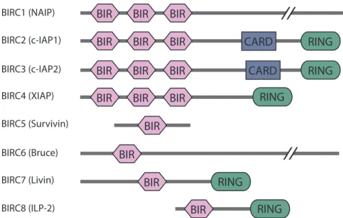
Members of the BIRC-family (baculovirus IAP repeat containing; also termed IAPs, for inhibi- tors of apoptosis) have mainly been investigated in respect of their roles in counter-acting apoptosis. BIRC proteins were initially found in baculoviruses and are highly conserved from viruses to yeast, nematodes and insects to humans (Uren et al., 1998). In humans there are eight members, BIRC1 (NAIP), BIRC2 (c-IAP1), BIRC3 (c-IAP2), BIRC4 (XIAP, ILP-1), BIRC5 (Survivin), BIRC6 (Bruce, Apollon), BIRC7 (Livin, ML-IAP) and BIRC8 (ILP-2). Members of the BIRC family are characterised by the presence of 1-3 tandem baculovirus inhibitory repeat (BIR) domains (Fig. 3) (reviewed in Gyrd-Hansen and Meier, 2010). The BIR domain is a zinc- binding fold containing ~70 amino acid residues that is known to promote protein-protein in- teractions. Most members also contain a really interesting new gene (RING) finger motif that has E3 ubiquitin ligase function (Vaux and Silke, 2005). BIRC2 and BIRC3 additionally contain a CARD domain of yet unknown function.
BIRC proteins are known to inhibit apoptosis either by direct binding of active caspases (Riedl et al., 2001; Deveraux et al., 1997), or by indirect means. Initially, it was thought that all BIRCs inhibit caspases by direct binding, as overexpression of BIRCs effectively protects cells against apoptotic stimuli. Nonetheless, recent studies indicate that only XIAP (BIRC4) is able to inhibit caspases by direct binding under physiological conditions (Eckelman and Salvesen, 2006;
Eckelman et al., 2006). BIRC8 shows a close homology to XIAP, but lacks the first two BIR- domains. In spite of its putative caspase-9 interaction domain, it is only a weak caspase-9 inhibi- tor on its own (Shin et al., 2005). However, it has been shown that over-expression of BIRC8 potently inhibits BAX-induced apoptosis (Richter et al., 2001).
Indirect means to inhibit apoptosis are thought to include mono- and polyubiquitination of
caspases (Morizane et al., 2005; Hao et al., 2004; Suzuki et al., 2001; Huang et al., 2000), or of the
pro-apoptotic factor SMAC (Ma et al., 2006; Morizane et al., 2005; Hao et al., 2004; Hu and Yang,
2003; MacFarlane et al., 2002), leading to proteasomal degradation of these factors. Other re-
ports indicate that BIRC7 and BIRC8 might exert their anti-apoptotic effects by binding and
neutralising SMAC, rather than by direct inhibition of caspases (Shin et al., 2005; Vucic et al.,
2005).
BIRC1 (NAIP) BIRC2 (c-IAP1) BIRC3 (c-IAP2) BIRC4 (XIAP) BIRC5 (Survivin) BIRC6 (Bruce) BIRC7 (Livin) BIRC8 (ILP-2)
BIR BIR BIR CARD RING
BIR BIR BIR CARD RING
RING
RING BIR RING BIR
BIR
BIR BIR BIR
BIR BIR BIR BIR
Figure 3. Domain architecture of the eight members of the BIRC family. The BIRC family is characterised by 1-3 N-terminal baculoviral inhibitory repeat (BIR) domains. Several members also contain caspase activation and recruitment (CARD) domains and/or really interesting new gene (RING) domains. For further details please refer to the main text (based on Salvesen and Duckett, 2002).
BIRC proteins are regulated by pro-apoptotic IAP-binding proteins, such as the mitochondrial proteins SMAC and HtrA2. SMAC has been shown to inhibit the association of XIAP with caspases and mediate the release of caspases to execute their downstream apoptotic functions.
This may also be the case for other BIRCs (Liu et al., 2000; Wu et al., 2000; Deveraux et al., 1997;
Roy et al., 1997), as SMAC also targets BIRC2 and BIRC3 for proteasomal degradation by ubiq- uitination (Yang and Du, 2004). Furthermore, HtrA2 has an intrinsic protease activity and can directly cleave BIRC proteins (Srinivasula et al., 2003; Yang et al., 2003; Hegde et al., 2002; Mar- tins et al., 2002; Verhagen et al., 2002).
Recent reports indicate that BIRC family members also have important functions in other cellular processes. Some members are involved in the regulation of cell division (Verhagen et al., 2001), cellular signalling (Salvesen and Duckett, 2002) and in immune detection of bacterial products. BIRC1, for example, which also belongs to the NLR family (Fig. 1, Fig. 3) , acts as PRR itself; it directly recognises bacterial flagellin and components ot the bacterial type III secretion apparatus to mediate caspase-1 activation and IL-1β processing via formation of a NLRC4- and ASC-dependent inflammasome (Kofoed and Vance, 2011; Zhao et al., 2011). BIRC5 is involved in the segregation of chromosomes in the G2/S-phase of the cell cycle (Skoufias et al., 2000;
Uren et al., 2000), and BIRC6 plays a role in cytokinesis (Pohl and Jentsch, 2008).
Importantly, recent studies indicate that some members of the BIRC family exert important
functions in innate immunity and pro-survival signalling. BIRC2 and BIRC3 are implicated as
key players in the TNFR1 (tumor necrosis factor receptor 1) signalling pathway. They were found to be associated with the TNFR signalling complex (Rothe et al., 1995) where they act as K63 E3 ubiquitin ligases mediating TNFR1-dependent NF-B activation and preventing TNFR1-mediated apoptosis (Bertrand et al., 2008). A recent study revealed that BIRC2 and BIRC3 play similar roles in NOD1 and NOD2 signalling (Bertrand et al., 2009). Furthermore, BIRC2 and BIRC3 also seem to positively regulate MyD88-dependent TLR signalling by medi- ating K48-linked ubiquitionation of TRAF3 (Tseng et al., 2010).
XIAP was also found to be implicated in pro-survival signalling events. Through interactions of its BIR1 domain with the TAB1/TAK1 complex it participates in activation of MAPKs and NF-B (Lu et al., 2007; Sanna et al., 2002; Sanna et al., 1998). Moreover, XIAP is essential for NOD2-mediated immune responses against the invasive pathogen L. monocytogenes (Bauler et al., 2008), indicating a role in innate immunity. This raises the questions how exactly the mentioned BIRC proteins are involved in the regulation of innate immune responses, and if also other BIRC family members contribute to innate immunity.
1.6 RNAi and systematic screening approaches
The principle of RNA-interference (RNAi)-mediated gene-silencing is widely used in research to determine the function of genes in certain biological processes by loss-of-function studies. In this work, we conducted an RNAi-based high-throughput screen (HTS) to identify novel factors involved in the regulation of the NOD1 signalling cascade.
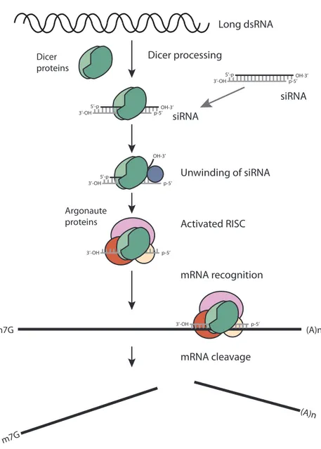
RNAi is a conserved mechanism common to eukaryotic cells. It serves to regulate gene ex-
pression by post-transcriptional gene silencing and provides protection against foreign nucleic
acids, such as transposons and viruses. The first RNAi-based processes, described in the early
1990s, were gene silencing in plants (Jorgensen, 1990) and quelling in the fungus Neurospora
crassa (Romano and Macino, 1992). Evidence for the exinstance of RNAi in animals was then
provided by Fire and colleagues in 1998. They demonstrated that injection of long dsRNA into
the nematode Caenorhabditis elegans triggers the degradation of complementary mRNA (Fire et al.,
1998). Only later it became evident that RNAi also occurs in mammalian cells (Elbashir et al.,
2001). Recently, a related RNA-based defence mechanism was also described for bacteria and
archea (Horvath and Barrangou, 2010), underlining the ubiquitous importance of RNA-based
mechanisms as protection systems against viruses and mobile genetic elements. Today, RNAi-
based methods are widely used as standard methods for reverse genetics, and also therapeutic
applications are being investigated.
Long dsRNA
mRNA recognition
mRNA cleavage Activated RISC Unwinding of siRNA siRNA
Dicer proteins
Dicer processing
OH-3’
3’-OH p-5’
5’-p
siRNA
OH-3’
3’-OH p-5’
5’-p
OH-3’
3’-OH p-5’
5’-p
3’-OH p-5’
3’-OH p-5’