Pathways in Class I Glutamine Amidotransferases
Dissertation
zur Erlangung des Doktorgrades der Naturwissenschaften (Dr. rer. nat.) der
Fakultät für Biologie und vorklinische Medizin der Universität Regensburg
vorgelegt von Florian Semmelmann
aus Deggendorf
Juli 2019
Die Arbeit wurde angeleitet von: Prof. Dr. Reinhard Sterner
Unterschrift: ...
Florian Semmelmann
chemie II des Institutes für Biophysik und Physikalische Biochemie der Fakultät für Biologie und Vorklinische Medizin der Universität Regensburg unter Leitung von Prof. Dr. Reinhard Sterner angefertigt.
Diese Arbeit wurde vom 01.01.2016 bis zum 31.12.2018 durch ein
Promotionsstipendium der Konrad-Adenauer-Stiftung gefördert.
Class I glutamine amidotransferases (GATases) represent a ubiquitous family of enzymes that catalyze the incorporation of ammonia within various metabolic pathways. GATases are het- eromeric enzyme complexes consisting of at least one pair of glutaminase and synthase subunits in various oligomeric associations. The activities of the glutaminase and synthase subunits are allosterically coupled in most class I GATases in a way that the presence of substrate at the synthase subunit stimulates glutamine hydrolysis at the glutaminase active site. The structural basis of allosteric communication between both subunits is, however, not well understood.
The first part of this thesis focuses on the molecular basis of allosteric coupling in 4-amino-4- deoxychorismate synthase (ADCS), an important but hitherto sparsely characterized member of class I GATases. Using ancestral sequence reconstruction, we generated a thermostable glu- taminase subunit, anc 2PabA, solved its crystal structure by molecular replacement and used it to compute for the first time a reliable model of the E. coli ADCS complex. Alanine scanning of conserved residues located between the glutaminase and synthase active sites revealed a net- work of mainly charged residues that lead to activity-inducing conformational changes at the glutaminase subunit. A central aspartate residue of the synthase subunit that is located at the synthase-glutaminase interface close to the active site of the glutaminase subunit, was identified as key residue for both complex formation and allosteric signaling.
The second part of this thesis describes the bioinformatic search and biochemical characterization
of similarly located aspartate residues in the remaining members of class I GATases. The
data confirms the important role of the identified aspartate residues for complex formation and
allosteric signaling and is integrated toward a unifying allosteric core mechanism for class I
GATases.
This thesis is composed of the following manuscripts:
A Adapted with permission from " Semmelmann, F. , Straub, K., Nazet, J., Rajendran, C., Merkl, R. & Sterner, R. (2019). Mapping the Allosteric Communication Network of Aminodeoxychorismate Synthase. Journal of Molecular Biology , 431(15), 2718-2728.
Copyright (2019) Elsevier."
B Adapted with permission from " Semmelmann, F. , Hupfeld, E., Heizinger, L., Merkl, R., & Sterner. R. (2019). A Fold-Independent Interface Residue is Crucial for Complex Formation and Allosteric Signaling in Class I Glutamine Amidotransferases. Biochem- istry , 58(22), 2584-2588. Copyright (2019) American Chemical Society."
In the course of this work, I contributed to further publications that are not part of this thesis:
C Semmelmann, F. , Kabeya, N., Malcicka, M., Bruckmann, A., Broschwitz, B., Straub, K., Merkl, R., Monroig, O., Sterner, R., Ruther, J. & Ellers, J. (2019). Func- tional characterisation of two ∆12-desaturases demonstrates targeted production of linoleic acid as pheromone precursor in Nasonia . Journal of Experimental Biology , 222(10), jeb201038.
D Semmelmann, F. , Hofferberth, J., Ruther, J. & Sterner, R. (2019). Mapping key amino acid residues for the epimerase efficiency and stereospecificity of the sex pheromone biosynthetic short-chain dehydrogenases/reductases of Nasonia . Scientific Reports , 9(1), 330.
E Rohweder, B., Semmelmann, F. , Endres, C. & Sterner, R. (2018). Standardized
cloning vectors for protein production and generation of large gene libraries in Es-
cherichia coli . BioTechniques , 64(1), 24-26.
Publication A
The research was designed by myself and Reinhard Sterner. Kristina Straub performed ancestral sequence reconstruction and Julian Nazet performed homology modeling. Chitra Rajendran collected diffraction data. All other experiments were performed by myself. The publication was written by myself, Rainer Merkl, and Reinhard Sterner with contributions from Kristina Straub and Julian Nazet.
Publication B
The research was designed by myself and Reinhard Sterner. Leonhard Heizinger and myself
provided computational tools and scripts. Enrico Hupfeld contributed data on the imidazole
glycerole phosophate synthase. All other experiments were performed by myself. The publication
was written by myself, Rainer Merkl, and Reinhard Sterner with contributions from Leonhard
Heizinger and Enrico Hupfeld.
Abstract iv
References of Published Manuscripts v
Personal Contributions vi
List of Figures ix
List of Tables x
1 General Introduction 1
1.1 A Short Introduction to Allostery . . . . 1
1.2 The Family of Glutamine Amidotransferases . . . . 2
1.2.1 Structural Diversity Within the Synthase Subunits of Class I GATases . . 4
1.2.2 Structural and Functional Insights into 4-Amino-4-Deoxychorismate Syn- thase . . . . 5
1.3 Aim and Scope of This Work . . . . 7
1.4 Guide to the Following Chapters . . . . 7
2 Mapping the Allosteric Communication Network of Aminodeoxychorismate Synthase 8 2.1 Abstract . . . . 8
2.2 Introduction . . . . 8
2.3 Results . . . 10
2.3.1 Crystal Structure Analysis of PabA and Homology Modeling of ADCS . . 10
2.3.2 Alanine Scanning for the Identification of Residues Involved in Allosteric Signal Propagation . . . 11
2.3.3 Identification of Four Branches of a Conserved Allosteric Communication Network . . . 13
2.3.4 Analysis of a PabA Mutant with Increased Basal Glutaminase Activity . 14 2.4 Discussion . . . 15
2.5 Material and Methods . . . 18
2.5.1 Cloning and Mutagenesis . . . 18
2.5.2 Gene Expression and Protein Purification . . . 18
2.5.3 Ancestral Sequence Reconstruction of PabA . . . 19
2.5.4 Crystallization, Data Collection, and Structure Determination . . . 20
2.5.5 Analysis of Complex Formation Between Different Synthases and Glutam- inases . . . 20
2.5.6 Steady-State Enzyme Kinetics . . . 20
2.5.7 Thermal Stability of Purified Proteins . . . 21
2.5.8 Accession Numbers . . . 21
2.5.9 CRediT Authorship Contribution Statement . . . 21
2.5.10 Acknowledgments . . . 22
2.5.11 Appendix A. Supplementary data . . . 22
2.5.12 Keywords . . . 22
2.5.13 Abbreviations Used . . . 22
2.6 Supplemental Information . . . 23
3 A Fold-Independent Interface Residue Is Crucial for Complex Formation and Al- losteric Signaling in Class I Glutamine Amidotransferases 43 3.1 Abstract . . . 43
3.2 Introduction, Results, and Discussion . . . 43
3.3 Supporting Information . . . 50
3.3.1 Experimental Procedures . . . 50
3.3.2 Supplementary Figure and Tables . . . 53
4 Comprehensive Summary, Discussion, and Outlook 58 4.1 Comprehensive Summary . . . 58
4.2 Discussion: Reconciliation of the Postulated Activation Mechanisms with an En- semble View of Allostery . . . 59
4.3 Outlook: Antimicrobial Potential of Class I GATases Involved in Chorismate Metabolism . . . 60
References 62
Acknowledgment 70
1.1 Model of hemoglobin . . . . 2
1.2 Reaction catalyzed by GATases and metabolic fates of ammonia . . . . 3
1.3 Structural diversity observed in class I glutamine amidotransferases . . . . 4
1.4 Crystal structures of autonomous PabB structures from E. coli and S. maltophilia 5 1.5 Proposed reaction mechanisms of PabB . . . . 6
2.1 Reaction catalyzed by ADCS. . . . 9
2.2 Structural comparison of anc 2PabA to homologous class I glutaminase subunits. 11 2.3 Allosteric residues in ADCS. . . 13
2.4 Activated variants of ADCS. . . 15
2.5 Reaction mechanism of class I glutaminases. . . 16
2.6 Endpoints of the allosteric communication pathways in ADCS. . . 18
2.7 Phylogenetic tree used for sequence reconstruction of ancestral PabA proteins. . 23
2.8 Inter-domain hydrogen bonding network of ADCS. . . 24
2.9 Standard size exclusion chromatography trace for E. coli ADCS. . . 25
3.1 Identification of d-Asp-s residues forming polar interactions with a-Res-g residues in the vicinity of c-His-g. . . 45
3.2 Analytical size exclusion chromatography with wild-type and d-Asp-s mutant GATases. . . 46
3.3 Sequence Logos of Experimentally Characterized Class I GATases. . . 53
4.1 Oxyanion hole flip in pyridoxal 5’-phosphate synthase . . . 59
4.2 Ensemble view of allostery . . . 60
4.3 Interaction hot-spot residues in ADCS, AS, and PhzE . . . 61
2.1 Thermal stability of anc 1-3PabA and modern PabA enzymes. . . 26
2.2 Multiple sequence alignment of ancestral and modern PabA enzymes . . . 27
2.3 Sequence identities/similarities between analyzed PabA enzymes. . . 28
2.4 SEC experiments to determine complex formation. . . 29
2.5 Stimulation of the PabA glutaminase activity by PabB or PabB + chorismate . . 31
2.6 Data collection and refinement statistics for anc 2PabA. . . 34
2.7 Sequence comparisons of enzymes used for molecular replacement and modeling. 35 2.8 Oligonucleotides used for cloning and site-directed mutagenesis. . . 37
2.9 Steady-state kinetic parameters for the glutamine-dependent ADCS activity. . . 39
2.10 Amino acid sequences of anc1-3 PabA . . . 41
2.11 Amino acid sequences of E. coli aminodeoxychorismate synthase . . . 42
3.1 Steady-state glutaminase activity kinetics of wild-type and d-Asp-s mutant GATases 47 3.2 Overview of Identified d-Asp-s and a-Res-g Residues in Class I GATases. . . 54
3.3 Oligonucleotides Used for Site-Directed Mutagenesis. . . 55
3.4 Amino Acid Sequences of Analyzed Enzymes. . . 56
General Introduction
"It is certain that all bodies whatsoever, though they have no sense, yet they have perception; for when one body is applied to another, there is a kind of election to embrace that which is agreeable, and to exclude or expel that which is ingrate; and whether the body be alterant or altered, evermore a perception precedeth operation;
for else all bodies would be like one to another."
Francis Bacon (about 1620)
1.1 A Short Introduction to Allostery
Allostery is the regulatory phenomenon by which proteins convey the effect of ligand binding at one site to a distant functional site (Motlagh et al., 2014). The term allostery (from greek allos for "other" and stereos for "solid") was first articulated in the 1960s by Monod, Changeux, and Jacob in their pioneer work on "Allosteric Proteins and Cellular Control Systems" (Monod et al., 1963). Ever since, allostery has elicited much attention both because of its exceptional physiological importance and the challenge its biophysical interpretation offered (Monod et al., 1965). For the latter, the advent of protein X-Ray crystallography techniques and especially the determination of the hemoglobin crystal structure (Figure 1.1) (Perutz et al., 1960), the prime example of allosteric proteins, preluded the era of the static view of allostery , which was to become the reigning paradigm to describe allostery for decades thereafter. This interpretation of allostery was centered around crystal structures of apo and holo forms of proteins and thus delivered a qualitative view of allostery that was greatly influenced by static structural images of end-point protein structures (i.e. unbound or tensed (T) vs. bound or relaxed (R) state). In order to describe the observed biochemical and crystallographic data, the (i) concerted model (Monod et al., 1965) and the (ii) sequential model (Koshland Jr et al., 1966) were developed.
Whereas the concerted model interprets the coupling of subunits as absolute, the sequential
model interprets the coupling between ligand binding and conformational change of an individual
subunit as absolute. More recently, however, it has been postulated that a functional state is
not necessarily linked to either the tensed or relaxed state of a protein. Rather, changes in the
conformational heterogeneity (local unfolding, intrinsic disorder, and conformational dynamics) of proteins seem to contribute to allosteric transitions (Hilser, 2010). This view of allostery is now commonly referred to as the ensemble view of allostery . Despite its importance, the physical basis of allosteric transitions has only been well understood for very few enzyme systems (Lisi and Loria, 2016) and a prerequisite for detailed analysis of allosteric transitions are structural data to interpret the observed allosteric regulation patterns. Studies on model systems are thus pivotal to further decipher the molecular basis of allosteric transitions and to eventually unravel potential universal patterns.
Figure 1.1: Model of hemoglobin. The haem groups are indicated as gray disks. The four protomers are shown in white and black. Figure reprinted by permission from Springer Nature: Nature (Perutz et al., 1960), copyright 1960.
1.2 The Family of Glutamine Amidotransferases
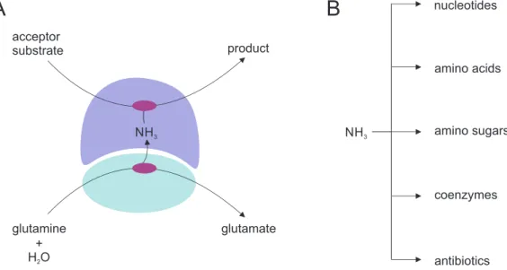
The heteromeric enzyme complexes of the family of glutamine amidotransferases (GATases) are well-established model systems to study the molecular basis of allosteric communication (Mouilleron et al., 2011; List et al., 2012b; Lisi et al., 2018; Negre et al., 2018). GATase enzyme complexes consist of at least one pair of glutaminase and synthase domains/subunits that are lo- calized either on the same or on different polypeptide chains. The glutaminase domain/subunit hydrolyzes glutamine to glutamate and nascent ammonia, which is channeled to the synthase domain/subunit active site where it is incorporated into specific acceptor substrates (Huang et al., 2001) (Figure 1.2 A). GATases catalyze the incorporation of ammonia into a variety of ac- ceptor substrates within different metabolic pathways (Figure 1.2 B) (Zalkin and Smith, 1998).
Whereas the synthase subunits belong to several, structurally unrelated protein families, the
glutaminase domains/subunits can be categorized into two classes according to their fold and
active site composition. Class I GATases comprise enzyme complexes whose glutaminase sub-
units possess a catalytic Cys-His-Glu triad and an α / β hydrolase fold (Ollis et al., 1992). This
class consists of the enzyme complexes imidazole glycerol phosphate synthase (ImGPS) (Chaud-
huri et al., 2003; Douangamath et al., 2002), anthranilate synthase (AS) (Knöchel et al., 1999;
Morollo and Eck, 2001; Spraggon et al., 2001), 4-amino-4-deoxychorismate synthase (ADCS) (Parsons et al., 2002; Bera et al., 2012), carbamoylphosphate synthetase (CPS) (Thoden et al., 1997), guanosin-5‘-monophosphate synthase (GMPS) (Tesmer et al., 1996), formylglycinamide ribonucleotide synthetase (FGARS) (Anand et al., 2004), cytidin 5’-triphosphate synthetase (CTPS) (Goto et al., 2004), 2-amino-2-deoxyisochorismate synthase (PhzE) (Li et al., 2011) and pyridoxalphosphate synthase (PS) (Strohmeier et al., 2006; Raschle et al., 2005; Zein et al., 2006; Bauer et al., 2004). Class II GATases, in contrast, comprise enzyme complexes whose glutaminase subunits possess a N-terminal catalytic cysteine residue and a Ntn-hydrolase fold. This class consists of the enzyme complexes glutamine phosphoribosylpyrophosphate ami- dotransferase (Kim et al., 1996), glucosamin-6-phosphate-synthase (Mouilleron and Golinelli- Pimpaneau, 2007), asparagine- and glutamate synthase (Nakatsu et al., 1998; Binda et al., 2000). To prevent the unproductive hydrolysis of glutamine, GATases evolved sophisticated al- losteric coupling mechanisms between their synthase and glutaminase subunits. These coupling mechanisms assure that ammonia is only efficiently generated at the glutaminase subunit when concomitantly there is acceptor substrate bound at the synthase subunit active site (Mareya and Raushel, 1994; Bera et al., 1999; Beismann-Driemeyer and Sterner, 2001). While conformational changes underlying the allosteric coupling of synthase and glutaminase subunits have been well characterized for glucosamin-6-phosphate-synthase, a member of class II GATases, less is known for class I GATases.
glutamate glutamine
H O
2+
NH
3product acceptor
substrate
A B
NH
3nucleotides
amino acids
amino sugars
coenzymes
antibiotics
Figure 1.2: Reaction catalyzed by GATases and metabolic fates of ammonia. (A) The glutaminase
subunit is depicted in cyan, the synthase subunit in blue. Active sites are indicated in purple. Glutamine
is hydrolyzed at the glutaminase active site to glutamate and ammonia (NH 3 ), which is channeled through
a tunnel to the active site of the synthase where it is incorporated into the synthase-specific acceptor
substrates. The products of this reaction are either released from the synthase subunit acitve site or
undergo additional chemical modifications. (B) The products of the GATase reaction are precursors for
the biosynthesis of various different metabolites.
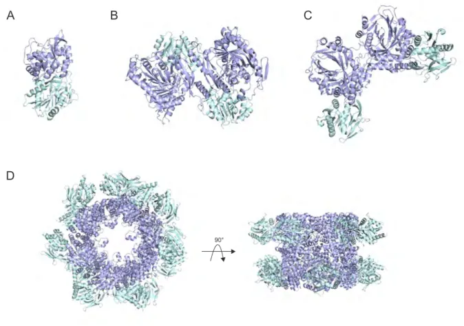
1.2.1 Structural Diversity Within the Synthase Subunits of Class I GATases
It is still unclear, however, if the allosteric regulation mechanisms of class I GATases evolved independently for the individual members of this enzyme family or if there are commonalities be- tween them. Whereas the similarities in structure and active site composition in the glutaminase subunits indicate a potential uniform allosteric regulation mechanism, the observed structural diversity within the synthase subunits does not support this notion. The latter ranges from ( β / α ) 8 -barrel folds (ImGPS, PS) to 4-layer sandwich-fold (AS, ADCS, and PhzE) (Figure 1.3 blue). Additionally, the different oligomeric states observed for these enzymes further compli- cate the identification of commonalities in allosteric regulation. These range from hetero-dimeric (ImGPS)(Douangamath et al. 2002), intertwined homo-dimeric fusions of glutaminase and syn- thase domain (PhzE) (Strohmeier et al., 2006), hetero-tetrameric (AS) (Knöchel et al., 1999), to dodecamers of hetero-dimeric (PS) (Zein et al., 2006; Strohmeier et al., 2006) associations (Figure 1.3).
Figure 1.3: Structural diversity observed in class I glutamine amidotransferases. (A) Imidazole glyc-
erol phosphate synthase from Thermotoga maritima (PDB ID: 1GPW) (B) Anthranilate synthase from
Salmonella typhimurium (PDB ID: 1I1Q) (C) 2-amino-2-deoxyisochorismate synthase from Burkholderi
lata (PDB ID: 3R74) (D) Pyridoxalphosphate synthase from Bacillus subtilis (PDB ID: 2NV2). Glutam-
inase subunits/domains are depicted in cyan and synthase subunits/domains are depicted in blue.
Strikingly, for all members of class I GATases except for 4-amino-4-deoxychorismate synthase (ADCS) there are high-resolution crystal structures of either the complexes of synthase and glutaminase subunits and/or of the autonomous subunits available. For ADCS, however, there are only two autonomous structures of its synthase subunit described in the literature. Studies on the allosteric mechanism underlying glutaminase activation in ADCS have thus been hampered by the absence of an autonomous glutaminase subunit structure for ADCS.
1.2.2 Structural and Functional Insights into 4-Amino-4-Deoxychorismate Synthase
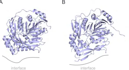
ADCS catalyzes the committed step in the biosynthesis of folate by incorporating ammonia into chorismate. Due to its sole appearance in bacteria, fungi, and plants, the enzyme has re- ceived significant interest as potential target of antimicrobial drugs (Davies et al., 1994; Bulloch et al., 2004). Drug development has halted though, most presumably due to missing struc- tural information on the glutaminase-synthase complex. To date, only two crystal structures of the autonomous PabB synthase subunits from Escherichia coli (Parsons et al., 2002) and Stenotrophomonas maltophilia (Bera et al., 2012) have been described in literature (Figure 1.4).
A B
interface interface
Figure 1.4: Crystal structures of autonomous PabB structures from (A) E. coli (PDB ID: 1K0E) and (B) S. maltophilia (PDB ID: 4GRH). The synthase subunits are depicted in blue, the potential (partially disordered) PabA binding sites are indicated as gray lines.
Both structures display a complex 4-layer sandwich fold similar to the synthase subunits of the homologous anthranilate synthases (Knöchel et al., 1999; Morollo and Eck, 2001). Even though both structures show significant disorder at the proposed PabA binding site (indicated in gray in Figure 1.4), they still allowed valuable insights into the reaction mechanism underlying ammonia incorporation into chorismate in these enzymes (He et al., 2004; He and Toney, 2006;
Viswanathan et al., 1995).
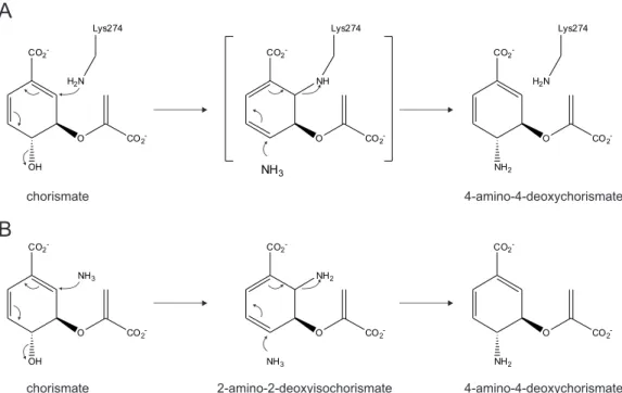
Mechanistically, PabB enzymes can be categorized into either the "PI K GT"- or "PI A GT"- motif group. According to the unifying core mechanism for chorismate utilizing enzymes (He et al., 2004), a central lysine residue (K274 in the enzyme from E. coli ) of the "PI K GT"-motif cova- lently binds to C2 of chorismate to initiate the reaction. The covalent attack of lysine leads to elimination of the hydroxyl-group at C4. The subsequent substitution of ammonia at C4 leads to an inversion of the stereochemistry at C4 and concomitant displacement of lysine ends the catalytic cycle (Figure 1.5 A). Enzymes of the "PI A GT" group utilize two equivalents of ammo- nia to generate 4-amino-4-deoxychorismate from chorismate. The first equivalent of ammonia attacks at C2 of chorismate with concomitant elimination of hydroxyl-group at C4 to yield 2- amino-2-deoxyisochorismate as intermediate. The subsequent addition of ammonia at C4 leads to a displacement of ammonia at C2 to end the catalytic cycle (Bera et al., 2012; Schadt et al., 2009) (Figure 1.5 B).
Figure 1.5: Proposed reaction mechanisms of PabB. (A) "PI K GT"-motif group: The catalytic cycle
is initiated by the attack of a highly conserved lysine residues (K274 from the enzyme of E. coli ) at
C2 of chorismate resulting in displacement of the hydroxyl-group at C4. In a second SN2 reaction,
ammonia attacks at C4 and lysine (K274) is displaced and 4-amino-4-deoxychorismate released from the
active site. (B) "PI A GT"-motif group: The catalytic cycle is initiated by the attack of one equivalent of
ammonia at C2 of chorismate resulting in displacement of the hydroxyl-group at C4 to yield 2-amino-
2-deoxyisochorismate as intermediate. A second equivalent of ammonia subsequently attacks at C4 and
ammonia at C2 is displaced and 4-amino-4-deoxychorismate released from the active site.
1.3 Aim and Scope of This Work
The aim of this work was to contribute to the understanding of the molecular basis of allosteric coupling in class I GATases. To this end, we sought to provide the missing structural information on the ADCS glutaminase subunit PabA to allow computation of a reliable homology model of the ADCS complex structure. To identify residues that are involved in allosteric communication, individual conserved amino acid side chains that are located between the PabA and the PabB active site should be replaced by alanine and their contribution to allosteric signaling studied by steady-state enzyme kinetics. Furthermore, we sought to consolidate these and previous findings on other class I enzyme systems to develop a generalized hypothesis of allosteric regulation.
1.4 Guide to the Following Chapters
The two following chapters are composed of the two first-author publications (A-B) that con- stitute this thesis.
The publication Mapping the Allosteric Communication Network of Aminodeoxycho- rismate Synthase describes an in-depth analysis of allosteric coupling in 4-amino-4-deoxy- chorismate synthase (ADCS). Ancestral sequence reconstruction was used to derive a ther- mostable PabA glutaminase subunit that was crystallized and its structure solved by molecular replacement. This PabA structure was then used to compute a reliable homology model of the E. coli PabA-PabB complex. Alanine-scanning at conserved residues located between the PabA and PabB active sites revealed four branches of an allosteric communication pathway that lead to activity-inducing conformational changes at the PabA active site as a consequence of chorismate binding at the PabB active site. Three conserved interaction hot-spot residues in the glutaminase-synthase interface were identified, one of which, a conserved aspartate-residue of the ADCS synthase subunit was identified as key residue for both complex formation and allosteric signaling.
The publication A Fold-Independent Interface Residue is Crucial for Complex For- mation and Allosteric Signaling in Class I Glutamine Amidotransferases corroborates the dual role of the conserved aspartate residue discovered in ADCS. Computational screening of the Protein Data Bank revealed the presence of similarly located aspartate residues in the syn- thase subunits of five class I glutamine amidotransferases with different folds/oligomeric states.
Mutational analysis of these aspartate residues in four different class I GATase enzyme systems
shows the critical role of this residue in complex formation (three out of four cases) and allosteric
signaling (all four cases).
Mapping the Allosteric Communication
Network of Aminodeoxychorismate Synthase
2.1 Abstract
Allosteric communication between different subunits in metabolic enzyme complexes is of utmost physiological importance but only understood for few systems. We analyzed the structural basis of allostery in aminodeoxychorismate synthase (ADCS), which is a member of the family of glutamine amidotransferases and catalyzes the committed step of the folate biosynthetic pathway. ADCS consists of the synthase subunit PabB and the glutaminase subunit PabA, which is allosterically stimulated by the presence of the PabB substrate chorismate. We first solved the crystal structure of a PabA subunit at 1.9-Å resolution. Based on this structure and the known structure of PabB, we computed an atomic model for the ADCS complex.
We then used alanine scanning to test the functional role of 59 conserved residues located between the active sites of PabB and PabA. Steady-state kinetic characterization revealed four branches of a conserved network of mainly charged residues that propagate the signal from chorismate at the PabB active site to the PabA active site. The branches eventually lead to activity-inducing transformations at (i) the oxyanion hole motif, (ii) the catalytic Cys-His-Glu triad, and (iii) glutamine binding residues at the PabA active site. We compare our findings with previously postulated activation mechanisms of different glutamine amidotransferases and propose a unifying regulation mechanism for this ubiquitous family of enzymes.
2.2 Introduction
Allostery is an integral property of enzymes that allows for the regulation of activity in response
to the physiological needs of a cell (Monod et al., 1965). Thereby, ligand binding to an allosteric
site induces signal propagation via protein backbone or amino acid side-chain movements to
affect function at a distant active site . Understanding the molecular details of signal propa-
gation between the two sites is an important biochemical challenge both for applied and basic
protein biochemistry. In applied research, molecular details of allosteric communication have the potential to reveal binding sites for novel drugs that perturb signal propagation (Chen et al., 2016; Monaghan et al., 2012; Fang et al., 2012; Selvy et al., 2011; Kenakin and Miller, 2010;
Rivalta et al., 2016; Goto et al., 2004). In basic research, the identification of signal propagation pathways is crucial for unraveling universal patterns of the underlying dynamic adaptability of enzymes (Gunasekaran et al., 2004).
A paradigm for studying the molecular basis of allosteric communication are glutamine amido- transferases (GATases). GATases form a large enzyme family, whose members are responsible for the incorporation of nitrogen within numerous metabolic pathways (Mouilleron and Golinelli- Pimpaneau, 2007; Raushel et al., 1999; Zalkin and Smith, 1998). Most GATases are bi-enzyme complexes, consisting of a synthase and a glutaminase subunit, whose activities are mutually coupled: Substrate binding to the synthase subunit allosterically induces glutamine hydrolysis to glutamate and ammonia at the glutaminase subunit. Nascent ammonia is then transported through an intermolecular channel to the active site of the synthase subunit, where it reacts with the “waiting” substrate to specific reaction products. While the synthase subunits of the various GATases are structurally diverse, the glutaminases can be categorized into two classes according to their fold and active site composition: class I glutaminases possess a α/β-hydrolase fold and a catalytic Cys–His–Glu triad (Ollis et al., 1992); in contrast, class II glutaminases possess an Ntn-hydrolase fold and a catalytic N-terminal Cys (Brannigan et al., 1995). Whereas the structural basis of allosteric communication is understood to a certain degree for class II GATases (Mouilleron et al., 2011), even a basic understanding of the allosteric communication pathways within class I GATases is still lacking.
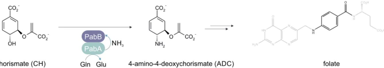
An interesting member of class I GATases is aminodeoxychorismate synthase (ADCS), which consists of the glutaminase subunit PabA and the synthase subunit PabB. It catalyzes the committed step in the biosynthesis of folate by incorporating nitrogen into chorismate to yield 4-amino-4-deoxychorismate (Figure 2.1).
OH
O CO
2-CO
2-PabB PabA Gln Glu
NH
3O CO
2-CO
2-NH
2chorismate (CH) 4-amino-4-deoxychorismate (ADC) folate
HN
N N
N NH
N H O
CO2H CO2H
H2N O
Figure 2.1: Reaction catalyzed by ADCS. The glutaminase subunit PabA hydrolyzes glutamine (Gln) to glutamate (Glu) and nascent ammonia (NH 3 ), which is channeled to the active site of the synthase subunit PabB where it reacts with chorismate (CH) to form 4-amino-4-deoxychorismate (ADC). ADC is a precursor in the biosynthesis of folate (shown in gray except the part of the molecule that stems from ADC).
Glutamine hydrolysis by PabA is activated upon complex formation with PabB and further
allosterically stimulated by chorismate binding to the synthase active site. Thus, the active site
of PabB acts as an allosteric site that, upon binding of its substrate, stimulates activity at the
PabA active site. In spite of the identification of key catalytic amino acid residues in PabA and PabB (Rayl et al., 1996; Roux and Walsh, 1992), the availability of two PabB crystal structures (Parsons et al., 2002; Bera et al., 2012), and a basic understanding of the chemical reaction mechanisms for PabA and PabB (He et al., 2004; Culbertson et al., 2015), the structural basis for allosteric communication in ADCS still awaits clarification.
Here, we addressed allostery in ADCS by a combination of experimental and computational approaches. We first solved the structure of a stable PabA subunit at high resolution. Together with the known structure of a PabB subunit, we then generated a homology model of the ADCS complex and performed a comprehensive alanine-scanning approach at conserved residues of both subunits. The results allowed us to define the allosteric communication pathway within ADCS. Specifically, we identified a conserved network of mainly hydrogen-bonded residues that propagates the signal from the allosteric site in PabB to the active site in PabA.
2.3 Results
2.3.1 Crystal Structure Analysis of PabA and Homology Modeling of ADCS
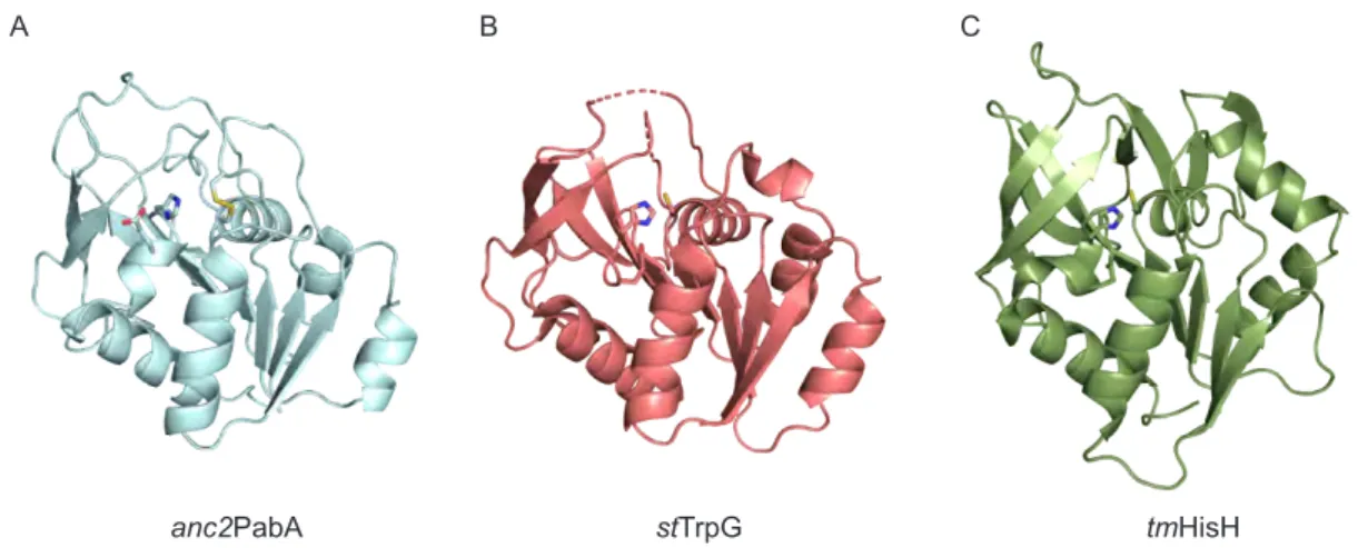
In order to map the allosteric communication network of ADCS, we first set out to provide the missing structural information for PabA. Despite extensive efforts, we were not able to crystallize modern PabA enzymes (from E. coli , Bacillus subtilis , Serratia marcescens , and Pseudomonas putida ) (Plach et al., 2017). Ancestral sequence reconstruction often yields thermostable pro- teins (Wheeler et al., 2016), which have a higher tendency to crystallize than their thermolabile homologs. Therefore, we reconstructed ancestral PabA sequences based on the sequences of 59 recent homologs from Firmicutes, Fusobacteria, Proteobacteria, and Halobacteriales (Figure 2.7). The genes encoding the three most ancient bacterial predecessors ( anc 1–3PabA) were expressed in E. coli , and the recombinant proteins were purified and biochemically character- ized. Interestingly, all three enzymes were more thermostable than modern PabA proteins (Tables 2.1, 2.2, 2.3), formed stable complexes with modern PabB subunits (Table 2.4), and were stimulated by them (Table 2.5). From these ancestral PabA enzymes, we were able to only crystallize anc 2PabA and solved its structure by molecular replacement using the homologous class I glutaminase subunit from Sulfolobus solfataricus anthranilate synthase (AS) (PDB ID:
1QDL; sequence identity: 51%, sequence similarity: 66%) as template (Table 2.6). The struc- ture of anc 2PabA (PDB ID: 6QUR) possesses the same α / β -hydrolase fold as the glutaminase subunits of the GATases AS and imidazole glycerol phosphate synthase (ImGPS) (Figure 2.2) (Morollo and Eck, 2001; Knöchel et al., 1999; Douangamath et al., 2002).
The catalytic triad consisting of a C79, a H168, and a E170 is located at the solvent-accessible
active site ( a connotes residues in PabA, and b connotes residues in PabB). Surprisingly, the
crystal structure of anc 2PabA features a disulfide bond between the catalytic a C79 residue
A B C
anc2PabA stTrpG tmHisH
Figure 2.2: Structural comparison of anc 2PabA to homologous class I glutaminase subunits. (A) Crystal structure of anc 2PabA (PDB ID: 6QUR); disulfide bond formed between a C54 and a C79 and the catalytic Cys–His–Glu triad is shown as sticks. (B) Crystal structure of the glutaminase subunit st TrpG from Salmonella typhimurium AS (PDB ID: 1I1Q). (C) Crystal structure of the glutaminase subunit tm HisH from T. maritima ImGPS (PDB ID: 1GPW). Sequence identity between anc 2PabA and st TrpG is 84/198 (42.4%) and sequence similarity is 115/198 (58.1%). Sequence identity between anc 2PabA and tm HisH is 50/231 (21.6%), and sequence similarity is 84/231 (36.4%). The structure of anc 2PabA superimposes well with those of st TrpG and tm HisH with all-atom RMSDs of 1.6 Å ( anc 2PabA- st TrpG) and 2.9 Å ( anc 2PabA- tm HisH), respectively.
and a C54, which is located in a loop covering the active site adjacent to a sequence stretch referred to as the oxyanion hole motif ( a P51– a G52– a P53– a C54) (Rivalta et al., 2012). Since our previous crystallization trials with a variety of modern PabA and PabA:PabB complexes failed, we assume that formation of this disulfide bond in anc 2PabA stabilizes the enzyme and thus ultimately allowed for its crystallization.
Based on the crystal structure of anc 2PabA, we generated a homology model for the E. coli ADCS complex by superimposing the previously solved ec PabB structure (PDB ID: 1K0E) and a model of ec PabA [that we generated based on the structure of anc2 PabA (PDB ID:
6QUR)] with the homologous AS complex from S. solfataricus (PDB ID: 1QDL) (Table 2.7).
To generate the final model of the holo enzyme, we superposed the substrates chorismate and glutamine from the structure of S. marcescens AS (PDB ID: 1I7Q) into the active sites of PabB and PabA, respectively. The model was energy minimized within YASARA’s “build model”
function (Krieger and Vriend, 2014) and further relaxed by molecular dynamics simulations for 100 ns with GROMACS (Berendsen et al., 1995; Lindahl et al., 2001).
2.3.2 Alanine Scanning for the Identification of Residues Involved in Allosteric Signal Propagation
To identify residues in the ADCS complex that propagate the allosteric signal induced by cho-
rismate binding to PabB onto PabA, we individually replaced 59 conserved residues located
between both active sites by alanines (wild-type alanines were replaced by valines, respectively)
using site-directed mutagenesis. We expressed the mutated genes in E. coli , purified the soluble recombinant enzymes (Table 2.8) and analyzed the effect of each individual mutation on complex formation and activity.
The results of analytical size exclusion chromatography showed that all mutant enzymes except those carrying the exchanges a Y127A, b D308A, and b R311A formed stable ADCS complexes with the corresponding wild-type partner subunit (Figure 2.9 and Table 2.4). These three “in- teraction hotspot residues” are embedded in an inter-domain hydrogen bonding network that comprises residues a Y18, a Y127, a E170 (backbone), a S171, b D308, and b R311 (Figure 2.8).
For the mutant enzymes that formed stable ADCS complexes with the corresponding wild-type partner subunits, we quantified the extent of allosteric PabA activation by determining the ap- parent glutamine hydrolysis rate (k app ). To this end, we incubated PabA with glutamine at saturating concentrations (20 mM) and monitored its hydrolysis before and after the addition of (i) PabB or (ii) PabB + chorismate (saturating concentrations) for all combinations of wild- type-PabA/mutant-PabB and mutant-PabA/wild-type-PabB. For the wild-type complex, the basal glutaminase activity of 0.004 s −1 [k app (PabA)] is elevated upon binding of PabB to 0.21 s −1 [k app (PabA + PabB)] and further allosterically stimulated in the presence of chorismate to 0.58 s −1 [k app (PabA + PabB + CH)]. Basal glutaminase activity was similar to wild-type for all mutant enzymes, except the one containing the a T125A exchange (will be discussed below).
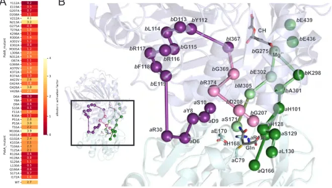
In contrast, PabA activation was very sensitive to mutations in both PabA and PabB: 24 out of the 49 assayed mutant enzymes showed a more than 2-fold change in k app (PabA + PabB) or k app (PabA + PabB + CH) values (Table 2.5).
In order to identify residues that contribute to allosteric signaling, we compared the ratio of the apparent rates in the presence and absence of chorismate, which we refer to as allosteric activation factor . For the wild-type enzyme, the allosteric activation factor is 2.7 [0.58 s −1 (k app (PabA + PabB + CH)]/0.21 s −1 (k app (PabA + PabB))]. In order to exclude minor sec- ondary effects that resulted from structural perturbations through the introduction of alanine, we defined residues as involved in allosteric signaling when their allosteric activation factor was below 1.5. Eleven mutants displayed allosteric activation factors of 1.0–1.5 indicating no or impaired allosteric activation. Thirteen mutants showed an allosteric activation factor ≤ 1.0, indicating allosteric deactivation (Figure 2.3 A).
We were interested to test whether the effects of mutations on k app for glutamine hydrolysis
are at least partially attributed to an increase in the Michaelis constant for glutamine (K M Gln )
or entirely due to a decrease of the turnover rate (k cat ). For this purpose, we followed the
glutamine-dependent conversion of chorismate (CH) to aminodeoxychorismate (ADC) for the
different ADCS complexes (Table 2.9). The results demonstrated that complexes containing
mutant enzymes with the exchanges a D6A, a Y8A, a R30A, a R126A, a H128A, a S129A, a L130A,
a Q166A, a I172A b E119A, and b G207A showed drastically elevated K M Gln values, which might
contribute to the observed decreases in k app . The complexes containing the other mutant en-
zymes showed only minor effects with respect to K M Gln , proving that the observed changes in
k app are entirely caused by changes in k cat . Interestingly, we also identified residues involved in ammonia channeling from the active site of PabA to the active site of PabB: Whereas in ADCS complexes containing mutant enzymes with the a D9A, a H101A, a R126A, a L130A, b D208A, b L309A, and b G369A exchanges, glutaminase activity was readily stimulated (Figure 2.3 A), no measurable or very low turnover was observed when the ADCS reaction was followed (Table 2.9). The unproductive loss of ammonia in these mutants indicates that the respective wild-type amino acids are involved in ammonia tunnel formation between active sites.
2.3.3 Identification of Four Branches of a Conserved Allosteric Communication Network
We subsequently mapped the 24 allosterically important residues on the model of E. coli ADCS and deduced the most plausible pathways for signal propagation between the two active sites. To this end, we drew connecting edges between allosteric residues when either (i) their side chains were in direct van der Waals or hydrogen-bonding contact or (ii) the residues were connected via the protein backbone (Figure 2.3 B).
A B
bY112
bI367
bE119
aR30
aD6 aY8
aD9 aS10
bG369 bR374
bD208 bG207
bG275
bE302 bK298
bA301 bM305
aH101
aQ166 aL130
aS129 aH128 aR126 aS171
CH
Mg
2+Gln aC79 aE170
aH168
bE439
bE436 bD113
bL114
bG115 bR116 bR117
bF118
Figure 2.3: Allosteric residues in ADCS. (A) Heat map indicating the ratio of the apparent glutaminase
rates in the presence and absence of chorismate (k app (PabA + PabB + CH)/k app (PabA + PabB) =
allosteric activation factor) of analyzed mutant enzymes. (B) Mapping of residues with an allosteric
activation factor <1.5 on the homology model of E. coli ADCS (PabA is depicted in cyan, and PabB
is depicted in blue). Allosterically important positions are shown as spheres (C α ), and pathways are
indicated as edges connecting them. Magnesium (Mg 2+ ) in the active site of PabB is shown as sphere
as well. The catalytic triad a C79– a H168– a E170 and the substrates glutamine and chorismate (CH)
are shown as sticks. All-atom RMSD between our E. coli ADCS model and the crystal structure of
S. solfataricus AS, which was used for modeling, is 1.7 Å.
Using this classification, we found a putative network of conserved, mostly hydrogen-bonded residues that reach from the chorismate binding site in PabB to the active site of PabA. Inter- estingly, this allosteric communication network appears to be split into four branches: Starting from the chorismate binding site in PabB, these branches lead to (i) the oxyanion hole motif a P51– a G52– a P53, to (ii) the catalytic triad a C79– a H168– a E170, and to (iii + iv) residues in- volved in glutamine binding at the PabA glutaminase active site.
The branch that leads to the oxyanion hole stretch a P51– a G52– a P53 supposedly senses the presence of chorismate via its hydrophobic interaction with b I367 located near the C4-atom of chorismate. Through a van der Waals interaction with b Y112, the signal might then be prop- agated via backbone interactions of the helix formed by residues b D113– b L114– b G115– b R116 - b R117– b F118 and ultimately to b E119. Residue b E119 is located at the PabB–PabA interface and is hydrogen bonded to a R30. Via a R30, the signal is further transferred to the sequence stretch a Y8– a D9– a S10 adjacent to the oxyanion hole motif (Figure 2.3 B, purple).
The branch that leads to the catalytic triad a C79– a H168– a E170 presumptively senses the pres- ence of chorismate via b E302, which is part of the conserved magnesium-binding site that coor- dinates the carboxylate group of chorismate. Via b E302 and the inter-domain hydrogen bonding network (Figure 2.8), the signal is ultimately transferred to a S171, which is located adjacent to the catalytic residues a H168 and a E170 (Figure 2.3 B, bright green).
One branch that leads to glutamine binding residues apparently senses the presence of choris- mate via b K298, which interacts with residues of the conserved magnesium-binding site (i.e., b E439 and b E436). The signal is then propagated via b A301 and a H101 to the glutamine binding residues a R126, a H128, a S129, and a Q166 as well as to a L130, which is involved in ammonia tunnel formation (Figure 2.3 B, dark green).
The second branch that leads to glutamine binding residues reputedly senses the presence of chorismate via b I367 (described above) and b G369 and transfers the allosteric signal via b R374 and b D208 and ultimately to b G207, which is involved in glutamine binding at the glutaminase active site (Figure 2.3 B, pink).
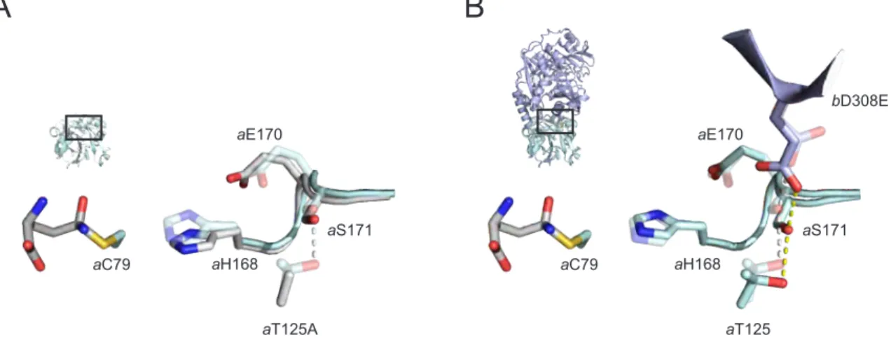
2.3.4 Analysis of a PabA Mutant with Increased Basal Glutaminase Activity
Strikingly, PabA mutant a T125A shows a 10-fold acceleration of basal glutaminase activity [k app (PabA) = 0.04 s −1 ] compared to the wild-type enzyme [k app (PabA) = 0.004 s −1 ] (Table 2.5).
As the allosteric activation factor (Figure 2.3 A) and catalytic parameters for the glutamine-
dependent chorismate turnover are almost identical to wild-type ADCS (Tables 2.5 and 2.9), the
wild-type a T125 residue seems not to be involved in the allosteric network, glutamine binding, or
ammonia tunnel formation. Residue a T125 is hydrogen-bonded to a S171, an allosteric residue
located adjacent to the catalytic residues a H168 and a E170 (Figure 2.4 A). The drastically
enhanced basal glutaminase activity upon removal of this hydrogen bond in the a T125A mutant
enzyme suggests that the relative positioning of a S171 with respect to the catalytic triad is
crucial for glutaminase activity. We thus further analyzed the interactions of a S171 to other residues in ADCS. As part of the inter-domain hydrogen bonding network, a S171 is also hydrogen bonded to the interaction hotspot b D308 (Figure 2.8). We generated mutant enzyme b D308E and monitored its ability to form stable ADCS complexes and to allosterically stimulate PabA activity. Strikingly, b D308E was not able to form stable ADCS complexes but led to a slightly enhanced k app (PabA + PabB) and an allosteric activation factor of 1.2 (Tables 2.4 and 2.5).
This finding indicates that this mutant might form a transient or weak interaction that affects the positioning of a S171 relative to the catalytic triad. We conclude that the corresponding wild-type residue b D308 undergoes a structural change in the course of signal propagation and might thus be a key residue for both complex formation and allosteric glutaminase activation within ADCS (Figure 2.4 B).
aT125A aS171 aE170
aH168 aC79
aT125
aS171 aE170
aH168 aC79
bD308E
A B
Figure 2.4: Activated variants of ADCS. (A) Putative structural changes in the a T125A mutant enzyme:
a T125 is hydrogen bonded to a S171 in wild-type PabA (transparent). The loss of the hydrogen bond to- ward a S171 in the a T125A mutant might allow for the movement of the loop a H168– a P169– a E170– a S171 and thereby enhance basal glutaminase activity by reducing the a C79– a H168 distance (solid). (B) Puta- tive structural changes in the b D308E mutant enzyme: b D308 is hydrogen-bonded to a S171 in wild-type ADCS (transparent). A potential movement of b D308 in the course of allosteric signaling is mimicked through the b D308E mutation and might result in bending of the loop a H168– a P169– a E170– a S171 to constitutively activate the glutaminase subunit (solid).
2.4 Discussion
Our study demonstrates that glutamine hydrolysis at the PabA subunit of ADCS is stimulated
by the presence of chorismate at the PabB subunit via a conserved allosteric communication
network. Starting from the chorismate binding site in PabB, the network splits into four branches
that propagate the allosteric signal via mostly hydrogen-bonded residues to (i) the oxyanion hole
motif a P51– a G52– a P53, (ii) the catalytic triad a C79– a H168– a E170, and (iii) glutamine binding
residues at the PabA active site, respectively. In more general terms, chorismate might shift
the enzyme complex from a less activated ensemble of conformational states to a more activated
ensemble (Motlagh et al., 2014). The deactivating effect observed in some mutants might thus result from the stabilization of the less activated ensemble by chorismate in these particular contexts.
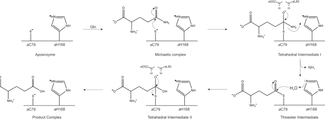
In the following, we will discuss our findings in light of previously hypothesized activation mechanisms for different class I glutamine amidotransferase complexes. A hallmark feature of class I glutaminases is the catalytic triad composed of Cys–His–Glu, where Cys acts as nucleophile and His as general acid/base in the catalytic cycle (Figure 2.5).
S
NH HN
O O
NH2 O
NH3+ S
NH HN
Gln
OO
NH2 O
NH3+ S
NH HN
O
O O
NH3+
S
NH N
O O
OH O
NH3+ S O
O
OH O
NH3+ S
H O
2aC79 aH168
NH
3Apoenzyme Michaelis complex Tetrahedral Intermediate I
Thioester Intermediate Tetrahedral Intermediate II
Product Complex
NH HNNH HN
N N
H H
aG52 aL80
N N
H H
aG52 aL80