Comparison between time-dependent and time-independent methods for the
calculation of inter-system crossing rates:
Application to uracil and its derivatives
Inaugural-Dissertation
zur
Erlangung des Doktorgrades der
Mathematisch-Naturwissenschaftlichen Fakult¨ at der Heinrich-Heine-Universit¨ at D¨ usseldorf
vorgelegt von Mihajlo Etinski aus Novi Sad (Serbien)
D¨ usseldorf 2010
Aus dem Institut f¨ ur Theoretische Chemie und Computerchemie der Heinrich-Heine-Universit¨ at D¨ usseldorf
Gedruckt mit Genehmigung der
Mathematisch-Naturwissenschaftlichen Fakult¨ at der Heinrich-Heine-Universit¨ at D¨ usseldorf
Referent:
Korreferent:
Prof. Dr. Christel M. Marian Jun.-Prof. Dr. J¨ org Tatchen
Tag der m¨ undlichen Pr¨ ufung:
Hiermit versichere ich, die hier vorgelegte Arbeit eigenst¨andig und ohne unerlaubte Hilfe angefertigt zu haben. Die Dissertation wurde in der vorgelegten oder in ¨ahnlicher Form noch bei keiner Institution eingereicht. Ich habe keine erfolglosen Promotionsver- suche unternommen.
D¨usseldorf, den
(Mihajlo Etinski)
List of papers included in the thesis
• Paper 1
Electronic and vibrational spectroscopy of 1-methylthymine and its water clus- ters: The dark state survives hydration
Matthias Busker, Michael Nispel, Thomas H¨aber, Karl Kleinermanns , Mihajlo Etinski, and Timo Fleig, Chem. Phys. Chem., 9(2008) 1570-1577
• Paper 2
Intersystem crossing and characterization of dark states in the pyrimidine nucle- obases uracil, thymine, and 1-methylthymine
Mihajlo Etinski, Timo Fleig, and Christel M. Marian, J. Phys. Chem. A 113, (2009) 11809-11816
• Paper 3
Ab initio investigation of the methylation and hydration effects on the electronic spectrum of uracil and thymine
Mihajlo Etinski and Christel M. Marian, Phys. Chem. Chem. Phys. 12, (2010) 4915 - 4923
• Paper 4
Overruling the energy gap law: Fast triplet formation in 6-azauracil
Mihajlo Etinski and Christel M. Marian, submitted to Phys. Chem. Chem.
Phys.
List of related papers not included in the thesis
• Paper 5
Theoretical investigation of the excited states of 2-nitrobenzyl and 4,5-methylendioxy- 2-nitrobenzyl caging groups
Klaus Schaper, Mihajlo Etinski, and Timo Fleig, Photochem. Photobio. 85 (2009) 1075-1081
I express my gratitude to Professor Dr. Christel Marian for her help to formulate the subject of my thesis, exploration of which brought me a lot of intellectual excite- ment. The collaboration with Professor Marian helped me to broaden my insights into electronic spectroscopy and photophysics. I am grateful to Junior Professor Dr. J¨org Tatchen for his support of my research. I would like to thank Professor Dr. Timo Fleig for his advice during the first part of my doctoral studies in Duesseldorf. I thank Dr. Martin Kleinschmidt for help to interface the code written in C as a subroutine to the VIBES program. Also, I am grateful to all experimentalists with whom I collabo- rated: Professor Dr. Karl Kleinermanns, Priv. Doz. Dr. Klaus Schaper, Dr. Matthias Busker, Dr. Michael Nispel and Dr. Thomas H¨aber. I would like to thank all col- leagues from the Institute of Theoretical and Computational Chemistry, and especially to Dr. Susanne Salzmann, Dr. Stefan Knecht, Dr. Lasse Sørensen, Dr. Vidisha Rai- Constapel, Kathleen Gollnisch, Dr. Stephan Raub, Karin Schuck and Klaus Eifert for creating a friendly and creative atmosphere. I appreciate very much all help and con- tinual support received from Professor Dr. Miljenko Peri´c and Professor Dr. Werner Jakubetz.
Zusammenfassung
Der erste Teil dieser Arbeit besch¨aftigt sich mit der theoretischen Formulierung des Problems, Interkombinationsraten in Molek¨ulen effizient zu evaluieren. Daf¨ur werden zeitunabh¨angige- und zeitabh¨angige N¨aherungen verwendet. Letztere wird ausf¨uhrlicher diskutiert. Das zentrale Objekt der zeitabh¨angigen Methode ist die Korrelationsfunk- tion. F¨ur die Berechnung der Korrelationsfunktion, welche sowohl spinfreie, vibro- nische Born-Oppenheimer-N¨aherungen als auch die Condon-N¨aherung f¨ur Spin-Bahn- Matrixelemente verwendet, wird ein geschlossener Ausdruck gefunden. Es wird eben- falls ein Ausdruck f¨ur die Rate, die eine Kumulantenentwicklung zweiter Ordnung nutzt, pr¨asentiert. Ein besonders einfacher Ausdruck wird bei Anwendung einer Kurzzeit- entwicklung der Kumulantenentwicklung erhalten. Alle drei Ausdr¨ucke f¨ur die Inter- kombinationsraten sind in einem Computerprogramm implementiert worden. Die f¨ur die Testmolek¨ule unter Nutzung der zeitabh¨angigen sowie zeitunabh¨angigen N¨aherungen erhaltenen Raten werden verglichen und diskutiert. Die zeitabh¨angige N¨aherung ist f¨ur F¨alle, in denen die Franck-Condon-gewichtete Dichte der vibronischen Zust¨ande groß ist, viel versprechend. Dies k¨onnte sowohl auf eine große adiabatische Energiel¨ucke zwischen den elektronischen Zust¨anden als auch einfach auf viele Normalmoden zur¨uck- zuf¨uhren sein.
Der zweite Teil der Arbeit behandelt die Anwendungen der quantenchemischen Metho- den auf die elektronische Relaxation nach Photoanregung in Uracil, seinen methylierten Derivaten und 6-Azauracil. Experimente mit Hilfe zeitaufgel¨oster Spektroskopie in einem Molekularstrahl haben gezeigt, dass die methylierten Uracile auf der Femto-, Piko- und Nanosekundenzeitskala relaxieren. Obwohl die ersten experimentellen Ergeb- nisse darauf hingedeutet haben, dass die Nanosekundenrelaxierung von dem tiefsten augeregten Singulettzustand herr¨uhrt, folgern wir in einer sp¨ateren Studie, dass sie vom tiefsten elektronischen Triplettzustand verursacht wird. Wir unterbreiten auch einen kontroversen Vorschlag, dass Hydratation Nanosekundenrelaxation l¨oscht. Un- sere Ergebnisse zeigen, dass Hydratation einen signifikanten Effekt auf die elektro- nischen Zust¨ande hat, so dass diese die Photostabilit¨at der Pyrimidinbasen modifizieren kann.
Von besonderem Interesse ist die Photorelaxierung von 6-Azauracil, da die Triplett- quantenausbeute sehr viel gr¨oßer als in Uracil ist. Die Azasubstitution erzeugt zus¨atz- liche, tiefliegende Singulett- und Triplett-nπ∗-Zust¨ande. Die Berechnung der Poten- zialenergieprofile entlang linear interpolierter Pfade ergibt, dass zwischen den elektro- nischen Singulett- und Triplettzust¨anden Kreuzungen und vermiedene Kreuzungen ex- istieren. Es werden m¨ogliche elektronische Relaxierungsmechanismen diskutiert.
Summary
The first part of this thesis is focused on the theoretical formulation of the problem how to efficiently evaluate inter-system crossing rates in molecules. The problem is addressed by time-independent and time-dependent approaches. The latter approach is presented in a more detail. The central object in time-dependent method is the correlation function. A closed-form expression for the calculation of the correlation function using spin-free Born-Oppenheimer vibronic states and the Condon approxi- mation for the spin-orbit matrix element is found. Also, the expression for the rate using a second-order cumulant expansion is presented. A particularly simple expres- sion is derived employing a short-time expansion of the cumulant expansion. All three expressions for the inter-system crossing rate are implemented in a computer code.
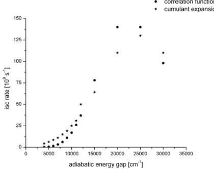
The rates obtained for test molecules using time-dependent and time-independent ap- proaches are compared and discussed. The time-dependent approach is promising in the cases where the Franck-Condon weighted density of the vibronic states is huge.
This may be due to a large adiabatic energy gap between electronic states or simply due to many normal modes.
The second part of the thesis is dedicated to applications of quantum-chemical meth- ods to the electronic relaxation upon photoexcitation in uracil, its methylated deriva- tives and 6-azauracil. Time-resolved spectroscopy experiments in the molecular beam showed that methylated uracils relax on the femto, pico and nanosecond time scales.
Although the first experimental results suggested that the nanosecond relaxation orig- inates from the lowest excited singlet state, in a later study we conclude that it should be due to the lowest triplet electronic state. Also, we address a controversal proposal that hydration quenches nanosecond relaxation. Our results show that hydration has a significant effect on the electronic states, so that it can modify the photostability of the pyrimidine bases.
Photorelaxation of 6-azauracil is particularly interesting because the triplet quantum yield is much larger than in uracil. The aza-substitution creates additional low-lying singlet and triplet nπ∗ states. The calculation of potential energy profiles along linearly interpolated paths reveals crossings and avoided crossings between singlet and triplet electronic states. Possible electronic relaxation mechanisms are discussed.
Contents
List of Tables 3
List of Figures 7
1 Introduction 9
1.1 Photochemistry of nucleic acids . . . 9
1.2 Motivation . . . 11
2 The molecular Hamiltonian and its approximations 13 2.1 Separation of electron and nuclear coordinates . . . 14
2.2 The solution of the electronic Schr¨odinger equation . . . 15
2.2.1 The Hartree-Fock method . . . 15
2.2.2 The coupled-cluster method . . . 17
2.3 The nuclear Schr¨odinger equation and the normal mode Hamiltonian . 19 2.4 Coupling of the electron spin and angular momentum . . . 23
3 Decay of the excited electronic state 25 3.1 Weak vibronic coupling . . . 26
3.2 Strong vibronic coupling . . . 29
3.3 The time-independent method for the calculation of the inter-system crossing rate . . . 30
3.4 The time-dependent method for the calculation of the inter-system cross- ing rate . . . 33
3.4.1 The correlation function . . . 33
3.4.2 Cumulant expansion . . . 36
3.4.3 Short-time approximation . . . 37
4 Implementation and testing of the time-dependent method for inter-system crossing rates 39 4.1 Implementation . . . 39
4.2 Test results . . . 42
4.2.1 Absorption spectrum of thioxanthone . . . 43
4.2.2 Inter-system crossing rates . . . 44
4.2.2.1 Thymine . . . 44
4.2.2.2 Phenalenone . . . 48
4.2.2.3 Flavone . . . 51
4.2.2.4 Free base porphyrin . . . 53
Contents
5 Applications 57
5.1 Dark electronic state in the pyrimidine bases uracil, thymine, and their methylated derivatives . . . 57 5.1.1 Overview of the experimental and theoretical results . . . 58 5.1.2 Electronic spectroscopy of 1-methylthymine and its water clusters 61 5.1.3 Effects of hydration and methylation on the electronic states of
uracil and thymine . . . 62 5.1.4 Inter-system crossing in uracil, thymine and 1-methylthymine . 66 5.2 Formation of the triplet electronic state in 6-azauracil . . . 71
6 Conclusions and outlook 77
Bibliography 79
List of Tables
1.1 Relative induction frequencies of the major photoproducts induced by UV radiation. Adapted from reference [1] . . . 10 3.1 Typical time scales of molecular photophysical processes [2, 3]. . . 26 4.1 Calculated inter-system crossing rates S1 T1y for phenalenone in s−1 . 51 4.2 Calculated inter-system crossing rates S1 T1x for flavone in s−1 . . . . 53 4.3 Calculated inter-system crossing rates S1 T1x for porphyrin in s−1 . . 55 5.1 Wavelengths of the pump and probe pulses, resolution and relaxation
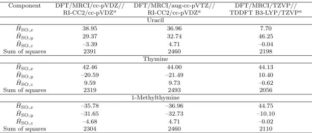
time constants obtained in femtosecond pump-probe experiments in molecular beams . . . 58 5.2 Spin-orbit matrix elements S1|HˆSO|T1 in cm−1 calculated at the S1
state geometry . . . 68 5.3 Spin-orbit matrix elements in cm−1 calculated at the TDDFT optimized
S2 geometry of uracil using the DFT/MRCI/TZVP method for gener- ating the wave function . . . 69 5.4 Calculated rate constants kISC [s−1] for the (S1 T1) ISC channels in
uracil, thymine and 1-methylthymine. ΔEad [cm−1] denotes the adia- batic electronic energy difference. . . 69 5.5 Rate constantskISC [s−1] for the (S2 T2) and (S2 T3) ISC channels
in uracil calculated at the DFT/MRCI/TZVP//DFT B3-LYP/TZVP level. ΔEad [cm−1] denotes the adiabatic electronic energy difference. . 69 5.6 Spin-orbit matrix elements calculated at the respective singlet minimum
geometry of 6-azauracil . . . 74 6.1 Comparison of calculated inter-system crossing rates with time-dependent
and time-independent approach (VIBES) in s−1. . . 77
List of Tables
List of Figures
1.1 UV action spectra for human cell killing and mutagenesis and carcino- genic action spectrum for mouse skin. Figure taken from reference [4] . 10 1.2 Nucleic bases . . . 10 2.1 Schematic two-dimensional (Q1, Q2) ground (lower, blue) and excited
(upper, red) state PES. Left panel: Displaced-distorted harmonic po- tential energy surfaces with (i) D = 0 , (ii) dQ1 = dQ1 and dQ2 = dQ2 , and (iii) J = 1, i. e. the excited state PES is (i) displaced, (ii) dis- torted, but (iii) not rotated relative to the ground state PES. Right panel: Displaced-distorted-rotated harmonic potential energy surfaces with (i) D = 0 (ii) dQ1 = dQ1 and dQ2 = dQ2 , and (iii) J = 1. For an explanation of the terms see text. Taken from ref. [5] . . . 21 2.2 Duschinsky matrix J for thymine: It is an orthogonal matrix close to a
unit matrix with the dimension equal to the number of normal modes (for discussion about translation and rotation see text); nondiagonal elements clearly show coupling between normal modes . . . 22 3.1 Photophysical (left) and photochemical path (right) in configuration space 25 3.2 Jablonski diagram: radiative processes - absorption (A), fluorescence (F)
phosphorescence (P); radiationless processes: internal conversion (IC), intersystem crossing (ISC), vibrational relaxation (VR) . . . 27 3.3 Level coupling scheme for the Wigner-Weisskopf model . . . 28 3.4 The singlet |S and triplet |T potential energy surfaces . . . 31 4.1 An example input file to start a time-dependent calculation of an inter-
system crossing rate with the VIBES program . . . 40 4.2 The pseudocode of the program. For explanation see text. . . 41 4.3 Chemical structure of thioxanthone . . . 43 4.4 Graphical representation of the Duschinsky matrix for the S0 →S2 tran-
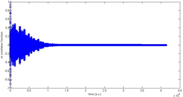
sition in thioxanthone . . . 43 4.5 Real part of the correlation function related to the absorption spectrum
of thioxanthone . . . 44 4.6 Absorption spectrum for the S2 ←S0transition in thioxanthone obtained
from the time-dependent and the time-independent (VIBES) approaches 45 4.7 The ground state structures of the test molecules. . . 45 4.8 Duschinsky matrix related to the transition between the S1 and T1states
of thymine. In order to visualize the normal mode mixing, absolute values of the matrix elementsJij are shown. . . 46
List of Figures
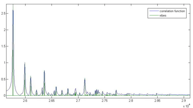
4.9 The real parts of the correlation function, second-order cumulant expan- sion and short-time approximation as functions of time for thymine . . 47 4.10 Dependence of the inter-system crossing rate on the adiabatic electronic
energy gap in thymine . . . 48 4.11 Duschinsky matrix related to transition between the S1 and T1 states
of phenalenone. In order to visualize the normal mode mixing, absolute values of the matrix elementsJij are shown. . . 49 4.12 The real parts of the correlation function, the second-order cumulant
expansion and the short-time approximation as functions of time for phenalenone (presented only the first 50 fs) . . . 49 4.13 Duschinsky matrix related to transition between the S1 and T1 states of
flavone. In order to visualize the normal mode mixing, absolute values of the matrix elements Jij are shown. . . 52 4.14 The real parts of the correlation function, the second-order cumulant
expansion and the short-time approximation as functions of time for flavone (presented only the first 50 fs) . . . 52 4.15 Duschinsky matrix related to transition between the S1 and T1 states
of porphyrin. In order to visualize the normal mode mixing, absolute values of the matrix elementsJij are shown. . . 54 4.16 The real parts of the correlation function, the second-order cumulant
expansion and the short-time approximation as functions of time for porphyrin (presented only the first 50 fs) . . . 55 5.1 Proposed potential energy surfaces and processes for the pyrimidine
bases. Ionization from the S1 state and the dark state Sd sample dif- ferent Franck-Condon regions of the ionic state, resulting in different ionization energies for these two states. From reference [6] . . . 59 5.2 Lifetimes of 1-methyluracil, 1,3-dimethyluracil, 1,3-dimethylthymine, and
thymine at different excitation wavelengths. From reference [6] . . . 60 5.3 Geometries of the conical intersection in 1-methylthymine: 1ππ/1nπ
(CI1),1ππ/S0 (CI2). . . 62 5.4 Potential energy profiles of the ground (squares), 1nπ∗ (triangles) and
1ππ∗state (circles) of 1-methylthymine, calculated at the CASSCF(10,8)/6- 31G* level of theory along the LIIC reaction path. A: from the equilib- rium geometry of the ground state to the CI1; B: from the equilibrium geometry of the ground state to the minimum of the1ππ∗ state and to the CI1; C: from the CI1 to the minimum of the1nπ∗ state; D: from the CI1 to the CI2. . . 63 5.5 Chemical structures of methylated uracils and thymines. Atom labels
are given for uracil. . . 64 5.6 Density distribution of the Hartree-Fock frontier molecular orbitals which
contribute to the lowest excited electronic states of methylated uracils and thymines (aug-cc-pVDZ basis set, isovalue=0.03). . . 65
List of Figures 5.7 Calculated difference IR spectra of thymine. Upper row: RI-CC2/cc-
pVDZ, middle row: B3-LYP/DFT/TZVP frequency calculations, The line spectra were broadened by Gaussian functions with a with of 50 cm−1 at half maximum, from ref. [7]. Lower row: TRIR spectrum of thymine in argon-purged acetonitrile-d3 at a delay of 0-1μs (solid curve).
Also shown is the inverted and scaled steady-state IR absorption spec- trum (dashed curve). The inset compares the TRIR spectra of thymine (solid) and thymine-d2 (dashed) at the same time delay, from ref. [8]. . 67 5.8 DFT/MRCI/TZVP single-point calculations along a linearly interpo-
lated path between the UDFT-optimized T1 geometry (RC = 0) and the TDDFT-optimized S1 geometry (RC = 1.0) of uracil. The paths are also extrapolated on both sides. T1 (ππ): upright open triangles;
T2 (nπ): upside down open triangles; S1 (nπ): upside down filled triangles; T3 (ππ): open squares; S2 (ππ): upright filled triangles. . . 70 5.9 6-azauracil: structure and numbering. . . 71 5.10 Single-point calculations along a linearly interpolated path between the
FC point (RC=0) and the S2 minimum (RC=10) of 6-azauracil. The curves are extended on both sides. . . 72 5.11 Single-point calculations along a linearly interpolated path between the
S2 minimum (RC=0) and the S1 minimum (RC=10) of 6-azauracil. The curves are extended on both sides. . . 73 5.12 Single-point calculations along a linearly interpolated path between the
S1 minimum (RC=0) and the T1 minimum (RC=10) of 6-azauracil. The curves are extended on both sides. . . 74 5.13 Sum of the squared spin-orbit matrix element components computed
along the linearly interpolated reaction path connecting the S2 minimum geometry (RC=0) and the S1 minimum geometry (RC=10) of 6-azauracil. 75
List of Figures
1 Introduction
1.1 Photochemistry of nucleic acids
Ultraviolet (UV) radiation comprises the electromagnetic spectrum from 10 to 380 nm.
The sun emits ultraviolet radiation in the UV-A (320-380 nm), UV-B (290-320 nm), and UV-C (290-100) bands. On Earth ground, only radiation from 290 to 380 nm is relevant because ozone or other atmospheric gases strongly absorb radiation below 290 nm. The UV-B radiation is partially absorbed by the ozone layer while it is transparent to UV-A radiation. Depletion of the ozone layer due to chlorofluorocarbon pollution increases the exposure to UV-B radiation. This is causing danger for the living organisms on Earth. Radiation can be toxic (kills living cells), mutagenic (changes the genotype of cells) and carcinogenic (removes the normal capacity to inhibit cell growth).
Biological effects upon absorption of UV light can be examined by measuring an action spectrum. It contains the biological response to a specific radiation wavelength. An action spectrum for mouse skin in the UV-B and UV-A regions is presented in Figure 1.1. It can be seen from the Figure 1.1 that in the UV-B region an action spectrum for mutagenesis and carcinogesis is overlapping with DNA absorption while in the UV- A region where DNA absorption is low, there is still a possibility for cell killing. It is believed that although the photon is not absorbed by DNA, it can be absorbed by an intermediate molecule and then its energy could be transferred to the DNA molecule. Hence, the DNA molecule is the main chromophore for UV absorption and its photophysical and photochemical properties are important for understanding the molecular origin of carcinogenesis and mutagenesis upon UV irradiation.
Nucleic acids absorb below 300 nm with maximum at 260 nm. Nucleic bases, Figure 1.2, are the main chromophores of nucleic acids. Upon absorption of a UV photon, an excited electronic state is created and it can decay through various pathways such as internal conversion to the ground state, vibrational relaxation, inter-system crossing to reactive triplet states and chemical reactions in the excited state. In the case of nucleic bases, the largest part of the excited electronic-state population decays to the ground state indicating a significant photostability. But a part of the population goes into chemical reactions. Major photoproducts of UV degradation of nucleic acids and their yields are given in Table 1.1.
Excited triplet electronic states live longer than singlet states due to the spin-forbidden character of their relaxation to the singlet ground electronic state. Since they are highly reactive, they present potential precursors for photochemical reactions. As can be seen from Table 1.1 the main mechanism of photodegradation is a dimerization of two pyrimidine bases yielding cyclobutyl structure. The detailed mechanism of this photoreaction is still under discussion: an ultrafast reaction in the singlet excited state [9] or a reaction in the triplet state [1].
1 Introduction
Figure 1.1: UV action spectra for human cell killing and mutagenesis and carcinogenic action spectrum for mouse skin. Figure taken from reference [4]
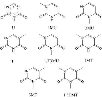
Figure 1.2: Nucleic bases
Table 1.1: Relative induction frequencies of the major photoproducts induced by UV radiation. Adapted from reference [1]
Photoproducts Lesions/108 Da J/m2 % of total photoproducts
UV-C UV-B UV-C UV-B
Cyclobutyl pyrimidine dimer 2.3 0.30 77 78
Pyrimidine pyrimidinone [6-4]-photoproduct 0.6 0.04 20 10 Dewar Pyrimidinone 2.3·10−2 ∼4·10−2 0.8 10
Adenine-thymine heterodimer 6·10−3 nd 0.2 nd
Cytosine photohydrate 5·10−2 6.6·10−3 ∼2 ∼2 Single-strand break ∼5·10−4 ∼4·10−6 <0.1 <0.1 DNA-protein crosslink ∼3·10−4 ∼1·10−6 <0.1 <0.1
1.2 Motivation
1.2 Motivation
This work is embedded into the Sonderforschungsbereich (Collaborative research cen- ter) 663 ”Molekulare Antwort nach electronischer Anregung” (Molecular response to electronic excitation). Within this center, several experimental and theoretical groups work on the understanding of photostability of biologically relevant molecules.
The group of Professor Kleinermanns was interested in the problem of the photo- stability of the pyrimidine bases uracil and thymine. They wanted to investigate whether their photostability is an intristic property or a consequences of the interac- tion with the solvent. They chose 1-methylthymine as a molecule for the experiment.
1-Methylthymine is a model system for the DNA-base thymine, since thymine is co- valently bound to the phosphate backbone in the 1-position in DNA double helices.
Electronic relaxation of 1-methylthymine involves an electronic state that lives about two hundred nanoseconds in the gas phase. This electronic state could be a precursor for the creation a cyclobutyl dimer. In order to characterize the structure and lifetimes, Professor Kleinermanns and his coworkers use resonance enhanced multi photon ioniza- tion (REMPI) and IR/UV double resonance spectroscopy in combination with time of flight mass spectrometry. Throughout expansion of the molecules in supersonic gas jets is employed. In order to interpret their experimental result a theoretical analysis was needed. My work started out as an ab initio examination of the electronic relaxation of 1-methylthymine [10]. As a next step we wanted to calculate inter-system crossing rates in uracil, thymine and 1-methylthymine [7]. For the vibrational contributions to the Fermi Golden Rule formula a method based on the summation of Franck-Condon integrals was used. It is implemented in the program VIBES [11]. During our work, we realized that the method we used for the calculation of inter-system rates is difficult to apply in situations where there are many normal modes and when the adiabatic elec- tronic energy gap is large. In those cases it takes much time to sum all Franck-Condon integrals and to obtain a rate constant. Due to that, Professor Marian proposed to develop a new method that can be applied in situations when molecules have no sym- metries and therefore all normal modes should be taken into account. A natural choice is to try to base the method in the time domain in order to avoid tedious summation of Franck-Condon integrals.
1 Introduction
2 The molecular Hamiltonian and its approximations
In order to describe molecular processes it is of fundamental importance to specify a molecular Hamiltonian. The complete molecular Hamiltonian contains three space and one spin degree of freedom per particle (an electron or a nucleus). The biggest problem concerning the description of the electronic and nuclear motion lies in the fact that electronic and nuclear degrees of freedom are coupled mutually through their interaction. Hence, the numerical solution of the Schr¨odinger equation for the complete Hamiltonian is usually not desirable and an analytical solution is impossible [12,13]. In fact, what we want to have is a clever choice of the important degrees of freedom that are involved in the concerned process. Then, we expect that the rest of the degrees of freedom will not significantly influence the dynamics of the selected ones, otherwise their influence can be included using effective Hamiltonian.
In the absence of an external field, the complete molecular Hamiltonian is given by [13]:
H = ˆTCM + ˆT + ˆT + ˆV + ˆHes+ ˆHhf s. (2.1) The first term ˆTCM is a kinetic energy operator of the molecular center of mass. The next two terms are related to the relative motion around the center of mass, ˆT = Tˆn+ ˆTe is a sum of the kinetic energy operators of the relative motion of nuclei and electrons and ˆT = is a term that couples the nuclear and electron momentum operators in their relative motion. ˆV = ˆVnn + ˆVee+ ˆVne is an electrostatic potential operator due to the Coulomb interaction between nuclei, electrons, and between electrons and nuclei. ˆHes = ˆHSO + ˆHsr+ ˆHss is a Hamiltonian operator related to the interaction of each electron spin magnetic moment with the magnetic moments generated by the orbital motions of the electrons (for instance, interaction with its own orbital moment is spin-same orbit interaction and that is a part of total spin-orbit interaction ˆHSO), the magnetic moments generated by the orbital motions of the nuclei ( ˆHsr) and the spin magnetic moments of the other electrons ( ˆHss). The last term in the molecular Hamiltonian ˆHhf s is related to the interaction of the magnetic and electric moments of the nuclei with the other electric and magnetic moments in the molecule. It gives very small energies compared to the other terms, so that its contribution can be freely neglected in the present context.
Motion of the center of mass is not relevant for internal motion, so that the operator TˆCM can be excluded. Also, the operator ˆT is weighted by the total mass of the molecule, so that its contribution is negligible. Therefore, the Hamiltonian of interest is given by:
Hˆ = ˆT + ˆV + ˆHes. (2.2)
2 The molecular Hamiltonian and its approximations
Concerning the ˆHes operator, in this thesis only the spin-orbit Hamiltonian will be considered.
2.1 Separation of electron and nuclear coordinates
In order to simplify the Hamiltonian we will consider the spin-free part and the influ- ence of the electronic spin will be treated later. The basic approximation in treating molecules is the adiabatic or Born-Oppenheimer approximation [14]. It is based on the fact that electrons and nuclei have significantly different masses. If the nuclei do not have large kinetic energies it is possible to separate electronic and nuclear motion.
Let us label the electron coordinates withr and the nuclear coordinates withR. Then, the spin-free Schr¨odinger equation related to equation 2.2 is
( ˆTn(R) + ˆTe(r) + ˆVnn(R) + ˆVne(R, r) + ˆVee(r))Ψ(R, r) = EΨ(R, r). (2.3) The total wavefunction depends on the nuclear and electronic coordinates. In the Born- Oppenheimer approximation, it is separated into an electronic wavefunction ψi(r;R) and a nuclear wavefunction χi(R), so that it is:
Ψ(R, r) =
i
χi(R)ψi(r;R). (2.4)
The former explicitly depends on the electron coordinates and parametrically on the nuclear coordinates. It is a solution of the electronic Schr¨odinger equation:
( ˆTe(r) + ˆVne(R, r) + ˆVee(r))ψi(r;R) = Ei(R)ψi(r;R). (2.5) Ei(R) is the energy of the electrons for a certain configuration of nuclei. Collecting its value for all configurations of nuclei we obtain the potential energy (hyper) surface (PES). Its significance is tremendous because it completely determines the motion of the nuclei. The nuclear wavefuntion χi(R) is a solution of the nuclear Schr¨odinger equation:
( ˆTn(R) +En(R)−E)χi(R) =
j
Λijχj(R) (2.6)
where E is the total energy of the nuclei and Λij is defined as Λij =−
ψi(r;R)Tn(R)ψj(r;R). (2.7) It represents non-adiabatic coupling in the adiabatic electronic representation and con- tains the first and second-order derivative coupling:
Λij =−
p
1 Mp
fij(p) ∂
∂Rp
−
p
1 2Mp
h(p)ij (R) (2.8)
fij(p)=
ψi∗(r;R) ∂
∂Rp
ψj(r;R)dr (2.9)
h(p)nm=
ψi∗(r;R) ∂2
∂R2pψj(r;R)dr. (2.10)
2.2 The solution of the electronic Schr¨odinger equation In the case where the non-adiabatic coupling is small, we can neglect it. This leads to uncoupled equations for the nuclear wavefunctions χi(R) :
( ˆTn(R) +En(R)−E)χi(R) = 0, i= 1,2, .... (2.11) This can be always done when the difference between the potential energy surfaces is much larger than the spacing between the energy levels related to the nuclear motion.
In cases when the electronic potential surfaces are not well-separated, the non-adiabatic coupling can become very strong and even singular.
2.2 The solution of the electronic Schr¨ odinger equation
The theoretical examination of molecular processes always starts with the construction of potential surfaces, based on equation 2.5. There are plenty of methods for the solu- tion of the electronic Schr¨odinger equation, like density functional theory, molecular- orbital based methods, valence-bond based methods. For an overview of the molecular orbital methods there is an excellent monograph Molecular Electronic-Structure The- ory by T. Helgaker, P. Jørgensen and J. Olsen [15].
The molecular orbital based method CC2 that was primarily used in this thesis will be explained here. The CC2 theory is built upon the Hartree-Fock method. The next section gives a short explanation of the Hartree-Fock method.
2.2.1 The Hartree-Fock method
The Hartree-Fock theory is the most elementary way for obtaining a molecular orbital basis while taking into account the Pauli exclusion principle [16]. It is a mean-field theory where the two-particle Coulomb interaction is substituted with an effective one- particle interaction. The electrons move in their spin orbitals independently of each other. The effective interaction represents an average field created by all other electrons and nuclei that interact with a given electron.
The orbitals which optimally represent the many-electron wavefunction are the orbitals that minimize the total electronic energy. They are found by employing a variational procedure to the Slater determinant:
Ψ(x1, x2, ..., xN) = (N!)−12det|ψ1(x1)ψ2(x2)...ψN(xN)| (2.12) where ψi(xi) is the i-th occupied spin-orbital. The total energy related to the Slater determinant is:
E =
i
ψi|h(1)ˆ |ψi+ 1 2
i,j
(ψiψj|ˆg(1,2)|ψiψj − ψiψj|gˆ(1,2)|ψjψi). (2.13) ˆh(1) is a one-electron operator that contains the electron kinetic energy and the interac- tion between electron 1 and all nuclei, ˆh(1) =−12∇21−
A ZA
R1A. ˆg(1,2) is a two-electron
2 The molecular Hamiltonian and its approximations
operator that represents the interaction between two electrons, ˆg(1,2) = |r1−1r2|. It is useful to define the Coulomb ˆJi and the exchange operator ˆKi in the following manner:
Jˆi(x1)ψj(x1) = [
ˆ
g(1,2)ψi(x2)ψi(x2)d3x2]ψj(x1) (2.14) Kˆi(x1)ψj(x1) = [
ˆ
g(1,2)ψi(x2)ψj(x2)d3x2]ψi(x1). (2.15) The Coulomb operator represents an average local potential at x1 arising from an electron inψi. The exchange operator is a space non-local operator that exchanges the electrons 1 and 2 with respect to their spin-orbitals. It is a manifestation of the Pauli principle. It is convenient to introduce the total Coulomb and exchange operators:
Jˆ=
iJˆi, ˆK =
iKˆi where the summation runs over all occupied spin-orbitals.
From the definition of the operators ˆJ and ˆK it follows that the total electronic energy is:
E =
i
ψi|ˆh+ 1
2( ˆJ −K)ˆ |ψi. (2.16) Using the variational principle, δE = 0, one can derive the Hartree-Fock equations:
F ψˆ i =iψi (2.17)
where the Fock operator is given by ˆF = ˆh+ ˆJ −K. Spin-orbitals that diagonalizeˆ the Fock operator are called canonical spin-orbitals. The Fock operator is an effective one-electron operator that contains no explicit two-electron interactions.
Expressed with the Fock operator, the total Hartree-Fock energy is:
EHF = 1 2
i
(ψi|Fˆ|ψi+ψi|ˆh|ψi). (2.18) To solve the Hartree-Fock equations is equivalent to diagonalizing the Fock matrix. But because the latter depends on the occupied orbitals the equations have to be solved iteratively. This iterative procedure is known as the self-consistent-field method. In practice, molecular spin orbitals are expanded as combinations of atomic orbitals. This defines the LCAO procedure.
Although the Hartree-Fock method gives typically more than 99% of the total elec- tronic energy, we usually have a need to accurately compute the remaining 1%. The difference between the total electronic energy and the Hartree-Fock energy (in a com- plete basis set) is the correlation energy. It presents the energy due to instantaneous electron interactions in the molecule. Usually it is divided into dynamic and static correlation energy. The former is a consequence of the fact that the Fock operator does not represent the complete electronic Hamiltonian. The difference between the complete electronic Hamiltonian and the Fock operator is a fluctuating potential. It produces virtual excitations from occupied to unoccupied orbitals. The static part is a consequence of the inadequacy of one Slater determinant to represent the elec- tronic wavefunction. It is usually considered as a long-range effect. Static correlation
2.2 The solution of the electronic Schr¨odinger equation is important in bond-breaking regions of nuclear configuration space but also for many excited states. Approaches to solve the correlation problem are divided into single- or multi-reference methods. Single-reference methods are based on one Slater determi- nant for subsequent perturbational or variational calculation and primary account for the dynamical correlation. Multi-reference methods are based on a linear combination of several Slater determinants and can address static as well as dynamic correlation.
2.2.2 The coupled-cluster method
The coupled-cluster method is a single-reference method that successfully solves the problem of dynamic electron correlation. It is employed in many benchmark calcu- lations of electronic energies. Dynamic correlations due to the fluctuating potential manifest themselves as virtual excitations from occupied to unoccupied orbitals. Ac- cording to the number of the occupied virtual orbitals, excitations can be divided into single (S), double (D), triple (T), ... These excitations have a certain amplitude and associated probability to occur. The Rayleigh-Schr¨odinger perturbation theory, where the fluctuating potential is a perturbation, accounts only for excitations up to a given order. For instance, the second-order perturbation produces double excitations from the occupied orbitals to unoccupied orbitals. But the perturbation expansion converges slowly. In order to speed up convergence it is possible to use an exponential ansatz for the wavefunction:
|CC=eTˆ|ΦHF (2.19)
where the cluster operator
Tˆ= ˆT1+ ˆT2+ ˆT3+...+ ˆTN (2.20) is a sum of operators that produce single ( ˆT1), double ( ˆT2), and higher excitations.
Using second quantization formalism they can be written as:
Tˆ1 =
AI
tAIa†AaI (2.21)
Tˆ2 =
A>B,I>J
tABIJ a†AaIa†BaJ. (2.22) IndicesI andJ are used for occupied orbitals andAandBfor unoccupied orbitals. tIA
and tIJ AB are cluster amplitudes for single and double excitations, respectively. The cluster operator can be written in shorthand notation as T = tτ =
μtμτμ, where t denotes the cluster amplitudes, τ the corresponding excitation operator and μ cover single, double and higher-order excitations. Retaining only certain classes of excitations creates a hierarchy of approximations. The ansatz for coupled-cluster singles (CCS) contains only the T1 operator, for singles and doubles (CCSD) it contains ˆT1+ ˆT2, for singles, doubles and triples (CCSDT) it contains ˆT1+ ˆT2 + ˆT3 etc. An advantage of using the exponential ansatz is that the wavefunction contains contributions from all states that can be created by excitations from occupied to unoccupied orbitals even
2 The molecular Hamiltonian and its approximations
at the truncated level. This can be shown by expanding the exponential ansatz into a Taylor expansion. For example, double excitations from the Hartree-Fock state can be obtained by acting with the double-excitation operator ˆT2 or by acting twice with the single-excitation operator ˆT1. Those different excitation processes have different amplitudes. So, the exponential ansatz represents some kind of a resummation of ordinary perturbation theory. The most used coupled-cluster approximation is singles and doubles (CCSD). The energy for that approximation is obtained from:
E =ΦHF|Hˆ|CCSD=ΦHF|Heˆ Tˆ1+ ˆT2|ΦHF. (2.23) The CCSD cluster amplitudes are obtained by solving the amplitude equations:
μ1|e−Tˆ1−Tˆ2Heˆ Tˆ1+ ˆT2|ΦHF= 0 (2.24) μ2|e−Tˆ1−Tˆ2Heˆ Tˆ1+ ˆT2|ΦHF= 0 (2.25) where |μ1 and |μ2 denote the single and double excitation manifold. It is useful to define the ˆT1 transformed operator as
ˆ˜
O =e−Tˆ1Oeˆ Tˆ1 (2.26)
so that the amplitude equations can be rewritten in the form:
μ1|Hˆ˜ + [H,ˆ˜ Tˆ2]|ΦHF= 0 (2.27) μ2|Hˆ˜ + [H,ˆ˜ Tˆ2] +1
2[[H,ˆ˜ Tˆ2],Tˆ2]|ΦHF= 0. (2.28) If we retain the singles equations but approximate the doubles equations to first order we obtain:
Ωμ1 =μ1|Hˆ˜ + [H,ˆ˜ Tˆ2]|ΦHF= 0 (2.29) Ωμ2 =μ2|Hˆ˜ + [ ˆF ,Tˆ2]|ΦHF= 0. (2.30) In the doubles equations, the Fock operator is introduced. These equations define the CC2 model [17]. The ground-state energy for a closed-shell molecule is:
ECC2 =ESCF +
IJ AB
(tABIJ +tAItBJ)(2(IA|J B)−(J A|IB)). (2.31) The CC2 excitation energies are obtained using a response theory. Here, the energies are obtained as eigenvalues of a Jacobian matrix, which contains derivatives of the vector function Ωμi with respect to the cluster amplitudes [18]:
Aμiνj = δΩμi
δtνi
=
μ1|[[Hˆ˜ + [H,ˆ˜ Tˆ2], τν1]|HF μ1|[H, τˆ˜ ν2]|HF μ2|[H, τˆ˜ ν1]|HF μ2|[ ˆF , τν2]|HF
. (2.32) The CC2 method is usually employed with the RI (resolution of identity) approximation [19]. This approximation simplifies the calculation of the two-electron integrals in
2.3 The nuclear Schr¨odinger equation and the normal mode Hamiltonian an atomic basis. Particularly computationally demanding are four-index integrals.
They are substituted by three-center integrals using an auxiliary basis set in the RI method. Products of the atomic orbitals pq and rs are expanded in the auxiliary basis set of atom-centered Gaussian functions. Hence, the two-electron integrals can be approximated by:
(pr|rs)≈
α
bαpqbαrs (2.33)
bαpq =
β
(pq|β)Vαβ−1/2 (2.34)
where α and β are the auxiliary basis functions and Vαβ = α(r1)β(r2)
r12 dr1dr2. The RI approximation greatly speeds up CC2 calculations, particularly for large molecules, i.
e. for a large number of basis functions.
2.3 The nuclear Schr¨ odinger equation and the normal mode Hamiltonian
After we have solved the electronic Schr¨odinger equation for a set of nuclear config- urations and therefore have obtained the potential energy surface, we can study the nuclear motion on it. The nuclear Schr¨odinger equation can be simplified by introduc- ing a rotating reference frame. In this way, rotations and vibrations can be separated.
If we are interested in the nuclear motion in the vicinity of the potential minimum then the normal model Hamiltonian is a natural choice.
Let us start from the nuclear Schr¨odinger equation in Cartesian coordinates in the cen- ter of mass system, equation 2.11. We will consider rigid molecules, that is molecules that have a well defined minimum of the potential energy surface. The coordinates of the nuclei in the minimum are (xeqi , yieq, zeqi ). We want to separate vibrational from rotational motion. The motion in the center-of-mass system has an angular momentum J. In order to decouple vibrational and rotational motion, we should minimize J. It is given by:
J =
i
miri×r˙i =
i
mi(reqi + Δri)×(r˙eqi + Δr˙i). (2.35) In a rigid molecule at low energies the nuclei move in the vicinity of the potential energy minimum, so that J can be approximated by:
J ≈
i
mireqi ×Δr˙i. (2.36)
This vector equation defines the Eckart condition:
i
mireqi ×Δri = 0. (2.37)
2 The molecular Hamiltonian and its approximations
that is employed in defining the body-fixed rotating molecular coordinate system. The three equations for the components give the three Euler angles θ, φ and χ that define the relative orientation of the rotating molecular system in comparison with the center of mass system.
In the vicinity of the potential energy minimum, the kinetic and potential energy operators can be diagonalized by introducing normal-mode coordinates. Thus, for a nonlinear molecule, the Cartesian body-fixed coordinate k of the i-th nucleus are related to the normal mode coordinate Ql by an orthogonal transformation L defined by:
rik =rikeq+m−i 12
l
LilQl. (2.38)
In nonlinear molecules, six normal modes correspond to translation and rotation and the rest correspond to vibrations. If we put the body-fixed frame in an orientation such that its axes are parallel to the principal axes of the molecular moment of inertia and assume that the angular momenta of vibrations are small, then we can separate the rotational and vibrational motion and write a rovibrational Hamiltonian as:
Hˆrv = ˆHrot+ ˆHvib (2.39) Hˆrot = 12(LI02x
x +LI02y
y + LI02z
z) (2.40)
Hˆvib = 12
i(Pi2+ω2iQ2i) + 3!1
klmΦklmQkQlQm+... (2.41) where Ix0, Iy0 and Iz0 are principal moments of inertia, ωi are normal-mode frequencies and Φklm, ... are anharmonic coupling constants.
Often, it is sufficient to consider a harmonic vibrational Hamiltonian as the first ap- proximation:
Hˆvib = 1 2
i
(Pi2+ωi2Ω2i). (2.42) Electronic transitions are usually followed by a change in normal modes. This change was first examined by Duschinsky [20] and the transformation is called Duschinsky transformation. The normal modes of the final electronic state are not orthonormal to the normal modes of the initial electronic state (for a two-dimensional case see Figure 2.1).
Consider, for instance, the ground- and some excited-state PES. In the ground state, the potential energy surface in the vicinity of the potential minimum can be represented as Eg = 12
iω2iQ2i +... while the excited state surface for the same nuclear geometry is Ee = Ee(0) +
iκeiQi + 12
i,jηeijQiQj +... where Ee(0) is the vertical electronic excitation energy, κei are the first-order intrastate coupling constants and ηeij are the second-order intrastate coupling constants. The body-fixed system in each electronic state is defined by the Eckart condition with respect to the equilibrium structure for that state. A calculation that involves both electronic states requires a common body frame, which is as the first approximation obtained by an affine transformation that
2.3 The nuclear Schr¨odinger equation and the normal mode Hamiltonian
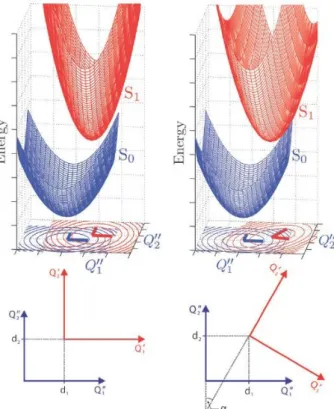
Figure 2.1: Schematic two-dimensional (Q1,Q2) ground (lower, blue) and excited (up- per, red) state PES. Left panel: Displaced-distorted harmonic potential energy surfaces with (i) D = 0 , (ii) dQ1 = dQ1 and dQ2 = dQ2 , and (iii) J = 1, i. e. the excited state PES is (i) displaced, (ii) distorted, but (iii) not rotated relative to the ground state PES. Right panel: Displaced-distorted- rotated harmonic potential energy surfaces with (i) D = 0 (ii) dQ1 = dQ1 and dQ2 =dQ2 , and (iii) J= 1. For an explanation of the terms see text.
Taken from ref. [5]
2 The molecular Hamiltonian and its approximations
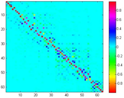
Figure 2.2: Duschinsky matrix J for thymine: It is an orthogonal matrix close to a unit matrix with the dimension equal to the number of normal modes (for discussion about translation and rotation see text); nondiagonal elements clearly show coupling between normal modes
removes the first-order intrastate coupling and diagonalizes the second-order intrastate coupling:
Q¯i =
j
JijQj+Di (2.43)
where Qi are normal modes of the final electronic state and Qj are normal modes of the initial electronic state. The Duschinsky matrix J is an orthogonal matrix and it produces a rotation of the normal modes. The rotation is followed by the translation D. If L and L are orthogonal matrices that transform Cartesian coordinates of the final and initial electronic states to the normal modes, then:
J =LTL (2.44)
D=LTm12(req−req) (2.45) wherereq andreq are Cartesian coordinates of the respective electronic minima, m12 is a diagonal matrix with square roots of the atomic masses. The two electronic states must be orientated such that they have the largest number of symmetry elements in common. The Duschinsky matrix J for thymine is shown in Figure 2.2
There are some possible complications for the determination of the Duschinsky matrix due to mixing of rotational and vibrational motion. In a case when the equilibrium geometries of the two electronic states are significantly different, then the Duschinsky matrix given by the product of the two normal mode transformation matrices cannot be decomposed into a vibrational and a rotational part. That is depicted as:
J =
⎛
⎝ Jvib Jvr 0 Jvr Jrot 0 0 0 Jtr
⎞
⎠. (2.46)
The transformation between normal modes of two electronic states given by equation 2.43 is an approximation. In the most general case it should be nonlinear. Those nonlinearities occur due to axis-switching [21–23].
![Table 4.1: Calculated inter-system crossing rates S 1 T 1y for phenalenone in s − 1 time interval [ps] # points correlation function cumulant expansion η cm − 1](https://thumb-eu.123doks.com/thumbv2/1library_info/4533127.1596400/65.892.109.757.157.513/calculated-crossing-phenalenone-interval-correlation-function-cumulant-expansion.webp)
![Table 4.2: Calculated inter-system crossing rates S 1 T 1x for flavone in s − 1 time interval [ps] # points correlation function cumulant expansion η cm − 1](https://thumb-eu.123doks.com/thumbv2/1library_info/4533127.1596400/67.892.108.758.165.273/calculated-crossing-flavone-interval-correlation-function-cumulant-expansion.webp)