F
ÜRP
LASTISCHE, H
AND-
UNDW
IEDERHERSTELLUNGSCHIRURGIEP
ROF. D
R. U
NIV. D
R. L
UKASP
RANTLDER
F
AKULTÄT FÜRM
EDIZIN DERU
NIVERSITÄTR
EGENSBURGAPPLICATION OF ADIPOSE TISSUE-DERIVED STEM CELLS FOR RESTORATION OF ACHILLES
TENDON ELASTICITY AFTER INJURY
DISSERTATION
zur Erlangung des Doktorgrades der Medizin
(doctor medicinae)
der
Fakultät für Medizin der Universität Regensburg
vorgelegt von Tobias Kügler geboren in Gräfeling
2017
GEWIDMET MEINEN ELTERN UND MEINEM BRUDER NIKOLAI
Dekan: Prof. Dr. Dr. Torsten E. Reichert
1. Berichterstatter: Prof. Dr. univ. Dr. Lukas Prantl
2. Berichterstatter: Prof. Dr. Dr. Torsten E. Reichert
Tag der mündlichen Prüfung: 29.11.2017
Teile dieser Arbeit wurden publiziert:
Sebastian Gehmert, Ernst-Michael Jung, Tobias Kügler, Silvan Klein, Sanga Gehmert, Katharina Zeitler, Markus Loibl, Lukas Prantl: Sonoelastography can be used to monitor the restoration of Achilles tendon elasticity after injury Ultraschall Med. 2012 Dec; 33(6): 581-6
Außerdem wurden die Ergebnisse beim 42. Jahreskongress der Deutschen Gesellschaft der Plastischen, Rekonstruktiven und Ästhetischen Chirurgen in Innsbruck, Österreich 2011 in einer Präsentation vorgestellt:
Sebastian Gehmert, Tobias Kügler, Silvan Klein, Eckhard Alt, Michael Jung,
Lukas Prantl. Autologe Stammzellapplikation unterstützt die Regeneration der
Elastizität von Sehnengewebe.
Dokumentationsblatt
Bibliografische Beschreibung
Kügler, Tobias
Application of adipose tissue-derived stem cells for restoration of achilles tendon elasticity after injury
-2016.- 72 Bl., 16 Abb., 18 Tab.
Medizinische Fakultät der Universität Regensburg
Zentrum für Plastische, Hand- und Wiederherstellungschirurgie (Direktor: Prof. Dr. univ. Dr. Lukas Prantl)
Kurzreferat:
Aktuelle Studien lassen darauf schließen, dass Stammzellen, isoliert aus dem Fettgewebe (adipose tissue-derived stem cells – ASCs), zur Behandlung von Sehnenrupturen verwendet werden können. Bisherige Ergebnisse belegen dies aufgrund von histologischen, immunhistologischen und verschiedensten biomechanischen Untersuchungen. Das Ziel dieser Arbeit war es zu untersuchen, ob die Behandlung von Achillessehnen mit ASCs zu einer Verbesserung der Elastizität führt und ob man diesen Effekt objektiv mit Hilfe der Sonoelastographie demonstrieren kann.
Es konnte gezeigt werden, dass Achillessehnen bei Behandlung mit ASCs auf einer Kollagenmatrix im Vergleich zu der Gruppe, behandelt nur mit Kollagenmatrix, einen signifikant niedrigeren Elastizitätsindex aufweisen konnten. Der Index-Wert entsprach dabei dem Niveau einer unverletzten Sehne. Weiterhin konnte dargestellt werden, dass die Sonoelastographie den Elastizitätsindex objektiv und valide abbilden kann.
Schlüsselwörter: [Achillessehne], [adipose tissue-derived stem cells],
[Elastizitaetsindex], [Sonoelastographie]Table of Contents
1. Zusammenfassung 2. Summary
3. Abbreviations 4. Introduction
4.1. Achilles tendon
4.1.1. Extracellular matrix and stem cells in tendon tissue 4.1.2. Tendon regeneration and repair
4.1.3. Application of growth factors for Achilles tendon injury 4.1.4. Stem cells for tendon tissue regeneration
4.1.5. ASCs for tendon tissue regeneration 4.2. Sonoelastography
5. Aim of the study
6. Materials und methods 6.1. Materials
6.1.1. Substances 6.1.2. Equipment 6.1.3. Software 6.2. Methods
6.2.1. Preparation of the fat body 6.2.2. Stem cell isolation
6.2.3. DAPI labeling of ASCs and fluorescence microscopy 6.2.4. Preparation of the Achilles tendon
6.2.5. Expansion and subculturing of ASCs
6.2.6. Adipogenic differentiation of ASCs
6.2.7. Osteogenic differentiation of ASCs 6.2.8. Histology
6.2.9. Sonoelastography 6.2.10. Statistical analysis 7. Results
7.1. ASC preparation and injection 7.2. Transdifferentiation of ASCs
7.2.1. Adipogenesis 7.2.2. Osteogenesis
7.3. Effects of ASCs on tendon’s elasticity 7.4. Fluorescence microscopy
7.5. Histological examination 8. Discussion
9. Limitations 10. Conclusion 11. References 12. Declaration
13. Acknowledgements 14. Curriculum vitae 15. Addendum
15.1 Figures
15.2 Tables
1. Zusammenfassung
Ziel: Die Sonoelastographie kann mechanische Eigenschaften von Gewebe darstellen und ist daher geeignet Defekt- und Narbenbildung von Sehnengewebe darzustellen. Ziel der Studie war es, die Elastizität von Achillessehnengewebe nach autologer mesenchymaler Stammzellapplikation unter Verwendung der Sonoelastographie zu untersuchen.
Material und Methodik: Die Achillessehne beider Hinterläufe wurde in neun Neuseeland Kaninchen vollständig durchtrennt. Anschließend wurden die Hinterläufe randomisiert drei Gruppen zugeteilt, wobei eine extrazelluläre Matrix mit Stammzellen (Gruppe 2, n=6) und ohne Stammzellen (Gruppe 3, n=6) verwendet wurde. In der Kontrollgruppe wurde eine Sham-Operation durchgeführt (Gruppe 1, n=6). Die Extraktion und Applikation der mesenchymalen Stammzellen erfolgte aus dem nuchalen Fettkörper zum gleichen Zeitpunkt wie die Achillessehnendurchtrennung, um die Untersuchung an einem autologen Sehnenregenerationsmodell zu untersuchen. Nach 8 Wochen wurden die Achillessehnen entnommen und die Elastizität mit einer hochauflösenden 6-15 MHz Matrix–Linear-Sonde untersucht. Für jede Sehne wurde eine 20 Sekunden farbkodierte Sonoelastographie-Sequenz aufgezeichnet und 10 Farbhistogramme untersucht. Definierte Regions of Interests (ROIs) wurden über den Sehnendefekt (n=3) und über angrenzendes vitales Sehnengewebe (n=3) gelegt. Für die semiquantitativen Auswertungen wurden 180 Einzelmessungen aufgezeichnet und ausgewertet.
Ergebnisse: In Gruppe 2 konnte für Achillessehnen mit beladener Matrix eine höhere Elastizität im Vergleich zu Achillessehnen mit unbeladener Matrix in Gruppe 3 gemessen werden (p<0.001; ANOVA). Hinsichtlich des Elastizitäts- Index (EI) von unbehandeltem Sehnengewebe (Gruppe 1) und Sehnengewebe mit beladener Matrix (Gruppe 2) konnte kein Unterschied gefunden werden (p>0.05). Für alle Einzelmessungen der verschiedenen Messzeitpunkte konnte kein signifikanter Unterschied festgestellt werden (p>0.05).
Schlussfolgerung: Unsere Ergebnisse zeigen, dass die Applikation von
autologen mesenchymalen Stammzellen des Fettgewebes zu einer
vollständigen Wiederherstellung der Elastizität des Sehnengewebes nach
Achillessehnenverletzung führt. Außerdem konnte gezeigt werden, dass die
Sonoelastographie eine geeignete Methode ist, um die Regeneration der Elastizität nach Achillessehnenverletzung darzustellen und zu beurteilen.
Schlüsselwörter:
[Achillessehne], [adipose tissue-derived stem cells], [ Elastizitaetsindex], [Sonoelastographie]
2. Summary
Purpose: Sonoelastography allows assessment of tissues’ mechanical properties and has recently been used to demonstrate the effects of Achilles tendon injury. The aim of the current study was to evaluate an ultrasound approach to depict elastic recovery after stem cell application on injured Achilles tendon.
Materials and Methods: A rabbit achilles tendon injury model was used and randomized hindlimbs received either extracellular matrix with autologous adipose tissue-derived stem cells (group 2, n=6) or without (group 3, n=6).
ASCs were harvested from the rabbits’ nuchal fat body at the same time as the tendon injury operation. Untreated Achilles tendon (group 1, n=6) served as controls but underwent sham-operation. Specimens were harvested after 8 weeks and were longitudinal analyzed for elasticity using a high resolution 6-15 MHz matrix linear probe. For each tendon, real-time color-coded sonoelastography sequences of 20 seconds were recorded and ten color histogram frames were obtained. Defined regions of interest (ROIs) were placed on the defect (n=3) and on adjacent uninjured tendon tissue (n=3). In total, 180 measurements were obtained for semiquantitative analysis.
Results: Repeated measures ANOVA demonstrated a higher elasticity for stem cell seeded matrix (group 2) in comparison to the unseeded matrix (group 3) (p<0.001; ANOVA). No significant difference was found between the injured tendon tissue treated with stem cell seeded matrix (group 2) and uninjured Achilles tendons (group 1) (p>0.05). Moreover, no differences were found between the measurements at different time-points (p>0.05).
Conclusion: The current study indicates that autologous mesenchymal stem cell application successfully restores mechanical properties of injured tendon tissue.
Furthermore, sonoelastography enables to monitor elasticity of injured Achilles tendon after stem cell application.
Keywords:
[Sonoelastography], [Achilles tendon], [Elasticity Index], [Adipose tissue-derived stem cells]3. Abbreviations
ASCs Adipose tissue-derived stem cells
ANOVA Analysis of variance
MHz Mega Hertz
n Number
ROI Regions of interest
EI Elasticity index
i.e. Id est
ECM Extracellular matrix
TSPC Tendon stem/progenitor cell
Bgn Biglycan
Fmod Fibromodulin
VEGF Vascular endothelial growth factor
IGF-1 Insulin-like-growth-factor 1
PDGF Platelet derived growth factor
TGF-β Transforming growth factor β
bFGF Basic fibroblast growth factor
MSC Mesenchymal stem cell
BMSC Bone marrow-derived mesenchymal stem
cell
ACL Anterior crucial ligament
AT Achilles tendon
BMMC Bone marrow mononuclear cells
BMC Whole bone marrow cells
PT Patellar tendon
RC Rotator cuff
SMSC Synovial mesenchymal stem cells
SE Sonoelastography
MRI Magnetic resonance imaging
PBS Phosphate bufferd saline
α-MEM α-modification of Eagle’s medium
FBS Fetal bovine serum
DAPI 4',6-diamidino-2-phenylindole
DMEM Dulbecco's Modified Eagle's medium
DAB 3,3′-Diaminobenzidine
HRP Horseradish peroxidase
BSA Bovine serum albumin
4. Introduction
4.1 Achilles tendon
4.1.1 Extracellular matrix and stem cells in tendon tissue
The Achilles tendon is the strongest and sturdiest tendon in the human body
1and serves as connective tissue which physically binds muscles (i.e. Musculus gastrocnemius) to skeletal structures
2(i.e. Calcaneus). This facilitates enhancing joint stability and locomotion
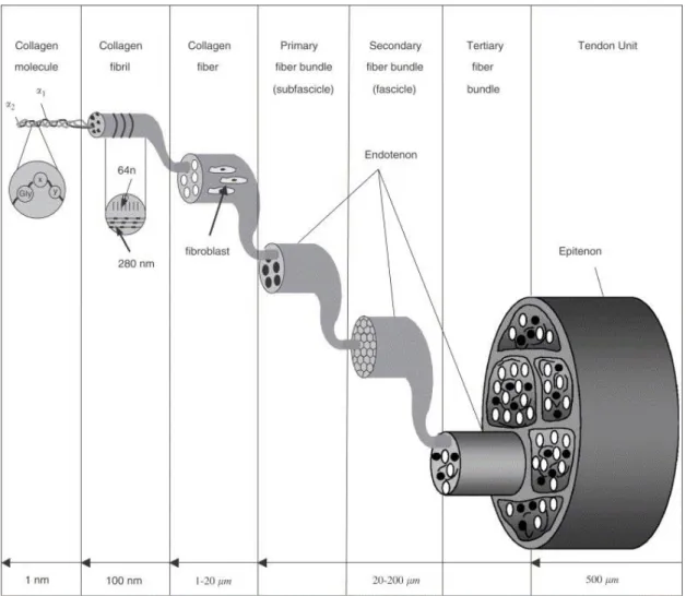
3. Tendon tissue has a multi-unit hierarchical organization of collagen molecules, fibrils, fibers, bundles, fascicles and tendon units, all running parallel to the tendon’s longitudinal axis
3designed to resist tensile load
4.
Figure 1: A drawing scheme of the multi-hierarchical structures of the tendon3
.
The structure of the extracellular matrix (ECM) molecules determines the physiological function and the mechanical strength of tendons
3. The predominant molecule of tendon tissue is collagen I, which constitutes about 95% of the total collagen
5. The overall content of cells is low
6and primarily comprises of tenocytes and fibroblasts also referred to as tenoblasts. Both cell types are of mesenchymal origin and represent 90-95% of the cellular elements
4, in tendon tissue. The premature fibroblast differentiates into a tenocyte
7, featured with a very limited proliferation capacity
8. Fibroblasts are responsible for synthesizing extracellular matrix proteins (e.g. collagen, fibronectin, proteoglycan), which aggregate to a collagen matrix. In addition, fibroblasts are responsible for remodeling the collagen matrix during tendon healing
3,8. This comprises localized matrix formation and degradation which is crucial for tendon healing, adaptation to exercise, and tendon growth
8. Recently, it has been shown, that tendon stem/progenitor cells (TSPCs) reside in ECM composed niche in tendon tissue and are controlled by biglycan (Bgn) and fibromodulin (Fmod). Moreover, isolated TSPCs could regenerate tendon- like tissues after extended expansion in vitro and transplantation in vivo
9. However, repair mechanism of these stem cells is limited due to low cell number in tendon tissue.
Thus, limited healing of tendon tissue is emphasized by poor vascularization and nerve supply
10as well as its low metabolic rate
11. Additionally, Åström
12et al. could demonstrate that mechanical loading but most important aging diminish the blood flow supplying the Achilles tendon.
4.1.2 Tendon regeneration and repair
A cascade of cellular and biochemical processes is initiated in order to restore
tendons elasticity after injury. The initial stage involves tissue inflammation
which attracts cells (e.g. inflammatory cells especially neutrophils, erythrocytes)
from tissue adjacent to the side of injury
13. Recruited fibroblasts maintain
production of extracellular matrix during the proliferative phase. But new
synthesized extracellular matrix is assembled in a random manner containing
large number of cells and high amount of type III collagen
14,15. Moreover,
angiogenesis occurs during this stage
13. Decrease of cellularity and type III
collagen starts 6 – 8 weeks after injury which is a characteristic feature of the remodeling stage. At the same time collagen I synthesis, as a crucial step for tensile strength
16, increases and provides longitudinal organized fibers along the lines of stress and a high rate of crosslinks which are responsible for tendon elasticity
15,17. Additionally, elastic fibers with increased elastin deposition are observed
13.
During the different stages of tendon repair various growth factors activate and regulate the cellular response in a temporal and spatial manner. Vascular endothelial growth factor (VEGF) stimulates angiogenesis
15and is produced at a maximum level after the inflammatory phase. In contrast, Insulin-like-growth- factor-1 (IGF-1) has been reported to be up-regulated highest at the inflammatory phase stimulating migration and proliferation of fibroblast and inflammatory cells
15,18. Moreover, IGF-1 enhances collagen synthesis during the stage of remodeling accompanied by improved tendon stiffness
18. In addition, platelet derived growth factor (PDGF) can facilitate IGF-1 expression during the inflammatory phase of tendon healing and is also involved in the remodeling process
18. Thus, a lack of IGF-1 has been shown to be associated with an insufficient repair response
15. Noteworthy, increased cell proliferation and synthesis of various ECM components especially collagen I during the remodeling phase has been reported for application of PDGF in a dose dependent manner
18. In addition, TGF-β initiates cell migration and collagen production
19and also effects the regulation of proteinases and fibronectin bindings
20. But restored tendon tissue has thinner collagen fibrils which remain
21and provides less mechanical properties than native tissue causing failure of tendons strength and eventual might lead to re-rupture
17.
4.1.3 Application of growth factors for Achilles tendon injury
Treatment options of Achilles tendon injury include nonsurgical treatment as
immobilization in a cast or a functional brace
22. Furthermore, surgical treatment
exists and can decrease re-rupture rate
23compared to conservative treatment
protocol with immobilization. Recent studies suggest that growth factor
application during tendon healing can enhance functional repair and might
reduce time of regeneration. To date various growth factors are investigated for
possible application to improve tendon healing whereas VEGF, IGF-1, TGF-β, PDGF and bFGF are best characterized and known for their role in tendon tissue regeneration
18.
The delivery of one growth factor or even a mixture is challenging since the dosage of these factors is critical during each stage of the regeneration process. Furthermore, the limited half-life of growth factors and their small size restrict their retention for a prolonged time at applied site of tissue. These claims are supported by Zhang et al.
24who showed that VEGF introduced exogenous to an injured tendon site can accelerate tensile strength just within the first 2 weeks postoperatively but no significant difference was seen after 4 weeks compared to the control group. Similar results were reported by treating patellar tendon defects in rabbits with IGF-1 and TGF-β1
25. 2 weeks after administration a significant increase in ultimate stress, energy uptake, stiffness and force at failure were documented compared to the control group.
Interestingly, 6 weeks after administration no significant difference was detected for all investigated parameters
25. Thus, mesenchymal stem cells (MSCs) were suggested as an appropriate delivery method due to their engraftment at applied site and constant secretion of cytokines.
4.1.4 Stem cells for tendon tissue regeneration
Injury or degeneration of tissue of multicellular organisms can be restored by either scar formation or tissue regeneration. The capacity of regeneration is very limited to specific tissue ( e.g. epidermis, intestinal mucosa
26). In contrast, tendon as a self-contained tissue lacks the property of adequate regeneration due to low cell numbers.
Regenerative medicine is utilizing MSCs as a cell based tool since they show
self-renewal and multi-lineage differentiation
27. Furthermore, these cells have
been shown to be hypo-immunogen due to the lack of the major
histocompatibility complex-II molecular expression
28. During the last decade
different types of stem cells have been applied to tendon defects to investigate
the potential for medical purposes in order to establish new experimental and
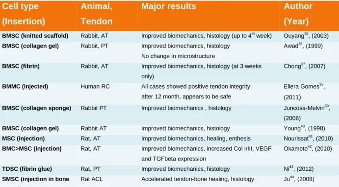
clinical opportunities as seen in the Table 1. Regenerative medicine depends on
stem cells that meet a number of qualities as 1. abundant appearance 2.
minimal invasive harvest 3. multi-linage differentiation and 4. safety and suitability for transplantation
29.
MSCs, as non-hematopoietic stem cells, have been primarily identified within the bone marrow stroma
30and were considered the most promising source for medical tissue engineering application for a long time
31. However, current studies showed that MSCs resides in almost all post-natal organs and tissues
32, including adipose tissue
33as well as tendon
34. In addition, Zuk et al.
33demonstrated that the differentiation potential of ASCs is as effective as of other MSCs.
Table 1. Compilation of cell therapies for tendon healing
Cell type (Insertion)
Animal, Tendon
Major results Author
(Year)
BMSC (knitted scaffold) Rabbit, AT Improved biomechanics, histology (up to 4th week) Ouyang35, (2003) BMSC (collagen gel) Rabbit, PT Improved biomechanics, histology
No change in microstructure
Awad36, (1999)
BMSC (fibrin) Rabbit, AT Improved biomechanics, histology (at 3 weeks only)
Chong37, (2007)
BMMC (injected) Human RC All cases showed positive tendon integrity after 12 month, appears to be safe
Ellera Gomes38, (2011)
BMSC (collagen sponge) Rabbit PT Improved biomechanics , histology Juncosa-Melvin39, (2006)
BMSC (collagen gel) Rabbit AT Improved biomechanics, histology Young40, (1998) MSC (injection) Rat, AT Improved biomechanics, healing, enthesis Nourissat41, (2010) BMC>MSC (injection) Rat, AT Improved biomechanics, increased Col I/III, VEGF
and TGFbeta expression
Okamoto42, (2010)
TDSC (fibrin glue) Rat, PT Improved biomechanics, histology Ni43, (2012) SMSC (injection in bone
tunnel)
Rat ACL Accelerated tendon-bone healing, histology Ju44, (2008)
ACL, anterior crucial ligament; AT, Achilles tendon; BMMC, bone marrow mononuclear cells; BMC, whole bone marrow cells; BMSC, bone marrow-derived mesenchymal stem cells; MSC, mesenchymal stem cells; PT, patellar tendon; RC, rotator cuff;
SMSC, synovial mesenchymal stem cells; TDSC, tendon-derived stem cells; TGF, transforming growth factor; VEGF, vascular endothelial growth factor; >, greater than;
4.1.5 ASCs for tendon tissue regeneration
The use of adipose tissue-derived stem cells, in particular, for tissue
engineering has obvious advantage compared to other stem cells. The
application of embryonic stem cells is very restricted due to potential problems of cell regulation, ethical problems and different national laws whereas autologous mesenchymal stem cells can be used without hesitation as there are no ethical or immunological matters
33.
On proliferation rate ASCs’ doubling time was 28 hours compared to BMSCs’
39 hours
45. ASCs became popular for research and clinical application in first line because of equal efficiency regarding multi-lineage differentiation capacity and minimal invasive method of harvesting (local excision or suction-assisted liposuction
46), low donor site morbidity
47and the abundance of adipose tissue in adult humans. Thus, a high number of stem cells can be obtained.
Furthermore, general or spinal anesthesia is often required
33for bone marrow aspiration due to painful procedure. In addition, bone marrow aspiration is also known for yielding low numbers of MSCs (approximately 1 out of 10
5adherent stromal cells)
30. From this practical point of view an insufficient number of cells may necessitate an additional step of in vitro proliferation to generate a sufficient amount of cells for adequate clinical use. Additionally, liposuction might be more tolerated by patients than bone marrow aspiration due to its aesthetic effects.
Based on these properties ASCs became a valuable tool for tissue engineering and regenerative medicine. Several studies have been carried out to investigate effect of ASCs on tendon healing. As one promising step Uysal et al.
47have enabled the differentiation of ASCs to tenocytes in vivo and furthermore demonstrated an improvement of tendons tensile strength after application of ASCs. In addition, tendons biomechanical properties enhance when combining MSCs with composite biomaterial scaffold
48. Taken together, many studies concern themselves with the topic how new approaches using MSCs influence tendon healing and repair. These studies were mainly focused on established methods for assessing the grade of tendon healing like histology
35,40,49, immunhistochemistry
16,40,50or biomechanical properties like tensile strength
47or maximum stress
49.
The current study did not investigated biomechanical surrogate endpoints (e.g.
tensile strength, maximum load or load to failure) since it has been extensively
applied by various studies in the recent past
16,51. This study centered on how
elasticity might serve as a surrogate marker for tendon healing linked to superior outcome since the elasticity of uninjured tendon tissue is accompanied by high energy uptake and the ability to deform.
4.2 Sonoelastography
Various animal models have been applied to investigate the role of MSCs on tendon regeneration. Interestingly, studies revealed that stem cell application can improve histological and biomechanical parameters but only in the first 6 weeks after tendon injury
37. Moreover, at 12 weeks post-surgery, maximum strength is almost comparable to vital uninjured tendon tissue
40. Macroscopical and histological scoring systems are already used to evaluate the quality of repaired tissue in tendon defect models
10. However, a high number of animals have to be included in a study to investigate maximum force load and histology at various time points in a tendon injury model in order to ensure sufficient power for statistical analysis.
Moreover, histological and immunohistological examinations as well as biomechanical tests for evaluation of different parameters are either not feasible or not established for in vivo use during the healing process. Therefore, these diagnostic tools are inadequate for clinical use in daily routine to monitor the healing process of tendons after stem cell application.
Nevertheless, several techniques for in vivo evaluation of soft tissue, including tendon, are in clinical use. Magnetic resonance tomography, computer tomography and as the most widely-used ultrasound examination are tools for examination. However, the above mentioned diagnostic methods can only provide detailed information of tissue’s morphology but not of biomechanical properties. However, biomechanical testing seems to be the gold standard for evaluating the efficiency of suture techniques or tendon strength, respectively
52,53. Among tensile strength, maximum load or load to failure
16elasticity, respectively stiffness
54, are biomechanical parameters of tendons.
Thus, evaluation of tendon’s elasticity might be a suitable marker to depict the
healing process of a tendon regardless whether tendon tissue was treated
conservatively, with suture or by regenerative methods like stem cell
application.
Sonoelastography (SE), as a new and appropriate diagnostic tool for soft tissue, was first introduced in the early 1990s for in vitro use and subsequently evolved into in vivo use for imaging
55,56to enable in vivo real-time measurement of tissue elasticity
57.
Sonoelastography is based on some basic principles allowing a qualitative determination of tissue elasticity. Manual palpation is one of the oldest medical examination methods
58. Even nowadays it continues to be of great value in medicine, both practiced by professionals and as a technique for self- examination for lymph nodes, breasts, thyroid or Achilles tendon rupture.
However, palpation is limited to superficial accessible structures and the interpretation is very limited due to a high subjectivity for information sensed by the fingers
59. Like manual palpation sonoelastography detects elastic properties of tissue by comparing the grade of deformation between different types of tissue or between different regions of interest. Therefore the examiner performs strain with the ultrasound probe on structures amenable to compression. The maximum of displacement between two image points of the ultrasound B-mode in a determined time interval is computed by the instrument’s software. The software generates a color-coded image, which displays certain displacements in a correspondent color which is superimposed on the B-mode image. The most common color definition depicts hard tissue as blue, intermediate as green and soft as red. The newest generation of sonoelastography device assigns a numerical value, known as elasticity index (EI) to each grade of color. This tool gives the examiner an opportunity to objectify elasticity in a more precise way.
Constant and uniform compression and decompression must be applied to the tendon during sonoelastography in order to avoid misinterpretation and artifacts as well as to assure most exact values. Therefore modern software provides a visual indicator displayed on the monitor.
Recent studies using SE showed that elastic measurements of different types of tissue provide promising and useful result for diagnosis
59–62.
For tendon, in particular, Pedersen et al.
63demonstrated that sonoelastography
seemed to be as feasible as ultrasound and MRI assessing tendon alteration
and furthermore was superior depicting subclinical alteration not detectable with
conventional ultra sound. In addition, in a different study sonoelastography was
able to distinguish between ruptured and healthy tendon by measuring
elasticity
64. However, this study only used a grading of tendon elasticity (i.e.
intermediate, hard, hardest) instead of EI. Moreover, every single tendon
suffering a rupture exhibited heterogeneous structure during SE, whereas all
healthy Achilles tendons had a homogeneous or relatively homogeneous
structure. However, this study admits limitations due to high influence of the
pressure of the probe on the tissue performed by the examiner. Taken together,
the above mentioned findings support various studies which describe
sonoelastography as a promising quantitative tool to characterize alteration in
morphology or biomechanics induced by previous injury
65.
5. Aim of the study
The current study aimed to investigate whether:
i) ASCs are suitable to improve Achilles tendon elasticity when placed at the site of injury
ii) Sonoelastography is an appropriate and examiner independent tool to investigate elasticity of tendon tissue
iii) The content of Collagen I is changed by ASCs application
iv) ASCs are able to engraft at the side of application and survive at least 8 weeks
v) ASCs harvested from rabbit’s nuchal fat body have multilineage pontential
vi) ASCs are able to change cell morphology and cell organization
6. Materials and methods
6.1 Materials
6.1.1 Substances
Povidone iodine Sigma-Aldrich, St.Loise, MO, USA
Cefazolin Pfizer, NYC, NY, USA
Blendzyme III Roche Diagnostics, Basel, CH
PBS Sigma-Aldrich, St.Loise, MO, USA
Hanks Balanced Salt Solution Cellgro, Corning, NY, USA α-modification of Eagle’s medium Cellgro, Corning, NY, USA 20%
FBS PAN Biotech, Aidenbach, Germany
Penicillin- Streptomycin Sigma-Aldrich, St.Louise, MO, USA DAPI stock solution Sigma-Aldrich, St.Louise, MO, USA
Buprenex Reckitt Benckiser, Slough, UK
Meloxicam Norbrook, Newry, North Ireland
Fentanyl citrate Sandoz, Holzkirchen, Germany
Buthanasia solution Virbac, Carros, France
Moist saline dressing Sigma-Aldrich, St.Louise, MO, USA
Trypsin PAN Biotech, Aidenbach, Germany
DMEM, low glucose with L-glutamine Life Technologies, Carlsbad, CA, USA Isobutyl-methylxanthine Sigma-Aldrich, St.Louise, MO, USA
Dexamathasone Sigma-Aldrich, St.Louise, MO, USA
Indomethacin Sigma-Aldrich, St.Louise, MO, USA
Bovine panceas insuline Sigma-Aldrich, St.Louise, MO, USA
Formalin Sigma-Aldrich, St.Louise, MO, USA
Red Oil O stock Sigma-Aldrich, St.Louise, MO, USA
Isopropanol 60% Sigma-Aldrich, St.Louise, MO, USA
beta-glycerophosphate disodium salt hydrate
Sigma-Aldrich, St.Louise, MO, USA
L-ascorbic acid Sigma-Aldrich, St.Louise, MO, USA
Alizarin Red S Sigma-Aldrich, St.Louise, MO, USA
Ammonium hydroxide solution Sigma-Aldrich, St.Louise, MO, USA
Ethanol 70% Sigma-Aldrich, St.Louise, MO, USA
Ethanol 95% Sigma-Aldrich, St.Louise, MO, USA
Xylene Sigma-Aldrich, St.Louise, MO, USA
Paraplast Paraffin Leica, Wetzlar, Germany
Hematoxylin Solution, Harris modified Sigma-Aldrich, St.Louise, MO, USA
Ethanol 100% Sigma-Aldrich, St.Louise, MO, USA
Lithium carbonate Sigma-Aldrich, St.Louise, MO, USA
Eosin Y Sigma-Aldrich, St.Louise, MO, USA
Phloxine B Sigma-Aldrich, St.Louise, MO, USA
Acetic acid Sigma-Aldrich, St.Louise, MO, USA
Cytoseal mounting media Thomas Scientific, Swedesboro, NJ, USA
Mouse anti-rabbit monoclonal antibody-against Collagen-I
Abcam, Cambridge, England
Goat anti-mouse IgG H&L (HRP) Abcam, Cambridge, England Goat anti-mouse IgG H&L (Texas Red) Abcam, Cambridge, England
Tris-EDTA buffer solution Sigma-Aldrich, St.Louise, MO, USA
Tween 20 Sigma-Aldrich, St.Louise, MO, USA
Bovine serum albumin Sigma-Aldrich, St.Louise, MO, USA Hydrogen peroxide 30% Sigma-Aldrich, St.Louise, MO, USA Trisodium citrate dehydrate Sigma-Aldrich, St.Louise, MO, USA
Triton X Sigma-Aldrich, St.Louise, MO, USA
Sodium azide Sigma-Aldrich, St.Louise, MO, USA
Gelatin from cold water fish skin Sigma-Aldrich, St.Louise, MO, USA 3,3’-Diaminobenzidine Sigma-Aldrich, St.Louise, MO, USA
6.1.2 Equipment
Autoclaving Systec, Linden, Germany
Vicryl 4-0 Ethicon, Somerville, New Jersey
50 ml plastic pipette Grainer bio-one, Kremsmünster,
Austria
Shaker HS 501 digital Ika Labortechnik, Staufen, Germany
50 ml plastic tube Grainer bio-one, Kremsmünster,
Austria
Centrifuge Multifuge 3S Heraeus, Hanau, Germany
100μm Steriflip Merck Millipore, Billerica, MA, USA Collagen matrix (Puracol® Plus) Medline Industries, IL, USA
Fume hood M18 Schulz Lufttechnik, Sprockhövel,
Germany
Olympus BX 40 Shinjuku, Tokio, Japan
Polypropylene 2-0 Ethicon, Somerville, New Jersey T75 culture flask Sarstedt, Nürnbrecht, Germany
Incubator Hera Cell 240 Thermo Scientific, Waltham, MA, USA Inverted microscope Wilovert S Helmut Hund GmbH, Wetzlar,
Germany
Refrigerator Liebherr medline Liebherr, Bulle, Schweiz
6 well-plate cellstar Grainer bio-one, Kremsmünster
Austria
HM 400 Microm, Heidelberg
Slides Engelbrecht GmbH, Edermünde,
Germany
Microscope Axiovert Carl Zeiss AG, Oberkochen, Germany
Camera Canon G7 Canon, Takio, Japan
Sonoelastographic device LOGIQ®E9 General Electrics, Fairfield, CT, USA Multifrequency probe 6-15 MHz General Electrics, Fairfield, CT, USA Counting chamber Neubauer-improved Paul Marienfeld GmbH,
Lauda-Königshofen, Germany
6.1.3 Software
Real-time sonoelastography software General Electrics, Fairfield, CT, USA GraphPad Prism 5 for Windows GraphPad Software Inc., La Jolla, CA,
USA
6.2 Methods
6.2.1 Preparation of the fat body
A total of nine male New Zealand white rabbits weighing 3.5±0.5 kg were used
to evaluate if sonoelastography is able to depict elastic recovery after
autologous adipose-derived stem cell application on injured Achilles tendon
following the guidelines of Veterinary Medicine & Surgery at MD Anderson
Cancer Center and US National Institutes of Health. The study was approved by
the IACUC of MD Anderson Cancer Center Houston. All the rabbits were aged
14 to 16 weeks and were randomly assigned to either a control group without
injury or a group treated with ASCs seeded collagen matrix or treated with
unseeded collagen matrix. Equipment for all operations was sterilized by
autoclaving at MD Anderson facility. All animals were anaesthetized by
administration of isoflurane received by mask. Anesthesia was monitored by
respiratory rate and heart rate, response to noxious stimulus, spontaneous
movement, pedal reflex, oxygen saturation and body temperature. Each rabbit
was placed in prone position for harvesting the nuchal fat body. The hair in the
field of operation was shaved and the skin was surgically prepared using
povidone iodine for disinfection. Subsequently, the animal was transferred to a
heated surgery table and the nuchal region was covered with a sterile surgical
drape. A small incision of 3 cm with a scalpel no. 15 was made to approach the
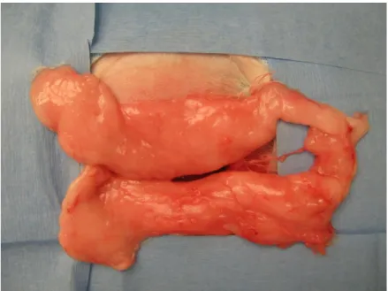
rabbit’s nuchal fat body (Figure 2), which was excised bilaterally and transferred
into a sterile container for stem cell harvesting. The mean weight of the fat
bodies was 15±2.4 grams. The subcutaneous layer and the skin were closed by
continuous suture technique with Vicryl 4-0. Every step was performed by using
aseptic techniques. All animals received perioperative doses of prophylactic
antibiotics (Cefazolin 15mg/kg, i.v.).
Figure 2: Nuchal adipose fat body that was used to harvest adipose tissue derived mesenchymal stem cells after surgical preparation.
6.2.2 Stem cell isolation
Nuchal fat tissue was extensively washed in PBS after harvesting and
subsequently minced in pieces less than 1mm³. Subsequently, the minced
tissue was incubated in PBS containing Blendzyme III (2U/mL) for 30 minutes at
37°C on a shaker at 100 rpm. After digestion, the suspension was
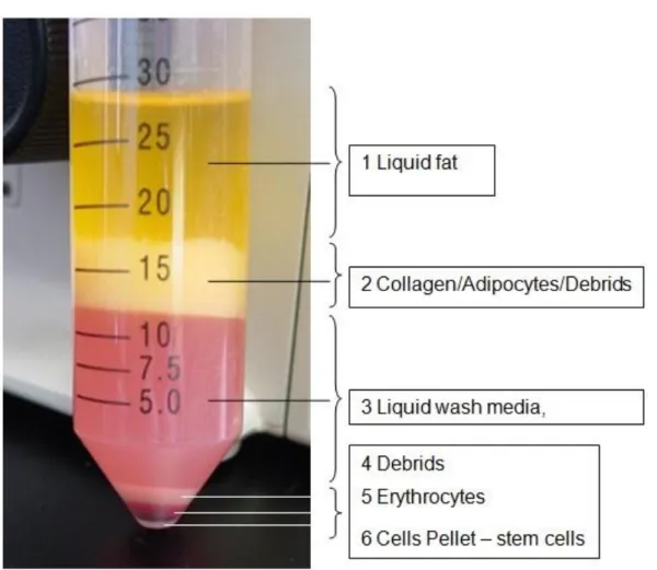
disaggregated by pipetting 5 times under sterile conditions and was transferred
into a 50 ml plastic tube followed by centrifugation at 450g for 10 minutes
(Figure 3). The supernatant was discarded and cells were washed twice with
PBS. Afterwards, cells were vacuum filtered through a 100 μm Steriflip
.The
filtered cell suspension was then centrifuged at 450g for 10 minutes. The
supernatant containing adipocytes and debris was discarded and the pelleted
cells were washed twice with 40 ml Hanks Balanced Salt Solution. The pellet
was resuspended in PBS for immediate application or for further in vitro
experiments in alpha-MEM supplemented with 20% fetal bovine serum (FBS),
100 U/ml penicillin and 100 µg/ml streptomycin. For immediate application cell
number was determined using a Neubauer chamber. Subsequently, the
suspension with the ASCs was seeded on a collagen matrix with 1x10⁶ stem
cells in 200µl PBS after labeling with DAPI. Except for digestion and
centrifugation all steps were performed under a fume hood to remain sterile conditions.
Table 2. Preparation of 500 ml growth medium
Reagents Amount
FBS Glutamine Streptomycin Penicillin alpha MEM
100 ml
14,61 g (2 mM) 50 mg
50000 U Up to 500 ml
Figure 3: 50 ml plastic tube after first centrifugation with 450 g for 10 minutes, fraction 6 shows the stem cell pellet
6.2.3 DAPI labeling of ASCs and fluorescence microscopy
Freshly isolated ASCs from the rabbit’s nuchal fat body were labeled with DAPI before seeded onto the collagen matrix. DAPI stock solution (Table 3) was diluted to final concentration of 50µg/ml in PBS and ASCs were incubated for 30 minutes at 37°C. Afterwards, cells were centrifuged at 500g for 5 minutes, and resuspended in PBS to further seed onto the matrix. Final DAPI concentration was revealed by testing ascending concentration without inducing apoptosis in previous tests.
For fluorescence microscopy 8 µm-thick sections were prepared from each tendon and 3 sections were microscoped to test for the presence of ASCs.
Tissue sections were microscoped using 358 nm wavelength and emission filter of 461 nm. All stained slides for fluorescence microscopy were analyzed using a Olympus BX 40 microscope equipped with a Canon G7 high-resolution digital camera adapter for image acquisition.
Table 3. Preparation of 100 ml DAPI working solution
Reagents Amount
DAPI stock solution PBS
5000 µg Up to 100 ml
6.2.4 Preparation of the Achilles tendon
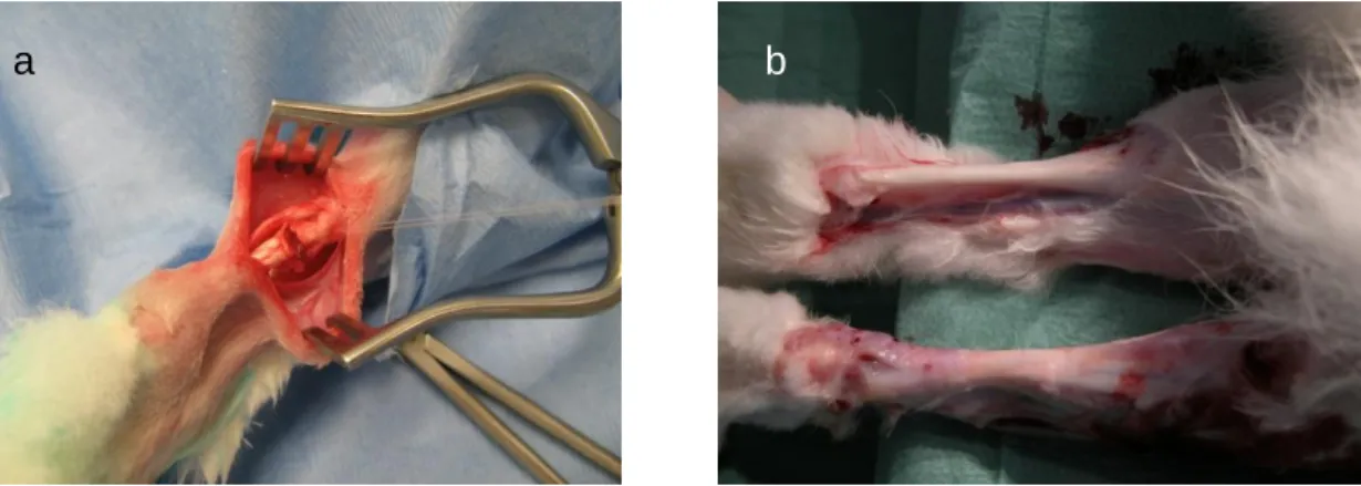
The hair of both hind limbs was shaved and the skin was disinfected with povidone iodine using aseptic techniques. Both hind limbs were covered with a steril surgical drape. Under aseptic conditions, a 3 cm small sharp skin incision was made with a scalpel no. 15 laterally to the Achilles tendon and the tendon was exposed. The peritendon was opened and the tendons of the musculus plantaris, soleus and gastrocnemius were identified and segmented (Figure 4a).
Only the gastrocnemius tendon was transected with a scalpel blade
perpendicular to the collagen fibers 3 cm above the tendon insertion of the
calcaneus. A gap of 10 mm was created on each limb and the defect randomly
received either a collagen matrix (Puracol® Plus) with 1x10
6stem cells (group 2, n=6 tendons) suspended in 200µl PBS or without stem cells (group 3, n=6 tendons). Collagen matrix was sutured in place using a modified Kessler pattern with 2-0 polypropylene. Control animals received no injury (group 1, n=6 tendons) but were sham operated. Skin was closed by suture using 4-0 Vicryl.
The leg was bandaged and postoperative analgesia were accomplished with Buprenex 0.5-2.5 mg/kg s.c./i.m. and Meloxicam (0.2 mg/kg, i.m. first day followed by 0.1 mg/kg once per day) or Fentanyl citrate. The rabbits were not immobilized postoperatively and were fed ad libitum. In a preliminary study we established the operation technique which provides enough movement of the animals to access food and water due to intact soleus and plantaris tendon.
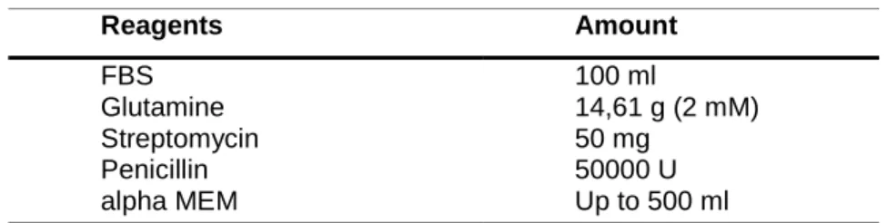
During the postoperative time, clinical parameters including activity, infection, bleeding, appetite and wound dehiscence were evaluated daily. All nine animals were anesthetized by isoflurane and sacrificed by an overdose of Buthanasia solution 8 weeks after tendon surgery to remove the whole tendons for sonoelastographical examination (Figure 4b). For harvesting the specimens, the calcaneus with Achilles tendon was displaced and the gastrocnemius muscle was transected. Tendons were packed in moist saline dressings, frozen and stored at -20°C. For sonoelastography tendon were slowly thawed over night in a refrigerator at 4°. One hour prior to the experiment tendons were equilibrate at room temperature and kept moist by saline solution. Tendons were fixed by pins on a cork board without tension. All sonoelastographic measurements were performed on the same day.
Figure 4: After gastrocnemius tendon dissection insertion of the collagen matrix and fixation with a modified Kessler suture technique (a). Scar formation of Achilles tendon after 8 weeks of gastrocnemius tendon dissection (b).
b f g h g h b b
a
6.2.5 Expansion and subculturing of ASCs
The cell pellet was resuspended after isolation in complete growth medium consisting of alpha-MEM, 20 % FBS, 2 mM L-glutamine, 100 U/ml penicillin and 100 μg/ml streptomycin. Plastic adherent ASCs were grown in appropriate cell culture flasks and placed in an incubator at 37 °C in a humidified atmosphere containing 5 % CO2. Daily washings on the first 2 days removed nonattached cells. Growth medium was exchange twice a week until 90 % confluency. Cell culture was examined daily under an inverted microscope. After reaching 90%
confluency the medium was discarded and cells were washed twice with PBS.
Trypsin was added (1ml for a T75 flask) and incubated for 3 minutes at 37°C in a humidified atmosphere containing 5 % CO2. Subsequently, the effect of trypsin was neutralized by adding growth medium. Cells were centrifuged at 500g for 5 minutes, resuspended in growth medium and splitted 1:3 for subculturing.
Table 4. Preparation of 500 ml growth medium
Reagents Amount
FBS Glutamine Streptomycin Penicillin alpha MEM
100 ml
14,61 g (2 mM) 50 mg
50000 U Up to 500 ml
6.2.6 Adipogenic differentiation of ASCs
Plastic adherent ASCs were washed twice with PBS and trypsinized by 0.25%
trypsin. Cells were seeded into 6 well-plates at a concentration of 1.5x10
4cells
per cm² and adipogenic differentiation was induced by differentiation medium
consisting of growth media (Table 4) with 0.5 mM Isobutyl-methylxanthine, 10
μM bovine insulin, 1 μM dexamathasone and 200 μM indomethacin (Table
5).The medium was changed every 3 days whereas only ASCs in passage
below 5 were used for adipogenic assay to ensure efficient differentiation. ASCs
were exposed to adipogenic induction media for a period of 14 days.
Table 5. Preperation of 100 ml adipogenic differentiation medium
Reagents Amount
Dexamethasone Isobutyl-methylxanthine Bovine insulin
Indomethacin Growth medium
39,2 µg (1 µM) 11,21 mg (0,5 mM) 5,78 mg (10 µM) 7,16 mg (200 µM) Up to 100 ml
Adipogenic transdifferentiation was detected by Oil Red O staining which detects intracellular oil droplets, triglycerides respectively. Oil Red O working solution was prepared by 6 parts of Oil Red O working solution and 4 parts of distilled water (Table 6). Induction medium was discarded and cells were fixed with 4% formalin for 5 minutes at room temperature. Formalin was discarded and fresh formalin was added for an incubation of 60 minutes. Afterwards formalin was removed and cells were washed with 60% isopropanol. Oil Red O working solution was added and incubated for 10 minutes followed by washing four times with distilled water. Finally 4 ml of distilled water was added to prevent the cells from drying.
Table 6. Preperation of 100 ml Oil Red O Working Solution
Reagents Amount
Oil Red O stock
dH2O
60 ml (0,21 g Oil Red O)
40 ml
6.2.7 Osteogenic differention of ASCs
Plastic adherent ASCs were washed twice with PBS and trypsinized by 0.25%
trypsin. Cells were seeded into 6 well-plates at a concentration of 1.0x10
4cells
per cm² and osteogenic differentiation was induced by differentiation medium
consisting of Growth media (Table 4) with 10 mM beta-glycerophosphate
disodium salt hydrate , 50 μM L-ascorbic acid, and 100 nM dexamethasone
(Table 7). The medium was changed every 3 days whereas only ASCs in passage below 5 were used for osteogenic assay to ensure efficient differentiation. ASCs were exposed to osteogenic induction media for a period of 14 days.
Table 7. Preparation of 100 ml osteogenic differentiation medium
Reagents Amount
Dexamethasone L-ascorbic acid
Beta-glycerophosphate disodium salt hydrate Growth medium
3,92 µg (100nM) 0,881 mg (50 µM) 0,461 g (10mM)
Up to 100 ml
Osteogenic transdifferentiation was detected by Alizarin Red S staining which detects osteocytes’ calcium deposits. Alizarin Red solution was prepared of 2g Alizarin Red with 100 ml of distilled water (Table 8). The pH-value was adjusted to 4,1-4,3 with ammonium hydroxide solution. Induction medium was discarded and cells were fixed by incubating in iced cold 70% ethanol for 60 minutes at room temperature. Ethanol was discarded and cells were rinsed twice for 5 minutes with distilled water. Afterwards 5 ml of the Alizarin Red S solution was added and incubated for 30 minutes at room temperature. Alizarin Red S solution was removed and each well was washed four times with distilled water.
Finally 4 ml of distilled water was added to prevent the cells from drying.
Table 8. Preparation of 100 ml Alizarin Red S Solution
Reagents Amount
Alizarin Red S dH2O
ammonium hydroxide solution
2 g
Up to 100 (50 µM) up to pH 4,1-4,3
6.2.8 Histology
After sonoelastography tendon tissue was immediately washed extensively with PBS to removed remaining ultrasound gel and placed in 4% formalin for 24 hours followed by ascending ethanol series starting at 70% for 60 minutes followed by 95% ethanol for 60 minutes, first 100% ethanol for 60 minutes, second 100% ethanol for 90 minutes, third 100% ethanol for 90 minutes and fourth 100% ethanol for 120 minutes. Next the tendons were processed twice with xylene as clearing agent for 60 minutes each. Ethanol and xylene were replaced with fresh reagents after every use. Fixed tissue specimens were embedded in paraffin wax for 60 minutes at 58°C. 3 μm serial sectioning of paraffin-embedded specimen blocks was performed with a HM 400. For histology 3 μm slides were deparaffinized by 2 changes of xylene for 10 minutes each and rehydrated in 2 changes of absolute ethanol for 5 minutes each, 95%
ethanol for 2 minutes and 70% ethanol for 2 minutes followed by rinsing in distilled water for 5 minutes.
Tissue sections were stained in hematoxylin (Table 13) solution for 6 minutes and rinsed for 20 minutes in tap water. Afterwards, slides were decolorized in acid alcohol (Table 12) for 1 second followed by rinsing again for 5 minutes in tap water. Next specimens were immersed in lithium carbonate (Table 14) for 3 seconds, rinsed in tap water for 5 minutes and counterstained in eosin solution (Table 9) for 15 seconds. Stained specimens were dehydrated with 2 changes of 95% ethanol for 3 minutes and 2 changes of 100% ethanol for 3 minutes.
Specimens were cleared in 2 changes of xylene for 5 minutes each. Ethanol and xylene was discarded after each use. Finally the stained specimens were mounted with cytoseal in a fume hood.
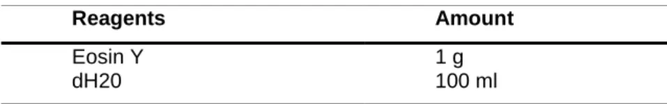
Table 9. Preparation of working solution eosin
Reagents Amount
Eosin stock
Phloxine B stock solution 95% ethanol
Acetic acid
100 ml 10 ml 780 ml 4 ml
Table 10. Preparation of eosin stock solution
Reagents Amount
Eosin Y dH20
1 g 100 ml
Table 11. Preparation of phloxine B stock solution
Reagents Amount
Phloxine B dH2O
1 g 100 ml
Table 12. Preparation of 0,25% working solution acid alcohol
Reagents Amount
95% ethanol dH2O HCL
2578 ml 950 ml 9 ml
Table 13. Preparation of Working solution hematoxylin
Reagents Amount
Hematoxylin Solution, Harris modified
1 L
Table 14. Preparation of working solution lithium carbonate
Reagents Amount
Lithium carbonate dH2O
47 g 3500 ml
A series of 3 μm-thick sections were utilized for immunohistochemical staining.
A mouse anti-rabbit monoclonal antibody against collagen I was used to detect the expression of collagen I in Achilles tendon tissue. For pretreatment heat- induced retrieval was conducted to break the methylene bridges, formed during fixation, to expose antigenic sites in order to allow the antibodies to bind.
Therefore antigen retrieval buffer (Tris-EDTA) was added to a pressure cooker
that was placed on a hotplate. Once boiling, the slides, placed in a metal rack, were transferred to the pressure cooker. Lid was secured and as soon as full pressure was reached, slides were processed for further 3 minutes. When time was elapsed pressure cooker was placed in an empty sink and pressure was released while cooling down in running tap water. Once depressurized lid was opened cold running water was applied into the pressure cooker for 10 minutes.
Next, pretreated slides underwent the staining procedure. First every slide was rinsed with 2 changes of PBS-Tween 20 (Table 17) for 2 minutes and then incubated in universal blocking buffer (Table 16) for 120 minutes at room temperature. Slides were rinsed in PBS Tween-20 before applying the mouse anti-rabbit monoclonal antibodies against collagen I diluted 1/200 in antibody dilution buffer (Table 18) for incubation at 4°C overnight. Next, slides were again rinsed with PBS-Tween 20 and incubated in 0.3% peroxidase in PBS blocking solution (Table 15) for 15 minutes at room temperature. After rinsing with PBS-Tween 20, slides were incubated with the secondary goat anti mouse HRP conjugated antibody diluted 1/500 in antibody dilution buffer for 1 hour at room temperature. Working solution of DAB was applied to tissue section for 10 minutes and monitored for chromogenic reaction. Slides were washed twice with distilled water for 2 minutes each followed by dehydration, clearing and mounting as described above. All stained slides were analyzed using an Axiovert microscope equipped with a Canon G7 high-resolution digital camera for image acquisition. For fluorescence histology a Texas Red conjugated goat anti-mouse antibody was used as secondary antibody. All steps of staining, except of the application of DAB, were performed in the same way. For analyzing the slides microscopy was performed in the dark using 586nm wavelength and an emission filter of 605 nm.
Table 15. Preparation of peroxidase blocking solution
Reagents Amount
30% Hydrogen peroxide PBS
10 ml 990 ml
Table 16. Preparation of 100 ml universal blocking buffer
Reagents Amount
BSA
Cold fish skin gelatin Triton X-100
Sodium azide PBS
1 ml 0,1 ml 0,5 ml 0,05 ml up to 100 ml
Table 17. Preparation of PBS-Tween 20
Reagents Amount
PBS Tween 20
95 ml 5 ml
Table 18. Preparation of 100 ml antibody dilution buffer
Reagents Amount
BSA PBS
1 ml 99 ml
6.2.9 Sonoelastography
The experimental set-up included a LOGIQ®E9 (General Electrics) (Figure 5, 6) using a linear high resolution multifrequency probe from 6-15 MHz (Figure 7).
The same conditions of brightness, contrast, intensity, color scale and frequency were used in all examinations. All measurements were performed by one experienced examiner (more than 5000 examinations per year). First an examination with the fundamental B-scan was performed in the longitudinal plane of the Achilles tendon to localize a potential irregularity of the tendon after the earlier disruption or as the development of the repair mechanism. Then the color coded ultrasound elastography was performed for whole tendon tissue up to a distance from 4 cm in the longitudinal plane and 5 mm in the axial plane.
The probe was perpendicular to the tendon in order to avoid anisotropy. The
aim was to find changes of the color coded evaluated tissue elasticitiy in relation
to surrounding normal tissue (red colour), tissue with good elasticity (yellow
colour), particularly fibrosis (green colour), or a scar (blue colour). For each
tendon real-time color-coded sonoelastography sequences of 20 seconds were
recorded. A quality marker was used to evaluate the best compression mode.
Only sequences with the highest image quality with five green points were used for an appositional evaluation by a quantification mode (Q-analysis) integrated in the ultrasound machine workstation. Ten color histogram frames were obtained for each tendon at ten randomly chosen time-points. Regions of interest (ROIs) were placed on the defect (n=3) and on adjacent uninjured tendon tissue. Real-time sonoelastography software calculated tendon’s elasticity in the region of interest by depicting certain local tissue displacement during a time shift in a certain color. A quantified value was assigned to each color, which serves as the elasticity index (EI) and ranges from 0.0 to 6.0.
Higher values represent higher stiffness and are visualized by a dominant blue color. This provides more objective information about the elasticity. Elasticity of the specimen was reconstructed by calculation of tissue displacement using the elastogram as a color overlay superimposed on the B-mode ultrasound image.
A visual indicator on the screen displayed ongoing tissue compression to assure correct technique of compression and decompression applied to the tendon. All tendons were covered with ultrasound gel for a longitudinal examination (Figure 8).
A total of 180 measurements were obtained and mean intensity of color
histograms were computed by a novel customized GE software. Ten time-point
measurements ensured that EI was not affected by compression variances
caused by the examiner. All of these images were recorded on a hard disk and
used for statistical evaluation.Figure 5: LOGIQ®E9 (General Electrics)
Figure 6: Experimental set up including a LOGIQ®E9
Figure 7: Linear high resolution multifrequency probe from 6-15 MHz during examination
Figure 8: Achilles tendon covered in ultrasound gel before examination
6.2.10 Statistical analyses
Statistics were calculated using GraphPad Prism 5 for Windows. Results are shown as means ± standard deviation. All data sets of metric variables were checked for Gaussian distribution (Kolmogorov-Smirnov test, alpha = 5 %).
Continuous variables were compared by means of one-way ANOVA with
Scheffe post hoc correction. Differences between the ten time-points for
sonoelastographic measurements were examined by repeated measures
ANOVA with a Greenhouse-Geisser correction since assumption of sphericity
had been violated (Mauchly's Test of Sphericity p < 0.0001). Values at p < 0.05
were considered as statistically significant.
7. Results
7.1 ASC preparation and injection
Nuchal adipose tissue was successfully harvested from all animals followed by enzymatic digestion in order to extract mesenchymal stem cells. Mean weight of harvested nuchal fat tissue was 15 g ± 2.4 g. Skin incisions healed without complications or defects in all rabbits. All cells were processed as described above and total yield of freshly isolated cells ranged from 27.5 x 10
6to 31.3 x10
6cells with a cell viability of 91%±3.2%. During the postoperative time, clinical parameters including activity, infection, bleeding, appetite and wound dehiscence were observed without any complications or irregularities.
7.2 Transdifferentiation of ASCs
7.2.1 Adipogenesis
Adipose derived stem cells of rabbits nuchal fat tissue were treated with adipogenic differentiation medium and showed differentiation into adipocytes.
Differentiated stem cells contained Oil Red O-positive lipid vacuoles
(triglyceride) clustered within cytoplasma after 14 days of adipogenesis (Figure
9a). A negative control, receiving growth medium instead of adipogenic
differentiation medium, did not show any Oil Red O positive lipid vacuoles
(Figure 9b).
Figure 9: (a) Adipogenic differentiation of ASCs after 14 days of incubation with adipogenic differentiation medium, stained with Oil Red O. (b) negative control of ASCs incubated with growth medium for 14 days, also stained with Oil Red O
7.2.2 Osteogenesis
Treatment of adipose derived stem cells from the nuchal fat tissue with osteogenic differentiation medium resulted in a differentiation of ASCs into osteocytes. Successfully differentiated stem cells produced Alizarin Red S- positive nodules of mineralized calcium phosphate matrix above the cell monolayer after 14 days of osteogenesis (Figure 10a). A negative control, receiving growth medium instead of osteogenic differentiation medium, did not show any Alizarin Red S-positive nodules (Figure 10b).
Figure 10: (a) Osteogenic differentiation of ASCs after 14 days of incubation with osteogenic differentiation medium, stained with Alizarin Red, (b) negative control of ASCs incubated with growth medium for 14 days, also stained with Alizarin Red
7.3 Effects of ASCs on tendon’s elasticity
Real time sonoelastography was performed in order to characterize the elasticity of Achilles tendons. Elasticity index (EI) was calculated from regions of interest, which were placed on injured (n=3) and non-injured (n=3) tissue on each tendon. The mean EI from Achilles tendons without injury (group 1), injured achilles tendons with ASC seeded matrix (group 2) and injured tendons with unseeded matrix (group 3) were compared to evaluate the effect of applied stem cells.
Post hoc tests using the Scheffe correction revealed a higher elasticity, measured by EI for the injured tendon tissue treated with ASC seeded matrix (group 2, Figure 11a) in comparison to the unseeded matrix (group 3, Figure 11b) (0.73±0.26 and 4.02±1.33, respectively; p<0.01; Figure 11c).
In addition, no difference was found between the injured tendon tissue treated
with ASC seeded matrix (group 2) and the uninjured Achilles tendons (group 1)
(0.73±0.26 and 1.05±0.40, respectively; p>0.05).
Figure 11: Example of sonoelastography for injured tendon tissue treated with stem cell seeded matrix (a) and unseeded matrix (b). Statistical analysis (c) revealed that autologous ASC treatment significantly lowered (**p<0,001) elasticity index of Achilles tendons (0.73, SD±0.26) compared to tendons treated with unseeded matrix (4.02, SD±1.33) but did not differ (n.s., p>0.05) from uninjured tendons (1.05, SD±0.40).