Modulation of Ca v 2.3 voltage-gated calcium channels by trace metal ions and trace metal chelators.
I n a u g u r a l – D i s s e r t a t i o n zur
Erlangung des Doktorgrades
der Mathematisch-Naturwissenschaftlichen Fakultät der Universität zu Köln
vorgelegt von Felix Neumaier
aus Köln
Köln, 2020
Modulation of Ca v 2.3 voltage-gated calcium channels by trace metal ions and trace metal chelators.
I n a u g u r a l – D i s s e r t a t i o n
zur
Erlangung des Doktorgrades
der Mathematisch-Naturwissenschaftlichen Fakultät
der Universität zu Köln
vorgelegt von
Felix Neumaier aus Köln
Köln, 2020
2
Berichterstatter/in: Prof. Dr. Ludwig Eichinger Prof. Dr. Peter Kloppenburg Prof. Dr. Stefan Herzig
Tag der letzten mündlichen Prüfung: 27.04.2020
3
For Mom, Dad and my brother Moritz.
4
This thesis presents publications as originally published, reprinted with permission from the corresponding publishers. The introduction and discussion sections contain illustrations from publications 1 and 4, reproduced with permission from the corresponding publishers. The copyright of the original publications is held by the respective copyright holders, see the following copyright notices.
Publication 1: © 2015 Elsevier Ltd. Reprinted with permission. The original
publication is available at ScienceDirect (
https://doi.org/10.1016/j.pneurobio.2014.12.003) Publication 2: © 2017 John Wiley and Sons. Reprinted with permission. The original publication is available at Wiley Online Library (
https://doi.org/10.1111/apha.12988) Publication 3: © 2018 Neumaier et al. The original publication is available at Rockefeller University Press (
https://doi.org/10.1085/jgp.201711880)
Publication 4: © 2015 Elsevier Ltd. Reprinted with permission. The original
publication is available at ScienceDirect (
https://doi.org/10.1016/j.bbamcr.2015.01.001)
Publication 5: © 2018 John Wiley and Sons. Reprinted with permission. The original
publication is available at Wiley Online Library (
https://doi.org/10.1111/jnc.14546)
Publication 6: © 2020 Neumaier et al. The original publication is available at
Rockefeller University Press (
https://doi.org/10.1085/jgp.202012585)
5
Table of contents
Summary ... 6
Zusammenfassung ... 8
1. Introduction ... 10
1.1. Voltage-gated Ca2+ channels ... 10
1.1.1. Classification and nomenclature ... 10
1.1.2. Subunit composition ... 11
1.1.3. Structure ... 11
1.1.4. Functional properties ... 12
1.2. Cav2.3 voltage-gated Ca2+ channels ... 15
1.2.1. Expression and general function ... 16
1.2.2. Role of Cav2.3 channels in the brain ... 16
1.2.3. Role of Cav2.3 channels for glucose homeostasis ... 17
1.2.4. Role of Cav2.3 channels in the vascular system ... 18
1.3. Endogenous trace metal ions ... 18
1.3.1. Trace metal speciation ... 18
1.3.2 Zn2+ and Cu2+ signaling in the brain... 19
1.3.3. Zn2+ signaling in pancreatic islets ... 20
1.3.4. Metal ion effects on voltage-gated Ca2+ channels ... 21
1.4. Aims of the present work ... 23
2. Results ... 24
2.1. Publication 1: Progress in Neurobiology 129:1-36. ... 24
2.2. Publication 2: Acta Physiologica 222(3): e12988. ... 61
2.3. Publication 3: Journal of General Physiology 150(3): 491-510. ... 80
2.4. Publication 4: Biochimica et Biophysica Acta 1853(5): 953-964. ... 101
2.5. Publication 5: Journal of Neurochemistry 147(3): 310-322. ... 114
2.6. Publication 6: Journal of General Physiology 152(9): e202012585. ... 128
3. Discussion ... 157
3.1. Mechanisms of Cav2.3 channel modulation by Zn2+ and Cu2+ ... 157
3.2. Trace metal chelators as functional Cav2.3 channel agonists ... 159
3.3. Cav2.3 channel modulation by endogenous brain Zn2+ and Cu2+ ... 159
3.4. Cav2.3 channels as a target for paracrine Zn2+ signals in the pancreas... 160
3.5. Conclusion ... 163
4. References ... 164
5. Declaration ... 179
6. Curriculum vitae ... 180
6
Summary
Background:
The trace metal ions Zn2+ and Cu2+ are increasingly recognized as endogenous modulators of neuronal transmission, hormone secretion and synaptic plasticity. Cav2.3-type voltage-gated Ca2+ channels (VGCCs) are among their most sensitive targets and have an expression pattern that coincides with the spatial distribution of histochemically reactive trace metals in the brain, suggesting that they could represent a main mediator for their reported neuro-modulatory effects. Although non-conserved histidine residues on the external side of domain I have been convincingly implicated in the effects of trace metals on Cav2.3 channel gating, the exact mechanisms involved and their (patho)physiological relevance remain incompletely understood.Aims:
Aim of the articles compiled in the present thesis was to shed some light on the exact mechanisms of Zn2+- and Cu2+-induced Cav2.3 channel modulation and their potential relevance under normal and pathophysiological conditions.Methods:
In publication 1, crystallographic data of a Ca2+-selective bacterial model channel was used as a framework to theoretically analyze eukaryotic VGCC structure, function and modulation by inorganic cations. In publication 2, general protocols for preparation and use of metal ion-buffered solutions were developed and a fluorescent Zn2+ sensor was used to illustrate the importance of proper metal ion-buffering. In publication 3, conventional and perforated patch-clamp recordings together with different inhibitors and cytosolic factors were used to study Cav2.3 channel run-down during electrophysiological recordings, which was critical to optimize the conditions for experiments performed in publication 6. In publication 4, the effects of intraperitoneal injection of the Zn2+ chelator DEDTC on blood glucose homeostasis, glucose tolerance and peptide hormone secretion in Cav2.3-deficient and -competent mice were analyzed, insulin secretion was examined in isolated islets of Langerhans from both genotypes and the Zn2+-dependence of DEDTC effects on cloned Cav2.3 channels was verified using whole-cell patch-clamp recordings. In publication 5, whole-cell patch-clamp and electroretinographic recordings were used to characterize a receptor-independent but Cu2+-dependent mechanism of Cav2.3 channel modulation by the glutamate-receptor agonist kainic acid (KA). In publication 6, whole-cell patch-clamp recordings were used to characterize Zn2+-induced changes in Cav2.3 channel function and to develop a Markov model for Cav2.3 channel gating under control conditions and in the presence of physiological Zn2+ concentrations.Results:
Publication 1 provided novel insights into eukaryotic VGCC function and modulation by trace metal ions. Publication 2 demonstrated the critical importance of proper metal ion buffering to avoid deviations between nominal and actual free metal ion concentrations.Publication 3 showed that run-down of Cav2.3 channel currents is associated with changes in channel gating and that it can be prevented or delayed by hydrolysable ATP through a mechanism that critically depends on protein phosphorylation by serine/threonine kinases.
Publication 4 revealed severe glucose intolerance in Zn2+-depleted Cav2.3-deficient but not vehicle-treated Cav2.3-deficient or Zn2+-depleted wildtype mice. In addition, fasting glucose and glucagon levels were significantly higher in Cav2.3-deficient mice, whereas Zn2+ chelation significantly increased blood glucose and glucagon concentrations in wildtype but not Cav2.3- deficient mice. Application of DEDTC significantly stimulated cloned human Cav2.3 channels when applied in the presence of Zn2+ but had no effect in the presence of the Zn2+ chelator CaEDTA. Publication 5 uncovered that KA can stimulate cloned human Cav2.3 channels in the absence of functional KA receptors by reversing Cu2+-induced suppression in vitro, presumably via formation of stable kainate-Cu2+ complexes. When the chelator tricine was used as a surrogate to study the receptor-independent effects of KA in the isolated bovine retina, it selectively reduced a late ERG b-wave component that was previously shown to be enhanced by
7
pharmacologic or genetic ablation of Cav2.3 channels. Publication 6 demonstrated that Zn2+- induced changes in Cav2.3 channel function are complex and inconsistent with a single mechanisms of action. Computer simulations were used to show that most, but not all of the effects can be reconciled by a simplified Markov model that involves Zn2+ binding to a first site with an associated electrostatic modification and mechanical slowing of one of the voltage- sensors and Zn2+-binding to a second, lower affinity site which blocks the channel and modifies the opening and closing transitions.
Discussion:
With regard toZn2+-induced Cav2.3 channel modulation, the results in publication 6 point to an intricate dependence on the prevailing neuronal properties and ionic conditions, which could profoundly influence and even invert the net Zn2+ action. Thus, due to Zn2+-induced parallel changes in activation and inactivation voltage-dependence, the net action is strongly affected by the holding potential, can be either inhibitory or stimulatory and may persist for several minutes after cessation of the Zn2+ signal. This could conceivably play a role for certain forms of synaptic sensitization or plasticity, and might also be relevant for e.g. the regulation of Cav2.3 channels in pancreatic islets, where sudden cessation of Zn2+ supply from β-cells is thought to serve as one of the switch-off signals for α-cell glucagon secretion. In support of the latter notion, the findings in publication 4 provide evidence for an involvement of Cav2.3 channels in the Zn2+-mediated suppression of glucagon secretion during hyperglycemia and indicate that Cav2.3 channel dysfunction could lead to severe disturbances in glucose homeostasis, especially under conditions of Zn2+-deficiency. Based on the results of publications 5 and 6, a decrease or reversal of Zn2+ and Cu2+-induced Cav2.3 channel suppression by endogenous (i.e. glutamate) or exogenous (i.e. KA) trace metal chelators, moderate acidification or depolarization of the neuronal resting membrane potential could also contribute to the pro- convulsive role of Cav2.3 channels demonstrated in previous investigations, although the pathophysiological relevance of these finding in vivo remains to be firmly established. Finally, the findings in publication 3 suggest that protein phosphorylation is required for normal Cav2.3 channel function and that it could modify the normal properties of currents carried by these channels.Conclusion:
The articles compiled in this thesis provide several novel insights into the mechanisms underlying reciprocal Cav2.3 channel modulation by trace metal ions and trace metal chelators as well as first evidence for their importance under (patho)physiological conditions.Moreover, while still far from complete, the model developed in publication 6 provides a quantitative framework for understanding Zn2+ effects on Cav2.3 channel function and a first step towards the application of computational approaches for predicting the complex action of Zn2+ on neuronal excitability.
8
Zusammenfassung
Hintergrund:
Die Spurenmetallkationen Zn2+ und Cu2+ werden zunehmend als endogene Botenstoffe erkannt, welche in die Regulierung von neuronaler Erregbarkeit, Hormonfreisetzung und synaptischer Plastizität involviert sind. Cav2.3 spannungsgesteuerte Ca2+ Kanäle gehören zu deren sensibelsten Targets und weisen ein Expressionsmuster auf, das mit der Verteilung von histochemisch reaktiven Spurenmetallkationen im Gehirn übereinstimmt, so dass sie einen Hauptmediator für deren neuro-modulatorischen Effekte darstellen könnten. Obwohl nicht- konservierte Histidinreste auf der extrazellulären Seite von Domäne I nachweislich eine Rolle für Spurenmetall-induzierte Veränderungen im Schaltverhalten von Cav2.3 Kanälen spielen, sind viele Fragen bezüglich der zugrundeliegenden Mechanismen und ihrer (patho)physiologischen Relevanz bisher unbeantwortet.Ziele:
Ziel der in dieser Arbeit zusammengestellten Artikel war es, einen besseren Einblick in die exakten Mechanismen der Zn2+- und Cu2+-vermittelten Modulation von Cav2.3 Kanälen sowie deren potentieller Bedeutung unter normalen und pathophysiologischen Bedingungen zu erhalten.Methoden:
In Publikation 1 wurden auf Basis kristallographischer Daten eines Ca2+-selektiven bakteriellen Modellkanals die Struktur, Funktion und Modulation von eukaryotischen Ca2+Kanälen theoretisch analysiert. In Publikation 2 wurden generelle Protokolle für die Herstellung und Verwendung von Spurenmetallionen-gepufferten physiologischen Lösungen erarbeitet und deren praktische Bedeutung mit einem fluoreszenten Zn2+ Indikator untersucht. In Publikation 3 wurden konventionelle und perforierte Ganzzell Patch-clamp Messungen sowie verschiedene Inhibitoren und zytosolische Faktoren verwendet um den Run-down von Cav2.3 Kanälen während elektrophysiologischer Messungen zu untersuchen und die Messbedingungen für die Versuche in Publikation 6 zu optimieren. In Publikation 4 wurde untersucht, wie intraperitoneale Injektionen des Komplexbildners DEDTC die Blutglukose-Homöostase, Glukosetoleranz und Peptidhormonfreisetzung in Cav2.3-defizienten und -kompetenten Mäusen beeinflussen.
Außerdem wurde die Insulinfreisetzung in isolierten Langerhans Inseln beider Genotypen verglichen sowie die Zn2+-Abhängigkeit der Effekte von DEDTC auf rekombinante Cav2.3 Kanäle in Ganzzell Patch-Clamp Messungen verifiziert. In Publikation 5 wurden Ganzzell Patch-clamp Messungen und elektroretinographische Ableitungen zur Charakterisierung eines Rezeptor- unabhängigen, Cu2+-abhängigen Mechanismus der Cav2.3 Kanal Modulation durch den Glutamatrezeptor Agonisten Kainsäure (KA) untersucht. In Publikation 6 wurden die elektrophysiologischen Effekte von Zn2+ auf rekombinante Cav2.3 Kanäle untersucht und ein Markov Model für deren Schaltverhalten unter Kontrollbedingungen sowie in Gegenwart physiologisch relevanter Zn2+ Konzentrationen entwickelt.
Ergebnisse:
Publikation 1 erbrachte neue Erkenntnisse zur Funktion eukaryotischer spannungsgesteuerter Ca2+ Kanäle und deren Modulation durch Spurenmetallkationen.Publikation 2 veranschaulichte die Wichtigkeit von Metallionen-Puffern zur Vermeidung von Abweichungen zwischen der nominellen und tatsächlichen freien Metallionenkonzentration in physiologischen Lösungen. Publikation 3 ergab, dass der Run-down von Cav2.3 Kanälen mit Veränderungen im Schaltverhalten verbunden ist, dass er durch hydrolisierbares ATP verhindert oder verlangsamt werden kann und dass Serin/Threonin Kinasen kritisch für die protektiven ATP Effekte sind. Publikation 4 ergab, dass Blutglukose und Serum Glukagon Spiegel in Cav2.3- defizienten Mäusen im Vergleich zu Cav2.3-kompetenten Mäusen signifikant erhöht sind, während Zn2+ Depletion mit DEPC zu einem signifikanten Anstieg von Blutglukose und Serum Glukagon Spiegeln in Cav2.3-kompetenten aber nicht in Cav2.3-defizienten Mäusen führt. DEPC Behandlung führte außerdem zu einer schweren Glukoseintoleranz in Cav2.3-defizienten Mäusen, während die Glukosetoleranz in unbehandelten Cav2.3-defizienten sowie behandelten
9
und unbehandelten Cav2.3-kompetenten Mäusen unbeeinträchtigt war. In in vitro Versuchen stimulierte DEPC den Ca2+ Einfluss durch rekombinante Cav2.3 Kanäle in der Gegenwart von Zn2+ aber nicht in der Gegenwart des Zn2+ Komplexbildners CaEDTA. Publikation 5 ergab, dass KA rekombinante Cav2.3 Kanäle durch eine Umkehr Cu2+-induzierter Inhibition, die vermutlich auf Bildung stabiler Kainat-Cu2+ Komplexe beruht, auch in Abwesenheit funktioneller Glutamat Rezeptoren stimulieren könnte. Simulation der Rezeptor-unabhängigen Effekte von KA durch Applikation des Komplexbildners Tricin in der isolierten Rinderretina führte zur selektiven Abnahme in der Amplitude einer späten Komponente der elektroretinographischen b-Welle, für welche in früheren Arbeiten eine Stimulation durch genetische oder pharmakologische Ablation von Cav2.3 Kanälen gezeigt wurde. Publikation 6 ergab, dass die Zn2+-induzierten Veränderungen im Schaltverhalten von Cav2.3 Kanälen komplex sind und mehr als einen einzelnen Wirkmechanismus umfassen. Mittels Computersimulationen wurde gezeigt, dass die meisten, aber nicht alle, Effekte durch ein vereinfachtes Modell reproduziert werden können, bei dem Zn2+ Interaktion mit einer ersten Bindungstelle zu einer elektrostatischen Modifikation und mechanischen Verlangsamung eines der Spannungssensoren führt und Zn2+ Interaktion mit einer zweiten Bindungstelle von niedrigerer Affinität den Kanal blockiert und zu einer Verlangsamung des Öffnungs- und Schließverhaltens führt.
Diskussion:
Bezüglich der Zn2+-induzierten Modulation von Cav2.3 Kanälen deuten die Ergebnisse aus Publikation 6 auf komplexe Wechselwirkungen mit den vorherrschenden neuronalen und ionischen Bedingungen hin, welche die Zn2+ Wirkung beeinflussen und sogar umkehren könnten. So hängt der Gesamt Zn2+ Effekt auf Cav2.3 Kanäle aufgrund paralleler Veränderungen in der Spannungsabhängigkeit von Aktivierung und Inaktivierung stark vom Haltepotential ab, kann sowohl inhibierend als auch stimulierend sein und für mehrere Minuten nach Beendigung des Zn2+ Signals andauern. Neben einer möglichen Rolle für bestimmte Mechanismen der synaptischen Sensibilisierung und Plastizität könnten diese Effekte für die Regulation von Cav2.3 Kanälen in den Langerhans Inseln des Pankreas relevant sein, wo eine plötzliche Einstellung der Zn2+ Freisetzung aus β-Zellen als eines der Switch-off Signale für die Glukagon Freisetzung aus α-Zellen zu dienen scheint. Letztere Annahme wird durch die Ergebnisse in Publikation 4 untermauert, welche erste Belege für eine Rolle von Cav2.3 Kanälen für die Zn2+-vermittelte Inhibition der Glukagonfreisetzung unter hyperglykämischen Bedingungen liefern und zeigen, dass Cav2.3 Kanaldysfunktion, besonders in Kombination mit Zn2+-Defizienz, zu schweren Störungen der Glukosehomöostase führen könnte. Basierend auf den Ergebnissen der Publikationen 5 und 6 könnte eine Abnahme bzw. Umkehr der Zn2+ und Cu2+ vermittelten Inhibition von Cav2.3 Kanälen durch endogene (z.B. Glutamat) oder exogene (z.B. KA) Komplexbildner, mäßige Acidose oder Depolarisation des neuronalen Ruhemembranpotentials möglicherweise auch zur pro-konvulsiven Rolle dieser Kanäle beitragen, welche in früheren Arbeiten belegt wurde. Schließlich legen die Ergebnisse aus Publikation 3 nahe, dass Proteinphosphorylierung kritisch für die normale Funktion von Cav2.3 Kanälen ist und ihre elektrophysiologischen Eigenschaften modifizieren könnte.Konklusion:
Die in dieser Arbeit zusammengestellten Artikel liefern verschiedene neue Erkentnisse über die Mechanismen der reziproken Modulation von Cav2.3 Kanälen durch Spurenmetallkationen und Komplexbildner sowie erste Erkenntnisse zu ihrer möglichen Bedeutung unter (patho)physiologischen Bedingungen. Obwohl das in Publikation 6 erarbeitete Model als vorläufig anzusehen ist, bietet es eine quantitative Grundlage zum Verständnis der Effekte von Zn2+ auf Cav2.3 Kanäle und einen ersten Schritt in Richtung der Anwendung computerbasierter Methoden zur Prognose der komplexen Wirkung von Zn2+ auf die neuronale Erregbarkeit.10
1. Introduction
1.1. Voltage-gated Ca
2+channels
Under physiological conditions, intracellular Ca
2+levels are tightly controlled and maintained in the low nanomolar concentration range. Ca
2+can enter the cell through voltage-gated Ca
2+channels (VGCCs), which shape the electrical properties of excitable cells and represent the key link between electrical signals and non-electrical processes, such as transmitter release, peptide hormone secretion or gene transcription. The following sections will briefly describe their classification, structure and function. A more detailed description that goes beyond a mere recap of the literature is provided in publication 1 (Neumaier et al., 2015).
1.1.1. Classification and nomenclature
Since their first recording in cardiomyocytes (Reuter, 1979, 1967), voltage-dependent Ca
2+currents in native membranes have been broadly classified into (I) low-voltage activated (LVA) currents which activate at membrane potentials near the resting level and display rapid inactivation kinetics and (ii) high-voltage activated (HVA) currents, which activate at somewhat more depolarized test potentials and display variable but typically less rapid inactivation kinetics (Carbone and Lux, 1984; Nowycky et al., 1985). Based on their biophysical and pharmacological properties, HVA currents were further subdivided into dihydropyridine-sensitive L-type (for long lasting), ω-conotoxin GVIA-sensitive N-type (for neuronal or neither T- nor L-type), ω -agatoxin IVA-sensitive P/Q-type (for Purkinje neurons) and partly SNX-482-sensitive R-type (for resistant or residual) currents, while LVA currents were assigned to a single group of T-type (for transient) currents (Llinás et al., 1992;
Nowycky et al., 1985; Randall and Tsien, 1995; Tsien et al., 1987). Purification (Curtis and
Catterall, 1984), reconstitution (Curtis and Catterall, 1986; Flockerzi et al., 1986) and
molecular cloning (Tanabe et al., 1987) of the skeletal muscle Ca
2+channel in the late 1980s
and subsequent homology screening of lambda-phage and cDNA libraries firmly established
the existence of multiple, distinct Ca
2+channel α
1-subunits (Catterall, 2011; Hille, 1992). They
were originally named based on an alphabetical nomenclature, which assigned the letter S to
the original skeletal muscle channel (i.e. α
1S) and the letters A-F to subsequently identified α
1-
subunits (Birnbaumer et al., 1994; Snutch et al., 1990; Snutch and Reiner, 1992). Today,
VGCCs are classified based on amino acid sequence identity and named according to the
same nomenclature used for other voltage-gated ion channels, where a given α
1-subunit is
identified by the principal permeating ion (i.e. Ca for Ca
2+), the main physiological regulator
(i.e. v for voltage) indicated as a subscript (i.e. Ca
v) and a numerical identifier that
corresponds to one of three subfamilies (Ca
v1 through Ca
v3) and the order of discovery
within that subfamily (Catterall et al., 2003; Ertel et al., 2000). Based on pharmacological and
functional criteria, the Ca
v1 family (Ca
v1.1 through Ca
v1.4) has been convincingly linked to
native L-type currents, the Ca
v2 family to native P/Q-type (Ca
v2.1), N-type (Ca
v2.2) and R-
type (Ca
v2.3) currents and the Ca
v3 family (Ca
v3.1 through Ca
v3.3) to native T-type currents.
11
1.1.2. Subunit composition
Similar to other voltage-gated ion channels, native HVA channels are multi-subunit complexes comprised of a principal, transmembrane-spanning Ca
vα
1-subunit, several auxiliary subunits of lower molecular weight and the ubiquitous intracellular Ca
2+-sensor calmodulin (CaM), which associates with a CaM-binding domain in the cytoplasmic C- terminal region. Expression levels and the properties of Ca
vα
1-subunits vary with co- expression of one of at least four Ca
vβ-subunits, one of at least four Ca
vα
2δ-subunits and in some cases one of several putative Ca
vγ-subunits. Additional diversity arises from a number of possible splice variants and posttranslational modification of the Ca
vα
1or other subunits (Jones, 1998; Lacinová, 2005). LVA channels on the other hand lack the CaM-binding domain and are typically thought to consist only of a pore-forming Ca
vα
1-subunit (Lacinová et al., 1999; Lambert et al., 1997; Leuranguer et al., 1998).
1.1.3. Structure
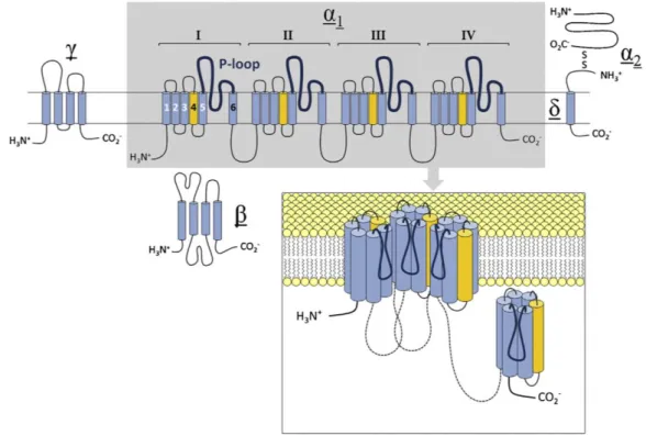
As illustrated in
Fig. 1, Cavα
1-subunits are made up of four homologous repeats termed domains I through IV, each comprising six membrane-spanning helical segments (S1-S6) and a pore forming p-loop between S5 and S6 (Zhen et al., 2005). The first four segments of each domain have been shown to form the voltage-sensor modules (VSMs) responsible for coupling changes in membrane potential to channel opening. The remaining two segments make up most of the internal pore lining, with the p-loops forming the extracellular mouth of
Figure 1. Subunit composition and transmembrane topology of high-voltage activated calcium channels.
Inset shows quadrameric arrangement of homologous domains of the pore-forming α1 subunit at the cell membrane. For clarity, domain IV is shown aside from the membrane. Reprinted with permission from (Neumaier et al., 2015).
12
the pore and the Ca
2+selective filter region (Kim et al., 1993; Yang et al., 1993). Ca
vβ-subunits are intracellular, hydrophilic proteins without transmembrane segments (Fig. 1), which bind to a high-affinity site located in the cytoplasmic linker between the first two homologous repeats of the Ca
vα
1-subunit (Buraei and Yang, 2013). Ca
vα
2δ-subunits are made up of two distinct subunits (α
2and δ), formed by post-translational cleavage of a single gene product and associated with each other via disulfide bonds (Ellis et al., 1988). As indicated in Fig. 1, the highly glycosylated α
2-subunit resides on the extracellular side and is thought to be anchored in the plasma membrane by the smaller δ protein (Gurnett et al., 1996).
1.1.4. Functional properties
The classical view of voltage-gated ion channels assumes that transfer of ions across the membrane (permeation) is independent of the transitions between non-conducting and conducting states (gating) and vice versa. This assumption is conceptually useful and often necessary for interpretation of experimental findings, but there is convincing evidence for an interaction between gating and permeation in VGCCs (for details see publication 1).
1.1.4.1. Voltage-dependent activation
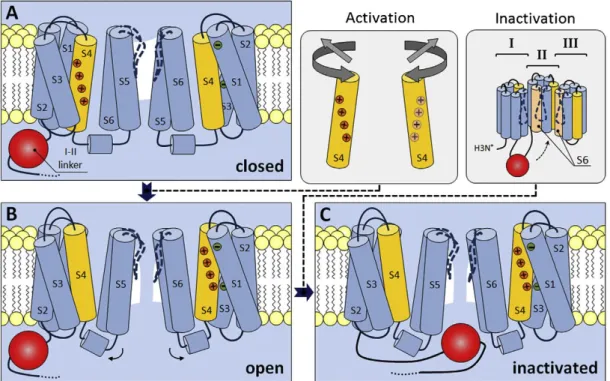
Activation of all voltage-gated ion channels involves charge-moving conformational changes that produce measurable gating currents and have been shown to depend on charged residues in the S2, S3 and especially S4 segments of all four domains (Hille, 1992; Vargas et al., 2012). A possible mechanism for the process based on models for activation of Shaker K
+- channels is shown in Fig. 2A & B. At resting membrane (i.e. hyperpolarized) potentials, the permeation pathway is occluded by parts of the S6 segments, which form a hydrophobic cavity that is inaccessible for ions. Depolarization forces the positively charged S4 segments to rotate or tilt through a protein-lined pathway formed by the rest of the VSM, which leads to relocation of the S5 and S6 segments with formation of a water-filled and ion permeable crevice. Evidence from mutational studies (Beyl et al., 2016; García et al., 1997) and optical tracking of voltage-sensor movement (Pantazis et al., 2014) indicates that activation of only two of the four non-identical VSM may be sufficient to drive channel opening in VGCCs.
1.1.4.2. Voltage- and Ca
2+-dependent inactivation
The extent of Ca
2+entry during prolonged depolarization is limited by voltage- or Ca
2+-
dependent inactivation, which may involve distinct conformational changes and can vary
with the exact subunit composition. Voltage-dependent inactivation (VDI) of HVA channels
has been linked to parts of the pore-forming segments and intracellular linkers (Hering et al.,
2000; Stotz et al., 2000) and proposed to involve a ‘hinged lid’ mechanism, where docking of
the I-II linker to the cytoplasmic ends of the S6 segments occludes the inner pore vestibule
(Fig. 2C)(Stotz et al., 2004, 2000). The process appears to be state- rather than truly voltage-
dependent and is subject to modulation by co-expressed Ca
vβ-subunits, which may modify
mobility of the I-II linker (Stotz et al., 2004). Ca
2+-dependent inactivation is largely restricted
to otherwise non-inactivating (Ca
v1.1-Ca
v1.4) or certain neuronal channels (Ca
v2.1 and
Ca
v2.2) and thought to be induced by formation of Ca
2+/CaM complexes (Budde et al., 2002).
13
Figure 2. Conformational changes during activation and inactivation of VGCCs. Cartoon illustrating possible mechanisms of channel activation and inactivation based on models of activation for Shaker K+- channels and fast VDI of Cav2.3 VGCCs. Only two of the four homologous domains are shown. (A) At resting membrane potentials, the permeation pathway is occluded by regions of the S6 segments which form parts of the channel lumen, resulting in a hydrophobic cavity that is inaccessible for ions. (B) During membrane depolarization, the positively charged S4 segments (yellow) rotate and decrease their tilt (see inset), leading to changes in the relative position of other segments and formation of a water-filled crevice, in which ions can traverse the channel. (C) Upon prolonged depolarization, the intracellular linker between domains I and II (highlighted in red) docks to a site composed, in parts, of the S6 segments of domains II and III (see inset), thereby physically occluding the inner vestibule of the channel and terminating the flow of ions. Reprinted with permission from (Neumaier et al., 2015).
1.1.4.3. Selective permeation
Under physiological conditions, VGCCs are almost exclusively selective for Ca
2+, with estimated rate coefficients for overall Ca
2+transfer of 7.5 x 10
7M
-1s
-1for HVA channels (Hess et al., 1986) and 0.3-0.5 x 10
8M
-1s
-1for LVA channels (Carbone and Lux, 1987; Lux et al., 1990). Selectivity is mainly conferred by a conserved high-affinity Ca
2+binding site in the pore region, which is formed by a ring of glutamate (and in LVA channels aspartate) residues located at equivalent positions in each of the four p-loops (EEEE/EEDD-locus) (Cens et al., 2007; Cibulsky and Sather, 2000; Ellinor et al., 1995; Shuba, 2014; Talavera et al., 2001;
Tang et al., 1993; Yang et al., 1993). Several mechanisms have been proposed to account for
the apparent paradox of high-affinity binding and high conductance, most of which are
based on a combination of ion-pore and ion-ion interactions in the permeation pathway. In
the classical ‘stairstep model’, low-affinity binding sites flanking the EEEE/EEDD-locus are
thought to allow for stepwise entry and exit of Ca
2+ions from the pore, thereby lowering the
energy required for dissociation from the high-affinity binding site (Dang and McCleskey,
14
1998). In the ‘car wash model’, the EEEE/EEDD-locus is thought to form a flexible, multi-ion binding site that can accommodate a single Ca
2+ion with high-affinity or multiple Ca
2+ions with lower affinity (Yang et al., 1993). Occupation by more than one Ca
2+ion could destabilize high-affinity binding due to electrostatic repulsion (Almers and McCleskey, 1984;
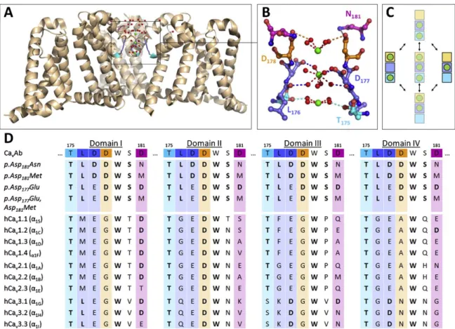
Hess and Tsien, 1984), competition for binding moieties (Armstrong and Neyton, 1991; Yang et al., 1993) and / or conformational changes (Lux et al., 1990; Mironov, 1992). Support for a mechanism akin to the stairstep model has been provided by crystallographic data on the Ca
2+-selective model channel Ca
vAb, which was derived from the bacterial sodium channel Na
vAB. In this channel and several related constructs, the selectivity filter is formed by a central high-affinity DDDD-locus and two flanking low-affinity sites (Fig. 3A-C), which are at least partly conserved in human VGCCs (Fig. 3D).
Figure 3. Functional organization of the selectivity filter in voltage-gated calcium channels. (A, B) Cartoon representation of the crystal structure of pAsp181Asn (a construct closely related to CavAb) showing three of the four identical domains with Ca2+ ions (green) bound to all three coordination sites in the selectivity filter. Note that the crystal structure likely reflects a mixed population of channels, in which either the central or the two flanking coordination sites are occupied. (C) State-diagram showing the two low energy states of occupation and possible intermediates. (D) Sequence alignment of residues making up the selectivity filter in CavAb and its derivatives with the corresponding residues in cloned human voltage- gated calcium channels. Conserved residues are highlighted in bold. Reprinted with permission from (Neumaier et al., 2015).
15
1.1.4.4. Run-up and run-down
Electrophysiological recordings associated with dialysis of the cytoplasm or excision of cell patches are almost invariably hampered by time-dependent changes in voltage-gated ion channel function. The most well-known form of these phenomena is a progressive decline of voltage activated currents after breakthrough into the whole-cell configuration (run-down).
The process is typically thought to result from dilution of the cellular contents by the pipette solution and an associated loss of second messengers and intracellular signaling cascades (Becq, 1996). In some cases, run-down is preceded by a transient current facilitation, which could reflect a relief of tonic suppression and / or voltage- and time-dependent repriming (i.e. recovery from inactivation) (Elhamdani et al., 1995, 1994; Tiaho et al., 1993). While they remain a major obstacle for studies on the pharmacology and physiology of voltage-gated channels, these time-dependent changes can also provide insight into the modulation of channels in intact cells. For this reason, the hallmarks and mechanisms of run-down of L- type Ca
2+currents have been subject to numerous investigations (Belles et al., 1988; Chad and Eckert, 1986; Hao et al., 1999; Hescheler et al., 1988; Martini et al., 2000; McDonald et al., 1994; Mironov and Lux, 1991). Probably owing to their minor contribution to the total Ca
2+current in many non-neuronal cells, much less work has been devoted to characterizing the process in other HVA VGCCs. For example, native R-type currents, which are mainly mediated by Ca
v2.3-type VGCCs, are well known to exhibit both run-up and run-down (Almog and Korngreen, 2009; Benquet et al., 1999; Cota, 1986; Hilaire et al., 1997). Although some findings indicate that run-down may be associated with changes in R-type-like Ca
2+channel gating (Cota, 1986), the rapid loss of these currents in conventional electrophysiological recordings has generally prevented further characterization of the phenomenon in native cells. Some of the experiments performed in the present work were also hampered by time-dependent changes in Ca
v2.3 channel currents, which prompted us to investigate the process in more detail. The results of these experiments allowed us to develop experimental conditions that significantly delayed the occurrence of run-down and provided interesting insights into the importance of protein phosphorylation for the normal function of Ca
v2.3 channels, as described in more detail in publication 3 (Neumaier et al., 2018b).
1.2. Ca
v2.3 voltage-gated Ca
2+channels
Ca
v2.3-type VGCCs are the only molecular counterpart of native R-type currents so far identified and remain one of the most enigmatic members of the family of VGCCs, not least because they share biophysical properties with both HVA (high activation threshold; large single channel conductance; rapid deactivation kinetics) and LVA (equal permeability to Ca
2+and Ba
2+; fast inactivation kinetics) channels (Bourinet et al., 1996; Soong et al., 1993;
Wakamori et al., 1994). They are resistant towards most commonly used organic Ca
2+channel
antagonists but highly sensitive to certain divalent trace metal ions (Zn
2+, Cu
2+and Ni
2+),
which has been linked to non-conserved histidine residues on the external side of the domain
I VSM (Kang et al., 2007; Shcheglovitov et al., 2012). In addition, Ca
v2.3 channels are sensitive
to the tarantula toxin SNX-482 (IC
50=20-60 nM), which is a relatively selective inhibitor when
16
compared to other VGCCs, but has been shown to be even more effective in suppressing A- type K
+-currents mediated by K
v4.3 channels (IC
50<3 nM) (Kimm and Bean, 2014).
1.2.1. Expression and general function
Ca
v2.3 channel expression is most prominent throughout the central and peripheral nervous system (Grabsch et al., 1999; Schneider et al., 1994; Soong et al., 1993; Williams et al., 1994), but has also been detected in neuroendocrine (Grabsch et al., 1999; Pereverzev et al., 2002b;
Vajna et al., 1998), cardiovascular (Galetin et al., 2013), reproductive (Cohen et al., 2014;
Wennemuth et al., 2000), and gastrointestinal tissues (Grabsch et al., 1999). Insight into the functional roles of Ca
v2.3 channels has been facilitated by the generation of Ca
v2.3-deficient mice, which exhibit no major pathologies but are characterized by altered spatial memory (Kubota et al., 2001) and nociception (Saegusa et al., 2000), increased anxiety (Lee et al., 2002), cardiac arrhythmias and impaired autonomic control (Galetin et al., 2013; Weiergräber et al., 2005), reduced susceptibility to chemically induced seizures and excitotoxicity (Dibué-Adjei et al., 2017; Weiergräber et al., 2007) and certain alterations in glucose homeostasis (Matsuda et al., 2001; Pereverzev et al., 2002a). Based on this relatively subtle phenotype and their preferential expression in rhythmically active tissues, a general function of Ca
v2.3 channels has been proposed to involve fine-tuning of rhythmogenesis in oscillatory networks (Schneider et al., 2015).
1.2.2. Role of Ca
v2.3 channels in the brain
In the CNS, highest densities of Ca
v2.3 channels can be detected in the hippocampus and
other limbic regions, in the retina and in cortical neurons (Sochivko et al., 2002; Weiergräber
et al., 2006b). They are expressed both presynaptically (Day et al., 1996) and postsynaptically
(Day et al., 1996; Yokoyama et al., 1995) and have been shown to be involved in transmitter
release and neuronal plasticity (Bloodgood and Sabatini, 2007; Dietrich et al., 2003; Gasparini
et al., 2001; Yasuda et al., 2003), somatodendritic integration and action potential burst firing
(Christie et al., 1995; Kavalali et al., 1997; Magee and Carruth, 1999; Magee and Johnston,
1995). In addition, Ca
v2.3 channels are the third most extensively phosphorylated ion
channels in mouse brain (Cerda et al., 2011) and depolarization has been shown to trigger
bulk changes of their phosphorylation state in intact hippocampal slices (Hell et al., 1995). In
the retina, Ca
v2.3 channels appear to be involved in GABA-ergic reciprocal inhibition of rod
bipolar cells (Siapich et al., 2009), and genetic ablation or pharmacological suppression of
these channels have been shown to selectively enhance a late b-wave component of the
eletroretinogram (ERG) (Alnawaiseh et al., 2011; Lüke et al., 2005; Siapich et al., 2010). The
exact physiological relevance of Ca
v2.3 channels in most other brain regions remains elusive,
but there is increasing evidence for their involvement in a number of pathophysiological
conditions. With regard to human patients, recent findings have implicated de novo gain-of-
function mutations in these channels with developmental and epileptic encephalopathies,
which are characterized by intractable seizures, abundant epileptiform EEG activity and
developmental impairments (Carvill, 2019; Helbig et al., 2018). A pro-ictogenic role of Ca
v2.3
channels in convulsive generalized tonic-clonic and hippocampal seizures has also been
17
demonstrated in a number of animal studies, where their genetic ablation significantly reduced the susceptibility to kainic acid (KA)-induced seizures and neurodegeneration (Dibué-Adjei et al., 2017; Weiergräber et al., 2007, 2006a). In addition, treatment with the broad-spectrum antiepileptic drugs lamotrigine (LTG) or topiramate (TPM), both of which have been shown to inhibit cloned Ca
v2.3 channels (Hainsworth et al., 2003; Kuzmiski et al., 2005), significantly reduced KA-induced seizures in wildtype but not Ca
v2.3-deficient mice (Dibué-Adjei et al., 2017), suggesting that Ca
v2.3 channels could be an important target for current antiepileptic treatments.
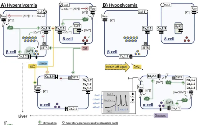
1.2.3. Role of Ca
v2.3 channels for glucose homeostasis
Animal studies have linked Ca
v2.3 VGCCs to α-cell glucagon secretion (Pereverzev et al.,
2005), β-cell insulin secretion (Jing et al., 2005; Matsuda et al., 2001; Pereverzev et al., 2002a)
and δ-cell somatostatin (SST) secretion (Zhang et al., 2007), suggesting that these channels
are critically involved in the regulation of blood glucose levels. Under hyperglycemic
conditions, glucose homeostasis is primarily maintained by glucose-stimulated insulin
secretion (GSIS) from pancreatic β-cells, which is thought to involve a triggering pathway of
fast insulin secretion (first phase insulin response), and an amplifying pathway of sustained
release (second phase insulin response). The molecular mechanisms underlying fast GSIS are
relatively well established and have been shown to involve β-cell glucose uptake and
metabolism followed by depolarization due to ATP-dependent closure of K
ATPchannels,
which triggers Ca
2+-influx through Ca
v1.2 channels and secretion of a readily releasable pool
of insulin vesicles. The sustained insulin response is much less well understood but thought
to involve mobilization of a reserve pool of insulin vesicles for release. Genetic or
pharmacological ablation of Ca
v2.3 channels selectively reduces the second phase insulin
response in mice (Jing et al., 2005; Pereverzev et al., 2005), suggesting that β-cell Ca
v2.3
channels are involved in the mobilization of insulin vesicles from the reserve pool. They
could also play a role for the paracrine effects of SST released from δ-cells, as SST suppresses
cloned Ca
v2.3 channels (Mehrke et al., 1997) and SNX-482 has been shown to prevent SST
inhibition of insulin secretion from insulinoma cells (Mergler et al., 2008). According to the
intra-islet insulin hypothesis, insulin released into the periportal circulation is carried to
nearby α-cells, where it provides tonic suppression of glucagon secretion during
hyperglycemia, while cessation of insulin secretion during hypoglycemia could serve as a
switch-off signal that initiates α-cell glucagon secretion (Banarer et al., 2002; Maruyama et al.,
1984). Interestingly, Ca
v2.3 channels appear to be important for the former (i.e. suppression
of glucagon release during hyperglycemia) but not directly involved in the latter (i.e. release
of glucagon during hypoglycemia), as glucose-induced suppression of glucagon release is
severely impaired or even reversed in Ca
v2.3-deficient mice (Jing et al., 2005), isolated islets
from Ca
v2.3-deficient mice (Jing et al., 2005; Pereverzev et al., 2005) and SNX-482 treated
islets from wildtype mice (Göpel et al., 2004; Jing et al., 2005) whereas SNX-482 fails to
reduce glucagon secretion from wildtype islets in low glucose. As discussed in more detail in
section 1.3.3 and assessed experimentally in publication 4 (section 2.4), there is increasing
evidence that Zn
2+co-released with insulin from pancreatic β-cells could serve as an
18
additional, non-insulin signal for crosstalk to neighboring α-cells, which might exert its action in part by suppressing Ca
v2.3 channels. The pathophysiological relevance of these findings is corroborated by several studies linking variants in the gene encoding Ca
v2.3 channels to impaired glucose homeostasis in human T2DM patients (Holmkvist et al., 2007;
Muller et al., 2007; Trombetta et al., 2012).
1.2.4. Role of Ca
v2.3 channels in the vascular system
There is also evidence for a role of Ca
v2.3 channels in the vascular system, although again mainly in the context of certain pathophysiological conditions. For example, hemoglobin, which can be released into the brain extracellular space during hemorrhagic stroke, has been shown to induce up-regulation of Ca
v2.3 channels in cerebral vessels (Ishiguro et al., 2005) and there is evidence for an involvement of these channels in delayed cerebral vasospasm and ischemia after subarachnoid hemorrhage (Ishiguro and Wellman, 2008). In addition, work from our lab shows that unconjugated bilirubin (UCB), a degradation product of hemoglobin, alters Ca
v2.3 channel function in a heterologous expression system and attenuates transretinal signaling in the isolated retina from wildtype but not Ca
v2.3-deficient when applied at pathophysiologically relevant UCB:albumin molar ratios (Albanna et al., 2019, 2017).
1.3. Endogenous trace metal ions
Trace metal ions like Zn
2+and Cu
2+are key structural components of numerous proteins and co-factors for enzymes involved in cellular respiration, catecholamine synthesis and antioxidant defense (Anzellotti and Farrell, 2008; Frederickson et al., 2005). In addition to their established structural and catalytic functions, endogenous Zn
2+and Cu
2+are increasingly recognized as potential modulators of neuronal transmission (Frederickson et al., 2005, 2000; Mathie et al., 2006) and synaptic plasticity (Pan et al., 2011). As noted above, Zn
2+has also been identified as a candidate auto- and / or paracrine signal for intra-islet crosstalk in the pancreas, which could be critically involved in the regulation of blood glucose homeostasis by insulin and glucagon (Ishihara et al., 2003).
1.3.1. Trace metal speciation
The biological effects of trace metal ions are determined by their chemical speciation, which
is defined as the distribution among all free and ligand-bound species in solution (for details
see publication 2). Under physiological conditions, the majority of Zn
2+and Cu
2+is tightly
bound by a range of metalloprotein ligands, so that the pool that remains
thermodynamically and kinetically accessible (i.e. loosely-bound) is usually orders of
magnitude below the total concentration. This is especially important in the intracellular
compartment, where even moderate increases in the loosely-bound Zn
2+or Cu
2+level are
deleterious and ultimately associated with cell death. A detailed review of the tightly
regulated intracellular trace metal homeostasis is beyond the scope of this work and can be
found elsewhere (Colvin et al., 2010, 2003; Krężel and Maret, 2006). The present article will
focus on the action of extracellular trace metal ions, noting that there is usually a large
concentration-gradient favoring their passive entry into postsynaptic neurons. The latter is
19
under study as potential signaling pathway (Li et al., 2001) and important mechanism that may contribute to hippocampal neurodegeneration under certain pathophysiological conditions (Frederickson et al., 1988; Mathie et al., 2006). With regard to the extracellular compartment, an important implication of chemical speciation is that loosely-bound trace metal concentrations could be subject to modulation by a number of small organic molecules that are capable of acting as trace metal chelators. This is exemplified in publication 5 (Neumaier et al., 2018a), where we provide
in vitro evidence for a receptor-independentmechanism of Ca
v2.3 channel modulation by the excitatory amino acid KA that involves reversal of Cu
2+-induced suppression. Additional implications are that suitable trace metal chelators can be used to antagonize the effects of endogenous trace metals, and that proper metal ion buffering should be used in experiments with metal ion-sensitive targets. The latter is important because even highly purified reagents contain small amounts of trace metal ions, so that estimated levels of Zn
2+, Cu
2+and / or other unwanted metal ions in typical physiological solutions are in the nano- to micromolar concentration range (for details see publication 2). As described in one of the later sections, this may be sufficient for significant tonic suppression of certain Ca
vα
1-subunits and could therefore also influence experimental findings.
1.3.2 Zn
2+and Cu
2+signaling in the brain
While 90% of total brain Zn
2+is tightly bound to zinc metalloproteins, much of the remaining
10% forms a loosely-bound pool located in the presynaptic vesicles of so-called zinc-enriched
neurons (Frederickson et al., 2000; Mathie et al., 2006). These neurons are not associated with
a single neurotransmitter and have been shown to release Zn
2+both spontaneously and in an
activity-dependent manner. Similar pools of loosely-bound Cu
2+that can be released
following membrane depolarization have been detected in certain neurons that appear to be
primarily associated with glutaminergic or adrenergic transmission (Kardos et al., 1989; Ono
and Cherian, 1999; Sato et al., 1994). The distribution of loosely-bound Zn
2+and Cu
2+in the
brain is not uniform, and highest concentrations are located in specific forebrain areas that
include the hippocampus and other limbic regions, the neocortex and the retina (Anastassov
et al., 2014, 2013; Charton et al., 1985; Hartter and Barnea, 1988; Kardos et al., 1989). Under
physiological conditions, estimated resting or ‘tonic’ levels in the brain extracellular fluid are
in the order of 5-25 nM for loosely-bound Zn
2+(Frederickson et al., 2006) and 0.1-0.8 µM for
loosely-bound Cu
2+(Mathie et al., 2006). These values are frequently exceeded at mossy fiber
synapses, where Zn
2+concentrations have been estimated to be in excess of 10-20 µM during
basal synaptic activity and >100 µM during repetitive electrical stimulation or periods of
intense activity (Kardos et al., 1989; Mathie et al., 2006; Vogt et al., 2000). During KA-induced
epileptiform (or ictal-like) discharges, Zn
2+levels in the order of 300 µM have been estimated
to be reached in the extracellular space (Assaf and Chung, 1984). Released metal ions could
modulate neuronal activity through effects on a variety of voltage- and ligand-gated ion
channels (Elinder and Arhem, 2003; Neumaier et al., 2015), which has made it difficult to
predict the net action of Zn
2+and Cu
2+. As such, their exact functional relevance in the brain
remains controversial, although a number of experimental observations indicate that
20
especially Zn
2+could dampen excitability in limbic regions and serve as an intrinsic anticonvulsant. In mice, synaptic release and extracellular accumulation of Zn
2+have been shown to limit propagation of cortical spreading depression
in vitro and in vivo (Aiba et al.,2012). Zn
2+chelation or deficiency increases the susceptibility to KA-induced seizures (Takeda et al., 2005, 2003) and induces excitotoxicity and convulsions in healthy rats (Blasco- Ibáñez et al., 2004) and rats subjected to non-lesioning over-excitation (Domínguez et al., 2003). In addition, Zn
2+treatment has been shown to reduce dentate granule cell hyperexcitability in epileptic human patients (Williamson and Spencer, 1995). On the other hand, massive release of synaptic Zn
2+and its translocation into postsynaptic neurons has been proposed as a mechanism for hippocampal neurodegeneration, which occurs following seizures, ischemia or traumatic brain injury (Frederickson et al., 1988; Mathie et al., 2006).
However, the presynaptic origin of Zn
2+involved in this process remains controversial, since postsynaptic Zn
2+accumulation and cell death after KA-induced seizures or traumatic brain injury were the same or even increased in mice lacking Zn
2+transporter 3 (ZnT
3), which are completely devoid of vesicular Zn
2+(Doering et al., 2010; Lee et al., 2000). Because intracellular metal ion buffering and sequestration are tightly coupled to the cells energetic and redox state, deleterious levels of Zn
2+could also be released from metallothioneins and other reservoirs located in damaged postsynaptic neurons themselves. The interpretation of experimental findings is further complicated by the fact that fluorescent probes used for Zn
2+staining have been shown to adhere non-specifically to the membrane of injured neurons (Hawkins et al., 2012). That notwithstanding and considering the many potential targets, it seems reasonable to assume that the effects of Zn
2+(and Cu
2+) could include both compensatory (i.e. neuroprotective) and causative (i.e. neurotoxic) actions and that a complex interplay of factors determines their respective contribution under pathophysiological conditions. As exemplified in publication 6 (section 2.6), the magnitude and even direction of Zn
2+effects on a single target like Ca
v2.3 channels can be affected by factors such as the resting membrane potential or the exact ionic conditions.
1.3.3. Zn
2+signaling in pancreatic islets
It is well known that Zn
2+is present at high (millimolar) concentrations in β-cell insulin
secretory vesicles and required for proper insulin biosynthesis, maturation and secretion
(Emdin et al., 1980; Li, 2014). During the maturation process, insulin mono- and dimers
aggregate in the presence of Zn
2+to form 2Zn-hexameric complexes, which significantly
lowers their solubility and causes crystallization within the vesicles (Emdin et al., 1980; Li,
2014). During hyperglycemia, the Zn
2+-insulin complexes are released into the periportal
circulation, where the higher pH of blood favors their rapid dissociation into free Zn
2+and
insulin monomers (Li, 2014; Robertson et al., 2011). The resulting local Zn
2+concentrations in
the extracellular space are estimated to be in the order of several hundred μM (Kim et al.,
2000), so that nearby cells are exposed to high levels of unbound Zn
2+. As such, Zn
2+has long
been recognized as a candidate signal for non-insulin paracrine crosstalk to neighboring α-
cells (Ishihara et al., 2003), and a number of recent findings provide support to this idea. For
example, Zn
2+was reported to suppress pyruvate- or glucose-induced glucagon release in
21