• Keine Ergebnisse gefunden

Iron-GalliumandCobalt-GalliumTetraphosphidoComplexes SHORTCOMMUNICATION

N/A
N/A
Protected

Academic year: 2021

Aktie "Iron-GalliumandCobalt-GalliumTetraphosphidoComplexes SHORTCOMMUNICATION"

Copied!
6
0
0

Wird geladen.... (Jetzt Volltext ansehen)

Volltext

(1)

Iron-Gallium and Cobalt-Gallium Tetraphosphido Complexes

Christoph G. P. Ziegler,

[a]

Felix Hennersdorf,

[b]

Jan J. Weigand,*

[b]

and Robert Wolf*

[a]

Dedicated to Professor Manfred Scheer on the Occasion of his 65th Birthday

Abstract. The synthesis and characterization of two heterobimetallic complexes [K([18]crown-6){(η

4

-C

14

H

10

)Fe(μ-η

4

2

-P

4

)Ga(nacnac)}]

(1) (C

14

H

10

= anthracene) and [K(dme)

2

{(η

4

-C

14

H

10

)Co(μ-η

4

2

-P

4

) Ga(nacnac)}] (2) with strongly reduced P

4

units is reported. Com- pounds 1 and 2 are prepared by reaction of the gallium(III) complex [(nacnac)Ga(η

2

-P

4

)] (nacnac = CH[CMeN(2,6-iPr

2

C

6

H

3

)]

2

) with

Introduction

Since the discovery of the first transition metal complex of white phosphorus (P

4

) by Ginsberg and Lindsell in 1970,

[1]

an extensive coordination chemistry has emerged for the P

4

molecule.

[2,3]

In numerous cases, the use of low-oxidation state metal complexes leads to formation of coordinated P

n

units with two to six P atoms (n = 2–6).

[2]

Thereby, the size of the phosphorus framework is mainly dictated by the electronic requirements of the metal atom. While formally anionic, these P

n

units are typically not very reactive, although selected re- ports in the literature show that functionalizations of transition metal polyphosphides are feasible with electrophiles

[3f,4]

and, as shown by more recent literature, with nucleophiles.

[3f,5]

The use of two electronically distinct metal atoms can generate strongly reduced P

n

fragments. However, such heterodinuclear complexes are relatively scarce.
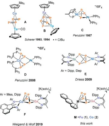
[6]

Some relevant examples are shown in Figure 1. [{Cp*Co}{Cp ⬙ (CO)Ta}(μ-η

2:2

-P

2

)

2

] (A, Cp* = C

5

Me

5

, Cp ⬘⬘ = C

5

H

3

{1,3-tBu

2

}) was synthesized by Scherer and co-workers by reaction of [Cp ⬘⬘ (CO)

2

Ta(η

4

-P

4

)]

with [Cp*Co(η

2

-C

2

H

4

)

2

],

[7]

while the related complex [{Cp*Fe}{Cp ⬘⬘ Ta}(μ-η

4:3

-P

5

)] (B) is formed by cothermolysis of two sandwich compounds [Cp*Fe(η

5

-P

5

)] and [Cp"Ta(CO)

4

].

[8]

Peruzzini and co-workers described the inser- tion of platinum(0) complexes into polyphosphido ligands.

[9]

* Prof. Dr. R. Wolf E-Mail: robert.wolf@ur.de

* Prof. Dr. J. J. Weigand

E-Mail: jan.weigand@tu-dresden.de [a] Institute of Inorganic Chemistry,

University of Regensburg 93040 Regensburg, Germany

[b] Faculty of Chemistry and Food Chemistry TU Dresden

01062 Dresden, Germany

Supporting information for this article is available on the WWW under http://dx.doi.org/10.1002/zaac.201900351 or from the au- thor.

© 2020 The Authors. Published by Wiley-VCH Verlag GmbH &

Co. KGaA. · This is an open access article under the terms of the Creative Commons Attribution-NonCommercial License, which permits use, distribution and reproduction in any medium, provided the original work is properly cited and is not used for commercial purposes.

bis(anthracene)ferrate(1–) and -cobaltate(1–) salts. The molecular structures of 1 and 2 were determined by X-ray crystallography and feature a P

4

chain which binds to the transition metal atom via all four P atoms and to the gallium atom via the terminal P atoms. Multinuclear NMR studies on 2 suggest that the molecular structure is preserved in solution.

Among other examples, such reactions resulted in complexes C and D shown in Figure 1. Akbayeva reported insertions into a P–P bond of η

1

-P

4

complexes of ruthenium(II) and iron(II).

[10]

Figure 1. Selected examples of heterodinuclear transition metal tetra- phosphido complexes.

Particularly relevant to the work described in this manuscript is a report by Driess and co-workers on hetero- dinuclear compounds of type E, which were synthesized from the silylene-activated P

4

ligand [(nacnac ⬘ )Si(η

2

-P

4

)]

(nacnac ⬘ = CH[(C=CH

2

)CMe][N(2,6-iPr

2

C

6

H

3

)]

2

) and the

nickel(I) species [({nacnac}Ni)

2

·toluene] (nacnac =

CH[CMeN(2,6-iPr

2

C

6

H

3

)]

2

) and [({nacnac ⬘⬘ }Ni)

2

·

toluene] (nacnac ⬘⬘ = CH[CMeN(2,6-Et

2

C

6

H

3

)]

2

).

[11]

These

(2)

complexes feature an [Si(μ,η

2:2

-P

4

)Ni] core, where the [(nacnac ⬘ )Si(η

2

-P

4

)] ligand is side-on coordinated to a tetrahe- dral nickel(I) center. Significantly, a reductive P–P bond cleav- age does not occur in this case.

We recently reported the synthesis of a related cobalt- gallium compound

[K(dme)

2

{(

Mes

BIAN)Co(μ-η

4

2

-P

4

)Ga(nacnac)}] (F) con- taining the α-diimine ligand bis(mesitylimino)acenaphthenedi- imine (

Mes

BIAN).

[4d]

Complex F is formed by reaction of[K(Et

2

O){(

Mes

BIAN)Co(η

4

-1,5-cod)}]

[12]

and the previously reported gallium tetraphosphido complex [(nacnac)Ga(η

2

-P

4

)]. The latter compound is readily accessible by reaction of (nacnac)Ga

I

with P

4

.

[13,14]

In contrast to the structures of the aforementioned Ni compounds E, a highly reduced catena-P

44–

unit resulting from the oxidative addition of a P–P bond to cobalt is observed in the structure of complex F. Reactions with chlorophosphanes R

2

PCl and RPCl

2

afford pentacyclic cyclo-P

5

R

2

ligands. Such a route thus offers the possibility to prepare new organofunctionalized polyphosph- orus ligands in the coordination sphere of transition metal atoms.

Building on this initial work, we subsequently investigated whether the range of accessible heterobimetallic phosphorus compounds can be expanded by using related transition met- alates. Herein, we describe the synthesis and structural charac- terization of

[K([18]crown-6){(η

4

-C

14

H

10

)Fe(μ-η

4

2

-P

4

)Ga(nacnac)}] (1) and [K(dme)

2

{(η

4

-C

14

H

10

)Co(μ-η

4

2

-P

4

)Ga(nacnac)}] (2).

These compounds show a similar M(μ-P

4

)Ga motif as com- pound F, but feature a distinct ancillary ligand at the transition metal center.

Results and Discussion

Expanding on the initial synthesis of F from [K(Et

2

O){(BIAN)Co(η

4

-1,5-cod)}]

[12]

and

[(nacnac)Ga(η

2

-P

4

)],

[13,14]

similar reactions of bis(anthracene) complexes [K([18]crown-6)(thf)

2

][Fe(η

4

-C

14

H

10

)

2

]

[15,16]

and [K(dme)

2

][Co(η

4

-C

14

H

10

)

2

]

[16,17]

were examined according to Scheme 1. Such complexes are useful sources of “naked” Fe

and Co

anions due to the presence of labile anthracene li- gands.

[15–18]

Scheme 1. Synthesis of 1 and 2; reagents and conditions for 1:

[K([18]crown-6){(η

4

-C

14

H

10

)Fe(μ-η

4

2

-P

4

)Ga(nacnac)}], for 2:

[K(dme)

2

{(η

4

-C

14

H

10

)Co(μ-η

4

2

-P

4

)Ga(nacnac)}].

Slow addition of a yellow solution of [(nacnac)Ga(η

2

-P

4

)]

(one equiv.) to a cooled rust-colored solution of

[K([18]crown-6)(thf)

2

][Fe(η

4

-C

14

H

10

)

2

] affords a brownish- yellow solution. After work-up, dark crystals of 1 suitable for X-ray diffraction analysis can be obtained by slow diffusion of n-hexane into a concentrated DME solution of the crude prod- uct. Solid 1 is readily soluble in polar coordinating solvents such as DME and THF, and it is highly sensitive toward oxy- gen and moisture. Unfortunately, all attempts to isolate 1 as a pure compound were unsuccessful until now. Resonances of residual free anthracene are found in

1

H NMR spectra, while the C, H, N combustion analysis results gave variable results, which strongly deviated from the values expected for pure 1.

Nevertheless, the molecular structure of 1 was confirmed by single-crystal diffraction (XRD) analysis.

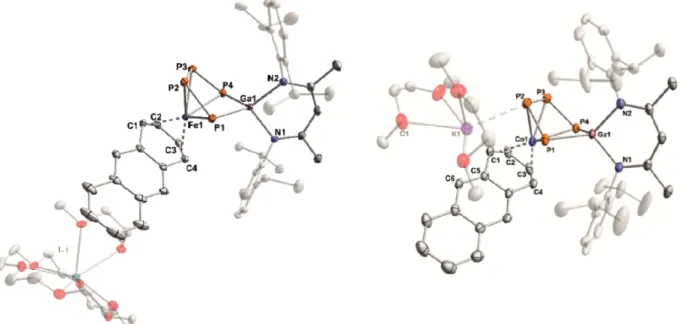
Complex 1 crystallizes in space group P1¯ with two formula units per cell. The molecular structure is depicted in Figure 2.

An ion-separated structure is observed where the potassium cation is coordinatively saturated by one molecule [18]crown- 6 and one DME molecule. The anion shows a bridging P

4

chain that binds to the iron center with all four P atoms, while only the terminal P atoms coordinate the gallium atom. As a result of the coordination by two P atoms and a nacnac ligand, the gallium atom adopts a nearly ideal tetrahedral arrangement with a twist angle of 87.57°. The P–P bond lengths are all shorter than typical P–P single bonds.

[25]

Notably, the terminal P–P bonds [P1–P2 2.1166(6) Å and P3–P4 2.1131(7) Å] are significantly shorter than the internal P–P bond [P2–P3 2.1801(7) Å], whereas the distance between the terminal P atoms [P1–P4 3.5473(6) Å] is long. The dihedral angle (P1–

P2–P3–P4 2.62°) shows that these bonds are nearly co-planar, while the P1–P2–P3 and P2–P3–P4 angles are almost identical [108.75(3)° and 108.93(3)°, respectively]. The anthracene li- gand is η

4

-coordinated to the iron center and shows the typical long-short-long pattern for the C–C bonds of transition-metal- coordinated anthracene molecules [C1–C2 1.4138(3) Å, C2–

C3 1.409(3) Å, C3–C4 1.421(3) Å]. Based on Fe–C and C–C distances, the coordination mode of the anthracene ligand seems identical with that in the starting material [K([18]crown- 6)(thf)

2

][Fe(η

4

-C

14

H

10

)

2

].

[15]

Cobalt complex 2 can be synthesized in a similar manner as 1 by adding a THF solution of [(nacnac)Ga(η

2

-P

4

)] to a solu- tion [K(dme)

2

][Co(η

4

-C

14

H

10

)

2

] in THF.

31

P{

1

H} NMR moni- toring of the reaction revealed the selective formation of 2 as the sole P-containing species. As a result, this compound was isolated purely and is fully characterized. Released anthracene can be removed by extraction of the crude product with diethyl ether and n-hexane, and subsequent crystallization from DME/

n-hexane affords 2 as thin, dark-violet plates, which were suit- able for single-crystal XRD. Due to the elaborate work-up pro- cedure, the yield of isolated compound is modest (22 %). Nev- ertheless, multinuclear NMR spectra and elemental analysis confirm the purity of the isolated compound.

Notably, the same compound 2 is formed as the major prod- uct even when an excess of [(nacnac)Ga(η

2

-P

4

)] is employed in the reaction. Apparently, a substitution of the second anthra- cene ligand does not occur.

Complex 2 likewise crystallizes in space group P1¯ with two

formula units per cell, and the molecular structure is shown in

(3)

Figure 2. Solid-state molecular structures of 1 and 2. Hydrogen atoms and some disordered parts are omitted for clarity; thermal ellipsoids are drawn at the 40 % probability level. Selected bond lengths /Å and angles /° for 1: P1–P2 2.1166(6), P2–P3 2.1801(7), P3–P4 2.1131(7), P1···P4 3.5473(6), Ga1–P1 2.3383(5), Ga1–P4 2.3307(5), Ga1–N1 2.018 (1), Ga1–N2 2.015(1), Fe1–P1 2.3879(5), Fe1–P2 2.3344(5), Fe1–P3 2.3368(7), Fe1–P4 2.3568(5), Fe1–C1 2.128(2), Fe1–C2 2.065(2), Fe1–C3 2.057(2), Fe1–C4 2.112(2), C1–C2 1.4138(3), C2–C3 1.409(3), C3–C4 1.421(3);

P1–P2–P3 108.75(3), P2–P3–P4 108.94(3), P3–P4–Ga1 101.36(2), P4–Ga1–P1 98.89(2), Ga1–P1–P2 100.54(2), C4–Fe1–P1 97.07(5), C1–Fe1–

P2 97.49(5), C2–Fe1–P3 101.14(5), C3–Fe1–P4 95.34(5); for 2: P1–P2 2.1095(9), P2–P3 2.1825(9), P3–P4 2.1057(9), P1···P4 3.4509(8), Ga1–

P1 2.3512(6), Ga1–P4 2.3297(6), Ga1–N1 1.993(2), Ga1–N2 2.008(2), Co1–P1 2.3635(7), Co1–P2 2.2922(7), Co1–P3 2.3368(7), Co1–P4 2.4108(7), Co1–C1 2.187(2), Co1–C2 2.026(2), Co1–C3 2.001(2), Co1–C4 2.82(2), C1–C2 1.4138(3), C2–C3 1.413(4), C3–C4 1.426(4), K1–

P1 3.5646(9), K1–P2 3.2981(8); P1–P2–P3 109.13(3), P2–P3–P4 105.88(3), P3–P4–Ga1 101.3(3), P4–Ga1–P1 94.99(2), Ga1–P1–P2 98.41(3), C4–Co1–P1 113.18(7), C1–Co1–P2 126.01(8), C2–Co1–P3 121.78(8), C3–Co1–P4 104.08(7) K1–C5 3.370(2) Å, K1–C6 3.175(2).

Figure 2. In case of 2, an ion-contact structure is observed [K1–P2 (3.298(1) Å], which is considerably shorter than the sum of the van der Waals radii (4.63 Å),

[19]

while it is slightly longer than the sum of the covalent radii (3.10 Å).

[20]

Further- more, the potassium cation interacts with the anthracene ligand [K1–C5 3.370(2) Å, K1–C6 3.175(2) Å] and two DME mol- ecules. The structure of the Co(μ-P

4

)Ga core is nevertheless similar to that of 1. The bridging P

4

unit is essentially in a syn conformation with a dihedral angle P1–P2–P3–P4 of 2.37°.

The P

4

chain displays short terminal P–P bonds [P1–P2 2.1095(9) Å and P3–P4 2.1057(9) Å], whereas the internal P2–

P3 bond [2.1825(9) Å] is closer to a single bond. The anthra- cene ligand coordinates to cobalt in an η

4

-fashion. The C–

C distances [C1–C2 1.4138(3) Å, C2–C3 1.413(4) Å, C3–C4 1.426(4) Å] indicate that there is considerable back-bonding between the metal atom and anthracene similar to the struc- tures of 1 and other related anthracene metalates.

[15,17]

In addition to complex F, several related structures with a P

4

fragment sandwiched between two metal atoms have been reported.

[12,21]

The most closely related compound appears to be [(Cp

R

Rh)(μ-η

4

2

-P

4

){Rh(CO)Cp

R

}] (Cp

R

= η

5

-C

5

Me

4

Et) reported by Scherer and co-workers.

[22]

In addition, it is noteworthy that Roesky, Konchenko, Scheer, and co-workers synthesized a trinuclear samarium-cobalt complex [(Cp ⬘⬘⬘ Co)

2

3

2

2

2

-P

4

)SmCp*

2

] (Cp* = η

5

-C

5

Me

5

, Cp ⬘⬘⬘

= η

5

-1,2,4-tBu

3

C

5

H

2

), which shows a similar structure mo- tif.

[23]

The

1

H NMR spectrum of the isolated crystals of 2 dis-

solved in [D

8

]THF displays five multiplets for coordinated an-

thracene with an integral ratio of 2:2:2:2:2. The chemical shifts

of these resonances are in the range δ = 6.90 ppm to 2.69 ppm,

which is common for anthracene coordinated to a transition

metal center.

[17,24]

The observed high-field shift of the anthra-

cene signals is explained by π-donation from the metal atom

to the π-acceptor ligand, which reduces the aromaticity of an-

thracene.

[17]

Two chemically distinct 2,6-diisoproplyphenyl

groups are observed in accordance with the single-crystal X-

ray structure, which are also discernible in the

13

C{

1

H} NMR

spectrum. The

31

P{

1

H} NMR spectrum shows two higher or-

der multiplets in a 1:1 ratio at δ = 89.2 ppm and δ = –94.7 ppm,

which can be assigned to an AA ⬘ XX ⬘ spin system shown in

Figure 3. The AA ⬘ XX ⬘ spin pattern observed is in line with a

C

2

symmetrical P

4

moiety. The high-field shifted multiplet at

δ = –94.7 ppm can be tentatively assigned to the terminal phos-

phorus atoms bound to the gallium atom based on the larger

line broadening and DFT calculations on the related compound

F.

[4d]

Conversely, the signal at δ = 89.2 ppm (τ

1/2

= 10.64 Hz)

can be assigned to the internal phosphorus atoms. The

1

J

AX

coupling constant is larger in magnitude than the

1

J

AA⬘

cou-

pling constant (

1

J

AA⬘

1

J

AX

= 126.2 Hz). This is in agreement

with a putative partial double bond character of the terminal

P–P bonds as suggested by the crystallographically obtained

P–P distances (vide supra). The previously reported diimine

complex F displays a similar AA ⬘ XX ⬘ pattern in the

31

P{

1

H}

(4)

NMR spectrum with

1

J

AX

= –450.5 Hz and

1

J

AA⬘

= –380.2 Hz.

[4d]

Nevertheless, it is noteworthy that the magni- tude of the

1

J

AX

coupling constant of 1 is about 45 Hz higher than that of compound F (

1

J

AX

=

1

J

A⬘X⬘

= –450.5 Hz), while the

1

J

AA⬘

constants are similar (2:

1

J

AA⬘

=

1

J

A⬘A

= –370.6 Hz;

C:

1

J

AA⬘

=

1

J

A⬘A

= –380.5 Hz).

[4d]

The large difference of the

1

J

AX

coupling constant might be caused by the shorter terminal P–P bonds in 2. The

2

J

PP

coupling constant is small (

2

J

AX⬘

=

2

J

A⬘X

= 4.9 Hz) and similar to those of F (

2

J

AX⬘

=

2

J

A⬘X

= 6.6 Hz).

[4d]

Remarkably, complex 1 shows a rather large

3

J

PP

coupling constant (

3

J

XX⬘

=

3

J

X⬘X

= –52.2 Hz) compared to that determined in compound F (

3

J

XX⬘

=

3

J

X⬘X

= –7.2 Hz), which might be explained by the more co-planar alignment of the P- atoms of the P

4

chain of 2. Overall, the multinuclear NMR spectra of 2 are in full agreement with the molecular structure observed in solid-state.

Figure 3.

31

P{

1

H} NMR spectrum of compound 2 (121.49 MHz, 300 K, [D

8

]THF) with nuclei assigned to an AA ⬘ XX ⬘ spin system;

insets: expanded spectrum (upwards) and simulated spectrum (down- wards); δ(P

AA⬘

) = 89.2 ppm, δ(P

XX⬘

) = –94.7 ppm,

1

J

AA’

=

1

J

A⬘A

= –370.6,

1

J

AX

=

1

J

A⬘X⬘

= –496.8,

2

J

AX⬘

=

2

J

A⬘X

= 4.9,

3

J

XX⬘

=

3

J

X⬘X

= –52.2 Hz; [Co] = (η

4

-C

14

H

10

)Co, [Ga] = (nacnac)Ga.

The UV/Vis spectrum of 2 was recorded in THF solution (see Figure S5, Supporting Information). The spectrum shows two very strong absorption bands in the UV region at 301 nm (ε = 42000 L·mol

–1

·cm

–1

) and 356 nm (ε = 39000 L·mol

–1

·cm

–1

), with the latter absorption tailing far into the visible region. The visible part shows a broad absorption at 524 nm. It is notable that the UV/Vis spectrum of F is similar in the UV region, while additional intense bands observed for F at 553 nm and 658 nm can be presumably assigned to charge transfer transi- tions involving the BIAN ligand.

[4d]

In summary, heterodinuclear complexes with a strongly re- duced P

4

unit were synthesized by oxidative P–P bond addition of [(nacnac)Ga(η

2

-P

4

)] with low-valent ferrate(–I) and co- baltat(–I) anions. The molecular structures of the resulting complexes 1 and 2 feature an η

4

2

-bridging P

4

chain between the transition metal atom and the gallium atom. Although bridging polyphosphide ligands are generally quite common in homodinuclear complexes,

[2]

this precise structural motif is rare, which underlines the flexible coordination behavior of polyphosphido ligands. In future work, substitution of the la- bile anthracene ligand in such complexes could enable the fur- ther derivatization of the P

4

ligand. In addition, it is note- worthy that polyphosphorus complexes containing the (nac-

nac)Ga moiety have recently emerged as promising platforms for the construction of extensive P

n

frameworks.

[4d,14]

There- fore, the complexes reported herein and related species might become a suitable starting point for further transformations leading to structurally novel species.

Experimental Section

General: All manipulations were performed under an atmosphere of dry argon using standard Schlenk techniques or an MBraun UniLab glovebox. Solvents were dried and degassed with an MBraun SPS800 solvent-purification system. THF, diethyl ether, and toluene were stored over molecular sieves (3 Å). n-Hexane was stored over a potas- sium mirror. 1,2-dimethoxyethane (DME) was stirred over K/benzo- phenone, distilled and stored over molecular sieves (3 Å). Deuterated tetrahydrofurane was purchased from Sigma–Aldrich and used as received. The starting materials [(nacnac)Ga(η

2

-P

4

)],

[13]

[K([18]crown-6)(thf)

2

][Fe(η

4

-C

14

H

10

)

2

],

[15,16]

and

[K(dme)

2

][Co(η

4

-C

14

H

10

)

2

],

[16,17]

were prepared according to pre- viously reported procedures.

NMR Spectroscopy: NMR spectra were recorded on Bruker Avance 300 and Avance 400 spectrometers at 300 K and internally referenced to residual solvent resonances (δ(TMS) = 0.00 ppm,

1

H,

13

C; δ(H

3

PO

4

) (85 %) = 0.00 ppm,

31

P, externally). Chemical shifts (δ) are reported in ppm. Coupling constants (J) are reported in Hz. For compounds, which give rise to a higher order spin system in the

31

P{

1

H} NMR spectrum, the resolution enhanced

31

P{

1

H} NMR spectrum was trans- ferred to the software gNMR, version 5.0, by Cherwell Scientific.

[25]

The full line shape iteration procedure of gNMR was applied to obtain the best match of the fitted to the experimental spectrum.

1

J(

31

P

31

P) coupling constants were set to negative values and all other signs of the coupling constants were obtained accordingly. The assignment of the

13

C signals was deduced from the cross-peaks in 2D correlation experiments (HMBC, HSQC).

Elemental Analysis: Elemental analyses were determined by the ana- lytical department of the University of Regensburg with a Micro Vario Cube (Elementar). UV/Vis spectra were recorded on an Ocean Optics Flame spectrometer.

Single-crystal X-ray Diffraction: Crystallographic data were re- corded on an Oxford Diffraction Gemini R Ultra with Atlas S2 CCD detector for 1 and an Agilent GV1000 diffractometer with a Titan S2 CCD detector for 2. In both cases, Cu-K

α

radiation (λ = 1.54184 Å) was used. Crystals were selected under mineral oil, mounted on micro- mount loops and quench-cooled using an Oxford Cryosystems open flow N

2

cooling device. Either semi-empirical multi-scan absorption corrections

[26]

or analytical ones

[27]

were applied to the data. Using Olex2,

[28]

the structures were solved with SHELXT

[29]

using intrinsic phasing and refined with SHELXL

[30]

using least-squares refinement on F

2

. The hydrogen atoms were located in idealized positions and refined isotropically with a riding model.

Crystallographic data (excluding structure factors) for the structures in this paper have been deposited with the Cambridge Crystallographic Data Centre, CCDC, 12 Union Road, Cambridge CB21EZ, UK. Copies of the data can be obtained free of charge on quoting the depository numbers CCDC-1972352 for 1 and CCDC-1972351 for 2 (Fax: +44- 1223-336-033; E-Mail: deposit@ccdc.cam.ac.uk, http://www.ccdc.

cam.ac.uk).

Crystallographic Data for 1: C

66

H

102

FeGaKN

2

O

10

P

4

(M =

1372.04 g·mol

–1

), triclinic, space group P1¯ (no. 2), a = 12.8013(4) Å,

(5)

b = 12.8477(3) Å, c = 22.8788(6) Å, α = 95.470(2)°, β = 99.362(2)°, γ = 105.796(2)°, V = 3533.66(17) Å

3

, Z = 2, T = 123(1) K, μ(Cu-K

α

)

= 3.933 mm

–1

, D(calcd) = 1.290 g·cm

–3

, 51666 reflections measured (7.23° ⱕ 2Θ ⱕ 133.694°), 12403 unique (R

int

= 0.0334, R

sigma

= 0.0246) which were used in all calculations. The final R

1

was 0.0314 [I ⬎ 2σ(I)], and wR

2

was 0.0816 (all data).

Crystallographic Data for 2: C

51

H

71

CoGaKN

2

O

4

P

4

(M = 1067.72 g·mol

–1

), triclinic, space group P1¯ (no. 2), a = 10.9891(2) Å, b = 12.0239(2) Å, c = 21.5532(3) Å, α = 90.3270(10)°, β = 92.4990(10)°, γ = 104.3790(10)°, V = 2755.63(8) Å

3

, Z = 2, T = 123.01(10) K, μ(Cu-K

α

) = 5.059 mm

–1

, D(calcd) = 1.287 g·cm

–3

, 71275 reflections measured (7.592° ⱕ 2Θ ⱕ 147.664°), 11035 unique (R

int

= 0.0509, R

sigma

= 0.0286) which were used in all calculations. The final R

1

was 0.0394 [I ⬎ 2σ(I)] and wR

2

was 0.1051 (all data).

Synthesis of [K([18]crown-6){(η

4

-C

14

H

10

)Fe(μ-η

4

2

-P

4

) Ga(nacnac)}] (1): A yellow solution of [(nacnac)Ga(η

2

-P

4

)] (12 mg, 0.018 mmol, 1.0 equiv.) in THF (2 mL) was slowly added to a rust- colored solution of [K([18]crown-6)(thf)

2

][Fe(η

4

-C

14

H

10

)

2

] (15.7 mg, 0.018 mmol, 1 equiv.) in THF (2 mL). The reaction mixture was stirred for 24 h at room temperature. Volatiles were removed under reduced pressure and the residual brown solid was washed with 8 mL n-hexane.

The crude product was dissolved in 4 mL DME and filtered through a Whatman glass filter (pore size: 0.1 μm). Crystals suitable for X-ray diffraction were obtained by diffusion of n-hexane into the concen- trated filtrate of the crude product at room temperature.

1

H NMR spec- tra of isolated samples of paramagnetic 1 show resonances of residual free anthracene. In addition, inconsistent and variable elemental analy- ses indicate the presence of large quantities of other unknown impuri- ties. Unfortunately, all attempts to purify 1 by fractional crystallization were unsuccessful so far.

Synthesis of [K(dme)

2

{(η

4

-C

14

H

10

)Co(μ-η

4

2

-P

4

)Ga(nacnac)}] (2):

[K(dme)

2

Co(η

4

-C

14

H

10

)

2

] (199 mg, 0.315 mmol, 1.0 equiv.) was dis- solved in THF (20 mL), resulting in a deep-violet solution, and a yel- low solution of [(nacnac)Ga(η

2

-P

4

)] (200 mg, 0.315 mmol, 1.0 equiv.) in 40 mL THF was slowly added at –80 °C. The reaction mixture was allowed to warm to room temperature while stirring. Volatiles were removed in vacuo and the remaining solid was washed with diethyl ether (20 mL) and n-hexane (40 mL). The brownish residue was redis- solved in DME and filtered through a P4 glass frit. The dark red-brown filtrate was concentrated in vacuo and layered with n-hexane (35 mL).

Pure [K(dme)

2

{(η

4

-C

14

H

10

)Co(μ-η

4

2

-P

4

)Ga(nacnac)}] (2) was iso- lated as dark violet thin plates after storage at room temperature for five days. Based on

1

H NMR spectroscopy and C,H,N analysis, 2 con- tains 1.9 molecules of DME per formula unit after drying under vac- uum (10

–3

mbar) for several hours. Yield: 86 mg (22 %). M.p. 178 °C (decomposition to black oil). UV/Vis (THF, λ

max

/nm, ε

max

/L·mol

–1

· cm

–1

): 301 (42000), 356 (39000), 524sh (8200).

1

H NMR (400.13 MHz, 300 K, [D

8

]THF): δ = 7.49 (br. m, 3 H, CH

Dipp

), 6.99 (m, 1 H, CH

Dipp

), 6.93 (m, 2 H, CH

Dipp

), 6.90 (m, 2 H, CH

(anth)

), 6.74 (m, 2 H,CH

(anth)

), 5.84 (s, 2 H, CH

(anth)

), 5.59 (m, 2 H, CH

(anth)

), 4,82 (s, 1 H, CH

nacnac

), 3.96 (sept,

3

J

HH

= 6.8 Hz, 2 H, CH(CH

3

)

2(nacnac)

), 3.44 (s, 7.6H DME), 3.27 (s, 11 H, DME), 2.88 [sept,

3

J

HH

= 6.8 Hz, 2 H, CH(CH

3

)

2(nacnac)

], 2.62 (m, 2 H, CH

(anth)1,4

), 1.79 (s, 3 H, (s, 3 H, CH

3(nacnac)

), 1.53 [d,

3

J

HH

= 6.8 Hz, 6 H, CH(CH

3

)

2(nacnac)

], 1.50 (s, 3 H, CH

3(nacnac)

), 1.38 [d,

3

J

HH

= 6.8 Hz, 6 H, CH(CH

3

)

2(nacnac)

], 1.25 [d,

3

J

HH

= 6.8 Hz, 6 H, CH(CH

3

)

2(nacnac)

], 0.94 [d,

3

J

HH

= 6.8 Hz, 6 H, CH(CH

3

)

2(nacnac)

] ppm.

13

C{

1

H} NMR (100.61 MHz, 300 K, [D

8

]THF): δ = 168.0 (NCCH

3

CH

(nacnac)

), 165.0 (NCCH

3

CH

(nacnac)

), 145.5 (C

(anth)

), 145.1 (C

(Dipp)

), 145.0 (C

(Dipp)

), 144.1 (C

(Dipp)

), 143.2 (C

(Dipp)

), 133.3 (C

(anth)

), 127.0 (C

Dipp

), 126.2 (CH

(Dipp)

), 125.8 (C

(anth)

), 125.6 (CH

(Dipp)

), 123.9 (CH

(Dipp)

), 123.1 (C

(anth)

), 113.5 (C

(anth)

), 95.8

(CH

(nacnac)

), 83.9 (C

(anth)

), 72.6 (DME), 58.8 (DME), 56.4 (C

(anth)

), 29.3 [CH(CH

3

)

2(Dipp)

], 28.7 [CH(CH

3

)

2(Dipp)

], 26.1 [CH(CH

3

)

2(Dipp)

], 25.9 [CH(CH

3

)

2(Dipp)

], 25.1 [CH(CH

3

)

2(Dipp)

], 24.6 (CH

3(Dipp)

), 24.5 (CH

3(Dipp)

) ppm.

31

P{

1

H} NMR (161.98 MHz, 300 K, [D

8

]THF):

(AA ⬘ XX ⬘ ) spin system δ = 89.2 (m, 2P

P

), –94.7 (m, 2P

Ga

) ppm; see Figure 3 for chemical shifts and coupling constants obtained by simu- lation. C

51

H

71

CoGaKN

2

O

4

P

4

·(C

4

H

10

O

2

)

1.9

(Mw = 1058.78 g·mol

–1

):

calcd. C 57.40, H 6.66, N 2.65 %; found C 57.51, H 6.52; N 2.59 %.

Supporting Information (see footnote on the first page of this article):

Further details of the refinement and the crystallographic data of 1 and 2.

Acknowledgements

Financial support by the Deutsche Forschungsgemeinschaft (WE4621/

3-1 and WO1496/7-1) and the European Research Council (CoG 772299) is gratefully acknowledged. Open access funding enabled and organized by Projekt DEAL.

Keywords: Heterobimetalic complexes; Phosphorus; Gal- lium; Cobalt; Iron

References

[1] a) A. P. Ginsberg, W. E. Lindsell, W. E. Silverthorn, Trans. Acad.

Sci. N. Y. 1970, 303; b) A. P. Ginsberg, W. E. Lindsell, J. Am.

Chem. Soc. 1971, 93, 2082–2084.

[2] a) M. Peruzzini, L. Gonsalvi, A. Romerosa, Chem. Soc. Rev.

2005, 34, 1038–1047; b) M. Scheer, G. Balázs, A. Seitz, Chem.

Rev. 2010, 110, 4236–4256; c) M. Caporali, L. Gonsalvi, A. Ros- sin, M. Peruzzini, Chem. Rev. 2010, 110, 4178–4235; d) B. M.

Cossairt, N. A. Piro, C. C. Cummins, Chem. Rev. 2010, 110, 4164–4177.

[3] Selected recent examples: a) S. Pelties, D. Herrmann, B. d. Bruin, F. Hartl, R. Wolf, Chem. Commun. 2014, 50, 7014–7016; b) C. C.

Mokhtarzadeh, A. L. Rheingold, J. S. Figueroa, Dalton Trans.

2016, 45, 14561–14569; c) E. Mädl, G. Balázs, E. V. Peresypkina, M. Scheer, Angew. Chem. Int. Ed. 2016, 55, 7702–7707; Angew.

Chem. 2016, 128, 7833–7838; d) F. Dielmann, A. Timoshkin, M.

Piesch, G. Balázs, M. Scheer, Angew. Chem. Int. Ed. 2017, 56, 1671–1675; Angew. Chem. 2017, 129, 1693–1698; e) C. Chan, A. E. Carpenter, M. Gembicky, C. E. Moore, A. L. Rheingold, J. S. Figueroa, Organometallics 2019, 38, 1436–1444; f) C. M.

Hoidn, T. M. Maier, K. Trabitsch, J. J. Weigand, R. Wolf, Angew.

Chem. Int. Ed. 2019, 58; Angew. Chem. 2019, 131, 19107–19112.

[4] a) G. Capozzi, L. Chiti, M. Di Vaira, M. Peruzzini, P. Stoppioni, J. Chem. Soc., Chem. Commun. 1986, 1799–1800; b) B. M.

Cossairt, M.-C. Diawara, C. C. Cummins, Science 2009, 323, 602; c) A. E. Seitz, M. Eckhardt, A. Erlebach, E. V. Peresypkina, M. Sierka, M. Scheer, J. Am. Chem. Soc. 2016, 138, 10433–

10436; d) C. G. P. Ziegler, T. M. Maier, S. Pelties, C. Taube, F.

Hennersdorf, A. W. Ehlers, J. J. Weigand, R. Wolf, Chem. Sci.

2019, 110, 4178.

[5] a) P. Barbaro, M. Di Vaira, M. Peruzzini, S. Seniori Costantini, P.

Stoppioni, Chem. Eur. J. 2007, 13, 6682–6690; b) E. Mädl, M. V.

Butovskii, G. Balázs, E. V. Peresypkina, A. V. Virovets, M. Seidl, M. Scheer, Angew. Chem. Int. Ed. 2014, 53, 7643–7646; Angew.

Chem. 2014, 126, 7774–7777; c) M. Piesch, S. Reichl, M. Seidl, G. Balázs, M. Scheer, Angew. Chem. Int. Ed. 2019, 58, 16563–

16568; Angew. Chem. 2019, 131, 16716–16721.

[6] a) M. Di Vaira, P. Stoppioni, Polyhedron 1994, 13, 3045–3051;

b) O. J. Scherer, T. Mohr, G. Wolmershäuser, J. Organomet.

Chem. 1997, 529, 379–385; d) M. Caporali, M. Di Vaira, M. Per-

(6)

uzzini, S. Seniori Costantini, P. Stoppioni, F. Zanobini, Eur. J.

Inorg. Chem. 2010, 2010, 152–158; d) V. Mirabello, M. Caporali, V. Gallo, L. Gonsalvi, A. Ienco, M. Latronico, P. Mastrorilli, M.

Peruzzini, Dalton Trans. 2011, 40, 9668–9671; e) M. Piesch, F.

Dielmann, S. Reichl, M. Scheer, Chem. Eur. J. 2020, 26, 1518–

1524; f) M. Scheer, M. Piesch, C. Graßl, Angew. Chem. Int. Ed.

2020 DOI: 10.1002/anie.201916622; Angew. Chem. DOI:

10.1002/ange.201916622.

[7] O. J. Scherer, R. Winter, G. Wolmershäuser, Z. Anorg. Allg.

Chem. 1993, 619, 827–835.

[8] M. Detzel, T. Mohr, O. J. Scherer, G. Wolmershäuser, Angew.

Chem. Int. Ed. Engl. 1994, 33, 1110–1112; Angew. Chem. 1994, 106, 1142–1144.

[9] a) M. Di Vaira, P. Stoppioni, M. Peruzzini, Polyhedron 1987, 6, 351–382; b) M. Caporali, P. Barbaro, L. Gonsalvi, A. Ienco, D.

Yakhvarov, M. Peruzzini, Angew. Chem. Int. Ed. 2008, 47, 3766–

3768; Angew. Chem. 2008, 120, 3826–3828.

[10] D. N. Akbayeva, Russ. J. Coord. Chem. 2007, 33, 661–668.

[11] Y. Xiong, S. Yao, E. Bill, M. Driess, Inorg. Chem. 2009, 48, 7522–7524.

[12] S. Pelties, T. Maier, D. Herrmann, B. de Bruin, C. Rebreyend, S.

Gärtner, I. G. Shenderovich, R. Wolf, Chem. Eur. J. 2017, 23, 6094–6102.

[13] G. Prabusankar, A. Doddi, C. Gemel, M. Winter, R. A. Fischer, Inorg. Chem. 2010, 49, 7976–7980.

[14] a) F. Hennersdorf, J. J. Weigand, Angew. Chem. Int. Ed. 2017, 56, 7858–7862; Angew. Chem. 2017, 129, 7966–7970; b) F. Hen- nersdorf, J. Frötschel, J. J. Weigand, J. Am. Chem. Soc. 2017, 139, 14592–14604.

[15] W. W. Brennessel, R. E. Jilek, J. E. Ellis, Angew. Chem. Int. Ed.

2007, 46, 6132–6136; Angew. Chem. 2007, 119, 6244–6248.

[16] W. W. Brennessel J. E. Ellis in Inorganic Syntheses (Ed.: P. P.

Power), vol. 37, chapter 4.8., Wiley, pp. 76–84.

[17] a) W. W. Brennessel, J. V. G. Young, J. E. Ellis, Angew. Chem.

Int. Ed. 2002, 41, 1211–1215; Angew. Chem. 2002, 114, 1259–

1263; b) W. W. Brennessel, J. E. Ellis, Inorg. Chem. 2012, 51, 9076–9094.

[18] a) J. E. Ellis, Inorg. Chem. 2006, 45, 3167–3186; b) R. Wolf, A. W. Ehlers, J. C. Slootweg, M. Lutz, D. Gudat, M. Hunger, A. L. Spek, K. Lammertsma, Angew. Chem. Int. Ed. 2008, 47, 4584–4587; Angew. Chem. 2008, 120, 5660–5663; c) R. Wolf, J. C. Slootweg, A. W. Ehlers, F. Hartl, B. de Bruin, M. Lutz, A. L.

Spek, K. Lammertsma, Angew. Chem. Int. Ed. 2009, 48, 3104–

3107; Angew. Chem. 2009, 121, 3150–3153; d) R. Wolf, N. Ghav-

tadze, K. Weber, E.-M. Schnöckelborg, B. d. Bruin, A. W. Ehlers, K. Lammertsma, Dalton Trans. 2010, 39, 1453–1456.

[19] P. Pyykkö, M. Atsumi, Chem. Eur. J. 2009, 15, 12770–12779.

[20] S. Alvarez, Dalton Trans. 2013, 42, 8617–8636.

[21] a) O. J. Scherer, G. Berg, G. Wolmershäuser, Chem. Ber. 1995, 128, 635–639; b) O. J. Scherer, S. Weigel, G. Wolmershäuser, Chem. Eur. J. 1998, 4, 1910–1916; c) V. A. Miluykov, O. G. Siny- ashin, P. Lönnecke, E. Hey-Hawkins, Mendeleev Commun. 2003, 13, 212–213; d) W. W. Seidel, O. T. Summerscales, B. O. Patrick, M. D. Fryzuk, Angew. Chem. Int. Ed. 2008, 47, 115–117; Angew.

Chem. 2008, 120, 121–123; e) S. Yao, Y. Xiong, C. Milsmann, E. Bill, S. Pfirrmann, C. Limberg, M. Driess, Chem. Eur. J. 2010, 16, 436–439; f) C. Schwarzmaier, M. Bodensteiner, A. Y. Timosh- kin, M. Scheer, Angew. Chem. Int. Ed. 2014, 53, 290–293; g) S.

Yao, N. Lindenmaier, Y. Xiong, S. Inoue, T. Szilvási, M. Adel- hardt, J. Sutter, K. Meyer, M. Driess, Angew. Chem. Int. Ed. 2015, 54, 1250–1254; Angew. Chem. 2015, 127, 1266–1270; h) S. Yao, T. Szilvási, N. Lindenmaier, Y. Xiong, S. Inoue, M. Adelhardt, J.

Sutter, K. Meyer, M. Driess, Chem. Commun. 2015, 51, 6153–

6156; i) F. Spitzer, C. Graßl, G. Balázs, E. M. Zolnhofer, K.

Meyer, M. Scheer, Angew. Chem. Int. Ed. 2016, 55, 4340–4344;

Angew. Chem. 2016, 128, 4412–4416; j) U. Chakraborty, J. Leitl, B. Mühldorf, M. Bodensteiner, S. Pelties, R. Wolf, Dalton Trans.

2018, 47, 3693–3697.

[22] O. J. Scherer, M. Swarowsky, H. Swarowsky, G. Wolmershäuser, Angew. Chem. Int. Ed. Engl. 1988, 27, 694–695; Angew. Chem.

1988, 100, 738–739.

[23] T. Li, N. Arleth, M. T. Gamer, R. Köppe, T. Augenstein, F. Diel- mann, M. Scheer, S. N. Konchenko, P. W. Roesky, Inorg. Chem.

2013, 52, 14231–14236.

[24] P. Büschelberger, D. Gärtner, E. Reyes-Rodriguez, F. Kreyen- schmidt, K. Koszinowski, A. Jacobi von Wangelin, R. Wolf, Chem. Eur. J. 2017, 23, 3139–3151.

[25] P. H. M. Budzelaar, gNMR for Windows (5.0.6.0), NMR Simula- tion Programm 2006.

[26] a) SCALE3ABS, CrysAlisPro, Aglient Technologies Inc. Oxford, GB 2015; b) G. M. Sheldrick, SADABS, Bruker AXS, Madison, USA 2007.

[27] R. C. Clark, J. S. Reid, Acta Crystallogr., Sect. A 1995, 51, 887.

[28] O. V. Dolomanov, L. J. Bourhis, R. J. Gildea, J. A. K. Howard, H.

Puschmann, J. Appl. Crystallogr. 2009, 42, 339–341.

[29] G. M. Sheldrick, Acta Crystallogr., Sect. A 2015, 71, 3.

[30] G. M. Sheldrick, Acta Crystallogr., Sect. C 2015, 71, 3.

Received: December 20, 2019

Published Online: February 26, 2020

Abbildung

Figure 1. Selected examples of heterodinuclear transition metal tetra- tetra-phosphido complexes.
Figure 2. Solid-state molecular structures of 1 and 2. Hydrogen atoms and some disordered parts are omitted for clarity; thermal ellipsoids are drawn at the 40 % probability level
Figure 3. 31 P{ 1 H} NMR spectrum of compound 2 (121.49 MHz, 300 K, [D 8 ]THF) with nuclei assigned to an AA ⬘ XX ⬘ spin system;

Referenzen

ÄHNLICHE DOKUMENTE

a Department Chemie und Pharmazie, Lehrstuhl f¨ur Anorganische und Allgemeine Chemie, Universit¨at Erlangen-N¨urnberg, Egerlandstraße 1, 91058 Erlangen, Germany.. b Department

The structural assign- ment shown in Scheme 3 was confirmed by NMR and MS data, elemental analysis and single crystal X-ray structural analysis for the compounds 12b, 12e, 13a and

Silica gel column chromatography of the EtOAc extract afforded compounds 1,4,5 while the n-hexane extract provided compounds 2, 3, 6 and 7.. Compound 1 was obtained as

The difference between Newtonian and general relativistic motion after 14 months is much larger for the four chaotic trajectories than it is for the three tra- jectories that are

Comparing the resulting prediction with the ac- tual structures derived by the three methods de- scribed above, we see that the predicted structure of ODSB is very well represented

The dihedral angles C-E-E-C range from 79(2) to 96(1) are consistent with the concept of minimized p lone-pair repulsion of adjacent chalcogen atoms.. The dependence of

The fluorescence depolarization of molecules is con- ditioned by their rotations (occurring during the lifetime of an excited state), which result in a change in the direction of

Wenn man in den 31 einfachen Artikelstrukturschemata für Artikeltypen, die den basalen Wörterbuchtyp mit dem Artikelstrukturschema WA  FK < SK erweitern,