Article
A Novel Language Paradigm for Intraoperative Language Mapping: Feasibility and Evaluation
Katharina Rosengarth
1,*, Delin Pai
1, Frank Dodoo-Schittko
2, Katharina Hense
1, Teele Tamm
3, Christian Ott
1, Ralf Lürding
4,†, Elisabeth Bumes
4, Mark W Greenlee
3, Karl Michael Schebesch
1, Nils Ole Schmidt
1and Christian Doenitz
1
Citation: Rosengarth, K.; Pai, D.;
Dodoo-Schittko, F.; Hense, K.; Tamm, T.; Ott, C.; Lürding, R.; Bumes, E.;
Greenlee, M.W; Schebesch, K.M.; et al.
A Novel Language Paradigm for Intraoperative Language Mapping:
Feasibility and Evaluation.J. Clin.
Med.2021,10, 655. https://
doi.org/10.3390/jcm10040655
Academic Editor: Francesco Acerbi Received: 29 December 2020 Accepted: 3 February 2021 Published: 8 February 2021
Publisher’s Note:MDPI stays neutral with regard to jurisdictional claims in published maps and institutional affil- iations.
Copyright: © 2021 by the authors.
Licensee MDPI, Basel, Switzerland.
This article is an open access article distributed under the terms and conditions of the Creative Commons Attribution (CC BY) license (https://
creativecommons.org/licenses/by/
4.0/).
1 Department of Neurosurgery, Regensburg University Hospital, 93053 Regensburg, Germany;
delin-wolfgang.pai@ukr.de (D.P.); katharina.hense@ukr.de (K.H.); christian.ott@ukr.de (C.O.);
karl-michael.schebesch@ukr.de (K.M.S.); nils-ole.schmidt@ukr.de (N.O.S.); christian.doenitz@ukr.de (C.D.)
2 Institute of Social Medicine and Health Systems Research, Otto von Guericke University Magdeburg, 39106 Magdeburg, Germany; frank.dodoo-schittko@ukr.de
3 Institute for Experimental Psychology, University of Regensburg, 93053 Regensburg, Germany;
teele.tamm@ymail.com (T.T.); mark.greenlee@ur.de (M.W.G.)
4 Department of Neurology and Wilhelm Sander-NeuroOncology Unit, Regensburg University Hospital, 93053 Regensburg, Germany; ralf.luerding@ukr.de (R.L.); elisabeth.bumes@ukr.de (E.B.)
* Correspondence: katharina.rosengarth@ukr.de; Tel.: +49-941-944-19006
† This author passed away a few months ago.
Abstract: (1) Background—Mapping language using direct cortical stimulation (DCS) during an awake craniotomy is difficult without using more than one language paradigm that particularly follows the demand of DCS by not exceeding the assessment time of 4 s to prevent intraoperative complications. We designed an intraoperative language paradigm by combining classical picture naming and verb generation, which safely engaged highly relevant language functions. (2) Methods—
An evaluation study investigated whether a single trial of the language task could be performed in less than 4 s in 30 healthy subjects and whether the suggested language paradigm sufficiently pictured the cortical language network using functional magnetic resonance imaging (fMRI) in 12 healthy subjects. In a feasibility study, 24 brain tumor patients conducted the language task during an awake craniotomy. The patients’ neuropsychological outcomes were monitored before and after surgery. (3) Results—The fMRI results in healthy subjects showed activations in a language-associated network around the (left) sylvian fissure. Single language trials could be performed within 4 s.
Intraoperatively, all tumor patients showed DCS-induced language errors while conducting the novel language task. Postoperatively, mild neuropsychological impairments appeared compared to the presurgical assessment. (4) Conclusions—These data support the use of a novel language paradigm that safely monitors highly relevant language functions intraoperatively, which can consequently minimize negative postoperative neuropsychological outcomes.
Keywords: intraoperative language mapping; direct cortical stimulation; awake surgery; neuropsy- chological outcome
1. Introduction
An awake craniotomy with language mapping in patients with brain tumors or metastasis in language-critical brain areas represents the gold standard for maximizing the resection of brain tumors and metastases in language-critical brain areas while minimizing the risk for postoperative functional deficits [1–3]. Both aspects improve postsurgical treatment responses, overall survival, and increase patients’ health-related quality of life [4,5].
Mapping the complete language capacity with its subfunctions during language testing with direct cortical stimulation (DCS) in the setting of an awake craniotomy can
J. Clin. Med.2021,10, 655. https://doi.org/10.3390/jcm10040655 https://www.mdpi.com/journal/jcm
be challenging because language with its subfunctions, such as phonology, morphology, semantics, syntax, and grammar, is organized as a complex network of cortical areas and fiber bundles around the sylvian fissure in the language-dominant hemisphere. There is a huge diversity of intraoperative language paradigms, such as object naming [6–8], sentence completion [9], action naming [10–12], or verb generation [7,13–16]. Unfortunately, these tasks only cover single aspects of language processing and might therefore insufficiently reflect patients’ linguistic capacity and skills. Rofes and Miceli reported huge differences between different intraoperative language tasks [17]. The most popular tasks, namely, intraoperatively applied picture-naming tasks, are extremely sensitive to phonological retrieval but they demand minimal linguistical processing compared to other tasks, such as verb generation or sentence completion, and therefore might fail to address more complex grammatical, syntactic, lexico-semantic, or morphophonological language processes [17].
This is in line with presurgical functional magnetic resonance imaging results that show that picture-naming tasks are incapable of reliably identifying Broca’s area in healthy subjects [18]. Especially for patients with a frontal tumor, it might therefore be helpful to increase the sensitivity of the object-naming task by adding a grammatical component to it.
This may increase the likelihood of detecting eloquent language regions that may currently be overlooked using lexico-semantic tasks. The lesion location might also influence the choice of intraoperative language paradigms; however, linguistic subprocesses (such as the production and perception of phonology, morphology, semantics, and syntax) are normally not bound to a single brain area but are rather organized as networks [19]. Therefore, some others suggest an individual language test battery with different language tasks [19–21], but this leads to increasing the surgery time and possibly to patients’ being overtaxed and exhausted. This might also bear the risk of false-positive language disturbances because of patients’ impaired attention [22].
Besides the validity of an intraoperative language task, the task must be designed to meet the technical demands of DCS by not exceeding the assessment time of 4 s in a single language trial to minimize the risk for DCS-induced seizures [10,12,23,24]. This also impacts the design of intraoperative language tasks because complex tasks are highly likely to need more than 4 s to be performed, even if those tasks might be able to cover more or even all linguistic subfunctions that are necessary for the generation of a correct language utterance compared to easy language paradigms, such as object naming or verb processing alone.
In this study, we aimed (1) to design and evaluate a novel single-language paradigm consisting of a combination of picture naming and sentences generation tasks that better reflect the highly relevant language functions and (2) to simultaneously restrain the assess- ment duration of single-task trials to up to 4 s to minimize intraoperative complications, such as seizures. Therefore, we investigated whether this language paradigm could be performed within 4 s in a healthy subject group to meet the criteria of DCS and whether the language task could stimulate the complete language system surrounding the sylvian fissure during a functional magnetic resonance imaging (fMRI) experiment. In the last step, (3) we investigated the novel language task intraoperatively in a group of brain tumor patients undergoing an awake craniotomy to test its feasibility and how it affected patients’
linguistical and cognitive outcome.
2. Materials and Methods 2.1. Language Paradigm
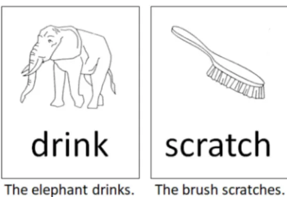
The language paradigm was designed as a single-stimulus trial comprising a com-
bination of the classical picture-naming task using the DO (Test de dénomination orale
d’images) 80 figures [25] and a semantically associated verb in the infinitive form. Both
stimuli were presented visually in black and white by being randomly placed one below
the other. We designed 80 different pairs of stimuli made up of a picture and a written
verb. Healthy controls and patients were asked to generate grammatically and semantically
correct sentences according to the given language stimuli (Figure 1). Linguistically, in
the German language, this task comprised the correct choice of determiner genus, the processing of the correct word order, and a correct subject–verb congruence structure to build a sentence that follows the subject–predicate formula. The performance of this task thereby required the processing of phonemic/phonological, semantic, morphological, and syntactic, perceptive, and productive aspects of language per se, as well as the transfer of this information into a surface structure (speech).
Figure 1. Two examples according to the intraoperative language task used in this study. Subjects and patients were asked to generate grammatically and semantically correct sentences according to the language stimulus as depicted.
2.2. Patient and Subject Sample
2.2.1. Healthy Subjects—Evaluation Study
The first aim of the pilot study was to test the intraoperative feasibility of the language paradigm. This intended to evaluate whether the performance speed of the rather complex language task did not exceed the assessment time of 4 s in a single language trial to minimize the risk for intraoperative DCS-induced seizures. Therefore, 30 healthy native German speakers were included (16 female: mean age 26.0 years (SD = 8.49)).
The second aim of the pilot study was to investigate whether the suggested language paradigm could sufficiently picture the cortical language network, including the inferior frontal gyrus, the ventral premotor area, the angular and supramarginal gyri, and the pos- terior superior and middle temporal gyri in the language-dominant hemisphere. Therefore, 12 additional healthy right-handed native German speakers were included to participate in an fMRI experiment (6 female: mean age 27.7 (SD = 1.69)). These subjects did not take part in the former behavioral experiment (as described above) to avoid familiarity effects.
2.2.2. Patient Group—Feasibility Study
The third aim of the study was the successful intraoperative application of the novel
language task. Twenty-three right-handed patients and one left-handed patient without
or with only mild presurgical language deficits with left lateral brain tumors in language-
associated brain areas were included in the study between the years 2015 and 2019. The
patients’ characteristics are shown in Table 1. An independent medical indication for awake
surgery was given in all patients. The presurgical fMRI data gathered using a battery of
verb and syntax generation tasks done by these patients showed a close spatial proximity
of language critical areas and brain tumors. Left-hemispheric language laterality was
computed in all patients. All patients underwent neuropsychological testing, including
language tests, before and after surgery.
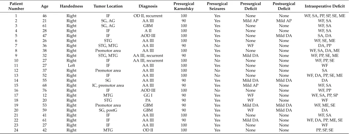
Table 1. Patients characteristics, including pre-, intra-, and postsurgical language deficits.
Patient
Number Age Handedness Tumor Location Diagnosis Presurgical
Karnofsky
Presurgical Seizures
Presurgical Deficit
Postsurgical
Deficit Intraoperative Deficit
1 46 Right IF OD II, recurrent 100 Yes None None WF, SA, PP, SP, SE, ME
2 21 Right SG, AG AA III 90 Yes Mild AP Mild AP WF, SA
3 61 Right SG, AG GBM 100 Yes None None WF, SA
4 28 Right IF A II 100 Yes None None WF, SA
5 47 Right IF AOD III 100 Yes None Mild DA SA, DA
6 26 Right STG AA III 100 Yes None None WF, SE, ME
7 36 Right STG, MTG AA III 90 No WF None DA, PP
8 38 Right Premotor area AA III 100 Yes None None WF, SA, DA, ME
9 32 Right STG, MTG AA III, recurrent 90 Yes Mild DA None WF, PP, SE, ME
10 27 Right IF AA III, recurrent 100 No None None WF, PP, SE
11 27 Left IF AA III 100 Yes None None WF
12 37 Right Premotor area AA III 100 No None None SA
13 52 Right IF AA III 100 No None None WF, DA, PP, SE, ME
14 55 Right SG AA III 90 Yes Mild DA Mild DA DA
15 68 Right IC, premotor area AA III 90 Yes Mild AP None WF, SA
16 76 Right IF AOD III 100 No None None WF, PP
17 12 Right MTG GG I 90 Yes WF None WF, SA, PP, SP
18 20 Right STG PA 90 Yes WF None WF
19 53 Right Premotor area GBM 90 Yes Mild DA Mild DA WF, ME, SE
20 59 Right SG, postG GBM 90 Yes WF Mild DA DA
21 41 Right IF AA III 100 Yes None None WF, SA
22 61 Right IF AA III 90 Yes Mild DA None WF, DA, PP, ME, SE
23 27 Right IF AA III 100 Yes None None WF
24 42 Right MTG OD II 100 Yes None None PP, SP, SE
Abbreviations: WFword finding error, AR—speech arrest, DA—dysarthria, PP—phonematic paraphasia; SP—semantic paraphasia, ME—morphological error, SE—syntactic error, AP—aphasia, OD—
oligodendroglioma, AOD—anaplastic oligodendroglioma, GBM—glioblastoma, A—astrocytoma, AA—anaplastic astrocytoma, GG—ganglioglioma, PA—pilocytic astrocytoma, IF—inferior frontal, SG—
supramarginal gyrus, AG—angular gyrus, STG—superior temporal gyrus, MTG—middle temporal gyrus, PostG—postcentral gyrus, IC—insular cortex.
2.3. Procedures and Data Analysis 2.3.1. Evaluation Study
To evaluate whether the performance speed of the novel language task exceeded the assessment time of 4 s in a single language trial, 30 healthy subjects were instructed to generate grammatically and semantically correct sentences according to the language stimulus in a time interval not exceeding 4 s. Stimulus presentation was done using the
“Presentation” software [26]. The 80 language stimuli were subsequently presented and randomized for each subject. The mean subjects’ performance speed during each trial was measured by a supervisor who pressed a button immediately after each subject stopped speaking. Data analysis was done using IBM SPSS Version 25 (IBM Deutschland GmbH, Ehningen, Germany) by calculating the mean performance speed.
To investigate whether the suggested language paradigm could sufficiently picture most parts of the cortical language network, 12 healthy right-handed native German speakers (6 female; mean age 27.7 (SD = 1.69)) were instructed to perform the novel lan- guage paradigm while stimulus-dependent changes in their blood oxygen level-dependent (BOLD) responses were recorded. The language task was analogous to the behavioral paradigm design with respect to the stimulus presentation. Using a block design, five stimulus trials, each lasting 4 s, were presented sequentially and were followed by a rest- ing period of 20 s. The data were collected using a 3T MRI scanner (Magneton Allegra, Siemens, Erlangen, Germany). The fMRI data parameter for T2*-images were 34 slices, repitition time (TR) = 2000 ms, echo time (TE) = 30 ms, voxel size = 3 × 3 × 3 mm
3, flip an- gle = 90
◦, and field of view = 192 × 192 mm
2. To visualize the results three-dimensionally, an additional T1 magnetization prepared rapid gradient echo (MPRAGE) image was acquired (TR = 2300 ms, TE = 2910 ms, flip angle = 9
◦, and slice thickness = 1 mm).
Data analysis was done using the Statistical Parametric Mapping 8 (SPM8) software run- ning (https://www.fil.ion.ucl.ac.uk/spm/ accessed on 7 February 2021) under Matlab 7.1 (Mathworks, Sherborn, MA, USA). Functional images were realigned, coregistered to the structural image, normalized to the Montreal Neurological Institute (MNI) space, and spatially smoothed using a full-width at half-maximum Gaussian kernel of 8 mm.
Afterward, based on the general linear model, we defined a single regressor according to the language task timing profile, which was then convoluted using a boxcar model function with the canonical hemodynamic response function. The resting period was not modeled explicitly and served as an implicit baseline. A post hoc t-test was calculated for every voxel to compare the fMRI signals and beta-weights according to the stimulus regressor. The resulting individual data were the basis of a second-level random effects analysis. Voxels were defined to be significant if they did not fall below a t-value of T = 7 at the voxel level and simultaneously did not exceed a p-value of p < 0.001 (uncorrected, T = 4.025) at the cluster level. Only clusters with more than 50 voxels were reported.
2.3.2. Feasibility Study
All 24 patients underwent an awake craniotomy without sedation following the wake–
awake–awake technique with therapeutic communication that had been established by our
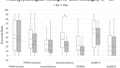
group [27]. Due to internal administrative affairs, the pre- and postoperative neuropsycho-
logical data of only 13 patients were analyzed in this study. Neuropsychological assess-
ment included the following cognitive domains: verbal and working memory (“number
repeating” forward and backward spans) [28], non-verbal working memory (“Corsi block
span”) [29], lexical and semantic verbal fluency [30], verbal [28] and visuo-constructive
capacity, visual long-term memory (“Rey Visual Design Learning Test”) [31], and executive
functions [32]. Long-term memory functions and visuo-constructive capacity were only
assessed prior to surgery to avoid potential training effects. Patients were tested one
day before surgery and 2–5 days after surgery. Performance differences between the pre-
and postsurgical neuropsychological outcomes were computed by comparing the median
percentile ranks of the single test scores with the nonparametric Wilcoxon–Mann–Whitney
Test because percentile ranks probably do not fulfill the criteria of a normal distribution.
The analysis was performed using IBM SPSS Version 25. The significance level was set to p < 0.05. Additionally, effect sizes were computed. To guarantee that patients could conduct a trial of the intraoperative language task within 4 s, all patients were intensively trained in advance to surgery. Accordingly, every single stimulus was first practiced by the patients. If patients were not familiar with some pictures shown during the language task, those stimuli would be left out during surgery. Intraoperatively, language stimuli were shown on an adjustable computer monitor that could be customized to each patient’s head position to increase the patient’s comfort as much as possible. Stimuli were displayed using Microsoft PowerPoint [33]. Every language trial was displayed for 4 s. For tem- poral coordination of the language stimuli presentation and DCS, a short acoustic tone was played at the beginning of each language trial to indicate to the surgeon to start the electrocortical stimulation. We used a negative-mapping technique with a limited and tailored craniotomy that exposed the tumor and a small margin of normal, peritumoral brain tissue. This approach allowed for stimulation mapping around the tumor margins but avoided extensive brain exposure only to force identifying all positive language sites.
This technique was proven safe [34], minimized surgery time, and prevented complica- tions of large skull flaps. During the language mapping with DCS, we used a rectangular biphasic pulse with a duration of 1.0 ms and a frequency of 50 Hz, bipolar electrodes for cortical stimulation and a monopolar electrode for subcortical stimulation, and currents varying between 1 and 6 mA. If a language disturbance referring to a single DCS locus was detected, the disturbance in association with the same stimulation locus had to be verified at least two more times. In turn, this stimulation site was defined as “language critical.”
Possible occurring language problems or deficits according to DCS were classified and documented in all 24 patients. Additionally, language-positive and -negative points were recorded using the Brainlab navigation system [35].
3. Results
3.1. Evaluation Study
The mean performance duration for the language paradigm was 2.53 s in healthy subjects, with a standard deviation of 0.39 s. Overall, only 2 of 80 language stimuli led to processing times that exceeded 3 s (M = 3.17, SE = 1.22 and M = 3.31 s, SE = 1.1 s, respectively).
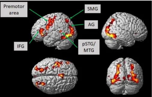
According to the fMRI experiment, we investigated which brain areas showed as- sociated activation according to the language paradigm introduced in this study. The language paradigm led to predominantly left-hemispheric activations in the opercular, triangular, and orbital part of the inferior frontal gyrus, the putamen, the pallidum, the pre-and postcentral gyri, the hippocampus, and the inferior parietal gyrus (T = 7, p < 0.001;
uncorrected). Moreover, we observed activation in the right hemisphere in the lingual
gyrus, the angular gyrus, and the medial temporal gyrus. There was also bilateral enhanced
activation in the fusiform gyrus, the occipital medial, inferior and superior gyri, the inferior
temporal and superior parietal gyri, the thalamus, the supplementary motor area, and the
insular cortex (T = 7, p < 0.001; uncorrected) (Figure 2, Table 2). Lowering the statistical
threshold of T = 5 at the voxel level, which was still assumed to be a very conservative
statistical threshold, there was also bilateral activation of the posterior superior and middle
temporal gyri (p < 0.001; uncorrected).
Figure 2. Brain activation in healthy subjects (n = 12) while constructing grammatically and semanti- cally correct sentences in response to the novel language task, which comprised a combination of picture naming and simple verb generation (T = 7, p < 0.001; uncorrected). Activation is depicted on a single-subject-normalized brain surface implemented in SPM8. Abbreviations: IFG—inferior frontal gyrus, SMG—supramarginal gyrus, AG—angular gyrus, pSTG—posterior superior temporal gyrus, MTG—middle temporal gyrus.
Table 2. Brain activation in healthy subjects (n = 12) while constructing grammatically and semantically correct sentences according to the language task, which comprised a combination of picture naming and simple verb generation (T = 7, p < 0.001; uncorrected).
MNI Coordinates Cluster
Size Region Brodmann
Area Hemisphere T-Value pFWE
x y z
34 −86 18 2598 Fusiform gyrus, MOG, IOG, SOG, lingual gyrus, SPG, MTG 7, 18, 19, 37 R 21.85 0.000
−18 2 8 342 Putamen, pallidum L 17.87 0.000
−38 2 34 835 Precentral gyrus, postcentral gyrus, IFG 4, 6 L 17.46 0.000
−8 −24 −10 881 Thalamus, hippocampus L 16.31 0.000
−40 −84 −4 2497 MOG, fusiform gyrus, IOG, SPG, ITG, IPG, SOG 7, 19, 37 L 16.16 0.000
24 −30 −2 268 Thalamus R 13.35 0.000
−6 14 44 424 SMA 6 L and R 13.19 0.000
−24 26 12 541 IFG (PT), IFG (PO),
Insula L 10.80 0.000
32 28 −2 96 Insula R 9.59 0.000
−2 −94 2 50 Fissura calcarina L 8.97 0.000
Abbreviations: MOG—middle occipital gyrus, SOG—superior occipital gyrus, SPG—superior parietal gyrus, MTG—middle temporal gyrus, ITG—inferior temporal gyrus, IFG—inferior frontal gyrus, PT—pars triangularis, PO—pars opercularis, IFG—inferior parietal gyrus, IOG—inferior occipital gyrus, SMA—supplemantary motor area, L—left, R–right, MNI—Montreal Neurological Institute, FWE—family- wise error. Anatomical labeling was done using the Automated Anatomical Labeling (AAL) atlas implemented in SPM8.