Research Collection
Report
Collateral sensitivity associated with antibiotic resistance plasmids
Author(s):
Herencias, Cristina; Rodríguez-Beltrán, Jerónimo; Leon-Sampedro, Ricardo; Alonso-del Valle, Aida;
Palkovičová, Jana; Cantón, Rafael; San Millán, Álvaro Publication Date:
2021-01
Permanent Link:
https://doi.org/10.3929/ethz-b-000473463
Originally published in:
eLife 10, http://doi.org/10.7554/eLife.65130
Rights / License:
Creative Commons Attribution 4.0 International
This page was generated automatically upon download from the ETH Zurich Research Collection. For more
information please consult the Terms of use.
*For correspondence:
jeronimo.rodriguez.beltran@
gmail.com (JR-B);
asanmillan@cnb.csic.es (A´SM)
†These authors contributed equally to this work Competing interests:The authors declare that no competing interests exist.
Funding:See page 10 Received:24 November 2020 Accepted:20 January 2021 Published:20 January 2021 Reviewing editor: Marc Lipsitch, Harvard TH Chan School of Public Health, United States
Copyright Herencias et al. This article is distributed under the terms of theCreative Commons Attribution License,which permits unrestricted use and redistribution provided that the original author and source are credited.
Collateral sensitivity associated with antibiotic resistance plasmids
Cristina Herencias
1†, Jero´nimo Rodrı´guez-Beltra´n
1,2†*, Ricardo Leo´n-Sampedro
1,2, Aida Alonso-del Valle
1, Jana Palkovicˇova´
3, Rafael Canto´n
1,4, A´lvaro San Milla´n
1,2,5*
1
Servicio de Microbiologı´a. Hospital Universitario Ramo´n y Cajal and Instituto Ramo´n y Cajal de Investigacio´n Sanitaria, Madrid, Spain;
2Centro de Investigacio´n Biolo´gica en Red Epidemiologı´a y Salud Pu´blica, Instituto de Salud Carlos III, Madrid, Spain;
3Department of Biology and Wildlife Diseases, Faculty of Veterinary Hygiene and Ecology, University of Veterinary and Pharmaceutical Sciences, Brno, Czech Republic;
4Red Espan˜ola de Investigacio´n en Patologı´a Infecciosa. Instituto de Salud Carlos III, Madrid, Spain;
5Centro Nacional de Biotecnologı´a-CSIC, Madrid, Spain
Abstract Collateral sensitivity (CS) is a promising alternative approach to counteract the rising problem of antibiotic resistance (ABR). CS occurs when the acquisition of resistance to one antibiotic produces increased susceptibility to a second antibiotic. Recent studies have focused on CS strategies designed against ABR mediated by chromosomal mutations. However, one of the main drivers of ABR in clinically relevant bacteria is the horizontal transfer of ABR genes mediated by plasmids. Here, we report the first analysis of CS associated with the acquisition of complete ABR plasmids, including the clinically important carbapenem-resistance conjugative plasmid pOXA- 48. In addition, we describe the conservation of CS in clinical
E. coliisolates and its application to selectively kill plasmid-carrying bacteria. Our results provide new insights that establish the basis for developing CS-informed treatment strategies to combat plasmid-mediated ABR.
Introduction
The rapid evolution of antibiotic resistance (ABR) in bacteria reduces the utility of clinically relevant antibiotics, making ABR one of the major challenges facing public health (Jim, 2016; MacLean and San Millan, 2019). There is therefore an urgent need for new treatment strategies to fight against resistant pathogens (Baym et al., 2016). Among the most promising alternatives, much attention has focused on collateral sensitivity (CS). CS occurs when the acquisition of resistance to one antibi- otic causes increased susceptibility to a second antibiotic (Szybalski and Bryson, 1952) and can be exploited for the design of multi-drug strategies that select against ABR (Imamovic and Sommer, 2013; La´za´r et al., 2013). Moreover, recent evidence suggests that the development of resistance might be prevented by treatments based on the sequential cycling of antibiotics whose resistance mechanisms produce reciprocal CS (Imamovic and Sommer, 2013). Several studies have cataloged CS networks emerging from mutations in chromosomal (Imamovic and Sommer, 2013; La´za´r et al., 2013; Maltas and Wood, 2019; Podnecky et al., 2018; Roemhild et al., 2020) and plasmid genes (Dortet et al., 2015; Fro¨hlich et al., 2019; Rosenkilde et al., 2019), but the CS effects produced by the horizontal acquisition of ABR plasmids remain poorly understood.
Mobile genetic elements, especially plasmids, play a crucial role in the dissemination of ABR
genes between clinical pathogens and are one of the major drivers of the alarming worldwide rise in
ABR (Partridge et al., 2018). Despite the evolutionary advantage conferred by plasmids in the pres-
ence of antibiotics, plasmid acquisition tends to produce common metabolic alterations in the host
bacterium (San Millan et al., 2018), that typically translate into a fitness cost (San Millan and
MacLean, 2017). We reasoned that the physiological alterations produced upon acquisition of ABR plasmids might lead to exploitable CS responses. To test this hypothesis, we analyzed the collateral effects associated with the acquisition of six natural plasmids carrying clinically relevant ABR genes in Escherichia coli MG1655. Our results reveal that most ABR plasmids, including the clinically impor- tant carbapenem-resistance conjugative plasmid pOXA-48, produce CS events of moderate effect.
To extend our results, we explored the degree of conservation of CS responses associated with pOXA-48 across phylogenetically diverse wild-type E. coli. Our results suggest that these responses can be exploited to preferentially kill plasmid-carrying bacteria.
Results and discussion
CS induced by ABR plasmids
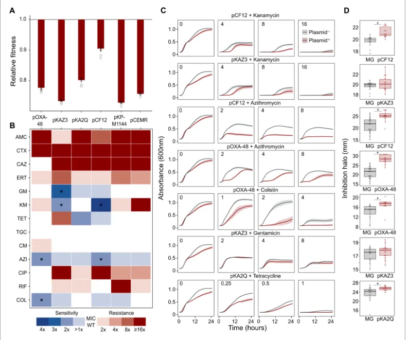
To test whether ABR plasmids elicit exploitable CS to other antibiotics, we selected six clinically rele- vant ABR plasmids (see Materials and methods, Table 1 and Figure 1—figure supplement 1). All plasmids carried important ß-lactamase genes, including extended-spectrum ß-lactamases (ESBLs), AmpC-type ß-lactamases, or carbapenemases, as well as other relevant ABR genes (Table 1 and Fig- ure 1—figure supplement 1). These plasmids belonged to a broad range of incompatibility groups, covered a range of different sizes, included conjugative and non-conjugative specimens, and pro- duced variable fitness effects, ranging from 10% to 27% reductions in relative fitness, when intro- duced into E. coli MG1655 (Figure 1A).
As a preliminary screening, we performed dose-response experiments with 13 antibiotics from eight drug families. For this analysis, we determined the minimal inhibitory concentration (MIC, see Materials and methods) using the broth microdilution method for plasmid-free MG1655 and each of its six plasmid-carrying derivatives (Supplementary file 1A). We analyzed the collateral antibiotic susceptibility effects associated with the presence of each plasmid, measured as the fold-change in the antibiotic MIC between plasmid-carrying and plasmid-free bacteria. As expected from their genetic content, all plasmids conferred resistance to ß-lactam antibiotics and most of them con- ferred additional resistance to other antibiotics from unrelated families (Figure 1B and Supplementary file 1A). Initially, we defined CS events as those plasmid-antibiotic combinations showing a minimum twofold reduction in MIC (calculated as the fold-change of the median MIC value obtained from four to five independent MIC determinations). We then performed additional MIC determinations (up to 10 replicates each) for the antibiotic-plasmid combinations that fulfilled this criterion to increase the robustness of our analysis (Supplementary file 1A). This led to the iden- tification of seven CS instances producing a consistent 2-fold reduction in antibiotic susceptibility, of which six showed significant reductions in antibiotic susceptibility (pOXA-48–azithromycin, pCF12–azithromycin, pOXA-48–colistin, pCF12–kanamycin, pKAZ3–kanamycin, and pKAZ3-gentami- cin; Mann-Whitney U test; p<0.015 in all cases; Figure 1B). Of note, CS to tetracycline elicited by
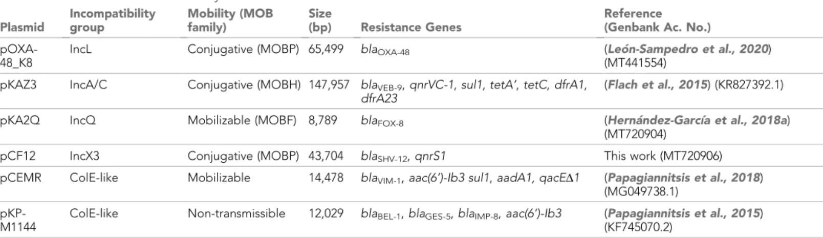
Table 1.Plasmids used in this study.
Plasmid
Incompatibility group
Mobility (MOB family)
Size
(bp) Resistance Genes
Reference (Genbank Ac. No.) pOXA-
48_K8
IncL Conjugative (MOBP) 65,499 blaOXA-48 (Leo´n-Sampedro et al., 2020)
(MT441554) pKAZ3 IncA/C Conjugative (MOBH) 147,957 blaVEB-9,qnrVC-1,sul1,tetA’,tetC,dfrA1,
dfrA23
(Flach et al., 2015) (KR827392.1)
pKA2Q IncQ Mobilizable (MOBF) 8,789 blaFOX-8 (Herna´ndez-Garcı´a et al., 2018a)
(MT720904)
pCF12 IncX3 Conjugative (MOBP) 43,704 blaSHV-12,qnrS1 This work (MT720906)
pCEMR ColE-like Mobilizable 14,478 blaVIM-1,aac(6’)-Ib3 sul1,aadA1, qacED1 (Papagiannitsis et al., 2018) (MG049738.1)
pKP- M1144
ColE-like Non-transmissible 12,029 blaBEL-1,blaGES-5,blaIMP-8,aac(6’)-Ib3 (Papagiannitsis et al., 2015) (KF745070.2)
*pOXA-48_K8 is a pOXA-48-like plasmid (see Materials and methods andFigure 1—figure supplement 1). For simplicity, we refer to this plasmid as pOXA-48 throughout the study.
Figure 1.Collateral sensitivity and fitness effects associated with antibiotic resistance (ABR) plasmids. (A) Competition assay determined fitness of plasmid-carrying clones relative to the plasmid-free MG1655 strain. Bars represent the means of six independent experiments (represented by dots), and error bars represent the standard error of the mean. (B) Heat-map representing collateral responses to antibiotics associated with plasmid
acquisition. The color code represents the fold change of MIC in plasmid-carrying derivatives compared to plasmid-free MG1655 (see legend). Asterisks indicate a significant2 fold decrease in MIC (Mann-Whitney U test; p<0.015). AMC, amoxicillin-clavulanic acid; CTX, cefotaxime; CAZ, ceftazidime;
ERT, ertapenem; GM, gentamicin; KM, kanamycin; TET, tetracycline; TGC, tigecycline; CM, chloramphenicol; AZI, azithromycin; CIP, ciprofloxacin; RIF, rifampicin; and COL, colistin. (C) Growth curves of MG1655 and plasmid-carrying MG1655 strains exposed to increasing antibiotic concentrations. The antibiotic concentration, in mg/L, is indicated at the top left corner of each panel. Lines represent the mean of 6 biological replicates, and the shaded area indicates standard error of the mean. (D) Boxplot representations of the inhibition halo diameters, in mm, obtained from disk-diffusion
antibiograms of plasmid-free and plasmid-carrying MG1655. Plasmid/antibiotic combinations in each row are the same as the ones indicated in panel C. Horizontal lines within boxes indicate median values, upper and lower hinges correspond to the 25th and 75th percentiles, and whiskers extend to observations within 1.5 times the interquartile range. Individual data points are also represented (11–15 biological replicates). Asterisks indicate statistically significant differences (unpaired t-test with Welch’s correction p<0.044).
The online version of this article includes the following source data and figure supplement(s) for figure 1:
Source data 1.Numerical data that are represented inFigure 1.
Figure supplement 1.Plasmids used in this study.
Figure supplement 2—source data 1.Numerical data that are represented inFigure 1—figure supplement 1.
Figure supplement 2.Analysis of bacterial growth curves.
pKA2Q did not reach statistical significance (Mann-Whitney U test; p=0.2), yet we decided to main- tain it for subsequent analysis.
To further validate MIC results, we conducted two complementary series of experiments for the seven cases of plasmid-induced CS: (i) complete growth curves under a range of antibiotic concen- trations and (ii) disk-diffusion antibiograms on agar plates. Analysis of the area under the growth curve, which integrates all growth parameters (maximum growth rate, lag duration and carrying capacity) (DelaFuente et al., 2020), confirmed significant plasmid-mediated CS effects at different antibiotic concentrations for six of the combinations (pOXA-48–azithromycin, pCF12–azithromycin, pOXA-48–colistin, pCF12–kanamycin, pKAZ3–kanamycin, and pKA2Q–tetracycline; Figure 1C, Fig- ure 1—figure supplement 2, and Supplementary file 1B). The disk-diffusion results largely agreed with the MIC and growth-curve data, showing a general trend toward an increased diameter of the antibiotic inhibition zone in plasmid-carrying strains compared with plasmid-free MG1655 (Figure 1D). These differences were statistically significant for pCF12–kanamycin, pCF12–azithromy- cin, pOXA-48–azithromycin, pOXA-48–colistin, and pKA2Q–tetracycline (unpaired t-test with Welch’s correction; p<0.044 in all cases). Overall, six of the seven CS instances initially identified were vali- dated with at least one of the additional methods (growth curves or disk-diffusion assays), and four of them showed full agreement across all three methods. The disk-diffusion assays produced notably robust results with minimum hands-on time, suggesting that this technique is appropriate for screen- ing CS responses in large strain collections. Together, these results demonstrate that ABR plasmids produce moderate but significant CS to different antibiotics, comparable to that generated by ABR chromosomal mutations in E. coli (Imamovic and Sommer, 2013; La´za´r et al., 2013;
Podnecky et al., 2018).
Conservation of pOXA-48-mediated CS
The success and applicability of CS-informed therapeutic strategies are crucially dependent on the conservation of CS across diverse genetic contexts (Podnecky et al., 2018). To address the phyloge- netic preservation of plasmid-induced CS, we focused on the plasmid pOXA-48, which confers resis- tance to last-resort carbapenem antibiotics, and whose prevalence in clinical settings is rising alarmingly (David et al., 2019). We tested the degree of conservation of pOXA-48-mediated CS patterns (to azithromycin and colistin) across phylogenetically diverse E. coli strains using disk-diffu- sion assays. We determined antibiotic susceptibility in nine diverse E. coli clinical isolates (Figure 2A) and their pOXA-48–carrying derivatives. CS to azithromycin showed a striking degree of conserva- tion across the strains, with eight out of nine plasmid-carriers showing significantly higher susceptibil- ity than their plasmid-free counterparts. In contrast, CS to colistin was conserved in only two of the tested strains (Figure 2B). Nevertheless, all strains showed CS responses to at least one antibiotic, suggesting that pOXA-48 elicits CS responses that could potentially be exploited therapeutically.
Exploitation of CS to selectively kill pOXA-48-carrying E. coli
We next tested the potential of exploiting CS effects to kill plasmid-carrying populations. We ratio-
nally selected four E. coli isolates and their pOXA-48 carrying derivatives showing the following
defined CS profiles: to colistin only (Ec10), to azithromycin only (Ec21 and Ec25), or to both antibiot-
ics (Ec18). MG1655 was included as a control. Plasmid-bearers and plasmid-free strains were propa-
gated separately in the presence of a single antibiotic or with sequential exposure to both
antibiotics over 2 days. On the first day of the experiment, 24 bacterial populations of each strain
were inoculated into media containing either colistin (4 mg/L) or azithromycin (8 mg/L). After 22 hr,
populations able to grow were transferred to fresh media containing colistin or azithromycin in a full
factorial experimental design. This approach gave rise to four antibiotic treatments: two single-drug
treatments (denoted Azi
!Azi and Col
!Col) and two treatments in which the antibiotic changed
(Azi
!Col and Col
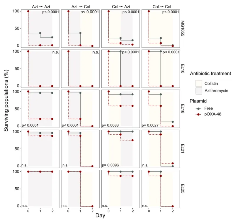
!Azi). In 11 of the 20 possible strain-treatment combinations, pOXA-48–carrying
populations showed higher mortality rates than their plasmid-free counterparts (Figure 3; log-rank
test p<0.001 in all cases). As we used fixed antibiotic concentrations, the results of this experiment
are likely to depend on the interplay between the particular resistance levels of each strain and the
order in which the antibiotics are used. This may explain why we failed to observe significant differ-
ences in the cases in which the antibiotic treatment produced a sudden drop in viability for both
plasmid-bearers and plasmid-free strains. Nevertheless, the mortality patterns observed were
consistent with the CS patterns obtained in the disk-diffusion technique (Figure 2B). For instance, plasmid-carrying MG1655 and Ec18 strains, which show CS to both antibiotics, exhibited significant reductions in survival in all treatments. In contrast, for strain Ec10, which shows CS exclusively to colistin, selective killing was observed only when the treatment included colistin as the first antibiotic (Col
!Col or Col
!Azi).
Concluding remarks
Our results reveal that the acquisition of clinically-relevant ABR plasmids induces CS to unrelated antibiotics. These findings thus serve as a stepping stone toward the development of new approaches aimed at blocking plasmid-mediated horizontal spread of ABR genes. These anti-plas- mid strategies could help to tackle the alarming clinical and community spread of ABR. However, the molecular basis of plasmid-associated CS is currently unknown. The feasibility of rational broad- spectrum anti-plasmid strategies will depend on gaining comprehensive knowledge about the molecular mechanisms that increase antibiotic susceptibility upon plasmid acquisition. Until these therapies are available, CS-informed empirical treatment of plasmid-carrying bacteria has the poten- tial to help resolve antibiotic resistant infections. In this regard, our results suggest that colistin and azithromycin may be valuable antibiotics for the treatment of pOXA-48–carrying enterobacteria.
Both antibiotics are already used to treat enterobacterial infections (Lu¨bbert, 2016; Morrill et al., 2015), and colistin in particular is currently used as a last resort antibiotic against carbapenem-
Figure 2.Phylogeny and collateral sensitivity profiles ofE. coliclinical isolates. (A) Phylogenetic tree ofE. colispecies, highlighting the strains used in this study and their sequence types (ST). Colors within tree branches represent different STs. The tree depicts the phylogenetic relationships of 1344 representativeE. coligenomes obtained from NCBI. (B) Representation of the inhibition halo diameters, in mm, obtained from disk-diffusion antibiograms of clinical isolates and their transconjugants carrying pOXA-48. Horizontal lines inside boxes indicate median values, upper and lower hinges correspond to the 25th and 75th percentiles, and whiskers extend to observations within 1.5 times the interquartile range. Individual data points are also represented (6–12 replicates, jittered to facilitate data visualization). Asterisks denote statistically significant differences (t-test with Welch’s correction p<0.035 in all cases).The online version of this article includes the following source data for figure 2:
Source data 1.Numerical data that are represented inFigure 2.
resistant enterobacteria (Rodriguez-Ban˜o et al., 2018). A limitation of our experimental setup is that plasmid-bearers and plasmid-free populations were subjected to antibiotic treatments sepa- rately. More experiments are thus required to confirm that CS-informed treatments can eradicate plasmid-carrying bacteria from mixed populations and to gauge the clinical relevance of our results.
Figure 3.Plasmid-mediated CS can be exploited to preferentially kill plasmid-carrying bacteria. Survival curves of plasmid-free (gray solid lines) and plasmid-carrying (red dashed lines) bacterial populations in response to constant or sequential antibiotic treatments. Plasmid-free or plasmid-carrying bacterial populations were cultured separately for 2 days with either constant antibiotic exposure or with a change of antibiotic after the first day;
treatments are specified by the background colors in each panel. Log-rank test p-values assessing statistically different survival curves are shown. n.s.
non-significant. For strains Ec10, Ec18, Ec21, and Ec25 n = 24; for MG1665 n = 48. AZI, azithromycin; COL, colistin.
The online version of this article includes the following source data for figure 3:
Source data 1.Numerical data that are represented inFigure 3.
In addition, future studies should focus on determining the best treatment option. Sequential treat- ments offer a promising strategy, as they better exploit evolutionary trade-offs (such as CS) to con- straint ABR development (Kim et al., 2014; Roemhild and Schulenburg, 2019). Combination therapies might also provide a valuable strategy as long as both antibiotics act synergistically to inhibit bacterial growth (Tamma et al., 2012). In summary, although extensive work will be needed before these therapies are available, the findings presented here open new avenues of research aimed at understanding plasmid-associated CS and pave the way for the development of new CS- based treatment strategies to combat plasmid-mediated ABR.
Materials and methods
Bacterial strains, plasmids, and culture conditions
All experiments were performed in liquid Lennox lysogeny broth (LB; CONDA) or on LB agar (15 g/
L) unless indicated. Mueller Hinton II broth (Oxoid) was used to verify that MIC values of E. coli MG1655 were comparable with those obtained in LB medium. The antibiotics used were amoxicillin- clavulanic acid, ceftazidime, cefotaxime, gentamicin (Normon), ertapenem (Merck Sharp and Dohme), kanamycin, chloramphenicol, tetracycline (Nzytech), tigecycline (Pfizer), azithromycin, colis- tin (Altan Pharmaceuticals), and rifampicin (Sanofi-aventis).
Plasmids pKAZ3 (Accession Number KR827392.1), pKP-M1144 (Acc. No. KF745070.2), pKA2Q (Acc. No. MT720904), and pEncl-30969cz (Acc. No. MG049738.1) were previously published (Flach et al., 2015; Herna´ndez-Garcı´a et al., 2018a; Papagiannitsis et al., 2015;
Papagiannitsis et al., 2018). Plasmid pCEMR is a derivative of pEncl-30969cz in which a spontane- ous deletion of
~19 kb had occurred (Acc. No. MT720903). Plasmids pOXA-48_K8 (Acc. No.MT441554) and pCF12 (Acc. No. MT720906) were isolated from rectal swabs of patients hospitalized in the Ramo´n y Cajal University Hospital in the framework of the R-GNOSIS project (Herna´ndez- Garcı´a et al., 2018b). pOXA-48_K8 is a pOXA-48-like plasmid that shares a 100% coverage and >99% identity with the first described pOXA-48 plasmid (Leo´n-Sampedro et al., 2020;
Poirel et al., 2012). For simplicity, we refer to this plasmid as pOXA-48 throughout the text. Plas- mids pKAZ3, pOXA-48 and pCF12 were conjugated into MG1655 using E. coli
b3914 as donorstrain, which is auxotrophic for diaminopimelic acid (Le Roux et al., 2007). Transconjugants were selected in LB plates containing ampicillin (100 mg/L) for plasmids pKAZ3 and pOXA-48 or aztreo- nam (25 mg/L) for pCF12 plasmid. Plasmids pCEMR, pKP-M1144 and pKA2Q were purified using a commercial mini-prep kit (Macherey-Nagel) and transformed into calcium chloride competent MG1655 cells. Transformant cells were selected on LB-ampicillin plates (100 mg/L). Strain construc- tions were verified by assessing that the ABR profile of plasmid-bearing MG1655 matched the expected resistance of the plasmid (Supplementary file 1A) and by sequencing the complete genomes of transconjugant clones, which also revealed that no significant chromosomal mutations accumulated during the process of plasmid acquisition (0–2 mutations between plasmid-carrying and plasmid-free complete genomes, see Supplementary file 1C).
The clinical E. coli strains used in this study were isolated during the R-GNOSIS project, which included representative ESBL-producing enterobacteria from hospitalized patients in the Ramon y Cajal University Hospital during a 2-year period (Herna´ndez-Garcı´a et al., 2018b; Leo´n- Sampedro et al., 2020). The E. coli strains used here, and their corresponding transconjugants, were described and characterized previously (Alonso-del Valle et al., 2020 and Supplementary file 1D).
Antimicrobial susceptibility testing
For the initial screening, single colonies of plasmid-free and plasmid-carrying bacteria were inocu-
lated in LB starter cultures and incubated at 37˚C for 16 hr at 225 rpm (4–5 biological replicates per-
formed on different days). Each culture was diluted 1:2000 in LB medium (~5 10
4colony forming
units) and 200
ml were added to a 96-well microtiter plate containing the appropriate antibiotic con-centration. MIC values were measured after 22 hr of incubation at 37˚C. Optical density at 600 nm
(OD
600) was determined in a Synergy HTX (BioTek) plate reader after 30 s of orbital shaking. MIC val-
ues corresponded with the lowest antibiotic concentration that resulted in OD
600<0.2. We found
that this threshold is qualitatively consistent with standard practice (i.e. visual determination of
growth inhibition), while providing higher accuracy and repeatability. MIC determinations of plas- mid-free and plasmid-carrying strains were performed in parallel to ensure reproducibility. Fold change in MIC was determined as the ratio between the median MIC from plasmid-carrying deriva- tives and that from the wild-type strain. To improve the robustness of our results, we performed five to six additional MIC determinations (to a total of n = 10) for the eight plasmid-antibiotic combi- nations that produced a 2 x reduction in ABR in the preliminary screening (Supplementary file 1A).
Disk-diffusion susceptibility testing
LB plates were swabbed with a 0.5 McFarland matched bacterial suspension and sterile disks con- taining the antibiotic were placed on the plates. Plates were incubated at 37˚C for 22 hr and pictures were taken using an in-house photographic system and a cell phone (Huawei Mate 20). Inhibition halos were measured using ImageJ software. Antibiotic disk content used in diffusion assays were as follows (all from Bio-Rad): gentamicin 10
mg, azithromycin 15mg, kanamycin 30mg, colistin 10mg,and tetracycline 30
mg.Bacterial growth curves
Starter cultures were prepared and incubated as described above. Each culture was diluted 1:2000 in LB medium and 200
ml were added to a 96-well microtiter plate containing the appropriate antibi-otic concentration. Plates were incubated 24 hr at 37˚C with strong orbital shaking before reading OD
600every 10 min in a Synergy HTX (BioTek) plate reader. Six biological replicates were performed for each growth curve. The area under the growth curve was obtained using the ‘auc’ function from the ‘flux’ R package. Data was represented using a R custom script and the ‘ggplot2’ package.
Competition assays
Competition assays were performed to measure the relative fitness of plasmid-carrying strains using flow cytometry as previously described (Rodriguez-Beltran et al., 2018). Briefly, competitions were performed between MG1655 clones carrying each of the six plasmids and plasmid-free MG1655 against a MG1655 derivative carrying an arabinose inducible chromosomal copy of gfp (MG1655::
gfp). Pre-cultures were incubated at 37˚C with 225 rpm shaking overnight in 96-well plates carrying 200
ml of LB broth per well. Pre-cultures were mixed at 1:1 proportion and diluted 1:400 in freshmedia. Initial proportions of GFP and non-fluorescent competitors were confirmed in a CytoFLEX Platform (Beckman Coulter Life Sciences) flow cytometer, recording 10,000 events per sample. To measure these proportions, we incubated a culture aliquot in NaCl 0.9% containing 0.5% L-arabinose for 1.5 hr to induce the expression of the chromosomal GFP. Mixtures were competed for 22 hr in LB medium at 37˚C with shaking (225 rpm). Final proportions were estimated again by flow cytome- try as described above. The fitness of each strain relative to MG1655::gfp was calculated using the formula: w=ln(N
final,gfp-/N
initial,gfp-)/ln(N
final,gfp+/N
initial,gfp+) where w is the relative fitness of the non GFP-tagged strain, N
initial,gfp-and N
final,gfp-are the numbers of non GFP-tagged cells before and after the competition and N
initial,gfp+and N
final,gfp+are the numbers of MG1655::gfp cells before and after the competition. To account for the possible cost of gfp insertion and/or its expression, plas- mid-free MG1655 was competed against MG1655::gfp and the data was normalized by dividing the relative fitness of plasmid-carrying strains by the relative fitness obtained for plasmid-free MG1655.
Six biological replicates were performed for each competition. We performed control experiments mimicking the growth conditions of the competition experiments to ensure that plasmid loss in plas- mid-carrying strains is negligible. Moreover, previous results indicated that plasmid conjugation under the experimental conditions used for the competition assays do not significantly affect relative fitness determinations (Alonso-del Valle et al., 2020).
CS-informed antibiotic treatments
Overnight bacterial cultures were diluted and seeded into 24 independent wells of a 96-well plate
filled with 200
ml of LB. After 16 hr of incubation at 37˚C, plasmid-free or plasmid-carrying bacterialpopulations were diluted 1:2000 into fresh medium containing either azithromycin (8 mg/L) or colis-
tin (4 mg/L) and grown separately at 37˚C for 22 hr. Although we used non-standardized media and
culture conditions, these concentrations are within the range of clinical breakpoints determined by
EUCAST, which are 16
mg/ml for azithromycin (clinical breakpoints only available forSalmonella typhi and Shigella spp) and 2
mg/ml for colistin (according to EUCAST clinical breakpoints version eleven;EUCAST, 2021). The following day, bacterial populations were diluted (1:2000) and inoculated into new plates containing, again, either azithromycin or colistin and allowed to grow as above. This approach led to four antibiotic treatments, two in which the antibiotic remains constant (Azi
!Azi and Col
!Col) and two in which the antibiotic treatment alternates (Azi
!Col and Col
!Azi). The growth of bacterial populations was assessed by measuring OD
600every day. Populations that did not reach an OD
600of 0.2 were declared extinct.
Whole genome sequencing
Genomic DNA was isolated using the Wizard genomic DNA purification kit (Promega) and quantified using the QuantiFluor dsDNA system (Promega) following manufacturer’s instructions. Whole genome sequencing was conducted at the Wellcome Trust Centre for Human Genetics (Oxford, UK), using the Illumina HiSeq4000 platform with 125 base pair (bp) paired-end reads. For plasmid pCF12, first described in this study, we performed additional long-read sequencing using PacBio technolo- gies. PacBio sequencing was performed at The Norwegian Sequencing Centre PacBio RSII platform using P5-C3 chemistry. Illumina and PacBio sequence reads were trimmed using the Trimmomatic v0.33 tool (Bolger et al., 2014). SPAdes v3.9.0 was used to generate de novo assemblies from the trimmed sequence reads with the –cov-cutoff flag set to ‘auto’ (Bankevich et al., 2012). Unicycler was used to generate hybrid assemblies from Illumina and PacBio data (Wick et al., 2017). QUAST v4.6.0 was used to generate assembly statistics (Gurevich et al., 2013). All the de novo assemblies reached enough quality including total size of 5–7 Mb, and the total number of contigs over 1 kb was lower than 200. Prokka was used to annotate the de novo assemblies (Seemann, 2014). The plasmid content of each genome was characterised using PlasmidFinder 2.1 (Carattoli et al., 2014), and the ABR gene content was characterised with ResFinder 3.2 (Zankari et al., 2012). MOB families were characterised using MOB-typer tool included in MOB-suite (Robertson and Nash, 2018). Vari- ant calling was performed using Snippy v4.6.0 (https://github.com/tseemann/snippy). The sequences generated and analysed during the current study are available in the Sequence Read Archive (SRA) repository, BioProject ID: PRJNA644278 (https://www.ncbi.nlm.nih.gov/bioproject/644278), and Genbank accession numbers are provided in Table 1.
To determine the distribution of the E. coli isolates across the phylogeny of the species, we obtained 1334 assemblies of E. coli complete genomes from the RefSeq database (https://www.
ncbi.nlm.nih.gov/assembly). Distances between genomes were established using Mash v2.0
(Ondov et al., 2016) and a phylogeny was constructed with mashtree v0.33 (Katz et al., 2019). The tree was represented with midpoint root using the phytools package in R and visualised using the iTOL tool (Letunic and Bork, 2011). To determine the phylotype of each genome, we used the Cler- monTyping tool (Beghain et al., 2018).
Statistical analyses
Data sets were analyzed using Prism six software (GraphPad Software Inc) and R. Normality of the data was assessed by visual inspection and the Shapiro-Wilk test. Mann-Whitney U test was used to assess the significance of MIC determinations (n = 10) for the 7 CS instances obtained after the pre- liminary screening. ANOVA test were perfomed to ascertain the effect of the ‘plasmid x antibiotic concentration’ interaction term in the analysis of growth curves (area under the growth curve). Statis- tical analyses of the disk-diffusion halos were performed using unpaired t-tests with Welch’s correc- tion. Survival curves were analyzed using the log-rank test within the ‘survminer’ R package.
Acknowledgements
We are grateful to thank Costas Papagiannitsis, Carl-Frederik Flach, Fabrice Elia Graf and Martin
Palm for generous gifts of plasmids. We also appreciate the technical support of Carmen de la Vega
and Laura Jaraba. This work was supported by the European Research Council under the European
Union’s Horizon 2020 research and innovation programme (ERC grant agreement no. 757440-PLAS-
REVOLUTION) and by the Instituto de Salud Carlos III (grant PI16-00860) co-funded by European
Development Regional Fund ‘a way to achieve Europe’. RC acknowledges financial support from
European Commission (grant R-GNOSIS-FP7-HEALTH-F3-2011-282512) and Plan Nacional de I+D
+i2013–2016 and Instituto de Salud Carlos III, Subdireccio´n General de Redes y Centros de Investi- gacio´n Cooperativa, Ministerio de Economı´a, Industria y Competitividad, Spanish Network for Research in Infectious Diseases (REIPIR D16/0016/0011) co-financed by European Development Regional Fund ‘A way to achieve Europe’ (ERDF), Operative program Intelligent Growth 2014–2020.
ASM is supported by a Miguel Servet Fellowship (MS15-00012). JRB is a recipient of a Juan de la Cierva-Incorporacio´n Fellowship (IJC2018-035146-I) co-funded by Agencia Estatal de Investigacio´n del Ministerio de Ciencia e Innovacio´n. CH is supported by Comunidad Auto´noma de Madrid (PEJD- 2018-POST/BMD-8016).
Additional information
Funding
Funder Grant reference number Author
European Research Council ERC-StG 757440- PLASREVOLUTION
A´lvaro San Milla´n
Instituto de Salud Carlos III PI16-00860 A´lvaro San Milla´n Agencia Estatal de Investiga-
cio´n
IJC2018-035146-I Jero´nimo Rodrı´guez-Beltra´n
Instituto de Salud Carlos III MS15-00012 A´lvaro San Milla´n Comunidad Auto´noma de
Madrid
PEJD-2018-POST/BMD- 8016
Cristina Herencias
European Commission R-GNOSIS-FP7-HEALTH-F3- 2011-282512
Rafael Canto´n
Instituto de Salud Carlos III REIPIR D16/0016/0011 Rafael Canto´n
The funders had no role in study design, data collection and interpretation, or the decision to submit the work for publication.
Author contributions
Cristina Herencias, Data curation, Formal analysis, Investigation, Methodology, Writing - original draft, Writing - review and editing; Jero´nimo Rodrı´guez-Beltra´n, Conceptualization, Formal analysis, Investigation, Methodology, Writing - original draft, Writing - review and editing; Ricardo Leo´n-Sam- pedro, Data curation, Formal analysis, Investigation, Writing - review and editing; Aida Alonso-del Valle, Resources, Writing - review and editing; Jana Palkovicˇova´, Investigation, Writing - review and editing; Rafael Canto´n, Resources, Investigation, Writing - review and editing; A´lvaro San Milla´n, Conceptualization, Supervision, Funding acquisition, Investigation, Methodology, Writing - original draft, Project administration, Writing - review and editing
Author ORCIDs
Cristina Herencias
https://orcid.org/0000-0002-1384-3109Jero´nimo Rodrı´guez-Beltra´n
https://orcid.org/0000-0003-3014-1229Ricardo Leo´n-Sampedro
https://orcid.org/0000-0001-5317-8310Aida Alonso-del Valle
https://orcid.org/0000-0002-5154-5778Jana Palkovicˇova´
https://orcid.org/0000-0003-4836-5239Rafael Canto´n
https://orcid.org/0000-0003-1675-3173A´lvaro San Milla´n
https://orcid.org/0000-0001-8544-0387 Decision letter and Author responseDecision letter
https://doi.org/10.7554/eLife.65130.sa1Author response
https://doi.org/10.7554/eLife.65130.sa2Additional files
Supplementary files
.
Supplementary file 1. This file includes antibiotic susceptibility data. (A) ANOVA results of the growth curves (B), information on chromosomal mutations accumulated during plasmid acquisition (C), and the characteristics of the E. coli clinical isolates used in this study (D).
.
Transparent reporting form
Data availability
All data generated or analysed during this study are included in the manuscript and supporting files.
Source data files have been provided for Figures 1, 2, 3 and S2. Sequencing data have been depos- ited in the Sequence Read Archive (SRA) repository, BioProject ID: PRJNA644278 (https://www.ncbi.
nlm.nih.gov/bioproject/644278).
The following dataset was generated:
Author(s) Year Dataset title Dataset URL
Database and Identifier Herencias C,
Rodrı´guez-Beltra´n J, Leo´n-Sampedro R, Valle AA-D, Palkovicˇova´ J, Canto´n R, Milla´n SA
2020 DNA Sequence Data https://www.ncbi.nlm.
nih.gov/bioproject/
PRJNA644278
NCBI BioProject, PRJNA644278
References
Alonso-del Valle A, Leo´n-Sampedro R, Rodrı´guez-Beltra´n J, Herna´ndez-Garcı´a M, Ruiz-Garbajosa P, Canto´n R, A´
SM, Spain M, Pen˜a-Miller R. 2020. The distribution of plasmid fitness effects explains plasmid persistence in bacterial.bioRxiv.DOI: https://doi.org/10.1101/2020.08.01.230672
Baym M, Stone LK, Kishony R. 2016. Multidrug evolutionary strategies to reverse antibiotic resistance.Science 351:aad3292.DOI: https://doi.org/10.1126/science.aad3292,PMID: 26722002
Beghain J, Bridier-Nahmias A, Le Nagard H, Denamur E, Clermont O. 2018. ClermonTyping: an easy-to-use and accurate in silico method for Escherichia genus strain phylotyping.Microbial Genomics4:1–8.DOI: https://doi.
org/10.1099/mgen.0.000192
Bolger AM, Lohse M, Usadel B. 2014. Trimmomatic: a flexible trimmer for illumina sequence data.Bioinformatics 30:2114–2120.DOI: https://doi.org/10.1093/bioinformatics/btu170,PMID: 24695404
Carattoli A, Zankari E, Garcı´a-Ferna´ndez A, Voldby Larsen M, Lund O, Villa L, Møller Aarestrup F, Hasman H.
2014.In silicoDetection and Typing of Plasmids using PlasmidFinder and Plasmid Multilocus Sequence Typing . Antimicrobial Agents and Chemotherapy58:3895–3903.DOI: https://doi.org/10.1128/AAC.02412-14
David S, Reuter S, Harris SR, Glasner C, Feltwell T, Argimon S, Abudahab K, Goater R, Giani T, Errico G, Aspbury M, Sjunnebo S. 2019. Epidemic of carbapenem-resistant Klebsiella pneumoniae in Europe is driven by
nosocomial spread.Nature4:1919–1929.DOI: https://doi.org/10.1038/s41564-019-0492-8
DelaFuente J, Rodriguez-Beltran J, San Millan A. 2020. Methods to study fitness and compensatory adaptation in Plasmid-Carrying Bacteria.Gene Transfer2075:371–382.DOI: https://doi.org/10.1007/978-1-4939-9877-7_
26
EUCAST. 2021.The European Committee on Antimicrobial Susceptibility Testing Breakpoint tables for interpretation of MICs and zone diameters.EUCAST. v11.0.http://www.eucast.org/clinical_breakpoints/
Flach C-F, Johnning A, Nilsson I, Smalla K, Kristiansson E, Larsson DGJ. 2015. Isolation of novel IncA/C and IncN fluoroquinolone resistance plasmids from an antibiotic-polluted lake.Journal of Antimicrobial Chemotherapy 70:2709–2717.DOI: https://doi.org/10.1093/jac/dkv167
Gurevich A, Saveliev V, Vyahhi N, Tesler G. 2013. QUAST: quality assessment tool for genome assemblies.
Bioinformatics29:1072–1075.DOI: https://doi.org/10.1093/bioinformatics/btt086,PMID: 23422339 Herna´ndez-Garcı´a M, Leo´n-Sampedro R, Pe´rez-Viso B, Morosini MI, Lo´pez-Fresnen˜a N, Dı´az-Agero C, Coque
TM, Ruiz-Garbajosa P, Canto´n R. 2018a. First Report of an OXA-48- and CTX-M-213-ProducingKluyvera Species Clone Recovered from Patients Admitted in a University Hospital in Madrid, Spain .Antimicrobial Agents and Chemotherapy62:e01238-18.DOI: https://doi.org/10.1128/AAC.01238-18
Herna´ndez-Garcı´a M, Pe´rez-Viso B, Carmen Turrientes M, Dı´az-Agero C, Lo´pez-Fresnen˜a N, Bonten M, Malhotra-Kumar S, Ruiz-Garbajosa P, Canto´n R. 2018b. Characterization of carbapenemase-producing Enterobacteriaceae from colonized patients in a university hospital in Madrid, Spain, during the R-GNOSIS project depicts increased clonal diversity over time with maintenance of high-risk clones.Journal of Antimicrobial Chemotherapy73:3039–3043.DOI: https://doi.org/10.1093/jac/dky284
Imamovic L, Sommer MO. 2013. Use of collateral sensitivity networks to design drug cycling protocols that avoid resistance development.Science Translational Medicine5:204ra132.DOI: https://doi.org/10.1126/
scitranslmed.3006609,PMID: 24068739
Jim O. 2016.Tackling Drug-Resistant Infections Globally: Final Report and Recommnedations: The Review on Antimicrobial Resistance.https://www.iica.int/pt/node/18560.
Katz L, Griswold T, Morrison S, Caravas J, Zhang S, Bakker H, Deng X, Carleton H. 2019. Mashtree: a rapid comparison of whole genome sequence files.Journal of Open Source Software4:1762.DOI: https://doi.org/
10.21105/joss.01762
Kim S, Lieberman TD, Kishony R. 2014. Alternating antibiotic treatments constrain evolutionary paths to multidrug resistance.PNAS111:14494–14499.DOI: https://doi.org/10.1073/pnas.1409800111, PMID: 25246554
La´za´r V, Pal Singh G, Spohn R, Nagy I, Horva´th B, Hrtyan M, Busa-Fekete R, Bogos B, Me´hi O, Cso¨rgo˝ B, Po´sfai G, Fekete G, Szappanos B, Ke´gl B, Papp B, Pa´l C. 2013. Bacterial evolution of antibiotic hypersensitivity.
Molecular Systems Biology9:700.DOI: https://doi.org/10.1038/msb.2013.57,PMID: 24169403 Le Roux F, Binesse J, Saulnier D, Mazel D. 2007. Construction of a Vibrio splendidus mutant lacking the
metalloprotease gene vsm by use of a novel counterselectable suicide vector.Applied and Environmental Microbiology73:777–784.DOI: https://doi.org/10.1128/AEM.02147-06,PMID: 17122399
Leo´n-Sampedro R, DelaFuente J, Dı´az-Agero C, Musicha P, Rodriguez-Beltran J, de la Vega C, Herna´ndez- Garcı´a M, Group S, Lopez-Fresnen˜a N, Ruiz-Garbajosa P, Canton R, Cooper BS, San A, Behnke M, Blok H, Brun-Buisson C, Fankhauser C, Gastmeier P, Goossens H, Hansen S, et al. 2020. Dissemination routes of the carbapenem resistance plasmid pOXA-48 in a hospital setting.bioRxiv.DOI: https://doi.org/10.1101/2020.04.
20.050476
Letunic I, Bork P. 2011. Interactive tree of life v2: online annotation and display of phylogenetic trees made easy.
Nucleic Acids Research39:W475–W478.DOI: https://doi.org/10.1093/nar/gkr201,PMID: 21470960 Lu¨bbert C. 2016. Antimicrobial therapy of acute diarrhoea: a clinical review.Expert Review of Anti-Infective
Therapy14:193–206.DOI: https://doi.org/10.1586/14787210.2016.1128824,PMID: 26641310
MacLean RC, San Millan A. 2019. The evolution of antibiotic resistance.Science365:1082–1083.DOI: https://
doi.org/10.1126/science.aax3879,PMID: 31515374
Maltas J, Wood KB. 2019. Pervasive and diverse collateral sensitivity profiles inform optimal strategies to limit antibiotic resistance.PLOS Biology17:e3000515.DOI: https://doi.org/10.1371/journal.pbio.3000515, PMID: 31652256
Morrill HJ, Pogue JM, Kaye KS, LaPlante KL. 2015. Treatment options for Carbapenem-Resistant
Enterobacteriaceae infections.Open Forum Infectious Diseases2:ofvo50.DOI: https://doi.org/10.1093/ofid/
ofv050
Ondov BD, Treangen TJ, Melsted P, Mallonee AB, Bergman NH, Koren S, Phillippy AM. 2016. Mash: fast genome and metagenome distance estimation using MinHash.Genome Biology17:1–14.DOI: https://doi.org/
10.1186/s13059-016-0997-x,PMID: 27323842
Papagiannitsis CC, Dolejska M, Izdebski R, Dobiasova H, Studentova V, Esteves FJ, Derde LPG, Bonten MJM, Hraba´k J, Gniadkowski M. 2015. Characterization of pKP-M1144, a novel ColE1-Like plasmid encoding IMP-8, GES-5, and BEL-1b-Lactamases, from a Klebsiella pneumoniae sequence type 252 isolate.Antimicrobial Agents and Chemotherapy59:5065–5068.DOI: https://doi.org/10.1128/AAC.00937-15
Papagiannitsis CC, Paskova V, Chudejova K, Medvecky M, Bitar I, Jakubu V, Zemlickova H, Jirsa R, Hrabak J.
2018. Characterization of pEncl-30969cz, a novel ColE1-like plasmid encoding VIM-1 carbapenemase, from an Enterobacter cloacae sequence type 92 isolate.Diagnostic Microbiology and Infectious Disease91:191–193.
DOI: https://doi.org/10.1016/j.diagmicrobio.2018.01.024,PMID: 29477275
Partridge SR, Kwong SM, Firth N, Jensen SO. 2018. Mobile genetic elements associated with antimicrobial resistance.Clinical Microbiology Reviews31:17.DOI: https://doi.org/10.1128/CMR.00088-17
Podnecky NL, Fredheim EGA, Kloos J, Sørum V, Primicerio R, Roberts AP, Rozen DE, Samuelsen Ø, Johnsen PJ.
2018. Conserved collateral antibiotic susceptibility networks in diverse clinical strains ofEscherichia coli.Nature Communications9:3673.DOI: https://doi.org/10.1038/s41467-018-06143-y
Poirel L, Bonnin RA, Nordmann P. 2012. Genetic features of the widespread plasmid coding for the
carbapenemase OXA-48.Antimicrobial Agents and Chemotherapy56:559–562.DOI: https://doi.org/10.1128/
AAC.05289-11
Robertson J, Nash JHE. 2018. MOB-suite: software tools for clustering, reconstruction and typing of plasmids from draft assemblies.Microbial Genomics4:206.DOI: https://doi.org/10.1099/mgen.0.000206
Rodriguez-Ban˜o J, Gutie´rrez-Gutie´rrez B, Machuca I, Pascual A. 2018. Treatment of infections caused by Extended-Spectrum-Beta-.Clinical Microbiology Reviews31:e00079-17.DOI: https://doi.org/10.1128/CMR.
00079-17
Rodriguez-Beltran J, Hernandez-Beltran JCR, DelaFuente J, Escudero JA, Fuentes-Hernandez A, MacLean RC, Pen˜a-Miller R, San Millan A. 2018. Multicopy plasmids allow Bacteria to escape from fitness trade-offs during evolutionary innovation.Nature Ecology & Evolution2:873–881.DOI: https://doi.org/10.1038/s41559-018- 0529-z,PMID: 29632354
Roemhild R, Linkevicius M, Andersson DI. 2020. Molecular mechanisms of collateral sensitivity to the antibiotic nitrofurantoin.PLOS Biology18:e3000612.DOI: https://doi.org/10.1371/journal.pbio.3000612,PMID: 31 986134
Roemhild R, Schulenburg H. 2019. Evolutionary ecology meets the antibiotic crisis Can we control pathogen adaptation through sequential therapy?Evol Med Public Heal2019:37–45.DOI: https://doi.org/10.1093/
emph/eoz008
Rosenkilde CEH, Munck C, Porse A, Linkevicius M, Andersson DI, Sommer MOA. 2019. Collateral sensitivity constrains resistance evolution of the CTX-M-15b-lactamase.Nature Communications10:1–10.DOI: https://
doi.org/10.1038/s41467-019-08529-y,PMID: 30728359
San Millan A, Toll-Riera M, Qi Q, Betts A, Hopkinson RJ, McCullagh J, MacLean RC. 2018. Integrative analysis of fitness and metabolic effects of plasmids inPseudomonas aeruginosaPAO1.The ISME Journal12:3014–3024.
DOI: https://doi.org/10.1038/s41396-018-0224-8,PMID: 30097663
San Millan A, MacLean RC. 2017. Fitness costs of plasmids: a limit to plasmid transmission.Microbiology Spectrum5:0016-2017.DOI: https://doi.org/10.1128/microbiolspec.MTBP-0016-2017
Seemann T. 2014. Genome analysis prokka: rapid prokaryotic genome annotation.Bioinformatics30:2068–2069.
DOI: https://doi.org/10.1093/bioinformatics/btu153
Szybalski W, Bryson V. 1952. Genetic studies on microbial cross resistance to toxic agents. I. cross resistance of Escherichia colito fifteen antibiotics.Journal of Bacteriology64:489–499.DOI: https://doi.org/10.1128/JB.64.4.
489-499.1952,PMID: 12999676
Tamma PD, Cosgrove SE, Maragakis LL. 2012. Combination therapy for treatment of infections with gram- negative Bacteria.Clinical Microbiology Reviews25:450–470.DOI: https://doi.org/10.1128/CMR.05041-11, PMID: 22763634
Wick RR, Judd LM, Gorrie CL, Holt KE. 2017. Unicycler: resolving bacterial genome assemblies from short and long sequencing reads.PLOS Computational Biology13:e1005595.DOI: https://doi.org/10.1371/journal.pcbi.
1005595,PMID: 28594827
Zankari E, Hasman H, Cosentino S, Vestergaard M, Rasmussen S, Lund O, Aarestrup FM, Larsen MV. 2012.
Identification of acquired antimicrobial resistance genes.Journal of Antimicrobial Chemotherapy67:2640–
2644.DOI: https://doi.org/10.1093/jac/dks261