Three new generic records and descriptions of four new species of jumping spiders (Araneae, Salticidae) from Sri Lanka
Nilani KANESHARATNAM 1 & Suresh P. BENJAMIN 2,*
1,2 National Institute of Fundamental Studies, Hantana road, Kandy, Sri Lanka.
1 Email: nilanik4@yahoo.com
* Corresponding author: suresh.benjamin@gmail.com
1urn:lsid:zoobank.org:author:2C782F97-2700-4B4F-94EC-B21F1D890DB6
2urn:lsid:zoobank.org:author:986CED51-9425-4CB6-84A4-A9464BB9207E
Abstract. The salticid genera Bristowia Reimoser, 1934, Habrocestum Simon, 1876 and Macaroeris Wunderlich 1992 are reported from Sri Lanka for the fi rst time. One new species of Bristowia, B. gandhii sp. nov. (♂♀), and three new species of Habrocestum, H. hantaneensis sp. nov. (♂♀), H. kodigalaensis sp. nov. (♂♀) and H. ohiyaensis sp. nov. (♂), are described and diagnosed. The male of Macaroeris nidicolens Walckenaer, 1802 is redescribed and illustrated, based on new material from Sri Lanka.
Keywords. Taxonomy, Sri Lanka, biodiversity, ground dwellers, diagnostic characters.
Kanesharatnam N. & Benjamin S.P. 2016. Three new generic records and descriptions of four new species of jumping spiders (Araneae, Salticidae) from Sri Lanka. European Journal of Taxonomy 228: 1–23. http://dx.doi.
org/10.5852/ejt.2016.228
Introduction
Jumping spiders are small, diurnal predators. They capture prey by actively searching and stalking (Foelix 2011). They are highly diverse in morphology, behavior and predatory ecology (Foelix 2011;
Jackson 1996; Jackson et al. 2001), which makes them attractive model organisms for the study of evolutionary phenomena. The jumping spider family (Salticidae) contains more than 595 genera and about 5838 described species placed in 7 subfamilies (Maddison 2015), making it the largest family with about 13% of all spider diversity (World Spider Catalog 2015). Currently, 64 species placed in 48 genera are known from Sri Lanka (World Spider Catalog 2015). However, recent fi eldwork and taxonomic work conducted by us suggests that this is only a fraction of the true diversity of the family on the island (Benjamin 2004, 2006, 2010, 2015). Here we report results of our endeavor to survey and describe this diversity, recording three new genera for the island as well as describing four new species.
The genus Bristowia Reimoser, 1934 currently comprises two species and Macaroeris Wunderlich, 1992 consists of 8 species with a widespread distribution. Ground-dwelling spiders of the genus Habrocestum Simon, 1876 are widely distributed and the genus currently contains 46 species.
http://dx.doi.org/10.5852/ejt.2016.228 www.europeanjournaloftaxonomy.eu 2016 · Kanesharatnam N. & Benjamin S.P.
This work is licensed under a Creative Commons Attribution 3.0 License.
R e s e a r c h a r t i c l e
urn:lsid:zoobank.org:pub:BB19E059-B45A-4665-A5A7-F4FBA53F2FB1
The aim of our present study is to report on these three new generic records (Bristowia, Habrocestum, Macaroeris) and describe four new species (B. gandhii sp. nov., H. hantaneensis sp. nov., H. kodigalaensis sp. nov. and H. ohiyaensis sp. nov). This paper is part of an ongoing island-wide study to collect and record Sri Lanka’s invertebrate biodiversity.
Material and Methods
Sampling was carried out in all major climatic-physiographic zones of Sri Lanka from February 2010 to October 2015. Spiders were collected by leaf litter sifting, general hand collecting, sweeping and beating of bushes and trees. Spiders were preserved in either 70% or 100% ethanol and examined under an Olympus SZX7 or a Leica M205C stereo microscope and identifi ed morphologically to species level using available literature such as Prószyński (2003), Prószyński & Deeleman-Reinhold (2012), Żabka (1985) and several databases (Prószyński 2006, 2015; Metzner 2015). Male palps were kept in methyl salicyclate for 4–5 hours prior to drawing. After clearing, palps were temporarily mounted on cavity slides in the same fl uid for examination and illustration. Female genitalia were excised by making holes around it using entomological pins and then the epigynal area was gently separated from the abdomen with a fi ne forceps (#5). Abdominal tissue was digested with Sigma Pancreatin LP 1750 enzyme complex, in a solution of sodium borate, prepared following the method described in Dingerkus & Uhler (1977).
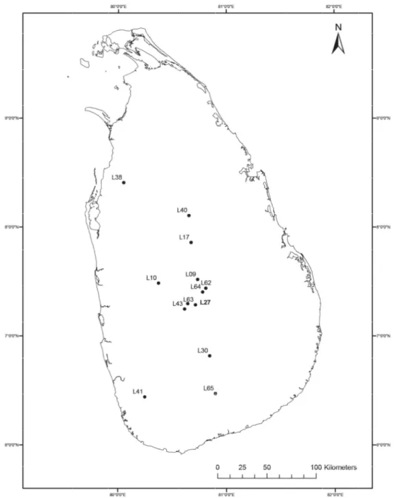
The specimen was then transferred to methyl salicylate and temporarily mounted for examination and illustration (Benjamin 2011). Drawings of male palps and internal structures of epigynes were made with the aid of a drawing tube attached to an Olympus BX51 microscope. Highly sclerotized or darker areas of palps and epigynum were shaded with a HB pencil. A Nikon D80 camera was used to take photographs of live spiders. Photographs of palps, epigynes and intact spiders were taken with a Leica MC170 HD camera mounted on a Leica M205C stereo microscope using Leica Application Suite, LAS version 4.6.2 software (Leica Microsystems Limited, Switzerland). Before photographing, palps and epigynes were embedded in Balea hygiene-hand gel (DM, Germany) to avoid shaking. For a single photo, between 30 and 40 images were taken in a Z-stack and merged to one image with the Helicon Focus stacking software (version 6, Helicon soft Ltd). Images were further edited with Adobe Photoshop CC and arranged in plates with Adobe Illustrator CS6. All measurements were taken with an Olympus SZX7 binocular stereo microscope with a graticule calibrated with a stage micrometer (1 mm / 100 divisions). After examination, the genitalia were transferred to micro-vials and kept in the same collection vial as the original specimen. Taxonomic histories follow the World Spider Catalog (2015). ArcGIS, version 9.3 software (Esri, USA) was used to create the GIS map for sampled localities of Sri Lanka (Fig. 1). All measurements are in millimeters. All specimens unless otherwise stated are deposited in the National Museum of Sri Lanka.
Abbreviations
AG = accessory gland AGH = accessory gland head AL = abdominal length ALE = anterior lateral eyes AME = anterior median eyes AW = abdominal width BL = body length CD = copulatory duct CL = Carapace length CO = copulatory opening CY = cymbium
CYD = cymbial depression
E = embolus
EB = embolic base FD = fertilization duct Fm = femur
L = Location
MI = median indentation Mt = metatarsus
MW = membranous window PLT = proximal lobe of tegulum PLE = posterior lateral eyes PME = posterior median eyes
Pt = patella
PW = prosoma width
RTA = retrolateral tibial apophysis S = spermatheca
SD = sperm duct T = tegulum
Ta = tarsus
Tb = tibia
TL = tegular ledge Tr = trochanter VR = vaginal roof
Results
Class Arachnida Cuvier, 1812 Order Araneae Clerck, 1757 Family Salticidae Blackwall, 1841
Bristowia Reimoser, 1934
Bristowia Reimoser, 1934: 17, fi gs 1–3. Type species: B. heterospinosa Reimoser, 1934.
Diagnosis
Spiders of this genus are easily distinguishable by the elongated coxa, trochanter and patella of the fi rst legs, the peculiar spination of the metatarsus (2 pairs of ventral spines) and tibia I (4 prolateral and 3 retrolateral spines), and the patella and tibia with a ventral fringe of black hairs (Ono et al. 2009;
Prószyński 1984; Reimoser 1934; Seo 1986; Szüts 2004). They have a longer prosoma with a thoracic fovea, and pluridentate chelicerae. The structure of the copulatory organs is relatively simple. The male palp has a single procurved retrolateral tibial apophysis, a thin embolus, and a pear-shaped tegulum.
The epigynum has large, globular, spermathecae with broad insemination ducts, and a circular hollow copulatory opening (Dobroruka 2004; Ikeda 1995).
Distribution
China, Congo, India, Japan, Korea, Krakatau and Sri Lanka (new record).
Bristowia gandhii sp. nov.
urn:lsid:zoobank.org:act:46066597-9E86-483A-BB19-6D08EE6DF7C9 Figs 1, 2A–B, 3–4
B. heterospinosa – Dobroruka, 2004: 14–17, fi gs 8–11, ♀ from India, Goa Province, S of Margao, Palolem env., leg. P. Sipek (specimens in the private collection of author), not examined. Misidentifi cation.
non B. heterospinosa Reimoser, 1934: 17, fi gs 1–3.
Fig. 1. Known distribution of Bristowia gandhii sp. nov. (L65, L38, L17, L62, L63), Habrocestum hantaneensis sp. nov. (L43, L27, L10, L64, L41), H. kodigalaensis sp. nov. (L40, L9), H. ohiyaensis sp. nov. (L30) and Macaroeris nidicolens (Walckenaer, 1802) (L17) in Sri Lanka. See text for details.
Diagnosis
This species can easily be distinguished from B. afra Szüts, 2004 by the high carapace with steeper thoracic slope, a thicker and longer embolus, copulatory openings anterior to the spermathecae, a large distance between copulatory ducts and the presence of a posterior epigynal plate with a median depression, and from B. heterospinosa by broader copulatory ducts, a comparatively shallow median indentation of posterior epigynal plate and a thicker embolus.
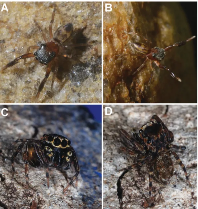
Fig. 2. Live specimens of Bristowia Reimoser, 1934 and Habrocestum Simon, 1876. A–B. B. gandhii sp. nov., ♀ from IFS Arboretum, Dambulla (IFS_SAL 543). C–D. H. kodigalaensis sp. nov., ♂ from Ritigala SNR, Kodigala, Sri Lanka (IFS_SAL 516).
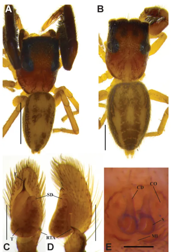
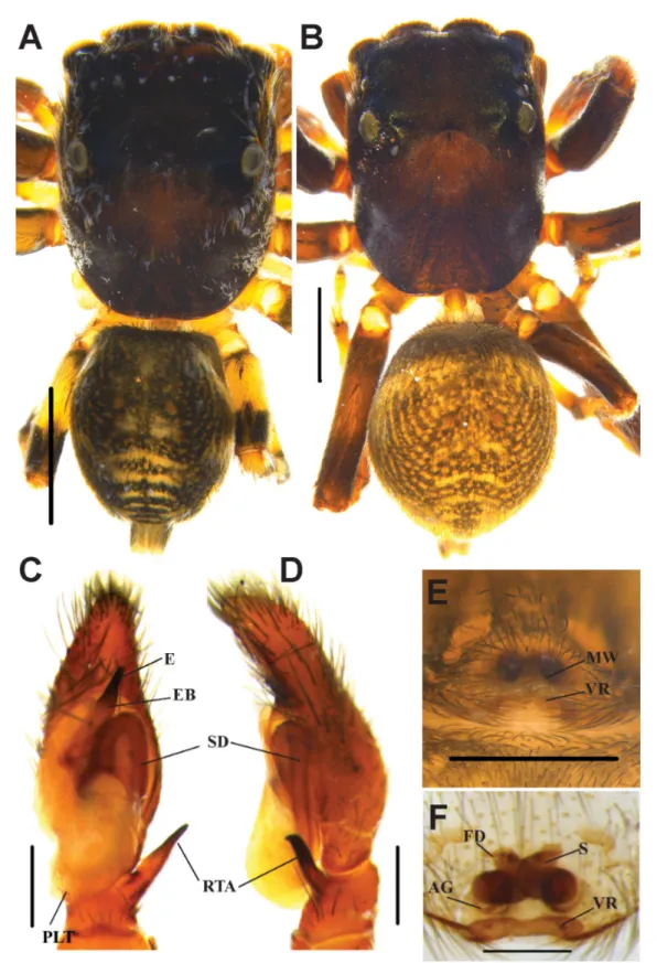
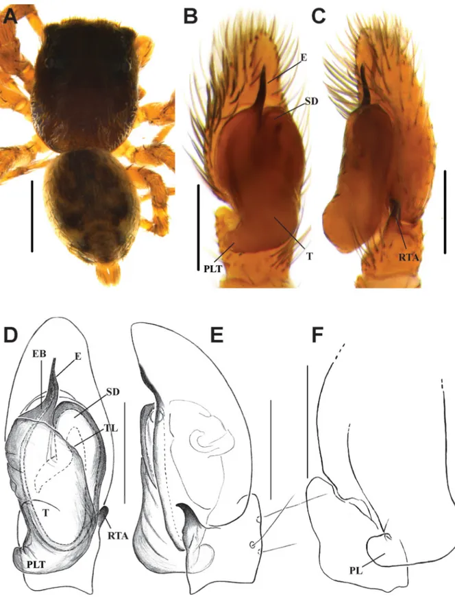
Fig. 3. Bristowia gandhii sp. nov. A, C–D. ♂ (IFS_SAL 228). B, E. ♀ (IFS_SAL 297). A–B. Habitus, dorsal view. C. Palp, ventral view. D. Palp, retrolateral view. E. Epigynum, ventral view. Abbreviations:
CD = copulatory duct; CO = copulatory opening; E = embolus; MI = median indentation; RTA = retrolateral tibial apophysis; S = spermathecae; SD = sperm duct; T = tegulum.Scale bars: A = 1 mm;
B= 0.5 mm; C–E = 0.2 mm.
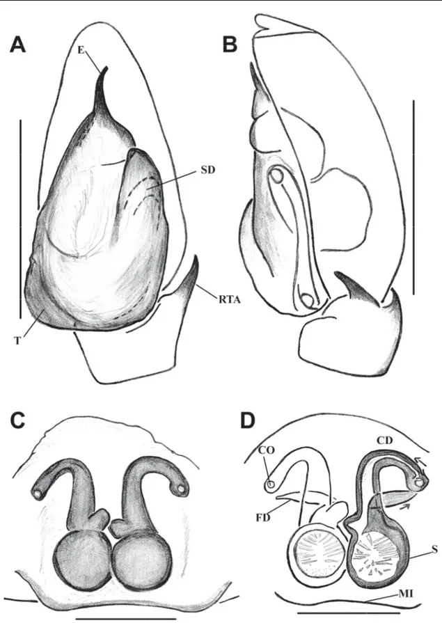
Fig. 4. Bristowia gandhii sp. nov. A–B. ♂ (IFS_SAL 228). C–D. ♀ (IFS_SAL 297). A. Palp, ventral view. B. Palp, retrolateral view. C. Epigynum, ventral view. D. Vulva, ventral view. Abbreviations:
CD = copulatory duct; CO = copulatory opening; E = embolus; FD = fertilization duct; MI = median indentation; RTA = retrolateral tibial apophysis; S = spermathecae; SD = sperm duct; T = tegulum. Scale bars: A–B = 0.2 mm; C–D = 0.1 mm.
Etymology
The species is named for Mohandas Karamchand Gandhi (1869–1948). He was the pre-eminent leader of the Indian Independence Movement in British-ruled India, eventually paving the way for independence of Sri Lanka as well.
Material examined Holotype
SRI LANKA: ♂, Central Province, Kandy District, Balagola, home garden (L63), 07°17′20.08″ N, 80°42′48.85″ E, 476 m, 10 Nov. 2013, leg. S.P. Benjamin (IFS_SAL 228).
Paratype
SRI LANKA: 1 ♀, Central Province, Matale District, IFS Arboretum (Ll7), 07°51′36.70″ N, 80°40′29″ E, 185 m, litter, 2 Nov. 2013, leg. S.P. Benjamin et al. (IFS_SAL 297).
Other material
SRI LANKA: 1 ♀, Central Province, Matale District, IFS Arboretum (Ll7), 07°51′36.70″ N, 80°40′29″ E, 185 m, litter, 19 Oct. 2015, leg. K. Kanesharatnam (IFS_SAL 543); 1 ♀, North Central Province, Anuradhapura District, Wilpattu National Park (L38), 08°24′37.98″ N, 80°03′03.99″ E, hand collection, 6 Apr. 2011, leg. S. Batuwita (IFS_SAL 157); 1 ♂, Sabaragamuwa Province, Udawalawe National Park (L65), 06°28′25.41″ N, 80°53′54.35″ E, 109 m, litter, 30 Dec. 2011, leg. N. Athukorala (IFS_
SAL 154); 3 ♂♂, Uva Province, Badulla District, environs of Kalupahana village (L62), 06°44′58″ N, 80°50′19.8″ E, 820 m, beating, 2 Jan. 2012, leg. S.P. Benjamin (IFS_SAL 357–359).
Description Male
MEASUREMENTS. BL 2.58, CL 1.42, PW at PLEs 0.74, AL 1.12, AW 0.53. Eye fi eld: Diameter of AME 0.24, PLE 0.19, ALE 0.16, PME 0.03, PME-PME 0.65, PLE-PLE 0.70, ALE-PME 0.30, ALE-PLE 0.68.
Leg I: Tr 0.35, Fm 0.93, Pt 0.44, Tb 0.70, Mt 0.40, Ta 0.22; Leg II: Tr 0.10, Fm 0.44, Pt 0.22, Tb 0.35, Mt 0.26, Ta 0.22; Leg III: Tr 0.10, Fm 0.44, Pt 0.22, Tb 0.31, Mt 0.26, Ta 0.22; Leg IV: Tr 0.13, Fm 0.57, Pt 0.22, Tb 0.44, Mt 0.39, Ta 0.22.
COLORAND BODY. Reddish brown carapace with punctured reticulate microsculpture (Fig. 3A). Chelicerae dark brown with two promarginal and four retromarginal teeth. Labium reddish brown with yellowish brown margin. Elevated ocular area blackish brown, median ocular quadrangle much broader than long and little wider behind than in front. Eye ratio AME > PLE > ALE > PME. Sternum oval-shaped, brownish yellow with sparse brown hairs, edges reddish brown. Prosoma longer than wide with thoracic fovea. Posterior margin of prosoma steep and slightly truncated (Fig. 3A). There are black stripes made up of minute tubercles behind PLEs.
ABDOMEN. Elongated oval-shaped and narrower than prosoma. Dorsum yellowish brown with several transverse black markings and black edges (Fig. 3A). Venter pale yellow, blackish brown near the yellowish black spinnerets.
LEGS. First pair of legs more strongly modifi ed in males than females, with elongated coxa, trochanter and patella. Patella and tibia with fringe of thick, long, black bristles (Fig. 3A). Leg I dark brown except for pale yellow tarsus, other legs yellowish brown. Tibia I with 4 prolateral and 3 retrolateral spines, metatarsus I with 2 pairs of fi ne, long spines. Legs III and IV spineless.
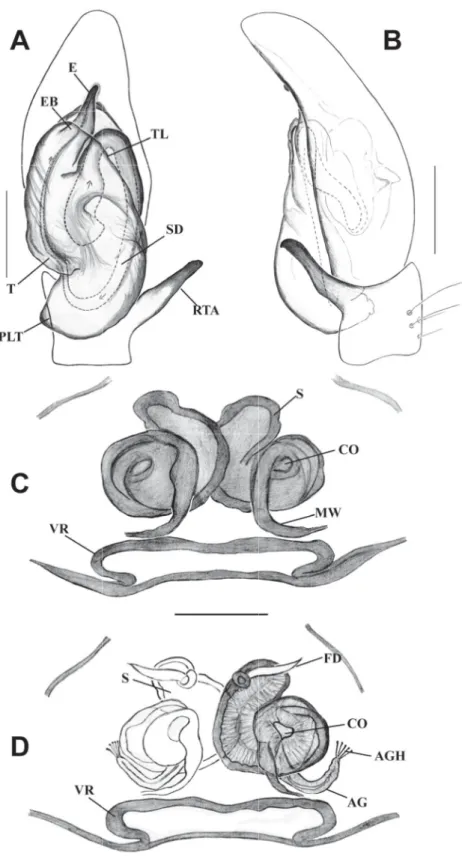
PALP. Relatively simple. Cymbium and palpal tibia pale yellow, but coxa, femur and patella dark brown.
Pyriform tegulum (Figs 3C–D, 4A–B). Thin and short embolus, tip of embolus slightly bent (Figs 3C, 4A), tibial apophyses hook-shaped and strongly bent towards venter.
Female
MEASUREMENTS. BL 3.20, CL 1.78, PW at PLEs 0.77, AL 1.40, AW 0.93. Eye fi eld: Diameter of AME 0.24, PLE 0.14, ALE 0.11, PME 0.03, PME-PME 0.65, PLE-PLE 0.73, ALE-PME 0.30, ALE-PLE 0.46.
Leg I: Tr 0.25, Fm 0.73, Pt 0.25, Tb 0.53, Mt 0.31, Ta 0.22; Leg II: Tr 0.12, Fm 0.46, Pt 0.15, Tb 0.31, Mt 0.25, Ta 0.22; Leg III: Tr 0.12, Fm 0.37, Pt 0.15, Tb 0.18, Mt 0.25, Ta 0.15; Leg IV: Tr 0.12, Fm 0.43, Pt 0.19, Tb 0.50, Mt 0.34, Ta 0.21.
COLORANDBODY. Similar to male except abdomen with four light brown, longitudinal stripes converging near spinnerets; less strongly modifi ed fi rst pair of legs (Figs 2A–B, 3B); leg spination resembles males except in tibia II which has a prolateral and a ventral spine.
EPIGYNUM. Circular hollow copulatory openings above the level of spermathecae (Fig. 4C–D). Long and broad copulatory ducts bent over 180°. Somewhat rounded accessory gland-like structures in front of spermathecae (Fig. 4C–D). Large and globe-like spermathecae. Posterior margin of epigynal plate with a median indentation (Figs 3E, 4C–D). Fertilization ducts lanceolate, originating from anterior wall of receptacles.
Distribution India, Sri Lanka.
Genus Habrocestum Simon, 1876
Habrocestum Simon, 1876: 132, pl. 11, fi g. 8f. Type species Habrocestum pullatum Simon, 1876.
Diagnosis
Small spiders. Prosoma longer than abdomen and broader behind PLEs. Anterior portion fl at, extending over 75–87% of prosomal length, posterior much steeper and slightly truncated (Prószyński 2003).
Short eye fi eld. Third eye row located on the edges of prosoma. Chelicerae with two teeth on promargin and single bicuspid tooth on retromargin. Abdomen rounded or oval in shape, narrower than prosoma, with a pair of large white spots and chevrons posteriorly (Prószyński 2003), fused medially in most species, dorsal view of cymbium with large, round spot of white scales (Fig. 2C). Third and fourth legs longer (tibia plus patella III ≥ tibia plus patella IV), femora III longer than others (Richman 1981).
Palp with single tibial apophysis which is hook shaped in most species. Bulbus elongated. Shape of embolus species specifi c, variable in length and originating under the tegulum. Conductor present or absent (Wunderlich 2008). Epigynum with small pocket-like sclerotized vaginal roof and shallow notch at posterior edge near epigastric furrow. Copulatory openings large, located laterally along the mid- length of epigyne. Large, transverse and membranous white “window”, with translucent dark internal structures, very complicated consisting of large channels running anteriorly and then reversing back, passing into spermathecae with thick walls connected by soft channels (Prószyński 2003). Some species with distinct accessory glands. Fertilization ducts narrow, originating from top of receptacles.
Distribution
Algeria, Congo, East Africa, Eastern Mediterranean to Near East, Ethiopia, France, Greece (incl. Crete), Hong Kong, Israel, Morocco, Namibia, Nigeria, Saudi Arabia, Socotra, Solomon Island, South Africa, Libya, Pakistan, Spain, Tanzania, Turkey, Western Australia, Yemen, Zimbabwe and Sri Lanka (new record).
Key to the species of Habrocestum in Sri Lanka
1. Cone-shaped, short and thick embolus, long tibial apophyses with minute teeth at tip, wide vaginal roof with shallow notch, large copulatory openings, curved accessory glands with pin like structures at the tip ………H. hantaneensis sp. nov.
– Comparably long embolus, short hook-shaped tibial apophyses ………2 2. Straight, thinner embolus tapers toward tip ………H. ohiyaensis sp. nov.
– Thick embolus with hook-like tip and broader at midlength, small vaginal roof with deep notch, large parentheses like copulatory openings, broader accessory glands ……H. kodigalaensis sp. nov.
Habrocestum hantaneensis sp. nov.
urn:lsid:zoobank.org:act:2804E80C-D3AC-4C5B-8620-E7D9EBAF5B6A Figs 1, 5–6
Diagnosis
This species can be distinguished from its congeners by the cone-shaped and comparatively short embolus (Figs 5C, 6A), long tibial apophyses with serrated tip, elongated tegulum with a large proximal lobe (Fig. 6A–B), epigyne with a wide vaginal roof, and characteristic shape of receptacles and accessory glands (Fig. 6C–D). This species is close to the female of H. laurae Peckham & Peckham, 1903, but differs in the shape of the spermathecae and presence of accessory glands.
Etymology
The specifi c epithet refers to the type locality.
Material examined Holotype
SRI LANKA: ♂, Central Province, Kandy District, Hantane (L43), 07°14′57″ N, 80°36′50″ E, 585 m, litter, 19 Nov. 2013, leg. M.K. Rathnayake & I. Sandunika (IFS_SAL 350).
Paratype
SRI LANKA: 1 ♀, same data as holotype (IFS_SAL 456).
Other material
SRI LANKA: 1 ♂, Central Province, Kandy District, Udawattakelle (L27), 07°17′54.71″ N, 80°38′37.42″ E, 580 m, litter, 21 Aug. 2012, leg. S.P. Benjamin (IFS_SAL 266); 1 ♂, same locality and collection data, beating, 8 Jun. 2015, leg. U.G.S.L. Ranasinghe & N. Kanesharatnam (IFS_SAL 484); 1 ♀, Gomaraya (L64), Road B205, nr. 26 km post, ca 07°28′17″ N, 80°22′30″ E, 600 m, hand collection, 2 Feb. 2010, leg. S. Batuwita, P.M.H. Sandamali et al. (IFS_SAL 514); 1 ♀, North Western Province, Kurunagala District, Ethagala (L10), 07°29′11.23″ N, 80°22′21.64″ E, 190 m, litter, 8 Apr.
2015, leg. S.P. Benjamin et al. (IFS_SAL 174); 1 ♀, Western Province, Kalutara District, Agalawatta- Kalutara Forest Reserve (L41), 06°26′35″ N, 80°14′52″ E, 40 m, 29 Jul. 2011, leg. S. Batuwita & P.M.H.
Sandamali et al. (IFS_SAL 298).
Description Male
MEASUREMENTS. BL 3.60, CL 2.04, PW 1.60, AL 1.04, AW 1.20. Diameter of AME 0.46, PLE 0.19, ALE 0.34, PME 0.06, PME-PME 1.33, PLE-PLE 1.12, ALE-PME 0.37, ALE-PLE 0.68. Leg I: Tr 0.24, Fm 1.10, Pt 0.40, Tb 0.80, Mt 0.48, Ta 0.36; Leg II: Tr 0.20, Fm 0.84, Pt 0.20, Tb 0.60, Mt 0.52, Ta 0.36;
Leg III: Tr 0.24, Fm1.28, Pt 0.28, Tb 0.80, Mt 0.72, Ta 0.40; Leg IV: Tr 0.24, Fm 1.04, Pt 0.24, Tb 0.72, Mt 0.68, Ta 0.40.
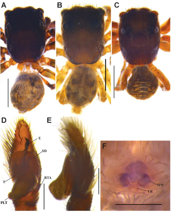
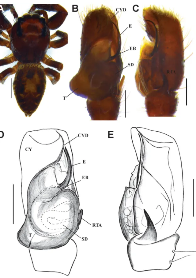
Fig. 5. Habrocestum hantaneensis sp. nov. A, C–D. ♂ (IFS_SAL 350). B, E–F. ♀ (IFS_SAL 456).
A.–B. Habitus, dorsal view. C. Palp, ventral view. D. Palp, retrolateral view. E. Epigynum, ventral view.
F. Vulva, ventral view. Abbreviations: AG = accessory gland; E = embolus; EB = embolic base; FD = fertilization duct; MW = membranous window; PLT = proximal lobe of tegulum; RTA = retrolateral tibial apophysis; S = spermathecae; SD = sperm duct; VR = vaginal roof. Scale bars: A–B = 1 mm;
C–F = 0.2 mm.
Fig. 6. Habrocestum hantaneensis sp. nov. A–B. ♂ (IFS_SAL 350). C–D. ♀ (IFS_SAL 456). A. Palp, ventral view. B. Palp, retrolateral view. C. Epigyne, ventral view. D. Vulva, dorsal view. Abbreviations:
AG = accessory gland; AGH =accessory gland’s head; CO = copulatory opening; E = embolus; EB = embolic base; FD = fertilization duct; MW = membranous window; PLT = proximal lobe of tegulum;
RTA = retrolateral tibial apophysis; S = spermathecae; SD = sperm duct; T = tegulum; TL = tegular ledge; VR = vaginal roof. Scale bars: A–B = 0.2 mm; C–D = 0.1 mm.
COLOR AND BODY. Carapace blackish brown. Chelicerae dark brown with two promarginal teeth and single bicuspid retromarginal tooth. Labium yellowish brown with black margin. Sternum oval with black colored spots in middle, edges reddish brown. Vicinity of eyes blackish brown and slightly elevated. Front eyes covered with long dark brown bristles. First eye row slightly curved upward. Eye fi eld comparatively longer than in congeners, occupying about half the length of prosoma (Fig. 5A).
Median ocular quadrangle much broader than long. White hairs interspersed on lateral sides of prosoma.
Prosoma high, longer than wide. Narrow fovea in midline of prosoma behind PLEs. Lateral and posterior sides of prosoma almost vertical and posterior margin slightly truncated. Abdomen: Rounded, smaller and narrower than prosoma. Dorsum with yellowish brown small dots on anterior portion, comparably large pair of yellowish brown spots at midpoint, transverse pale yellow stripes extending laterally as dashed line at posterior half (Fig. 5A). Venter with longitudinal pale yellow dashed-line pattern from spinnerets to epigastric furrow. Spinnerets dirty yellow.
LEGS. All legs yellow with black lateral bands. Tibia I with 3 spines on prolateral and 3 spines on retrolateral sides.
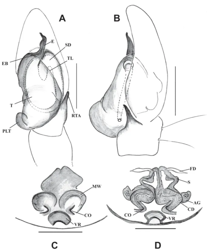
PALP. Yellowish brown palp with black hairs. Palpal tibia short. Retrolateral tibial apophyses with minute tooth-like structures at tip (serrated), strongly curved retrolaterally (Figs 5C–D, 6A–B). Long cymbium narrowing toward tip. Bulbus elongated. Tegulum long with a large and broad proximal lobe extending posteriorly up to half of the palpal tibia. Embolus cone-shaped with slightly curved blunt tip, originating from under tegular ledge (Figs 5C, 6A). Part of the spermophor clearly visible at shoulder of tegulum.
Female
MEASUREMENTS. BL 4.13, CL 2.11, PW 1.58, AL 1.76, AW 1.54. Diameter of AME 0.56, PLE 0.31, ALE 0.37, PME 0.06, PME-PME 1.61, PLE-PLE 1.40, ALE-PME 0.28, ALE-PLE 0.65. Leg I: Tr 0.22, Fm 1.05, Pt 0.35, Tb 0.88, Mt 0.53, Ta 0.39; Leg II: Tr 0.13, Fm 0.97, Pt 0.49, Tb 0.84, Mt 0.44, Ta 0.30;
Leg III: Tr 0.18, Fm 0.97, Pt 0.48, Tb 0.84, Mt 0.48, Ta 0.26; Leg IV: Tr 0.26, Fm 1.58, Pt 0.66, Tb 0.70, Mt 0.92, Ta 0.22.
COLORAND BODY. All characters are as in males except all legs dark brown without the distinguishable bands found in males, sternum light brown in middle with dark brown edge.
EPIGYNUM. Wide sclerotized pocket-like vaginal roof with shallow notch at posterior edge near epigastric furrow. Large copulatory openings like windows situated posterolaterally (Figs 5E–F, 6C–D). Curved accessory glands with pin-like structures at the tip (Fig. 6D). Copulatory ducts short. Peculiarly shaped, heavily sclerotized spermathecae with thick walls. Fertilization ducts narrow arising from anterodorsal side of receptacles (Fig. 6D).
Habrocestum kodigalaensis sp. nov.
urn:lsid:zoobank.org:act:DD31A278-E248-45C4-8871-75DB4F3B9FC9 Figs 1, 7–8
Diagnosis
This species can be distinguished from known congeners by the peculiar shape of embolus (Figs 7D, 8A), short hook-shaped RTA, shape of proximal lobe of tegulum and small and deep vaginal roof, oval shape accessory glands and triangular shape receptacles (Figs 7F, 8C–D). The epigynum of this species is similar to those of H. dubium Wesolowska & van Harten, 2002 and H. ignorabile Wesolowska & van Harten, 2007; it, however, differs by the presence of accessory glands, and the shape of the spermathecae and seminal ducts.
Fig. 7. Habrocestum kodigalaensis sp. nov. A. Habitus, ♂ from Ritigala SNR, Kodigala, dorsal view (IFS_SAL 516). B. Habitus, ♂ from Riverston, Knuckles, dorsal view (IFS_SAL 345). C. Habitus, ♀ from Ritigala SNR, Kodigala, dorsal view (IFS_SAL 467). D. Male palp, ventral view. E. Male palp, retrolateral view. F. Female epigynum, ventral view. Abbreviations: E = embolus;MW = membranous window; SD = sperm duct; T = tegulum; PLT = proximal lobe of tegulum; RTA = retrolateral tibial apophysis; VR = vaginal roof. Scale bars: A–C = 1 mm; D–F = 0.2 mm.
Fig. 8. Habrocestum kodigalaensis sp. nov. A–B. ♂ (IFS_SAL 516 and IFS_SAL 345). C–D. ♀ (IFS_
SAL 467). A. Palp, ventral view. B. Palp, retrolateral view. C. Epigyne, ventral view. D. Vulva, ventral view. Abbreviations: AG = accessory gland; CD = copulatory duct; CO = copulatory opening; E = embolus; EB = embolic base; FD = fertilization duct; MW = membranous window; S = spermathecae;
SD = sperm duct; T = tegulum; TL = tegular ledge; PLT = proximal lobe of tegulum; RTA = retrolateral tibial apophysis; VR = vaginal roof. Scale bars: A–B = 0.2 mm; C–D = 0.1 mm.
Etymology
The specifi c epithet refers to the type locality.
Material examined Holotype
SRI LANKA: ♂, North Central Province, Anuradhapura District, Ritigala Strict Nature Reserve, Kodigala summit (L40), 08°06′33″ N, 80°39′16″ E, 716 m, hand collection, 28 Jun. 2011, leg. S.P.
Benjamin, S. Batuwita et al. (IFS_SAL 516).
Paratype
SRI LANKA: 1 ♀, sama data as holotype (IFS_SAL 467).
Other material
SRI LANKA: 1 ♂, Central Province, Matale District, Riverstone (L9), Knuckles Range, 07°31′17″ N, 80°44′04″ E, 1220 m, hand collection, 2 Feb. 2010, leg. S. Batuwita & P.M.H Sandamali (IFS_SAL 345).
Description Male
MEASUREMENTS. BL 3.80, CL 1.88, PW at PLEs 1.92, AL 1.48, AW 1.20. Diameter of AME 0.43, PLE 0.19, ALE 0.25, PME 0.05, PME-PME 1.18, PLE-PLE 0.96, ALE-PME 0.19, ALE-PLE 0.34.
Leg I: Tr 0.32, Fm 1.04, Pt 0.40, Tb 1.16, Mt 0.60, Ta 0.40; Leg II: Tr 0.16, Fm 1.00, Pt 0.40, Tb 0.60, Mt 0.48, Ta 0.36; Leg III: Tr 0.16, Fm 1.20, Pt 0.32, Tb 0.60, Mt 0.60, Ta 0.28; Leg IV: Tr 0.16, Fm 0.8, Pt 0.24, Tb 0.52, Mt 0.72, Ta 0.28.
COLOR AND BODY. Carapace dark brown. Dark brown bristles near front eyes. Anterior median eyes covered with tuft of pale yellow hairs. Dark ocular region. Chelicerae blackish brown. Sternum oval shaped with dispersed black colored spots. White and brown hairs sparsely dispersed on lateral prosoma.
Distance between posterior eyes slightly shorter than distance between anterior lateral eyes. Carapace somewhat high in the ocular region. Eye fi eld short and wide occupying about one-third length of prosoma. Median ocular quadrangle much broader than long. Round and middle depression around short fovea behind PLEs. Prosoma longer than wide. Lateral and posterior sides of prosoma much steeper.
Posterior margin of prosoma truncated (Fig. 7A–B). Abdomen: Oval shaped, smaller and narrower than prosoma. Dorsum greyish beige color with black stripes and spots. Venter with longitudinal pale yellow dash line from spinnerets to epigastric furrow. Spinnerets blackish yellow. Anterior venter with black blotches.
LEGS. All legs with yellow and black banding pattern. Femora III much longer than femora IV. Front legs slightly darker than other pairs.
PALP. Dark brown palp except for tibia and cymbium. Dorsum of cymbium with large, round spot of pale yellow scales. Palpal tibia with short hook-shaped tibial apophysis (Figs 7E, 8B). Elongated bulbus.
Tegulum with a long lateral proximal lobe, protruding posterolaterally beyond base of cymbium. Thick embolus with hook-like tip and broader at midlength, arising from under tegular ledge (Figs 7D, 8A).
Part of sperm duct clearly visible at shoulder of tegulum.
Female
MEASUREMENTS. BL 3.40, CL 1.92, PW 1.24, AL 1.46, AW 0.88. Diameter of AME 0.34, PLE 0.19, ALE 0.25, PME 0.05, PME-PME 1.02, PLE-PLE 0.93, ALE-PME 0.25, ALE-PLE 0.50. Leg I: Tr 0.20, Fm 0.65, Pt 0.48, Tb 0.60, Mt 0.28, Ta 0.20; Leg II: Tr 0.16, Fm 0.80, Pt 0.36, Tb 0.48, Mt 0.36, Ta 0.24;
Leg III: Tr 0.20, Fm 1.12, Pt 0.44, Tb 0.48, Mt 0.40, Ta 0.40; Leg IV: Tr 0.20, Fm 0.80, Pt 0.20, Tb 0.56, Mt 0.44, Ta 0.28.
COLORAND BODY. Most characters are similar to the male except: posterior margin of prosoma slightly more truncated than in the male, dorsal abdomen with black and yellow stripes distributed from anterior portion to spinnerets (Fig. 7C), venter light grey colour with some transverse light brown markings near spinnerets, anterior venter with pattern similar to dorsum.
EPIGYNUM. Small sclerotized pocket-like vaginal roof with deep notch at posterior edge of epigynum (Figs 7F, 8C–D). Large parentheses-like copulatory openings located posterolaterally. Copulatory ducts with narrower anterior part and broader portion entering into spermathecae. Heavily sclerotized spermathecae with thick walls connected by soft channels. Oval accessory glands, fertilization ducts narrow, originating from top of spermathecae (Fig. 8C–D).
Habrocestum ohiyaensis sp. nov.
urn:lsid:zoobank.org:act:3441025A-C967-4A9B-9BE2-6D53747AC3E4 Figs 1, 9
Diagnosis
This species can be distinguished from known congeners by its straight, tapering embolus (Fig. 9B–D), medium sized, hook-shaped tibial apophysis (Fig. 9C, E) and the shape of the proximal tegular lobe (Fig. 9F). The male palp of this species is close to that of H.bovei Lucas, 1846, H. ibericum Dalmas, 1920 and H. lepidum Dalmas, 1920, but it differs in the shape proximal lobe of tegulum and tegular ledge.
Etymology
The specifi c epithet refers to the type locality.
Material examined Holotype
SRI LANKA: ♂, Central Province, Badulla District, Ohiya (L30), 06°49′08.32″ N, 80°50′41.35″ E, 1280 m, beating, 26 May 2012, leg. N. Athukorala (IFS_SAL 267).
Description Male
MEASUREMENTS. BL 3.40, CL 1.48, PW 1.76 at PLEs, AL 1.20, 1.12. Diameter of AME 0.31, PLE 0.19, ALE 0.22, PME 0.02, PME-PME 1.15, PLE-PLE 0.90, ALE-PME 0.22, ALE-PLE 0.43. Leg I: Tr 0.40, Fm 0.80, Pt 0.60, Tb 0.92, Mt 0.40, Ta 0.32; Leg II: Tr 0.20, Fm 0.80, Pt 0.32, Tb 0.64, Mt 0.44, Ta 0.40;
Leg III: Tr 0.20, Fm 0.60, Pt 0.36, Tb 0.60, Mt 0.52, Ta 0.32; Leg IV: Tr 0.20, Fm 0.84, Pt 0.24, Tb 0.56, Mt 0.44, Ta 0.28.
COLOR AND BODY. Carapace dark brown. Chelicerae blackish brown. Labium yellowish brown with black edges. Dark brown bristles in vicinity of front eyes. Ocular region blackish brown, slightly elevated. PLEs surrounded by grey hairs. White and grey hairs intermingled on lateral and posterior sides of prosoma. Sternum oval shaped with tiny blackish dots. Prosoma longer than wide. First eye row slightly curved upward. Eye fi eld short and wide occupying about one–third length of prosoma. Median ocular quadrangle much broader than long. Posterior margin truncated. Abdomen. Oval shaped, smaller and narrower than prosoma. Anterolateral dorsum with black blotches, posterior dorsum with pale white chevrons just above spinnerets (Fig. 9A). Venter pale yellow medially, blackish yellow laterally.
Spinnerets yellow.
Fig. 9. Habrocestum ohiyaensis sp. nov. A. ♂, habitus, dorsal view. B. Palp, ventral view. C. Palp, retrolateral view. D. Palp, ventral view. E. Palp, retrolateral view. F. Palp, prolateral view. Abbreviations:
E = embolus; EB = embolic base; SD = sperm duct; T = tegulum; TL = tegular ledge; PLT = proximal lobe of tegulum; RTA = retrolateral tibial apophysis. Scale bars: A = 1 mm; B–F = 0.2 mm.
LEGS. All legs yellowish brown without any banding pattern.
PALP. Yellowish brown palp. Cymbium covered with dark brown hairs. Comparatively thin and straight embolus that tapers toward tip, and base of embolus emerging from under tegular ledge (Fig. 9B–D).
Elongated bulbus. Tegulum with a proximal lobe, protruding posterolaterally beyond base of cymbium.
Part of sperm duct clearly visible at distal end of tegulum. Short palpal tibia with medium sized and hook-shape tibial apophysis.
Female Unknown.
Genus Macaroeris Wunderlich, 1992
Macaroeris Wunderlich, 1992: 611–613, fi gs 829–852. Type species Macaroeris nidicolens (Walckenaer, 1802).
Diagnosis
Morphologically, this genus is similar to Dendryphantes Koch, 1837, except for the structure of the genitalia (Prószyński 2003). Long prosoma broader at mid-length, fl at anterior and gently sloped posterior prosoma. Eye fi eld short and trapezoid shaped. Second row of eyes very small, rather closer to ALE than PLEs, diameter of AME twice that of ALE. Abdomen long and broad, tapering posteriorly with marginal, medial blotches and chevrons. Enlarged front legs, rather longer than other legs. Embolus long, gently curved to form a very loose coil with a wide base originating on anterolateral bulbus (Prószyński 2003; Wunderlich 2008). Tegulum distended prolaterally. Tip of tibial apophysis bent forward. Copulatory openings located antero-laterally in a depression, but not in a semilunar groove as in Dendryphantes, postero-median copulatory ducts converging diagonally, posteriorly and ending in complicated spermathecal chambers.
Distribution
Canary Islands, Central Asia, China, Cyprus, Madeira, Mediterranean to Azerbaijan, Portugal, Romania, Salvage Islands, Sri Lanka (new record), and Turkey.
Macaroeris nidicolens (Walckenaer, 1802) Figs 1, 10
Diagnosis
This species is easily distinguishable from known congeners by the prosoma being broader at mid- length, its massive front legs, and its broad and posteriorly tapered abdomen with an anterior white band with blotches laterally and medially and chevrons posteriorly. It has a long and gently curved embolus (Fig. 10B–D), its tegulum is distended prolaterally and the tibial apophysis bent forward. (Merrett &
Milner 2004).
Material examined
SRI LANKA: 1 ♂, Central Province, Matale District, IFS Arboretum (L17), 07°51′36.70″ N, 80°40′29″ E, 180 m, litter, 1 Dec. 2011, leg. S.P. Benjamin et al. (IFS_SAL 330).
Description Male
MEASUREMENTS. BL 3.42, CL 1.54, PW 1.25 at PLEs, AL 1.88, AW 1.20. Eye fi eld: Diameter of AME 0.4, PLE 0.16, ALE 0.24, PME 0.04, PME-PME 1.32, PLE-PLE 1.36, ALE-PME 0.16, ALE-PLE 0.64.
Fig. 10. Macaroeris nidicolens (Walckenaer, 1802). A. ♂, habitus, dorsal view. B, D. Palp, ventral view.
C, E. Palp, retrolateral view. Abbreviations: CY = cymbium; CYD = cymbial depression; E = embolus;
EB = embolic base; RTA = retrolateral tibial apophysis SD = sperm duct; T = tegulum. Scale bars: A = 1 mm; B–E = 0.2 mm.
Leg I: Tr 0.40, Fm 1.16, Pt 0.72, Tb 1.12, Mt 0.60, Ta 0.48; Leg II: Tr 0.36, Fm 0.88, Pt 0.48, Tb 0.52, Mt 0.52, Ta 0.44; Leg III: Tr 0.40, Fm 0.88, Pt 0.40, Tb 0.52, Mt 0.52, Ta 0.44; Leg IV: Tr 0.36, Fm 0.96, Pt 0.52, Tb 0.72, Mt 0.52, Ta 0.44.
COLOR ANDBODY. Carapace dark reddish brown with black marking in middle of cephalic area, black colour around eyes and brown hairs surrounding AMEs. Clypeus with a few long white hairs. Chelicerae dark brown. Sparse white hairs on lateral sides of prosoma. Posterior margin of prosoma gently inclined with black colored striped pattern. Sternum dark yellow-brown. Short, trapezoid shaped eye fi eld (Fig. 10A). Cephalic region almost square and slightly elevated. Prosoma oval shaped, broader laterally behind eyes. Abdomen: long and broad in shape, tapering posteriorly. Dorsum with yellowish brown blotches and chevrons. Anterior margin with white colored net-like pattern extending laterally. Posterior margin with yellowish brown chevrons (Fig. 10A). Venter whitish grey with brown broad median longitudinal stripe from spinnerets to epigastric furrow. Spinnerets dark brown.
LEGS. Legs I and II darker brown than the others. Front legs, strikingly massive with enlarged coxa, trochanter, femur, patella and tibia (Fig. 10A). Tibia I with 3 pairs of short stout ventral spines, tibia II with 2 pairs, metatarsi I and II with 2 pairs. Femora I–IV with 2–3 long curved dorsal spines and additionally, femora I–II with 2 short stout spines distomesally.
PALPS. Dark brown, densely clothed with pale hairs at distal end of cymbium. Retrolateral tibial apophysis strong, tip slightly bent forward, base straight (Fig. 10E). Tegulum bulbous, distended prolaterally. Long embolus with a wide basal part, originating anterolaterally on anterior part of bulbus and gently bent to form a curve that runs along depression in cymbium (Fig. 10B–D).
Distribution
Cyprus, Portugal, Sri Lanka (new record), Turkey.
Discussion
Our main objective was to report on the high diversity of jumping spiders of Sri Lanka. While we added four new species and three new generic records for the island, our unpublished data, however, suggest that a huge diversity still remains to be discovered and described. We intend to provide data on the ecology and phylogenetic relationships of this unknown component of the islands’ biodiversity in future contributions.
Acknowledgments
This study was funded by the National Institute of Fundamental Studies with additional funding coming from a fellowship from the Alexander von Humboldt Foundation to SPB. We wish to thank all those who collected and made available many specimens used in this study: S. Batuwita, P.M.H.
Sandamali, N. Athukorala, U.G.S.L. Ranasinghe, M.K. Rathnayake and I. Sandunika; without their efforts this publication would have been impossible. The Department of Wildlife Conservation and the Department of Forest Conservation of Sri Lanka facilitated fi eldwork; their assistance is very gratefully acknowledged. Thanks to Mr. D.G.R.M.M. Kaushalya Rathnayake (Environmental Offi cer, Operations and Monitoring Support Unit, Ministry of Provincial Councils and Local Government) for assistance in the preparation of the map of Sri Lanka. Thanks to Don Buckle and two anonymous reviewers for useful suggestions that greatly improved this manuscript.
References
Benjamin S.P. 2004. Taxonomic revision and phylogenetic hypothesis for the jumping spider subfamily Ballinae (Araneae, Salticidae). Zoological Journal of the Linnean Society 142: 1–82. http://dx.doi.
org/10.1111/j.1096-3642.2004.00123.x
Benjamin S.P. 2006. The male of Marengo nitida with the description of M. rattotensis new species from Sri Lanka (Araneae: Salticidae). Zootaxa 1326: 25–36.
Benjamin S.P. 2010. Revision and cladistic analysis of the jumping spider genus Onomastus (Araneae:
Salticidae). Zoological Journal of the Linnean Society 159: 711–745. http://dx.doi.org/10.1111/j.1096- 3642.2009.00580.x
Benjamin S.P. 2011. Phylogenetics and comparative morphology of crab spiders (Araneae: Dionycha, Thomisidae). Zootaxa 3080: 1–108.
Benjamin S.P. 2015. Model mimics: antlike jumping spiders of the genus Myrmarachne from Sri Lanka.
Journal of Natural History 49 (43–44): 2609–2666. http://dx.doi.org/10.1080/00222933.2015.1034209 Dingerkus G. & Uhler L.D. 1977. Enzyme clearing of alcian blue stained whole small vertebrates for demonstration of cartilage. Stain Technology 52: 229–232. http://dx.doi.
org/10.3109/10520297709116780
Dobroruka L.J. 2004. One new species and one new record of jumping spiders (Araneae: Salticidae) from India. Acta Arachnologica Sinica 13: 14–17.
Foelix R.F. 2011. Biology of Spiders. Oxford University Press, New York.
Ikeda H. 1995. Two poorly known species of salticid spiders from Japan. Acta Arachnologica 44: 159–
166.
Jackson R.R. 1996. Portia spider mistress of deception. National Geographic Magazine 190: 104–115.
Jackson R.R., Pollard S. D., Nelson X.J., Edwards G.B. & Barrion A.T. 2001. Jumping spiders (Araneae: Salticidae) that feed on nectar. Journal of Zoology 255: 25–29. http://dx.doi.org/10.1017/
S095283690100108X
Maddison W.P. 2015. A phylogenetic classifi cation of jumping spiders (Araneae: Salticidae). Journal of Arachnology 43: 231–292.
Merrett P. & Milner E. 2004. Macaroeris nidicolens (Walckenaer, 1802) in Britain (Araneae: Salticidae).
Bulletin of the British Arachnological Society 13: 63–64.
Metzner H. 2015. Worldwide database of jumping spiders (Arachnida, Araneae, Salticidae) [online].
Available from http://www.jumping-spiders.com/index.php [accessed 12 Dec. 2015]
Ono H., Ikeda H. & Kono R. 2009. Salticidae. In: Ono H. (ed.) The Spiders of Japan with Keys to the Families and Genera and Illustrations of the Species: 558–588, Tokai University Press, Kanagawa.
Prószyński J. 1984. Atlas rysunków diagnostycznych mniej znanych Salticidae (Araneae). Wyższa Szkola Rolniczo-Pedagogiczna, Siedlcach 2: 1–177.
Prószyński J. 2003. Salticidae (Araneae) of the Levant. Annales Zoologici 53: 1–180.
Prószyński J. 2006. Salticidae (Araneae) of the World Part I: Diagnostic Drawings Library [online].
Available from http://salticidae.org/salticid/diagnost/salticid.htm [accessed 07 Dec. 2015].
Prószyński J. 2015. Monograph of the Salticidae (Araneae) of the World 1995–2014 Chapter II: Global species database of Salticidae [online]. Available from http://www.peckhamia.com/salticidae/index.
html [accessed 26 Dec. 2015].
Prószyński J. & Deeleman-Reinhold C.L. 2012. Description of some Salticidae (Aranei) from the Malay Archipelago. II. Salticidae of Java and Sumatra, with comments on related species. Arthropoda Selecta 21: 29–60.
Reimoser E. 1934. The spiders of Krakatau. Proceedings of the Zoological Society of London 1: 13–18.
Richman D.B. 1981. A Revision of the Genus Habrocestum (Araneae, Salticidae) in North America.
Bulletin of the American Museum of Natural History 170: 197–206.
Seo B.K. 1986. One unrecorded species of salticid spider from Korea (II). Korean Arachnology 2:
23–26.
Simon E. 1876. Les arachnides de France. Paris 3: 1–364.
Szüts T. 2004. A revision of the genus Bristowia (Araneae: Salticidae). Folia Entomologica Hungarica 65: 25–31.
World Spider Catalog 2015. World Spider Catalog, version 16.5. Natural History Museum Bern [online].
Available from http://wsc.nmbe.ch [accessed 25 Nov. 2015].
Wunderlich J. 1992. Die Spinnen-Fauna der Makaronesischen Inseln: Taxonomie, Ökologie, Biogeographie und Evolution. Beiträge zur Araneologie 1: 1–619.
Wunderlich J. 2008. Identifi cation key to the European genera of the jumping spiders (Araneae:
Salticidae). Beiträge zur Araneologie 5: 698–719.
Żabka M. 1985. Systematic and zoogeographic study on the family Salticidae (Araneae) from Viet- Nam. Annales Zoologici 39: 197–485.
Manuscript received: 16 December 2015 Manuscript accepted: 12 April 2016 Published on: 9 September 2016 Topic editor: Rudy Jocqué
Desk editor: Kristiaan Hoedemakers
Printed versions of all papers are also deposited in the libraries of the institutes that are members of the EJT consortium: Muséum national d’Histoire naturelle, Paris, France; Botanic Garden Meise, Belgium;
Royal Museum for Central Africa, Tervuren, Belgium; Natural History Museum, London, United Kingdom; Royal Belgian Institute of Natural Sciences, Brussels, Belgium; Natural History Museum of Denmark, Copenhagen, Denmark; Naturalis Biodiversity Center, Leiden, the Netherlands.