Invasion of HEp-2 cells by Shigella spp. isolated from acute pediatric diarrhea
Abstract
Aim:Shigellainfection is an important global health problem in devel- oping countries where hygiene is poor and hence shigellosis is a main
Sajjad Omidi
1Mohammad Mehdi Soltan Dallal
2,3cause of diarrhoea-associated mortality and morbidity, particularly in children under the age of five. The bacterial entry into colon and rectal
Abolfazle Davoodabadi
4epithelial cells has been named ‘bacterium-directed phagocytosis’. This
term highlights that the bacteria actively stimulate their own uptake
Ramin Mazaheri Nezhad Fard
2into non-professional phagocytes. The aim of this study was to demon- strate the invasion of HEp-2 cells byShigellaspp. isolated from acute
pediatric diarrhea in Tehran, Iran.
Marayam Usefi
5Methods:Three-hundred and ten non-duplicative diarrheal stool samples
were collected from the children admitted to Children’s Medical Center
Ronak Bakhtiari
1in Tehran, Iran. Samples were cultured and suspected colonies were
1 Division of Microbiology, Department of Pathobiology, identified by routine microbiological and biochemical tests. The invasion
of the two isolatedShigellaspp. to HEp-2 cells was studied.
School of Public Health, Results:Of 310 stool samples, 16 (5.2%)Shigellaspp. were isolated,
including seven (43.7%)S. sonneiand nine (56.3%)S. flexneri. Four Tehran University of Medical Sciences, Tehran, Iran (44.4%) S. sonnei and seven (42.8%) S. flexneri showed invasive
phenotype to HEp-2. 2 Division of Food Microbiology,
Department of Pathobiology, Conclusion:Shigella sonneiandS. flexneriare reported as the most
prevalent Shigella spp. in nature which infect humans. Invasion of School of Public Health, Tehran University of Medical Sciences, Tehran, Iran various cell lines gives the chance of survival toShigellaspp. This ability
causes more virulent infections in the host. Despite costly and time
3 Food Microbiology Research Center, Tehran University of consuming cell culture techniques, the current method described in
this paper is reliable for detecting invasive behavior ofShigella spp.
Medical Sciences, Tehran, Results have also shown that not all theShigellaspp. are able to invade Iran
intestinal epithelial cells.
4 Department of Microbiology, Medical School, Babol Keywords:cell invasion, cell culture, diarrhea, Shigella spp., HEp-2
University of Medical Science, Babol, Iran 5 Department of Virology,
School of Public Health, Tehran University of Medical Sciences, Tehran, Iran
Introduction
Shigella infection or shigellosis is a significant global health problem in developing countries where hygiene is poor. Shigellosis is a main cause of diarrhoea-associated mortality and morbidity, particularly in under five-year-old children [1]. Shigella spp. are categorized in four serogroups, including S. dysenteriae (serogroup A), S. flexneri (serogroup B), S. boydii (serogroup C) and S. sonnei(serogroup D). The bacteria are responsible for causing gastroenteritis in the host that may progress to mucoid bloody diarrhea, known as bacillary dysentery [2].
Shigella flexneriandS. sonneihave been described as the most common causes of shigellosis in tropical areas.
Furthermore,S. sonneiis mainly isolated in developed countries. Recently, it has been estimated that 91 million cases of Shigella infections occur every year. In Asia, 410,000 children, commonly undernourished, die every year due toShigellainfections.Shigellaspp. are transmit- ted through the fecal-oral route and enter the human body via the ingestion of contaminated food and water [3].Shigellaspp. cause bacillary dysentery in humans by invading epithelial cells of the colon. The bacterial entry into colon and rectal epithelial cells has been named
‘bacterium-directed phagocytosis’. This term describes that the bacteria actively stimulate non-professional phagocytes to engulf them. Bacterial invasion proteins IpaB, IpaC and IpaD are necessary for the process [4].
Shigellaspp. have a large (100–140 MDa) plasmid, which is critical for their virulence and at least three chromo- somal loci are required for the bacterial pathogenesis.
The intracellular entry ofShigellaspp. into the host cells is not passive and needs the expenditure of energy by the bacteria and their host cells. Following entry,Shigella quickly lyses phagosomal membrane and replicates in the host cell cytoplasm [5]. Bacteria move efficiently in infected cell cytoplasm by polymerization of actin at bacterial pole, which also allows formation of protrusions in cell membrane leading to invasion of adjacent cells [6]. The aim of the current study was to demonstrate in- vasion of HEp-2 cells byShigellaspp. isolated from acute pediatric diarrhea in Tehran, Iran.
Methods
Clinical samples and bacterial isolation
Three-hundred and ten non-duplicative diarrheal stool samples were collected from January to December 2015 from 0- to 12-year-old children (165 males and 145 fe- males) admitted to Children’s Medical Center in Tehran, Iran. Suspected colonies were identified by routine micro- biological and biochemical tests, including API-20E system kit (BioMerieux, France) and Shigella polyvalent aggluti- nating antisera (MAST, UK).
Cell culture adherence and penetration
HEp-2 cells were chosen because of their extensive use and accessibility.Shigellaisolates were cultured in brain heart infusion (BHI) agar. HEp-2 cells were preserved in Dulbecco modified Eagle medium (DMEM) with 10% fetal calf serum (FCS). Confluent monolayers of 5.0×10⁴–105 HEp-2 cells per ml were grown for 18 h in 6-well tissue plates at 37°C in humidified incubator containing 5%
CO2. To infect HEp-2 cells, bacterial isolates were suspen- ded in DMEM with 10% FCS without antibiotics to give a final concentration of approximately 5×10⁵cells per ml.
One milliliter of this suspension was added to the mono- layers. The infected monolayers were incubated for 2 h and then washed three times with 2 ml of phosphate buffered saline (PBS). For intracellular growth step, fresh DMEM containing 100 µg/ml of gentamicin was added to the monolayers and incubated for further 3 h and then washed three times with PBS. Cell monolayers were washed in PBS, fixed in a mixture of 3:1 methanol/acetic acid for 10 min and stained with Giemsa, then examined under an invert microscope. Invasion index (penetration to the cells) was recorded as 10–30 (1+), 30–70 (2+) and 70–100 (3+).S. flexneriATCC 12022 andS. sonneiATCC 9290 were used as positive andEscherichia coliK1 as negative control.
Results
Clinical samples and bacterial isolation
Of 310 stool samples from children with diarrhea, 16 (5.2%) Shigella spp. were isolated. Ten isolates were identified in male and six in female children. Slide agglu- tination test using monovalent antisera showed that seven (43.7%)S. sonneiand nine (56.3%)S. flexneriwere identified out of the 16 positive samples. The mean age of the patients was six years with 165 (53.2%) male and 145 (46.7%) female participants. Nine (56.2%) strains were isolated from children in ages ranged from one month to two years and seven (43.7%) from those in ages from two to 12 years.
Cell culture adherence and penetration
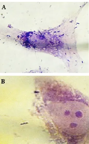
Four (44.4%) isolates of S. sonnei and seven (42.8%) isolates of S. flexneri showed invasive phenotype to HEp-2. Ability of Shigella isolates to invade HEp-2 cell monolayers was first assessed using microscopic exam- ination of the monolayer and detection of Giemsa-stained intracellular bacteria. Invasion index (penetration in the cells) was recorded as 10–30 (1+), 30–70 (2+) and 70–100 (3+). After 2 h of incubation,Shigella isolates could invade cells, but replication in and destruction of the cells occurred after 3 h of incubation (Figure 1 and Figure 2).
Figure 1: Gentamicin-HEp-2 cell invasion assay. Intracellular localization ofS. sonnei (A) and S. flexneri (B) in HEp-2 cell
monolayers after 2 hours
Figure 2: Gentamicin-HEp-2 cell invasion assay. Intracellular localization ofS. sonnei (A) and S. flexneri (B) in HEp-2 cell
Discussion
Approximately 91 million people are infected byShigella spp. worldwide each year [7]. It is well established that S. flexneriis the predominant bacterial isolate in develop- ing countries; in contrast,S. sonneiis the most common bacterial isolate in developed countries [8]. There has been dramatic change in predominant bacterial strain in some Asian and African countries such as Bahrain, Iran, Thailand and Vietnam, in whichS. flexneripredominance has shifted to S. sonneipredominance [9], [10], [11], [12], [13]. In the current study, 16 (5.2%)Shigellaspp.
were isolated from 310 non-duplicative stool samples, from which seven (43.7%) isolates included S. sonnei and nine (56.3%) isolates included S. flexneri. These results differ from the results by Eftekhari et al. [14]. In a study by Eftekhari et al., a total number of 32 (4.5%) Shigellaspp. were recognized in 700 stool samples from patients with diarrhea in two provinces in Iran.S. sonnei (70.8%) andS. flexneri(62.5%) were the most prevalent species in Tehran and Khorasan Razavi Provinces, respec- tively [14]. However, the difference between the results of the two studies might be seen due to the sample size.
However, the prevalence rates of Shigellaspp. in both studies are relatively similar (5.2% compared to 4.5%).
Invasion of gastrointestinal epithelia is one of the major virulence mechanisms, by which Gram-negative bacteria cause diarrheal diseases [15]. A majority of enteric pathogens, includingShigellaandSalmonellaspp., have been shown to possess such mechanisms. These mech- anisms can be reproducedin vitroby indicating the ability of virulent strains to invade mammalian cell lines such as HEp-2 or HeLa [16]. Although CHO cell line has been used to study the bacterial invasion, this cell line is sensitive to the effects of bacterial toxins. The susceptib- ility of CHO cells to toxins has made these cells inappro- priate for the investigation of elongated bacterial infec- tions [17]. Another cell line, Madin-Darby canine kidney (MDCK), seems more appropriate for the study, but is less susceptible to bacterial invasion [18]. A comparative study has shown that Henle 407 cell line is highly efficient in adherence and invasion ofS. flexneri. Although HeLa cells are used to investigate invasion of Shigella spp., Henle 407 cells have been shown to be more efficient [19]. Research has shown thatShigellaspp. degrade cell cultures in replication phase. Furthermore, attachment and invasion ofShigellaspp. is inhibited by low concen- tration of IgA [20], [21], [22]. In the current study, Hep-2 cells were used to demonstrate the invasion ofShigella spp. Of 16 Shigella spp., four (44.4%) S. sonnei and seven (42.8%) S. flexneri isolates showed invasive phenotype to HEp-2 after 2 h of incubation. However, replication in and destruction of the cells occurred com- pletely after 3 h. These results are relatively similar to those of a study by Soltan Dallal et al. in 2013 [23]. They isolated 36 (8.6%)Shigellaspp. from 280 rectal swabs (140 dysentery and 140 watery diarrheal samples) and reported that 14 (38.8%) isolates demonstrated invasive
it is essential to primarily grow the bacteria at 37°C for the optimal expression of invasion phenotype ofS. flexneri on the HEp-2 cell line. Results from the current study have revealed the invasive ability of Shigella spp. and demonstrated that a 5-h incubation time is sufficient for complete destruction of the cell layers byShigellaspp.
Conclusions
Despite costly and time consuming cell culture tech- niques, the current described technique is a reliable technique for detecting invasive behavior of theShigella spp. This study actually provides a good model for further studies on the invasive properties of these bacteria. In conclusion, although cell invasion is described as a common feature of the highlightedShigella species as detected in a majority of the isolates within the current study, this feature was not detected in a minor portion of the total bacterial samples.
Notes
Acknowledgments
This work was supported by a Vice-Chancellor for Re- search grant (No. 23125), Tehran University of Medical Science, Tehran, Iran. We thank Children’s Medical Center in Tehran for providing bacterial isolates and related data for use in this study.
Competing interests
The authors declare that they have no competing in- terests.
References
1. Mulatu G, Beyene G, Zeynudin A. Prevalence of Shigella, Salmonella and Campylobacter species and their susceptibility patters among under five children with diarrhea in Hawassa town, south Ethiopia. Ethiop J Health Sci. 2014 Apr;24(2):101- 8.
2. Bassa A, Dadie A, Guessennd Ne, Gbonon V, Dako E, Dje M , Dosso M. Virulence Factors and Resistance Profile of Shigella Isolated During Infectious Diarrhea in Abidjan, Côte D’Ivoire. J Appl Sci Res. 2010;6(6):594–9.
3. Joh RI, Hoekstra RM, Barzilay EJ, Bowen A, Mintz ED, Weiss H, Weitz JS. Dynamics of shigellosis epidemics: estimating individual-level transmission and reporting rates from national epidemiologic data sets. Am J Epidemiol. 2013 Oct;178(8):1319- 26. DOI: 10.1093/aje/kwt122
4. Adam PR, Dickenson NE, Greenwood JC 2nd, Picking WL, Picking WD. Influence of oligomerization state on the structural properties of invasion plasmid antigen B from Shigella flexneri in the presence and absence of phospholipid membranes. Proteins.
2014 Nov;82(11):3013-22. DOI: 10.1002/prot.24662
5. Bongrand C, Sansonetti PJ, Parsot C. Characterization of the promoter, MxiE box and 5’ UTR of genes controlled by the activity of the type III secretion apparatus in Shigella flexneri. PLoS ONE.
2012;7(3):e32862. DOI: 10.1371/journal.pone.0032862 6. Carayol N, Tran Van Nhieu G. The inside story of Shigella invasion
of intestinal epithelial cells. Cold Spring Harb Perspect Med.
2013 Oct;3(10):a016717. DOI: 10.1101/cshperspect.a016717 7. Soltan Dallal MM, Ranjbar R, Pourshafie MR. The study of
antimicrobial resistance among Shigella flexneri strains isolated in Tehran, Iran. J Pediatr Infect Dis. 2011;6(2):125-9. DOI:
10.3233/JPI-2011-0307
8. Kotloff KL, Nataro JP, Blackwelder WC, Nasrin D, Farag TH, Panchalingam S, Wu Y, Sow SO, Sur D, Breiman RF, Faruque AS, Zaidi AK, Saha D, Alonso PL, Tamboura B, Sanogo D,
Onwuchekwa U, Manna B, Ramamurthy T, Kanungo S, Ochieng JB, Omore R, Oundo JO, Hossain A, Das SK, Ahmed S, Qureshi S, Quadri F, Adegbola RA, Antonio M, Hossain MJ, Akinsola A, Mandomando I, Nhampossa T, Acácio S, Biswas K, O’Reilly CE, Mintz ED, Berkeley LY, Muhsen K, Sommerfelt H, Robins-Browne RM, Levine MM. Burden and aetiology of diarrhoeal disease in infants and young children in developing countries (the Global Enteric Multicenter Study, GEMS): a prospective, case-control study. Lancet. 2013 Jul 20;382(9888):209-22. DOI:
10.1016/S0140-6736(13)60844-2
9. Ashkenazi S, Levy I, Kazaronovski V, Samra Z. Growing antimicrobial resistance of Shigella isolates. J Antimicrob Chemother. 2003 Feb;51(2):427-9. DOI: 10.1093/jac/dkg080 10. Vinh H, Nhu NT, Nga TV, Duy PT, Campbell JI, Hoang NV, Boni
MF, My PV, Parry C, Nga TT, Van Minh P, Thuy CT, Diep TS, Phuong le T, Chinh MT, Loan HT, Tham NT, Lanh MN, Mong BL, Anh VT, Bay PV, Chau NV, Farrar J, Baker S. A changing picture of shigellosis in southern Vietnam: shifting species dominance, antimicrobial susceptibility and clinical presentation. BMC Infect Dis. 2009 Dec;9:204. DOI: 10.1186/1471-2334-9-204 11. Jamsheer AE, Bindayna KM, Al-Balooshi NA, Botta GA. Trend of
antibiotic resistance in 1316 Shigella strains isolated in Bahrain.
Saudi Med J. 2003 Apr;24(4):424-6.
12. Chompook P, Samosornsuk S, von Seidlein L, Jitsanguansuk S, Sirima N, Sudjai S, Mangjit P, Kim DR, Wheeler JG, Todd J, Lee H, Ali M, Clemens J, Tapchaisri P, Chaicumpa W. Estimating the burden of shigellosis in Thailand: 36-month population-based surveillance study. Bull World Health Organ. 2005
Oct;83(10):739-46. DOI: /S0042-96862005001000010 13. Ranjbar R, Soltan-Dallal MM, Pourshafie MR, Mammina C.
Antibiotic resistance among Shigella serogroups isolated in Tehran, Iran (2002-2004). J Infect Dev Ctries. 2009 Sep;3(8):647-8. DOI: 10.3855/jidc.560
14. Eftekhari N, Bakhshi B, Pourshafie MR, Zarbakhsh B, Rahbar M, Hajia M, Ghazvini K. Genetic diversity of Shigella spp. and their integron content. Foodborne Pathog Dis. 2013 Mar;10(3):237- 42. DOI: 10.1089/fpd.2012.1250
15. Willer Eda M, Lima Rde L, Giugliano LG. In vitro adhesion and invasion inhibition of Shigella dysenteriae, Shigella flexneri and Shigella sonnei clinical strains by human milk proteins. BMC Microbiol. 2004 Apr;4:18. DOI: 10.1186/1471-2180-4-18 16. Rahman M, Monira S, Nahar S, Ansaruzzaman M, Alam K, Alam
M, Albert MJ. TnphoA mutants of Providencia alcalifaciens with altered invasiveness of HEp-2 cells. J Med Microbiol. 2002 Aug;51(8):682-6. DOI: 10.1099/0022-1317-51-8-682 17. Noh SM, Sathyamurthy M, Lee GM. Development of recombinant
Chinese hamster ovary cell lines for therapeutic protein production. Curr Opin Chem Eng. 2013 Nov;2(4):391–7. DOI:
10.1016/j.coche.2013.08.002
18. Furuse M, Furuse K, Sasaki H, Tsukita S. Conversion of zonulae occludentes from tight to leaky strand type by introducing claudin- 2 into Madin-Darby canine kidney I cells. J Cell Biol. 2001 Apr 16;153(2):263-72. DOI: 10.1083/jcb.153.2.263
19. Guhathakurta B, Sasmal D, Ghosh AN, Kumar R, Saha P, Biswas D, Khetawat D, Datta A. Adhesion and invasion of a mutant Shigella flexneri to an eukaryotic cell line in absence of the 220- kb virulence plasmid. FEMS Microbiol Lett. 1999 Dec 15;181(2):267-75. DOI: 10.1111/j.1574-6968.1999.tb08854.x 20. Turbyfill KR, Joseph SW, Oaks EV. Recognition of three epitopic
regions on invasion plasmid antigen C by immune sera of rhesus monkeys infected with Shigella flexneri 2a. Infect Immun. 1995 Oct;63(10):3927-35.
21. Sansonetti PJ, Kopecko DJ, Formal SB. Involvement of a plasmid in the invasive ability of Shigella flexneri. Infect Immun. 1982 Mar;35(3):852-60.
22. Willer Eda M, Lima Rde L, Giugliano LG. In vitro adhesion and invasion inhibition of Shigella dysenteriae, Shigella flexneri and Shigella sonnei clinical strains by human milk proteins. BMC Microbiol. 2004 Apr;4:18. DOI: 10.1186/1471-2180-4-18 23. Soltan Dallal MM, Rahimi-Forushani A, Aminharati F, Ohadian-
Moghadam S, Nikmanesh B, Rastegare-Lari A. Investigation the Shigella serotypes invasive cells isolated from patients with diarrhea in HEp-2 cell culture. J Shahrekord Univ Med Sci.
2013;15(6):100–8.
Corresponding author:
Mohammad Mehdi Soltan Dallal
Food Microbiology Research Center, Department of Pathobiology, School of Public Health, Tehran University of Medical Sciences, P.O. Box 6446-14155, Tehran, Iran, Phone: +98-21-42933082
msoltandallal@gmail.com
Please cite as
Omidi S, Soltan Dallal MM, Davoodabadi A, Mazaheri Nezhad Fard R, Usefi M, Bakhtiari R. Invasion of HEp-2 cells by Shigella spp. isolated from acute pediatric diarrhea. GMS Infect Dis. 2017;5:Doc05.
DOI: 10.3205/id000031, URN: urn:nbn:de:0183-id0000312
This article is freely available from
http://www.egms.de/en/journals/id/2017-5/id000031.shtml Published:2017-09-15
Copyright
©2017 Omidi et al. This is an Open Access article distributed under the terms of the Creative Commons Attribution 4.0 License. See license information at http://creativecommons.org/licenses/by/4.0/.