Regulation of PIN-FORMED-mediated auxin transport and the role of auxin and cytokinin responses
in plant-nematode interactions
DISSERTATION
ZUR ERLANGUNG DES DOKTORGRADES DER NATURWISSENSCHAFTEN (DR. RER. NAT.)
DER FAKULTÄT FÜR BIOLOGIE UND VORKLINISCHE MEDIZIN
UNIVERSITÄT REGENSBURG
Vorgelegt von
Birgit Absmanner
aus Göming
im Juni 2013
Das Promotiongesuch wurde eingereicht am: 21.06.2013
Die Arbeit wurde angeleitet von: Dr. Ulrich Hammes
I
Table of contents
1 General introduction ... 1
1.1 Auxin – a major regulator of plant growth and development ... 1
1.2 Auxin - the mode of action... 2
1.2.1 Metabolism... 2
1.2.2 Polar transport... 2
1.2.3 Perception and transcriptional output... 3
1.2.4 Crosstalk with other hormones ... 4
1.3 The role of auxin in vascular development ... 5
1.3.1 Vascular development in the embryo ... 5
1.3.2 Vascular development in the primary root ... 6
1.3.3 Vascular development in organismic interactions ... 8
1.4 Aims of the work ... 9
2 Regulation of PIN-FORMED-mediated auxin transport by the AGCVIII kinases D6 and PINOID ... 11
2.1 Introduction... 11
2.1.1 The PIN protein family of auxin efflux carriers... 11
2.1.2 Polarity dynamics of PIN proteins ... 13
2.1.3 Regulation of polar targeting by phosphorylation ... 14
2.1.4 The AGCVIII kinase family ... 16
2.1.5 The AGCVIII kinase D6PK regulates auxin transport ... 16
2.1.6 Aims of the project ... 17
2.2 Results ... 19
2.2.1 Co-expression of PIN1 and YFP-D6PK leads to enhanced IAA efflux in X. laevis oocytes... 19
2.2.2 Efflux is sensitive to the auxin transport inhibitor NPA ... 22
2.2.3 Determination of transport rates ... 24
2.2.4 A kinase-inactive version of D6PK does not activate PIN1... 26
2.2.5 PINOID also enhances PIN1 mediated IAA export from oocytes, while other tested AGCVIII kinases have no effect ... 27
2.2.6 D215 and S271 are important for PIN1 activation ... 31
II
2.2.7 Described PID target sites contribute to PIN1 activity ... 34
2.2.8 PIN3 is also activated by YFP-D6PK and PINOID... 37
2.3 Discussion ... 41
2.3.1 The AGCVIII kinases D6PK and PID activate PIN1 and PIN3 by phosphorylation ... 41
2.3.2 NPA interferes with auxin efflux mediated by D6PK - activated PIN1 ... 42
2.3.3 Phosphorylation of PIN1 and PIN3 by D6PK and PID has different consequences in plants ... 43
2.3.4 D6PK and PID activate PIN1 by preferential phosphorylation of different serine residues ... 45
3 Phytohormone responses during vascular development in nematode induced feeding sites... 47
3.1 Introduction... 47
3.1.1 Plant parasitic nematodes ... 47
3.1.2 Life cycles of cyst and root knot nematodes ... 48
3.1.3 Sedentary nematodes induce the formation of specialized feeding sites ... 49
3.1.4 The role of phytohormones in early events of feeding site establishment and development ... 50
3.1.5 Nutrient supply and vascularization of feeding sites ... 52
3.1.6 Aims of the project ... 54
3.2 Results ... 55
3.2.1 Identification of auxin responsive cells in nematode infected roots ... 55
3.2.1.1 Auxin response in uninfected roots ... 55
3.2.1.2 Auxin response in M. incognita infected roots ... 57
3.2.1.3 Auxin response in mutants with defects in auxin transport and signaling was not affected in root knots... 63
3.2.1.4 Auxin response in H. schachtii infected roots ... 65
3.2.2 Identification of cytokinin responsive cells in nematode infected roots ... 67
3.2.2.1 Cytokinin response in uninfected roots ... 67
3.2.2.2 Cytokinin response in M. incognita infected roots ... 67
3.2.2.3 Cytokinin response in H. schachtii infected roots ... 68
3.2.3 Expression of a phloem identity marker in root knots ... 70
3.3 Discussion ... 72
III
3.3.1 Hormone response in the differentiated root of A. thaliana... 72
3.3.2 Differentiated giant cells and syncytia do not respond to auxin and cytokinin ... 74
3.3.3 Auxin response and vascularization in M. incognita induced root knots are tightly linked ... 75
3.3.4 Sieve elements and companion cells in phloem induced by H. schachtii respond differentially to auxin and cytokinin... 77
3.3.5 Giant cells and syncytia are surrounded by different types of phloem ... 79
4 Comprehensive discussion and outlook ... 80
5 Summary... 84
6 Zusammenfassung... 88
7 Material and Methods... 92
7.1 Molecular biological work ... 92
7.2 Confocal microscopy ... 92
7.3 Work with plants ... 92
7.3.1 Plant material ... 92
7.3.2 Growth conditions ... 92
7.3.3 Crossing of transgenic lines ... 93
7.4 Work with M. incognita ... 94
7.4.1 Cultivation of M. incognita on tomato ... 94
7.4.2 Isolation of second-stage juveniles of M. incognita... 94
7.4.3 Surface sterilization of second-stage juveniles ... 95
7.4.4 Infection of A. thaliana with M. incognita second-stage juveniles... 95
7.4.5 Infection of A. thaliana with M. incognita by egg mass transfer ... 96
7.5 Work with H. schachtii ... 96
7.5.1 Isolation of second-stage juveniles of H. schachtii ... 96
7.5.2 Infection of A. thaliana with H. schachtii second-stage juveniles ... 97
7.6 Work with nematode infected plant material ... 97
7.6.1 GUS staining... 97
7.6.2 Sectioning of GUS-stained tissue... 97
7.6.3 Immunohistochemistry ... 98
7.6.3.1 Fixation and embedding of plant tissue in methacrylate... 98
7.6.3.2 Sectioning of embedded tissue ... 99
IV
7.6.3.3 Immunolocalization ... 99
7.7 Work with X. laevis oocytes ... 100
7.7.1 Oocyte material ... 100
7.7.2 Synthesis of mRNA for injection ... 101
7.7.3 mRNA injection ... 101
7.7.4 Efflux assay with [3H-IAA] ... 102
7.7.5 Membrane preparation from oocytes ... 103
7.7.6 Western Blot analysis ... 104
7.7.6.1 SDS-PAGE ... 104
7.7.6.2 Wet Blot ... 104
8 Bibliography ... 106
1
1 General introduction
1.1 Auxin – a major regulator of plant growth and development
In order to compensate for their sessile lifestyle, plants exhibit enormous developmental plasticity which allows them to adapt to changing environmental conditions and nutrient supply. Phytohormones act as mobile signals between cells, tissues and organs and play a crucial role in the regulation and coordination of plant growth and development. They include the five “classical” phytohormones auxin, cytokinin, giberellic acid, abscisic acid and ethylene as well as compounds that have been identified more recently like brassinosteroids, jasmonic acid, salicylic acid and strigolactones. Processes that are influenced by phytohormone action include the control of plant size and architecture, tropisms, responses to biotic and abiotic stresses, organismic interactions as well as flower and embryo development (Davies, 2010).
The most investigated phytohormone and also the first one to be described is auxin. Based on classical experiments studying the phototropism of canary grass coleoptiles, Charles Darwin postulated the existence of a transmissible signal (Darwin and Darwin, 1880) which was later chemically identified as indole-3-acetic acid (IAA) and termed auxin (Kögl et al., 1934; Went and Thimann, 1937). Nowadays, the term auxin does not only describe IAA but also several other chemical compounds - naturally occurring and synthetic ones - that exhibit auxin activity. However, IAA is by far the most prominent endogenous auxin in plants (Sauer et al., 2013).
Since its discovery, extensive research revealed that auxin is involved in the regulation of an enormous variety of biological processes. On a cellular level, auxin influences cell division, elongation and differentiation. Additionally, it plays a role in a seemingly never-ending list of developmental and growth processes including embryogenesis, organogenesis, flower development, root meristem maintenance, vascular tissue differentiation, apical dominance as well as various growth responses to environmental stimuli like light, gravity and pathogen attack (Vanneste and Friml, 2009; Sauer et al., 2013).
How is it possible that the rather simple indolic molecule auxin is such a crucial determinant for the plant’s structure and functioning? The unique property of auxin that distinguishes it from other plant hormones is that its action is not only mediated through signaling and transcriptional changes but also through the formation of local maxima and gradients within
2
tissues. This differential distribution is interpreted on the level of the individual cell via a signaling pathway that can lead to distinct outputs, depending on temporal and spatial context. Therefore, auxin is probably better described as a morphogen rather than a hormone because very often a concentration gradient within a tissue governs the developmental output (Friml, 2003). An overview of the mode of action of auxin including metabolism, transport and signaling will be given in the following chapters.
1.2 Auxin - the mode of action
1.2.1 Metabolism
Cellular auxin levels are strongly influenced by the complex interplay between biosynthesis, storage by conjugation and degradation. IAA, which is structurally related to tryptophan, is synthesized mainly in rapidly dividing and growing tissues of the shoot, but also in the meristematic root tip by several redundant biosynthetic pathways (Ljung et al., 2001; Ljung et al., 2005). So far, one tryptophan-independent as well as four tryptophan-dependent synthesis pathways have been described (Woodward and Bartel, 2005; Mano and Nemoto, 2012). Characterization of mutants with defects in key enzymes of the different biosynthetic pathways and analyses of their distinct expression patterns suggest that local biosynthesis contributes to differential auxin distribution. Additionally, IAA can be catabolized or conjugated to various amino acid and sugar moieties, which allows temporal inactivation and storage of the hormone. Finally, excess cellular auxin is degraded by several different oxidation pathways (Woodward and Bartel, 2005; Sauer et al., 2013).
1.2.2 Polar transport
Although local differences in auxin metabolism contribute to its asymmetric distribution within the plant body, it has become obvious over the last years that the major determinant for establishing auxin maxima and gradients is cell-to-cell transport. From the places of synthesis, auxin is distributed via two different pathways. Long distance transport throughout the plant occurs in the phloem (Cambridge and Morris, 1996). The local accumulation observed in different developmental contexts on the other hand is controlled by directional intercellular transport of the phytohormone termed polar auxin transport
3
(PAT). PAT is mediated by a system of influx and efflux carriers, whose differential and often polar subcellular localization defines the direction of auxin flow (Tanaka et al., 2006; Vieten et al., 2007).
IAA is a weak acid which is partly protonated at the apoplastic pH of ~ 5.5. In this form, it can enter the plant cell via lipophilic membrane diffusion. Additionally, proton symporters of the AUXIN-RESISTANT1 (AUX1) / LIKE-AUX1 (LAX1) family mediate the uptake of the anionic form of IAA. Auxin influx carrier activity can exceed diffusive influx enormously in certain cell types and is essential for developmental processes like gravitropism or lateral root formation (Marchant et al., 1999; Marchant et al., 2002; Yang et al., 2006; Swarup et al., 2008). At neutral cytosolic pH, IAA is predominantly dissociated and is therefore unable to pass through the plasma membrane. Consequently, export of auxin from cells is fully dependent on efflux carriers. Members of the P-GLYCOPROTEIN/MULTIDRUG-RESISTANCE subfamily of ATP-binding cassette proteins (MDR/PGP/ABCB) play a role both in auxin influx and efflux (Geisler et al., 2005; Santelia et al., 2005; Terasaka et al., 2005). However, ABCB transporters generally exhibit apolar localization and high stability at the plasma membrane (Blakeslee et al., 2007; Titapiwatanakun et al., 2009). Their efflux activity is therefore mainly nondirectional and they might rather fulfill more general functions in auxin transport like controlling the amount of auxin that is available on certain transport routes (Zazimalova et al., 2007; Vanneste and Friml, 2009). The main players in PAT-related auxin efflux are the PIN-FORMED (PIN) proteins which facilitate vectorial auxin transport and, by their distinct expression and dynamic polar localization, provide the molecular basis for the establishment of auxin maxima and gradients during different developmental processes (Tanaka et al., 2006; Zazimalova et al., 2007). A detailed introduction to PIN-mediated auxin efflux will be given in Chapter 2.1.
1.2.3 Perception and transcriptional output
Biosynthesis, metabolism and transport ensure appropriate and distinct auxin levels within plant tissues. In order to trigger a biological response, auxin signals must be perceived and interpreted at the level of the individual cell. Several independent auxin receptors and their corresponding signaling systems have been described in Arabidopsis, adding further flexibility to the great variety of auxin responses. The best-studied receptor is the F-box protein TRANSPORT INHIBITOR RESISTANT 1 (TIR1) which is a subunit of a Skp1-Cullin-F-box
4
(SCF) class E3 ubiquitin ligase complex (Ruegger et al., 1998; Dharmasiri et al., 2005). Upon binding of auxin, this ubiquitin ligase promotes the degradation of AUXIN / INDOLE ACETIC ACID (Aux/IAA) transcriptional repressors via the proteasome pathway by enhancing their ubiquitination. In the absence of auxin, the Aux/IAAs form inhibitory heterodimers with AUXIN RESPONSE FACTOR (ARF) transcription factors. Therefore, auxin-enhanced proteolysis of the Aux/IAA repressors leads to release and thus activation of the ARFs and subsequent early auxin-responsive gene expression (Chapman and Estelle, 2009). These auxin-responsive genes contain specific sequence motifs, so called auxin response elements (AuxRE) in their promoters, which are recognized and bound by the ARFs (Ulmasov et al., 1995). The components of the described pathway belong to large protein families. There are 5 homologs of TIR1, AUXIN SIGNALIN F-BOX-PROTEIN 1 (AFB) to AFB5. Additionally, 29 Aux/IAAs and 23 ARFs provide a huge number of combinatorial possibilities and consequently of potential transcriptional outputs (Santner and Estelle, 2009). It seems likely that defined pairs of ARFs and Aux/IAAs can mediate specific developmental responses by acting on distinct sets of target genes. One such a pair is for example formed by the auxin response factor MONOPTEROS (MP) / ARF5 and its corresponding AUX/IAA protein BODENLOS (BDL) /IAA12 which are involved in the initiation of the root meristem, the specification of provascular cells in the embryo and vascular differentiation in the post- embryonic plant (Berleth and Jürgens, 1993; Przemeck et al., 1996; Hardtke and Berleth, 1998; Hamann et al., 1999; Hamann et al., 2002).
1.2.4 Crosstalk with other hormones
Auxin action is very often dependent on input provided by other hormones. A famous example is the modulation of auxin responses by cytokinin and vice versa. Cytokinins are adenine derivates originally identified in the 1950ies as compounds which promote cell division and act antagonistically to auxin by promoting shoot growth from tobacco suspension cultured cells (Skoog and Miller, 1957). Since then they were shown to be involved in the regulation of numerous aspects of development like root growth, root architecture and vascular development, all of which are also influenced by auxin (To and Kieber, 2008). Several studies provide evidence for the extensive crosstalk and feedback loops between the auxin and cytokinin signaling pathways on the molecular level and further substantiate their role as regulatory opponents (Ioio et al., 2008; Müller and Sheen, 2008;
5 Moubayidin et al., 2010; Bishopp et al., 2011b).
Apart from cytokinins, a number of other phytohormones, among them gibberelic acid, ethylene and brassinosteroids, have been shown to be closely linked to auxin (Woodward and Bartel, 2005), adding further complexity and flexibility to the hormonal control of plant life.
1.3 The role of auxin in vascular development
1.3.1 Vascular development in the embryo
The delicate auxin regulatory network plays a crucial role in many – if not all – aspects of plant development. A well characterized example is the initiation of vascular development during embryogenesis and the development of the root.
In the course of embryogenesis, the basic organization of the plant body is established, resulting in a seedling that consists of shoot apical meristem, cotelydons, hypocotyl, embryonic root and root meristem. The correct arrangement of these organs and the different cell types within them is tightly regulated and strongly dependent on auxin (Weijers and Jürgens, 2005). Vascular precursor cells, also termed procambial cells, are initiated during the globular stage of embryogenesis and subsequently develop as continuous files through the embryo, providing the basis for the vascular network of the mature plant. Specification of xylem and phloem from the vascular initials starts either during embryogenesis or after germination, depending on the plant species (Esau, 1965;
Scheres et al., 1995). The above mentioned auxin responsive transcription factor MP (see 1.2.3) plays a critical role in the initiation of the procambium. Mutations in the genes encoding for MP and also its corresponding repressor BDL result in rootless seedlings with strongly reduced vascular systems and occasionally fused cotyledons (Berleth and Jürgens, 1993; Hamann et al., 1999). These phenotypes are partially caused by a lack of polar auxin transport in the procambial cells of the embryo. Indeed, MP positively regulates expression of the auxin efflux carrier PIN1, which by its polar localization mediates basally directed auxin flow in the preprocambial cells of the proembryo from the globular stage on.
Therefore, a positive feedback loop between auxin, MP and PINs defines the auxin fluxes in the embryo (Friml, 2003, Wenzel). Such a feedback loop was also found to be essential during vein formation in leaves where likewise auxin flow directed by the polar localization
6
of PIN1 precedes procambium differentiation (Scarpella et al., 2006b). The so called auxin- canalization hypothesis that was formulated over 30 years ago (Sachs and Woolhouse, 1981) states that directional auxin flow in a self-reinforcing canalization defines the position of future vein strands. This means that auxin maxima which are established in procambial cells enhance the transport to adjacent cells in a polar manner. These adjacent cells consequently perceive high auxin levels and, turn into procambium cells and repeat the process with the next cell. Like in the embryo, MP plays an important role in the canalization of auxin flow in the leaves. Additionally, it was shown that in leaves the auxin dependent regulatory network is complemented by the HD-ZIP III family transcription factor ARABIDOPSIS THALIANA HOMEOBOX 8 (ATHB8). ATHB8 acts in the specification of the procambial cell fate and its expression is directly activated by MP via binding of an AuxRe in the promoter. The described auxin flows lead to the formation of continuous and interconnected veins which are arranged in a specific pattern (Scarpella et al., 2006a; Wenzel et al., 2007; Donner and Scarpella, 2009)
After establishing procambial strands and thereby the sites of vascular tissue formation, the tissue is patterned, defining the domains of the future xylem, phloem and intervening pluripotent procambium in an organ specific manner. In the embryo, the vascular initials devide periclinally, thereby giving rise to the radial pattern that is highly similar to that of the primary root (Scheres et al., 1994). After germination the primary vascular pattern is propagated by the apical meristems, i.e. the shoot apical meristem and the root apical meristem.
1.3.2 Vascular development in the primary root
In the root apical meristem, a single layer of multipotent stem cells surrounds the quiescent center, a population of slowly dividing cells that represents the organizing center of the meristem. After stem cell division, the daughter cell that is located next to the quiescent center retains stem cell identity while the second daughter cell acquires a specific cell fate depending on its position. As the root grows, this daughter cell undergoes several rounds of division before it enters a stage of elongation and differentiation (Dolan et al., 1993; van den Berg et al., 1995). The activity and maintenance of the stem cell population are strongly dependent on an apical-basal auxin gradient which is brought about and maintained by the concerted action of PIN proteins (Blilou et al., 2005). Additionally, other hormonal inputs
7
Figure 1-1 Schematic representation of a cross-section through an Arabidopsis root showing the vascular organization during the primary development. The stele consists of a central xylem axis and two phloem poles. The different cell lineages are separated by procambium and surrounded by the pericycle. Picture taken from Elo et al. (2009).
contribute to meristem size and function. For example, cytokinin and auxin have antagonistic functions in the root meristem, thereby balancing cell differentiation with cell division and determining meristem size. While auxin supports cell proliferation in the division zone of the meristem, cytokinin promotes cell differentiation in the transition zone (Dello Ioio et al., 2007; Perilli et al., 2012).
Vascular tissues which are continuously produced from the respective procambial stem cells are organized in a cylindrical structure which is schematically depicted in Figure 1.1. The Arabidopsis root exhibits a diarch vascular pattern with a central xylem axis and two phloem poles in perpendicular positions. The phloem and xylem cell lineages are separated by cambial cells. Xylem vessels that are formed in the primary root are termed according to their position. The first xylem elements to be specified, the so called protoxylem elements, are located at the outermost position of the stele adjacent to the pericycle and can be identified due to their annular cell wall thickenings. Metaxylem elements, which are characterized by their reticulate cell wall thickenings, are located between the protoxylem elements, thereby forming the central xylem axis of the primary root. The phloem which develops at the positions perpendicular to the xylem axis, can also be divided in proto- and metaphloem. Phloem generally comprises two cell types, sieve elements and companion cells which in angiosperms are derived from one common mother cell. The protophloem elements of the primary root exhibit sieve element characteristics but do not contai n
8
companion cells. In contrast, the metaphloem elements which are specified slightly later during root growth consist of both sieve elements and companion cells (Esau, 1969; Scheres et al., 1995; Bauby et al., 2007)
It is a complex regulatory network that controls the specification and patterning of the vascular tissues, however a number of key players have been identified. Most of these so far characterized regulators, for example the members of the HD-ZIP III and GRAS transcription factor family, work in xylem cell type specification, while little is known about how phloem identity is defined (reviewed in Cano-Delgado et al., 2010). The wooden leg (wol) mutant which carries an amino acid exchange in the cytokinin receptor protein WOL/CRE1/AHK4, is characterized by a stele that consists of fewer cells which exclusively possess protoxylem identity (Mähönen et al., 2000). These observations suggest that cytokinins are required for cell divisions in the stele and suppress protoxylem identity. Phloem initiation does not appear to require cytokinins because the introduction of fass, a mutant that restores the cell division defect but has no impact on cytokinin signalling, results in the protophloem specification (Torres-Ruiz and Jürgens, 1994; Scheres et al., 1995). Recently, it has been described that the combinatorial effects of auxin and cytokinin in the primary root meris tem contribute to the specification of the xylem axis, thereby defining spatial constraints in which phloem development can take place (Bishopp et al., 2011b). ALTERED PHLOEM DEVELOPMENT (APL), a MYB-type transcription factor, is the only known regulator of phloem identity so far. An apl mutant fails to specify phloem. APL seems to be essential for the first asymmetric cell divisions that lead to phloem formation. Apart from that, it obviously also suppresses xylem differentiation, as in the mutant, cells with xylem characteristics are found at the phloem positions (Bonke et al., 2003). The question how the different cell types within the phloem are specified and whether their differentiation is under hormonal control remains unsolved.
1.3.3 Vascular development in organismic interactions
In contrast to the initiation of vascular tissues from the apical meristems, little is known about how existing vascular systems are connected to each other for example after wounding, during the formation of lateral organs or during organismic interactions, including symbiotic and parasitic ones.
Nodules which are formed during the beneficial interaction between legumes and nitrogen
9
fixing bacteria are vascularized to ensure the exchange of assimilates and fixation products between the plant and the bacteria (Schultze and Kondorosi, 1998).
Among the parasitic interactions, haustoria formed by plants like Cuscuta sp. or Viscum sp.
or crown galls induced by A. tumefaciens display a high degree of vascularization. The tissue in crown galls for a long time was considered an unorganized mass with varying degrees of organization reflecting the random movement of stimulating substances within the tumorous tissue (Sachs, 1975, 1991). However, it is now generally accepted that crown galls exhibit a well-organized vascular system to satisfy the high demand of the tumor for nutrients (Aloni et al., 1995). A further agronomically important organismic interaction during which sophisticated vascularization occurs is the interaction of sedentary plant parasitc nematodes and their host plant`s roots. The plant parasitic nematodes will be introduced in detail in chapter 3.1.
It is well known that parasites as well as symbionts interfere with the phytohormone households of their host plant in order to serve their purposes. This can be achieved by secretion of phytohormones, by transfer of hormone biosynthesis genes like in the case of A.
tumefaciens or by manipulation of the hosts signaling and transport machinery. In the first instance, the modification of phytohormone pathways serves the induction and establishment of the symbiont- or parasite induced tissues, for example nodules or galls. The vascularization events that take place during these organismic interactions also seem to depend on phytohormone action, with auxin as the most important determinant. (Aloni et al., 1995; Ullrich and Aloni, 2000). However, detailed studies on the latter aspect are missing and nothing is known about whether and how auxin is involved in the vascularization of nematode induced tissues.
1.4 Aims of the work
The phytohormone auxin is involved in the regulation of numerous aspects of growth and development in plants. The basis for its multiple functions is provided by its complex mode of action. Asymmetric distribution of the molecule between and within tissues leads to different downstream responses in individual cells. In this work, two different aspects of auxin biology were studied.
In the first part of the thesis, new insights into the regulation of PIN-mediated polar auxin
10
transport by phosphorylation should be gained. Therefore, the impact of PIN phosphorylation by several members of the plant specific AGCVIII kinase family should be studied in the heterologous X. laevis oocyte expression system.
A second goal of this thesis was to shed light on the role of phytohormone responses during the vascularization of nematode-induced feeding sites. Therefore, auxin and cytokinin responses in infected Arabidopsis plants should be studied with the help of reporter constructs and responsive tissues should be identified in order to gain first insights about possible functions of the two phytohormones in the specification of the vascular tissues around the feeding sites.
11
2 Regulation of PIN-FORMED-mediated auxin
transport by the AGCVIII kinases D6 and PINOID
Parts of the data presented in this chapter will be included in the following publi cation:
Zourelidou#, M., Absmanner#, B., Weller, B., Barbosa, I., Willige, B. C., Fastner, A., Streit, V., Port, S., Colcombet, J., van Bentem, S., Hirt, H., Küster, B., Schulze, W. X., Hammes, U. Z. and Schwechheimer, C.: PIN-FORMED-mediated auxin efflux is activated by D6 PROTEIN KINASE.
(#contributed equally. Manuscript in preparation).
2.1 Introduction
2.1.1 The PIN protein family of auxin efflux carriers
A function of the PIN proteins in mediating auxin efflux was first proposed based on a phenotypic analysis of the Arabidopis pin1 mutant which is defective in organ initiation and phyllotaxy, resulting in the eponymous pin-shaped influorescence that does not form flowers. The mutant shows drastically reduced PAT and its phenotypes can be mimicked by application of auxin efflux inhibitors (Okada et al., 1991). Cloning of the gene lead to the identification of PIN1, a transmembrane protein that shares limited similarity to some bacterial transporters and localizes polarly to the basal end of cells in the vasculature (Gälweiler et al., 1998).
PIN1 belongs to a land plant specific protein family which, in Arabidopsis, consists of eight members that can be subdivided into two clades distinguished by their predicted structure and their subcellular localization. The long PINs, PIN1-4 and PIN7, are characterized by a central hydrophilic loop separating two hydrophobic domains of five transmembrane regions each (Gälweiler et al., 1998; Paponov et al., 2005; Zazimalova et al., 2007). These PINs are localized at the plasma membrane in a polar manner which corresponds to the direction of auxin flow (Palme and Gälweiler, 1999). Functional analysis of the long PINs revealed that they act in tropic responses, meristem patterning, vascular differentiation, lateral root formation and early embryogenesis (Chen et al., 1998; Luschnig et al., 1998; Friml et al., 2002a; Friml et al., 2002b; Benkova et al., 2003; Friml et al., 2003; Scarpella et al., 2006b).
The three PINs that form the second clade of the family, PIN5, 6 and 8, have a strongly
12
reduced central hydrophilic loop and are localized to the endomembrane system, suggesting a function in intracellular auxin distribution and the regulation of cellular auxin homeostasis (Mravec et al., 2009; Ding et al., 2012). A similar function was also proposed for the members of the recently identified family of the PINL-LIKE (PILS) proteins that are structurally similar to the short PINs (Barbez et al., 2012).
The fact that pin mutants are defective in PAT, their asymmetric subcellular localization as well as the observation that auxin transport inhibitors such as naphthylphthalamic acid (NPA) can phenocopy loss-of-function pin mutants strongly pointed towards a common molecular function of PINs as efflux carriers. According to predictions derived from sequence analysis, PINs work as secondary transporters which gain the energy for the transport process from an electrochemical gradient. Consequently, none of the PIN sequences contains an ATP-binding domain (Zazimalova et al., 2007; Zazimalova et al., 2010).
First biochemical evidence for an actual transport activity of PINs came from heterologous expression of PIN2 in yeast which resulted in reduced accumulation of auxin as well as in decreased sensitivity towards toxic auxin-like molecules, suggesting that PIN2 is involved in the export of these compounds (Chen et al., 1998; Luschnig et al., 1998). In 2006, Petrasek et
al. used different plant cell cultures as well as the heterologous expression systems S. cerevisiae and HeLa in order to prove the direct role of PINs as efflux carriers. Indeed they
could show that overexpression of different PINs resulted in reduced accumulation or retention of auxin compounds and that this effect was partially sensitive to auxin transport inhibitors. Similar findings were described by Yang and Murphy (2009) who used S. pombe for heterologous expression of PIN1 and PIN2.
Although different systems have been used to characterize single PINs it seems that none of them is appropriate for the analysis of all PINs under controlled conditions. This highlights the fact that very little is known about the actual mechanism of the transport, about additional factors that might be required and about processes that differentially control the efflux activity of PIN proteins.
13
Figure 2-1 Schematic overview of the pattern of PIN protein localization in the Arabidopsis root tip.
Arrows indicate polar PIN localization at the plasma membrane and direction of auxin flow. Picture taken from Kleine-Vehn and Friml (2008).
2.1.2 Polarity dynamics of PIN proteins
The differential polar localization of PIN proteins at apical, basal or lateral cell faces depends on the protein itself, on the cell type as well as the developmental context. The complexity and dynamics can be illustrated impressively by looking at auxin streams and PIN locali zation in the root apex (Figure 2-1). Here, the concerted action of the different PIN proteins provides the basis for the establishment and maintenance of a stable auxin maximum around the quiescent center that is required for root meristem organization and growth (Sabatini et al., 1999; Blilou et al., 2005; Petrasek and Friml, 2009). Localization of certain PINs can even differ in neighboring cell files as seen for PIN2 which is found at the basal cell face in cortical cells and is localized apically in the epidermis and the lateral root cap (see arrows in Figure 2-1, Müller et al., 1998). Dynamic changes of PIN localization in the root apex can be observed for example during gravitropic response as was shown for PIN3. After a gravitropic stimulus, PIN3 localization in the columella rapidly changes from non-polar to polar, facing the lower side of the cells. As a consequence, auxin flow is redirected which results in asymmetric growth and ultimately in downward bending of the root (Friml et al., 2002b).
14
How is PIN polarity regulated? It was shown that, following protein synthesis , PINs are delivered to the membrane in a nonpolar fashion, are then internalized and subsequently sorted polarly (Dhonukshe et al., 2008). The rapid dynamic switches observed during responses to developmental or environmental stimuli are achieved by constant endocytosis, transcytosis and exocytosis which allow polar retargeting after each internalization event.
Depending on the destination of the specific PIN, different pathways are used (Feraru and Friml, 2008). Brefeldin A (BFA) is an inhibitor of subcellular vesicle trafficking. In its presence, PIN1 is no longer found at the plasma membrane but aggregates in so-called BFA- compartments inside the cell (Steinmann et al., 1999; Geldner et al., 2001). One of the molecular targets of BFA is GNOM, which belongs to the so ARF-GEFs. ARF-GEFs are GDP- GTP exchange factors and activate ADP-ribosylation factor (ARF) GTPases, thereby mediating vesicle budding processes at different subcellular compartments (Donaldson and Jackson, 2000; Geldner et al., 2001). The endosomal ARF-GEF GNOM is crucial for trafficking of PINs to basal membranes. Consequently, a pharmacological or genetical reduction of GNOM activity leads to dynamic basal-to-apical PIN transcytosis. Apical or lateral cargo on the other hand seems to use alternative pathways that require distinct sets of BFA-insensitive ARF- GEFs (Kleine-Vehn et al., 2008).
2.1.3 Regulation of polar targeting by phosphorylation
The decision whether a specific PIN protein is recruited into the apical or basal sorting pathway is strongly dependent on its phosphorylation status (Michniewicz et al., 2007;
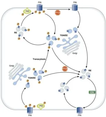
Kleine-Vehn et al., 2008; Dhonukshe et al., 2010). The Ser/Thr kinase PINOID was the first identified molecular determinant in PIN polar targeting (Friml et al., 2004). It was shown to phosphorylate PIN proteins in their hydrophilic loop region and this phosphorylation was antagonized by the action of the protein phosphatase subunit PP2A (Michniewicz et al., 2007; Dai et al., 2012). Overexpression of PID as well as inhibition of PP2A causes apicalization of PIN proteins whereas loss-of function pid mutants exhibit preferentially basal PIN targeting. The mislocalization of PINs in thes e mutants goes along with strong phenotypes that correlate with the changes in the direction of auxin flow (Benjamins et al., 2001; Friml et al., 2004; Michniewicz et al., 2007).
The recruitment of PINs to the apical recycling pathway is instructed by a phosphorylation of the serine residues in three conserved TPRXS(N/S) motifs which are highly conserved among
15
Figure 2-2 Schematic illustration of phosphorylation-dependent polarity changes of PIN proteins.
Unphosphorylated PINs are constantly recycled to the basal cell face. PIN-dependent phosphorylation results in apicalization by transcytosis. The phosphatase PP2A acts as a PID –antagonist. TGB/EE: trans- golgi-network / early endosomes, RE: recycling endosomes. Picture taken from Grunewald and Friml (2010).
the long PIN proteins. Analysis of loss-of phosphorylation and phosphomimicking mutants of PIN1 and PIN2 revealed that the reversible phosphorylation of these three residues is required and sufficient for proper PIN localization. Consequently, lack of phosphorylation leads to basal localization due to higher affinity of these variants to the GNOM-dependent sorting pathway (Dhonukshe et al., 2010; Huang et al., 2010). The current model for the phosphorylation-dependent sorting of PIN proteins is depicted in Figure 2-2. Two closely related kinases WAVY ROOT GROWTH 1 (WAG1) and WAG2, have been shown to target the same residues and act redundantly to PID in the apical targeting of PINs (Dhonukshe et al., 2010).
16
2.1.4 The AGCVIII kinase family
PID, WAG1 and WAG2 belong to the plant specific AGCVIII family of Ser/Thr protein kinases (Galvan-Ampudia and Offringa, 2007). The AGC kinases were named based on their homology to the mammalian protein kinase A, cyclic GMP-dependent protein kinase G and protein kinase C, which are all involved in receptor-mediated growth factor signal transduction in animals. They contain a highly conserved catalytic core consisting of two lobes which are interconnected by a linker domain. The binding sites for ATP, Mg2+ and for
the substrate are localized in this catalytic core. AGC kinase activity is regulated by (auto-)phosphorylation in a specific loop as well as by interacting proteins (Bögre et al.,
2003; Rademacher and Offringa, 2012). The characteristic features of the AGCVIII subfamily are a substitution of DFG to DFD in the conserved catalytic domain VII as well as a specific insertion between the subdomains VII and VIII which was speculated to be essential for the proper subcellular localization of the kinases that ranges from cytosolic, nuclear to plasma membrane associated (Zegzouti et al., 2006; Galvan-Ampudia and Offringa, 2007;
Rademacher and Offringa, 2012). In Arabidopsis, the AGCVIII family consists of 23 members which can be divided into four subgroups, one of which includes the above mentioned PID and WAG kinases. The members of this clade are the only AGCVIII kinases demonstrated to be involved in the regulation of PIN polarity. However, the D6 protein kinases, which belong to the largest subclade of the AGCVIII kinases, have also been shown to phosphorylate PINs and to regulate auxin transport (Zourelidou et al., 2009).
2.1.5 The AGCVIII kinase D6PK regulates auxin transport
The family of the D6 protein kinases consists of the founding member D6PK and its three close homologs D6PKL1, D6PKL2 and D6PKL3. First indications for a role of the D6PKs in auxin transport came from a phenotypic analysis of d6pk mutants which exhibit a number of developmental defects that are typically associated with reduced auxin transport, among them fused cotyledons, impaired lateral root initiation, agravitropic root growth as well as defects in phototropic responses. Consistently, auxin transport in stems of mutant plants is strongly decreased (Zourelidou et al., 2009; Willige et al., 2013). A synergistic genetic interaction between PIN1 and the D6PK genes as well as a colocalization of D6PK and different PINs at the basal sides of cells in various tissues suggested a functional link
17
between PINs and D6 kinases and indeed it could be shown that PIN proteins are phosphorylation targets of D6PK in vitro (Zourelidou et al., 2009). However, unlike the related PID and WAG kinases, D6PKs do not influence the polar targeting of PIN proteins (Zourelidou et al., 2009; Dhonukshe et al., 2010; Willige et al., 2013). Also, PID and D6PK overexpression results in different phenotypes, further suggesting different molecular functions of the two kinases.
As D6PK phosphorylates PINs and regulates auxin transport efficiency without influencing PIN polarity, a function in the regulation of transport activity of PIN proteins was suggested (Zourelidou et al., 2009). This hypothesis is further substantiated by the recent finding that lateral auxin transport during phototropic bending of hypocotyls is dependent on D6PKs (Willige et al., 2013). An essential process in phototropism is redirection of the auxin flow to the shaded side of hypocotyls which is achieved by a relocalization of PIN3 to lateral cell faces (Friml et al., 2003; Ding et al., 2011). In d6pk mutants, a gradual loss of PIN3 phosphorylation and a loss in lateral auxin transport was observed, however PIN3 lateralization took place normally. The data suggested that PIN3 transport activity was stimulated by D6PK mediated phosphorylation (Willige et al., 2013).
The major phosphorylation targets of PID that are required for apical sorting of PIN proteins have been characterized and are located within conserved sequence motifs (Dhonukshe et al., 2010; Huang et al., 2010). Recently, candidate residues that are phosphorylated by D6PK in vitro and in vivo were identified in PIN1, however their relevance for PIN1 activity and activation was not clarified to date (Zoureidou et al., unpublished).
2.1.6 Aims of the project
The D6 protein kinases have been implicated in the regulation of PIN protein function by phosphorylation (Zourelidou et al., 2009, Zourelidou et al., unpublished, Willige et al., 2013).
However, biochemical proof that D6PK phosphorylation directly influences the efflux activity of PIN proteins was missing. The main goal of this project therefore was to establish an auxin transport assay that would allow the investigation of the activatory effect of D6PK and possibly also of other AGCVIII kinases on PIN proteins in the heterologous X. laevis oocyte expression system.
Amino acids that are targeted by D6PK have been identified in vitro and in vivo (Zourelidou et al., unpublished). Focussing on PIN1, the importance of these amino acid residues in
18
terms of transport activity should be addressed by quantifying auxin efflux in oocytes upon co-expression of the respective mutants with D6PK.
Moreover, as a functional link between D6PK and PIN3 was recently shown (Willige et al., 2013), the heterologous system should also be used to demonstrate activation of PIN3 by D6PK.
19
2.2 Results
2.2.1 Co-expression of PIN1 and YFP-D6PK leads to enhanced IAA efflux in X. laevis oocytes
The D6 Ser/Thr kinases have been implicated in the regulation of PIN-mediated auxin efflux (Zourelidou et al., 2009). In order to find out whether D6PK directly regulates PIN1 transport activity by phosphorylation, an IAA efflux assay was established in X. laevis oocytes. Oocytes are a versatile and well established expression system especially for membrane proteins. As they store large amounts of protein, they do not depend on extracellular resources for nutrition and therefore express only a very limited number of own membrane proteins.
Consequently, background activity of endogenous transporters is low (Bröer, 2010).
Additionally, no PIN-related genes or compounds of the auxin signaling and transport pathways that might interact with the proteins of interest in any way are present. The auxin importer AUX1 has been characterized successfully in this system (Yang et al., 2006), but no data for PIN mediated auxin efflux in X. laevis oocytes were available so far.
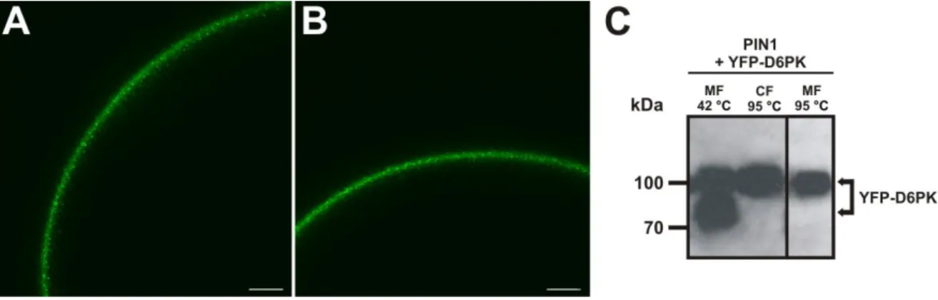
For heterologous expression of proteins in oocytes, mRNAs including a 5’-cap and a poly-A- tail (synthesized in vitro as described in 7.7.2) encoding for PIN1 and a YFP tagged version of D6PK (YFP-D6PK) which was shown to be functional (Zourelidou et al., 2009) were injected into oocytes which were then incubated for 5 days to allow production of the mature proteins. First, it had to be confirmed that the proteins were produced and correctly localized in the oocytes. In case of the D6PK, the protein could be detected by fluorescence microscopy due to the N-terminal YFP-tag. Figure 2-3 A and B shows confocal images of oocytes expressing YFP-D6PK in absence (Figure 2-3 A) or presence of PIN1 (Figure 2-3 B). In either case, YFP fluorescence was detected in the periphery of the oocytes. D6PK does not contain any transmembrane domains, but it is localized polarly in plant cells and appears to be associated with the plasma membrane in a so far unknown fashion (Zourelidou et al., 2009). It seems likely that the attachment of the kinase to the membrane is needed for the correct function of the protein. To confirm the membrane association also in oocytes, protein extracts were prepared and microsomal and cytosolic fractions were separated by ultracentrifugation (see 7.7.5). Figure 2-3 C shows a Western Blot where YFP-D6PK was detected in protein extracts from oocytes that were co-expressing PIN1 and YFP-D6PK.
Before loading samples of microsomal and cytosolic fractions on a SDS gel, they are typically
20
Figure 2-3 Expression and subcellular localization of YFP-D6PK in X. laevis oocytes 5 days after mRNA injection. (A,B) YFP fluorescence in oocytes expressing YFP-D6PK (A) or PIN1 + YFP-D6PK (B). In both cases, the kinase is localized i n the periphery of the oocyte. Scale bars in (A) and (B) represent 50 µm.
(C) Western blot analysis of YFP-D6PK in protein extracts. The fusion protein was detected with an anti-GFP antibody. Membrane fraction (MF) and cytosolic fraction (CF) were incubated at 42 °C or 95 ° prior to protein separation. The fusion protein was detected both in the MF and in the CF. The lower band in panel 1 results from incomplete solubilization of the protein and disappeared when the sample was treated like the CF (panels 2 and 3).
solubilized in a different way, i.e. cytosolic proteins are boiled at 95 °C for 5 min while membrane proteins which are more temperature sensitive are solubilized at 42 °C for 15 min. If gels containing samples that were treated like this were blotted and decorated with an anti-GFP antibody to detect the fusion protein which has a calculated size of 82 kDa, strong signals were observed both in the microsomal and in the cytosolic fraction ( Figure 2-3 C, panel 1 and 2). An additional band with higher mobility appeared in the microsomal fraction. However, this band disappeared when the microsomal fraction was boiled at 95 °C prior to loading (Figure 2-3 C, panel 1 and 3). Therefore, the additional band in the microsomal fraction that was incubated at 42 °C most likely represents incompletely solubilized protein. Taken together it can be concluded that at least a part of the fusion protein YFP-D6PK is associated with membranes in X. laevis oocytes like it is the case in the plant and can consequently act in direct proximity to membrane localized proteins.
PIN1 was used in an untagged version for the transport assay and was therefore only detected on a Western Blot. Figure 2-4 shows the analysis of protein extracts from oocytes that were separated into microsomal and cytosolic fractions and labeled with an anti -PIN1 antibody by western blotting (see 7.7.5, 7.7.6). As expected, PIN1 which contains 10 transmembrane domains was mainly found in the microsomal fractions in protein extracts
21
Figure 2-4 Western blot analysis of PIN1 in protein extracts from X. laevis oocytes expressing PIN1 or PIN1 + YFP-D6PK. PIN1 was detected with an anti -PIN1 antibody and was found mainly in the micro- somal fraction (MF), only weak signals were detected in the cytosolic fraction (CF). The additional bands with lower mobility (asterisks) in protein extracts from oocytes co-expressing PIN1 + YFP-D6PK represent phosphorylated PIN1.
from oocytes expressing PIN1 exclusively as well as in extracts from oocytes co-expressing PIN1 and YFP-D6PK. In the microsomal fraction of protein extracts from the latter samples, a smear of additional bands of higher mobility was observed that was not seen in the corresponding fraction from oocytes lacking the kinase (asterisks in Figure 2-4). This is the typical pattern described for phosphorylated PIN1 (Michniewicz et al., 2007; Zourelidou et al., 2009) indicating that PIN1 is phosphorylated by YFP-D6PK in X. laevis oocytes.
After the five day incubation period of the mRNA-injected oocytes, efflux assays were performed as described in 7.7.4. Briefly, oocytes were injected with defined amounts of the [3H]-labeled auxin IAA, resulting in an internal concentration of 1 - 1.5 µM IAA which corresponds to a physiologically relevant concentration (Petersson et al., 2009). Directly after injection, oocytes were incubated on ice for 10 min to allow homogenous distribution of the IAA inside the cell and the closure of the injection wound. The reduction of the IAA content over time was then determined by sampling 10-12 oocytes at 5 different time points and measuring the residual radioactivity in each cell by liquid scintillation counting. Initial experiments were performed with oocytes expressing PIN1 or the YFP-D6PK alone or co- expressing both proteins. Water-injected oocytes were used as a control. At least 5 biological replicates were analyzed for each tested construct. Figure 2-5 clearly shows that co-expression of PIN1 and YFP-D6PK in oocytes resulted in a much higher decrease of the IAA content than expression of PIN1 or the kinase alone which in turn could not enhance auxin efflux at all as the respective samples were indistinguishable from water-injected cells.
While in water, PIN1- or YFP-D6PK injected oocytes, about 65 % of the initially injected amount of [3H]-IAA was retained after 60 min, [3H]-IAA content in the PIN1 / YFP-D6PK
22
Figure 2-5 Co-expression of PIN1 and YFP-D6PK leads to enhanced IAA efflux in X. laevis oocytes.
Reduction of [3H]-IAA content in oocytes after direct injection of the substrate. CPM at time point 0 min were set to 1. Oocytes expressing PIN1 or YFP-D6PK alone were indistinguishable from water- injected control oocytes, whereas oocytes co-expressing PIN1 + YFP-D6PK contained app. 20 % less [3H]-IAA after 60 min. Error bars show SEM of biological replicates (n=23 for PIN1 and PIN1 + YFP-D6PK, n=17 for H2O, n=5 for YFP-D6PK). 10-12 technical replicates per time point were analyzed in every biological replicate.
co-expressing oocytes was reduced to app. 44 %. This means that while PIN1 alone is obviously not able to actively efflux auxin from oocytes, it can do so upon co-expression of the D6PK kinase. As it was shown that PIN1 is a phosphorylation target of the D6PK (Zourelidou et al., 2009) and is also phosphorylated by YFP-D6PK in oocytes (Figure 2-4), it can be assumed that phosphorylation of PIN1 is crucial for its auxin transport activity.
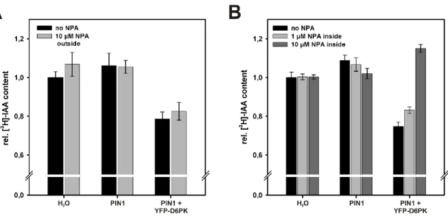
2.2.2 Efflux is sensitive to the auxin transport inhibitor NPA
Auxin transport inhibitors are important tools in studying the role of polar auxin transport in plant developmental processes. One well known inhibitor of auxin efflux is naphthylphthalamic acid (NPA) which belongs to the so-called phytotropins (Katekar and Geissler, 1980; Rubery, 1990). The molecular mechanism of NPA action is still unclear, but its application causes increased auxin accumulation in tobacco suspension cultured cells (Delbarre et al., 1996) and leads to pin1 like phenotypes in Arabidopsis (Okada et al., 1991;
23
Gälweiler et al., 1998), suggesting that it acts in close proximity to PINs. Additionally, phenotypes of d6pk mutants can be mimicked by NPA application (Zourelidou et al., 2009).
In order to test whether NPA has an influence on auxin efflux from oocytes mediated by D6PK-activated PIN1, changes in the IAA content in oocytes expressing PIN1, PIN1 + YFP- D6PK and in control water-injected oocytes were determined under different conditions.
mRNA injection and efflux assays were basically performed as described in 7.7.3 and 7.7.4.
Oocytes were incubated in Barth’s solution supplemented with 10 µM NPA out of a 10 mM stock in DMSO or with the equivalent amount of DMSO only as a control. After 60 min, residual IAA content was determined by liquid scintillation counting. The relative IAA content after 60 min compared to time point 0 min was normalized to the data for water-injected oocytes incubated in buffer without NPA. As Figure 2-6 A shows, the relative IAA content in oocytes co-expressing PIN1 and YFP-D6PK was not affected by 10 µM NPA in the buffer. Both in the presence and in the absence of NPA the IAA content was about 20 % lower than in PIN1 expressing oocytes or in water-injected control oocytes. However, the observation that NPA had no effect when present in the buffer is very likely due to the inability of the chemical to enter the cells under the given experimental conditions, i.e. a pH of 7.4 in the incubation solution. NPA is a weak acid with a pKa of 4.6 which is mainly present in the deprotonated form at neutral pH and can therefore not pass the plasma membrane by diffusion. Consistently, different results were obtained when NPA, instead of adding it to the buffer, was co-injected into the oocytes together with the substrate [3H]-IAA (Figure 2-6 B).
Control oocytes were injected with [3H]-IAA supplemented with the respective amount of DMSO. While the rel. IAA content in water-injected and PIN1-expressing oocytes, which do not actively transport auxin, was not altered by co-injection of NPA, a clear effect was observed for oocytes co-expressing PIN1 and YFP-D6PK. As expected, IAA content in oocytes that were injected with [3H]-IAA only was about 20 % lower than in PIN1- or water-injected oocytes after 60 min, reflecting the enhanced auxin efflux from these cells. Co-injection of NPA leading to a concentration of app. 1 µM inside the oocytes caused a slight increase in residual IAA, while 10 µM NPA resulted in a very strong increase of the residual IAA content.
Unexpectedly, the IAA retention in these cells was even slightly higher than in the corresponding PIN- or water-injected oocytes. The last observation cannot be explained at the moment, however the experiments on NPA inhibition were performed with only one biological replicate and should be repeated in order to verify the data.
24
Figure 2-6 Effect of NPA on PIN1 mediated IAA efflux from X. laevis oocytes. (A) Rel. [3H]-IAA content in oocytes after 60 min incubation in buffer without (black bars) or with 10 µM NPA (NPA outside, grey bars). Rel. IAA content in water-injected oocytes incubated in buffer without NPA was set to 1. 10 µM NPA in the buffer had no effect on the IAA content in water -injected oocytes, PIN1-expressing oocytes or oocytes co-expressing PIN1 + YFP-D6PK. Co-expression of PIN1 + YFP-D6PK resulted in significantly enhanced IAA export under both tested conditions. 10-12 oocytes were collected for each sampling point. (B) Rel. [3H]-IAA content in oocytes 60 min after co-injection of different amounts of NPA together with the substrate (no NPA: black bars, 1 µM NPA inside: light grey bars, 10 µM NPA inside:
dark grey bars). Rel. IAA content in water-injected oocytes injected with [3H]-IAA only was set to 1. Co- injection of 10 µM NPA caused a strong increase o the rel. IAA content in oocytes co-expressing PIN1 and YFP-D6PK indicating that IAA export was reduced.
2.2.3 Determination of transport rates
The establishment of the transport assay in the X. laevis oocyte expression system and the presented evidence that PIN1 needs to be phosphorylated in order to efficiently mediate auxin efflux (see chapter 2.2.1) provided a basis for addressing a number of further questions for example about the influence of other kinases on PIN1 activity and of course the effect of mutations at target phosphorylation sites. In order to be able to compare results from different experiments efficiently and to present the data more clearly, transport rates were calculated from the obtained time courses. Therefore, only those parts of the curve in which the auxin content decreased in a linear fashion, i.e. all time points up to 30 min, were considered. A representative experiment for PIN1 and PIN1 co-expressed with YFP-D6PK including the corresponding linear regressions is shown in Figure 2-7 A. Transport
25
Figure 2-7 Determination of transport rates from efflux assays in X. laevis oocytes. (A) Reduction of [3H]-IAA content in oocytes after direct injection of the substrate. Exemplary linear regression graph showing measurements of one biological replicate for PIN1 (r2 = 0.981) and PIN1 + YFP-D6PK (r2 = 0.988). The linear regression graphs served as a basis for the calculation of transport rates and hence rel. IAA efflux. Error bars show SEM of technical replicates (n=12). (B) Rel. [3H]-IAA efflux in oocytes after direct injection of the substrate. Rel. efflux from oocytes expressing PIN1 alone was set to 1. Error bars show SEM of biological replicates (n=23 for PIN1 and PIN1 + YFP-D6PK, n=17 for H2O, n=5 for YFP- D6PK). Different letters indicate significant differences. Statistical analysis was performed by means of a one way ANOVA followed by Bonferroni’s post hoc test (p<0.001).
rates were determined from the negative value of the slopes. Blotting of the rates from the data sets shown in Figure 2-5 and subsequent normalization to “PIN1 without kinase” results in a graph as shown in Figure 2-7 B. Hence, auxin efflux is increased app. 1.6 fold when PIN and YFP-D6PK are co-expressed compared to PIN1 or the kinase expressed alone or when oocytes were injected with water only. This efflux in the latter samples is considered as background caused by diffusion or endogenous transporters that might transport auxin unspecifically. The difference in the transport rates was highly significant (p<0.001).
26
2.2.4 A kinase-inactive version of D6PK does not activate PIN1
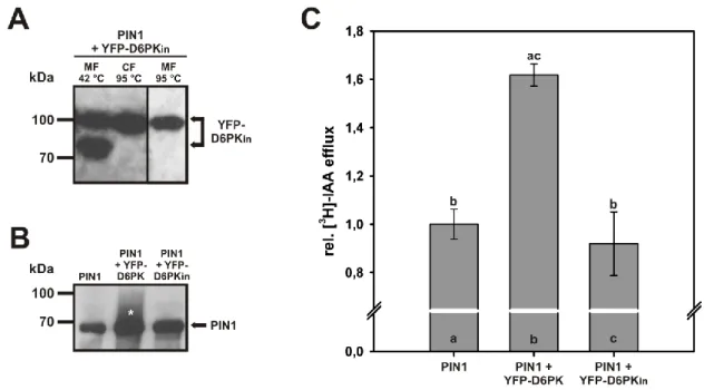
In order to make sure that the activation of PIN1 upon co-expression with D6PK was indeed due to the phosphorylation and not to a phosphorylation-independent interaction between the two proteins, oocytes were co-injected with mRNAs encoding for PIN1 and a mutant version of YFP-D6PK which carries an amino acid substitution of K to E in the ATP-binding pocket, leading to total loss of the kinase activity (YFP-D6PKin, (Zourelidou et al., 2009).
Western Blot analysis of protein extracts from oocytes five days after mRNA injection confirmed the expression of YFP-D6PKin and showed that the protein was localized to the microsomal as well as in the cytosolic fraction, indicating a membrane association of at least part of the protein (Figure 2-8 A). A double band that was observed in the microsomal fraction disappeared upon boiling the sample at 95 °C prior to SDS gel loading like it is typically done with the cytosolic fraction. The observed pattern was identical to the one described for the functional version of YFP-D6PK in chapter 2.2.1 (compare Figure 2-8 A to Figure 2-3 C). Additionally, an identical localization of YFP-D6PK (Figure 2-3 A,B) and YFP- D6PKin in the periphery of oocytes was confirmed by confocal microscopy (data not shown).
In oocytes co-expressing PIN1 and the kinase-dead version of YFP-D6PK, no phosphorylated PIN1 which is represented by a smear of lower mobility bands as seen in samples derived from PIN1 / YFP-D6PK co-expressing oocytes was detected (Figure 2-8 B, compare panels 2 and 3).
In efflux assays with [3H]-labeled IAA (Figure 2-8 C), oocytes co-expressing PIN1 and YFP- D6PKin did not show any differences in IAA efflux compared to cells expressing PIN1 alone, but differed significantly from PIN1 / YFP-D6PK co-expressing oocytes (p<0.001). Taken together, these results clearly show that the activation of PIN1 which leads to enhanced auxin efflux depends on phosphorylation by D6PK.
27
Figure 2-8 A kinase-inactive version of YFP-D6PK does not activate PIN1. (A) Western blot analysis of YFP-D6PKin in protein extracts 5 days after mRNA injection co-expressing the YFP-D6PKin + PIN1. The fusion protein was detected with an anti -GFP antibody. Membrane fraction (MF) and cytosolic fraction (CF) were incubated at 42 °C or 95 ° prior to SDS gel loading. The fusion protein was detected both in the MF and in the CF. The lower band in panel 1 results from incomplete denaturation of the protein and disappeared when the sample was treated like the CF (panels 2 and 3). (B) W estern blot analysis of PIN1 in the MF of protein extracts 5 days after mRNA injection expressing PIN1 alone or PIN1 in combination with YFP-D6PK and YFP-D6PKin, respectively. The smear of bands with lower mobility (asterisk) in protein extracts from oocytes co-expressing PIN1 + YFP-D6PK represents phosphorylated PIN1. (C) Rel. [3H]-IAA efflux in oocytes after direct injection of the substrate. Rel. efflux from oocytes expressing PIN1 alone was set to 1. Co-expression of YFP-D6PK, but not of YFP-D6PKin resulted in enhanced IAA efflux. Error bars show SEM of biological replicates (n=23 for PIN1 and PIN1 + YFP-D6PK, n=4 for PIN1 + YFP-D6PKin). Different letters indicate significant differences. Statistical analysis was performed by means of a one way ANOVA followed by Bonferroni’s post hoc test (p<0.001).
2.2.5 PINOID also enhances PIN1 mediated IAA export from oocytes, while other tested AGCVIII kinases have no effect
The AGCVIII kinase family comprises 23 members in Arabidopsis which can be subdivided into four groups. D6PK and its three close homologs D6PKL1, D6PKL 2 and D6PKL3 belong to the AGC1 group (Bögre et al., 2003; Galvan-Ampudia and Offringa, 2007). The AGC3 group includes PID as well as WAG1 and WAG2 which are, apart from D6PKs, the only AGCVIII
28
Figure 2-9 Influence of other AGCVIII kinases on PIN1 transport activity in X. laevis oocytes. Rel. [3H]- IAA efflux in oocytes after direct injection of the substrate. Rel. efflux from oocytes expressing PIN1 without a kinase was set to 1. Co-expression of YFP-D6PK or PID, but not of PHOT1 or UNC resulted in enhanced IAA efflux. Error bars show SEM of biological replicates (n=23 for PIN1 and PIN1 + YFP -D6PK, n=14 for PIN1 + PID, n=5 for PIN1 + PHOT1, n=3 for PIN1 + UNC). Different letters indicate significant differences. Statistical analysis was performed by means of a one way ANOVA followed by Bonferroni’s post hoc test (p<0.05).
kinases shown to directly phosphorylate PIN proteins, thereby regulating PIN polar localization (Michniewicz et al., 2007; Dhonukshe et al., 2010; Huang et al., 2010).
PHOTOTROPIN1 (PHOT1) and PHOT2, which form the AGC4 group, are blue light receptors and play an important role in phototropic response (Sakai et al., 2001). In this context, they act upstream of PIN proteins, as relocalization of the auxin efflux carriers leads to lateral auxin transport and consequently to bending (Friml et al., 2002b; Ding et al., 2011).
However, there is no evidence that PINs are direct phosphorylation targets of phototropins.
The fourth group of the AGCVIII kinases includes the recently characterized UNICORN (UNC), which is involved in the regulation of cell growth and division in various organs, but has not been implicated in auxin transport regulation so far (Enugutti et al., 2012).
In order to find out more about the specificity of the activatory effect of D6PK on PIN1, representative members of each subgroup of the AGCVIII kinases, i.e. PID, PHOT1 and UNC, were chosen and the respective mRNAs were injected into oocytes together with the PIN1- encoding mRNA. Efflux assays revealed that co-expression of PIN1 with PHOT1 or UNC did not have any effect on auxin efflux, whereas co-expression with PID lead to a significant