Kur J Clin Clwm Clin Biochcm 1996; 34:199-205 © 1996 by Walter de üruytor · Berlin · New York
Azathioprine Pharmacogenetics:
The Relationship between 6-Thioguanine Nuclcotides and Thiopurinc Methyltransferase in Patients after Heart and Kidney Transplantation
Ekkelmnl Schütz1, Jan Glimmert2, Victor William Armstrong*, Friedrich Wilhelm Mohr2
and Michael Ocllerlcli
11
Abteilung Klinische Chemie, Zentrum Innere Medizin
2
Abteilung Thorax-, Her/- und Gcfiißehirurgie, Zentrum Chirurgie Gcorg-August-Universitat Götlingüii, Göttingen, Germany
Dedicated Prof. Dr. Dr. J. ßiittncr on tlw occasion of his 65th birthday
Summary: The commonly used immunosupprcssivc regimen after solid organ transplantation consists of eyclo- sporine A, azathioprinc and steroids. Azathioprine, which is known to carry the risk of severe myelosuppression, is catabolized in vivo by xanthine oxidase and thiopurinc methyltransferase, an enzyme which exhibils a common genetic polymorphism; 11% of Caucasians are heterozygous and 0.3% are homozygous wilh respect to thiopurinc methyltransferase deficiency. Toxicity and immunosuppressive effects have been attributed to the 6-thioguaninc nucleotides generated from azathioprinc. We have studied thiopurine methyltransferase activity and 6-thioguaninc nucleoiide concentrations in erylhrocytcs from 39 heart and kidney recipients. Brythrocylc thiopurine methyl- transferase was determined by a radioen/ymatic assay and erylhrocyte 6-thiogimninu niicleotidc concentration with I1PLC. Thiopurinc methyltransferase activity [median (range, l()
lll~90
thpercentile)] was significantly (p < 0,05) higher in patients (n - 39) receiving axathioprine [285 (218-362) vs, 262 (160-352) inU/l erythrocylcs] than in healthy blood donors as controls (n = 120). When stratified according to thiopurinc methyltransferase phenotypc, one patient homozygous for the low allele exhibited an excessive crylhrocyte 6-lhioguaninc nuclcotide concentration (2210 pmol/0.8 · I0
9erylhrocytcs). Heterozygous patients had significantly higher 6-thioguaninc niicleotidc concen- trations (median: 435 pmol/0.8 · 1 erythrocytes) compared with concentrations in patients homoxygous for the high allele (median: 86 pmol/0.8 · 10
(Jerythrocytes; < 0.01), although the azalhioprine dosage did not differ (p = 0.66). Erythrocytc thiopurine methyltransferase determination therefore identifies patients at high risk of accu- mulating 6-thioguanine nucleotides. The monitoring of this enzyme may contribute to the safer management of immunosuppressive therapy with axathioprine. Alternative regimens such as cyclosporin A/mycophenolate mofet i I or tacrolimus should also be considered for this patient group.
Introduction
In immunosuppressive cyclosporin-based triple therapy transferase. Xanthine oxidase is not genetically poly-
aficr organ transplantation, axathioprine is often used us morphic and if not pharmacologically blocked this path-
an antiproliforative agent. Azathioprine represents the S- way leads to 6-thiourie acid. In contrast, thiopurinc
imidaxolc derivative of 6-mercaptopurine and is rapidly melhyltransferase displays a common genetic polymor-
cleaved to this drug in vivo by glutathione-S-transfcrasc phism (11 — 13). This polymorphism leads in 0.3% of
(1). It is generally agreed that 6-thioguaninc nueleotidos, Caucasians to a complete deficiency of the enzyme, and
which are synthcsiscd from 6-mercaptopurine through in about 11% to a heterozygous form with an enzyme
several enxymatic steps including hypoxanthine-gua- activity of 40—60%, compared with individuals with the
nine-phosphoribosyl transferase (2), are the major toxic two high allelcs (14). An accumulation of 6-thioguaninc
species of 6-thiopurine derivatives (fig. 1) (3, 4). The nucleotides is therefore likely to occur if one of these
determination of 6-thioguanine nucleotides in crythro- eatabolising pathways is altered. Thiopurine methyl-
cytes is therefore an accepted means of estimating toxic- transferase is generally determined in erythrocytes,
ity and efficacy of 6-mercaptopurinc treatment in pa- which are readily accessible, and there is a close correla-
tients with acute leukaemia (5—7) and after solid organ tion between activities measured in erylhrocytes and in
transplantation (8-10). 6-Thiopurines are catabolised by other cells and organs e. g. liver, kidney and leukocytes
two enzymes, xanthine oxidase and thiopurine methyl- (15-17). Brythrocyte thiopurine methyltransferase of
200 Schütz et al.: Thiopurine methyltransferase and 6-thioguanine nuclcotides in heart and kidney recipients
Azathioprine 6-Mercaptopurine 6-ThIolnoslnlc acld
IMPDH (EC 1.1.1.205) | 6-Thioxanthyllc acld Ribosa-P
?
H 6-Thioguanylicacid
Ribose-P
6-Thioguanosine di phosphate
6-Thioguanosine triphosphate
-OH
6-Methyl-thiouric acid Fig. l Metabolie pathway of azathioprine
GST: Glutathione-S-transferase; HGPRT: hypoxanthine-guanine phosphoribosyl transferase; IMPDH:Inosine monophosphate dehy-
randomly selected healthy Europeans typically shows a biphasic frequency distribution, consistent with a co- dominant inheritance (12, 13). One single point muta- tion, which leads to a substantial reduction of catalytic activity has been described (18) and recently a predomi- nant 2-point mutation, which is present in about 70%
of thiopurine methyltransferase deficient subjects was discovered (19). Further factors complicating therapeutic management of thiopurines are their widely variable bio- availability (20, 21) and an increase of thiopurine methyltransferase activity during therapy consistent with an induction phenomenon (10, 22). These might be the main reasons for the fact, that until now, only one study has shown a close negative correlation between thiopur- ine methyltransferase and 6-thioguanine nucleotides (23).
In patients receiving thiopurines for malignancies, i. e.
where leukocyte suppression is desired, the dosage is mainly adjusted to achieve low white blood cell count.
In contrast, in immunosuppressive triple therapy, azathioprine is most commonly used in a protocol-based standard dosage and only reduced if leukopenia occurs.
On the other hand, transplant recipients, compared with acute lymphatic leukaemia and acute myeloic leukaemia patients under maintenance therapy, are at higher risk for complications of leukopenia since they receive a combination of immunosuppressive drugs. They may therefore particularly profit from therapeutic drug moni- toring of the toxic 6-thioguanine nucleotides. Further- more, in these patients it would be of benefit to investi- gate the correlation of 6-thioguanine nucleotides and
drogenase; XO: Xanthine oxidase; TPMT: Thiopurine methyl transferase; SAH: S-Adenosyl homocysteine; SAM: S-Adenosyl methionine
thiopurine methyltransferase in vivo, since if thiopurine methyltransferase phenotype is correlated with toxic me- tabolites, a stratification of each patient according to thiopurine methyltransferase activity prior to therapy may aid in planning subsequent immunosuppressive therapy, particularly with respect to the new immuno- suppressants.
We studied erythrocyte thiopurine methyltransferase ac- tivity and 6-thioguanine nucleotide concentrations in 39 patients after heart and kidney transplantation under sta- ble clinical conditions at least 3 months after the last blood transfusion, to elucidate the relationship between erythrocyte 6-thioguanine nucleotides and thiopurine methyltransferase activity.
Patients and Methods
Probands
Twenty-one heart transplant recipients and 18 kidney recipients with generally uncomplicated postoperative courses were included in this study. Thirty-eight patients had undergone transplantation at least 3 months prior to the investigation and none had blood transfusions within this time. The only exception was one thiopur- ine methyltransferase-deficient patient who was tested 2 months after transplantation. The immunosuppressive regimen consisted of cyclosporin A, the dosage being adjusted according to its concen- trations in blood (200-350 g/l in heart recipients, 150-200 g/l in kidney recipients), steroids, which were tapered during the post- operative period, and azathioprine at a daily dosage median of 1 mg/kg body weight. Some patients received antilymphocyte anti- bodies as therapy induction for a maximum of 7 days immediately after transplantation. Patients with comedication with drugs which can potentially influence thiopurine metabolism, particularly xan- thine oxidase inhibitors, and patients with» unstable kidney function (e. g. due to rejection episodes) were excluded. If patients were
Sch tz et al.: Thiopurine methyltraiisferase and 6-thioguanine nucleotides in heart and kidney recipients 201
tested several times, the latest time point was considered for statis- tical evaluation unless creatinine concentrations were increased compared with earlier measurements in the same patient.
For comparison, blood samples of 120 healthy blood donors were also analysed for their erythrocyte thiopurine methyltransferase ac- tivity.
Methods
Erythrocyte preparation
Erythrocytes were obtained from NH4-heparinate anticoagulated whole blood by centrifugation at 2000 g for 10 min. Two washing steps were performed with Hank's balanced salt solution under the same conditions. Cells were finally rediluted to a haematocrit of about 0.40 and the exact haematocrit and red blood cell count for calculations of thiopurine methyltransferase activity and 6-thiogua- nine nucleotide concentration were determined with a Coulter STKS cell counting device. Samples were stored at -90 °C until analysis of thiopurine methyltransferase or 6-thioguanine nucleo- tides.
Tliiopurine methyltransferase assay
Erythrocyte lysates were analysed for their thiopurine methyl- transferase activity by a modified method according to Weinshil- boum et al. (24). As proposed elsewhere, S-[me/Ay/-3H]adenosyl- methionine was used as methyl donor (12) and the chelating step was omitted since erythrocytes lack interfering enzymes requiring bivalent cations (15). Lysates were incubated for l h at 37 °C with 6-mercaptopurine, S-[we///y/-3H]adenosyl-methionine, dithiothrei- tol, and allopurinol. The formed [mef/iy/-3H]6-mercaptopurine was liquid/liquid extracted with 120 mmol/1 isoamyl alcohol in toluene and the radioactivity was determined by -scintillation in a model LS3801 counter (Beckman Instruments, Munich, Germany). The radioactivity of the total reaction mixture was also determined. Re- sults were corrected for"quench, chemical and radiochemical purity of S-adenosylmethionine and extraction efficacy (56-65%; deter- mined in each run), and expressed as mU/1 packed erythrocytes).
These modifications yielded an improvement of between day varia- tion of the erythrocyte thiopurine methyltransferase assay from 9.5% to 5.9%. Within-series imprecision was 4.1-9.0% (100-350 mU/1 erythrocytes) in our laboratory with a dynamic range of this procedure of 17-920 mU/1 erythrocytes.
6-Thioguanine nucleotide assay
Erythrocyte lysates were hydrolysed in 1.5 mol/1 H2SO4 at 98 °C for 1 hour, in order to convert the respective nucleotides to 6- thioguanine. A saturated solution of phenyl mercuric acetate in tol- uene/isoamyl alcohol at alkaline pH was used to extract com- pounds with sulfhydryl groups. After back-extraction into 100 mmol/1 HC1, samples were analysed by C18 reversed phase HPLC (Hypersil ODS 5 μπι 250 · 4.6 mm, MZ Analysentechnik, Mainz, Germany, guard column: RP select B 5 μπι, Merck, Darmstadt, Germany) under isocratic conditions (25). 6-Thioguanine spiked drug-free erythrocyte lysates were used for construction of a cali- bration curve with UV detection at 342 nm. 4-Mercapto-lH pyra- zolo [3,4-d] pyrimidine was used as internal standard, and was detected at 322 nm (Shimadzu SPD 10 AV, Shimadzu Europe, Gro- burgwedel, Germany). If deviation from the assigned haematocrit (0.35) of sample preparation was > 0.05, a correction was per- formed with donor blood or Hank's balanced salt solution prior to the hydrolysis step. This is necessary since extraction efficacy decreases with the number of erythrocytes, with a resulting opti- mum at an haematocrit of about 0.35. Thioguanine concentrations were expressed as pmol/0.8 · 109 erythrocytes. Between-days im- precision was < 7% at concentrations of 100 pmol/0.8 · 109 eryth- rocytes and above, with a detection limit of 20 pmol/0.8 · 109 eryth- rocytes.
All reagents were of highest available purity. S-[we//?y/-3H]adeno- syl-methionine was purchased from Amersham (Braunschweig, Germany), isoamyl alcohol from J. T. Baker (Devender, The Neth- erlands) and toluene, borate, H2SO4, HC1 and NaOH from Merck (Darmstadt, Germany). All other chemicals were from Sigma Chem. (Deisenhofen, Germany).
Statistical analyses were performed with SAS statistical package, Ver.6.01 (SAS Inst. New York, USA).
This study was approved by the local Ethics Committee.
Results
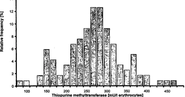
Erythrocyte thiopurine methyltransferase activity of 120 healthy donors showed a typical biphasic distribution (fig. 2) with a cut-off for the putative heterozygous group at 167 mU/1 erythrocytes. The calculated cut-off
100 150 200 250 300 350 400
Thiopurine methyltransferase [mU/1 erythrocytes] 450
Fig. 2 Frequency distribution of erythrocyte thiopurine methyl- Typical biphasic distribution with calculated lower limit for the transferase in 120 healthy blood donors without thiopurine admin- intemiediate group of 162 mU/1 erythrocytes
istration. Groups are defined by their upper limit.
202 Schütz et al.: Thiopurine melhyltransferase and 6-thioguanine nucleotides in heart and kidney recipients
83 117 150 183 217 250 283 317 350
Thiopurine methyltransferase [mU/l erythrocytes]
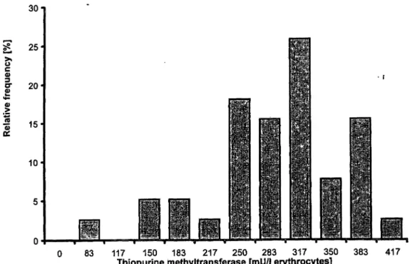
383Fig. 3 Frequency distribution of erythrocyte thiopurine methyltransferase in 39 kidney and heart
•recipients under chronic azathioprine administration. Groups are defined by their upper limit.
point, based on the llth percentile of this donor group was 162 mU/l erythrocytes. The median for the whole group of healthy donors was significantly lower than in the group of patients under long-term therapy with azathioprine (262 vs. 285 mU/l erythrocytes; p < 0.05).
In the patient group, one subject showed a total thiopur- ine methyltransferase deficiency and five patients were identified with an intermediate phenotype (fig. 3). The upper limit of this intermediate group was 217 mU/l erythrocytes, defined as the 11th percentile of the patient group with the exclusion of the thiopurine methyl-
transferase deficient patient (since taking this case under consideration would over-represent this group, which has a frequency of 0.3% in a normal population).
Erythrocyte 6-thioguanine nucleotide concentrations displayed a broad distribution from 31 to 599 pmol/0.8
• 10
9erythrocytes in patients with detectable erythrocyte thiopurine methyltransferase activity (fig. 4). The high- est 6-thioguanine nucleotide concentration observed was 2210 pmol/0.8 · 10
9erythrocytes in the thiopurine methyltransferase-deficient patient. This was associated with an episode of leukppenia.
0 50 100 150 200 250 300
ü400 500 600 1000 2000 >2000 6-Thioguanine nucleotides [pmol/0.8 · 10
9erythrocytes]
Fig. 4 Frequency distribution of erythrocyte 6-thioguanine nucleotides in 39 kidney and heart»recipients under chronic azathioprine administration. Groups are defined by their upper limit.
Sch tz et al.: Thiopurine methyltransferase and 6-thioguanine nucleotides in heart and kidney recipients 203
S
"? 600·
2
l 500 S 400 300
CO
σ> 200 .2
100
ο
«β
100 150 200 250 300 350
Thiopurine methyltransferase [mU/l erythrocytes]
400
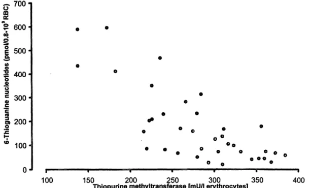
Fig. 5 Scatterplot of 6-thioguanine nucleotides vs. thiopurine prine. One patient with thiopurine methyltransferase deficiency is methyltransferase in erythrocytes in patients receiving azathio- not displayed (n = 38; r = -0.785).
Tab. 1 Erythrocyte 6-thioguanine nucleotide concentrations in heart and kidney recipients stratified according to erythrocyte thio- purine methyltransferase activity.
Thiopurine methyltransferase (mil/I erythrocytes)
<17 17-210 >210 (n = 1 ) (n = 5) (n = 33) Thioguanine nucleotide concentration (pmol/0.8 · 109 erythrocytes) Median
84th percentile 16thpercentile
2210_
—
436*593 321
22899 46
* p < 0.05 vs high thiopurine methyltransferase group
When stratified according to thiopurine methyltransfer- ase activity, significantly higher erythrocyte 6-thiogua- nine nucleotide concentrations were observed in the groups with low thiopurine methyltransferase activity (tab. 1).
Azathioprine dosage did not differ between these groups (median 75 mg/day; p = 0.88). Furthermore there was no correlation between azathioprine daily dose and erythrocyte-6-thioguanine nucleotide concentrations (r < 0.01; p = 0.431). A highly significant negative cor- relation was seen between thiopurine methyltransferase activity in erythrocytes and the respective concentrations of 6-thioguanine nucleotides (fig. 5), even when the pa- tient with total thiopurine methyltransferase deficiency was excluded (r = -0.785; p < 0.01).
Discussion
The detoxification of azathioprine, used in immunosup- pressive management after organ transplantation, is de- pendent on the common genetic polymorphism of thio- purine methyltransferase (11 — 13, 14, 27). This study showed that thiopurine methyltransferase activity is ob- viously influenced by thiopurines and shows an increase under therapy. This is in close agreement with results from studies in which thiopurine methyltransferase was measured in patients prior to and under treatment with azathioprine or 6-mercaptopurine, and thereafter (5, 22).
Despite the well known metabolism of thiopurines, only one study has shown a close relationship between thio- purine methyltransferase activity and 6-thioguanine nu- cleotide concentrations (23). This might be due to sev- eral uncertainties of azathioprine therapy, such as the induction phenomenon mentioned above, different dos- ages, especially if dosage is adjusted according to leuko- cyte count, and the variable bioavailability of this drug (20, 21) which is influenced by food intake (26). In this study, we observed a close negative correlation of 6- thioguanine nucleotides and thiopurine methyltransfer- ase in patients taking azathioprine. In these heart and kidney recipients, the observed highly significant corre- lation (r = —0.785) was probably due to the relatively constant dosage of 75 mg/day in this patient group.
High concentrations of erythrocyte 6-thioguanine nucle-
otides are associated with the risk of myelosuppression
(5-7, 10), with related complications, such as septicae-
mia and consecutive multiple organ failure. As reported
204 Schütz et al.: Thiopurine methyltransferase and 6-thioguanine nucleotides in heart and kidney recipients
(27), the patient with complete thiopurine methyl- transferase deficiency and excessive 6-thioguanine nu- cleotide concentration subsequently died from this com- plication. Furthermore in a previous study, we showed that patients with 6-thioguanine nucleotide concentra- tions > 600 pmol/0.8 · 10
9erythrocytes may develop leukopenia (10). These findings suggest that one might expect an increased risk of toxic side effects in such patients under treatment with higher azathioprine dos- age.
In therapeutic drug monitoring for azathioprine after solid organ transplantation the lower limit of erythrocyte 6-thioguanine nucleotides is hard to define, since the immunosuppressive regimen consists of a combination of drugs and there is a lack of simple quantities for as- sessing the immunosuppressive state of an individual patient. The data of this study demonstrate a close rela- tionship between thiopurine methyltransferase activity and 6-thioguanine nucleotide concentration in transplant recipients under azathioprine therapy. It would therefore
seem advisable to measure erythrocyte thiopurine methyltransferase prior to transplantation, to identify those patients with low or absent activity, who obviously carry an increased risk for toxic complications due to elevated 6-thioguanine nucleotides. In patients with lower or absent thiopurine methyltransferase, the con- centrations of 6-thioguanine nucleotides should be mon- itored during azathioprine therapy to avoid toxic effects, and dosage could be adjusted at the start of therapy by taking erythrocyte thiopurine methyltransferase activity into account. Alternatively, determination of erythrocyte thiopurine methyltransferase prior to transplantation could be used to identify patients at high risk of azathio- prine toxicity, who might more appropriately be treated with one of the new immunosuppressants such as tacrol- imus or mycophenolate mofetil.
Acknowledgements
We thank Jutta Engelmayer and Iris Brandel for their skilful tech- nical assistance.
References
1. Elion G. Biochemistry and pharmacology of purine analogues.
FedProc 1967; 26:898-903.
2. Kelley WN, Rosenbloom FM, Seegmiller JE. Effects of azathioprine on purine synthesis in clinical disorders of purine metabolism. J Clin Invest 1967; 46:1518-29.
3. Tidd DM, Paterson ARP. A biochemical mechanism for the delayed cytotoxic reaction of 6-mercaptopurine. Cancer Res
1974; 34:738-46.
4. Elion GB. The purine path to chemotherapy. Science 1989;
244:41-53.
5. Lennard L, Lilleyman JS, Van Loon J, Weinshilboum RM.
Genetic variation in response to 6-mercaptopurine for child- hood acute lymphoblastic leukaemia. Lancet 1990;
336:225-9.
6. Evans WE, Homer MH, Chu YQ, Kalwinsky D, Roberts WM.
Altered mercaptopurine metabolism, toxic effects and dosage requirements in a thiopurine methyltransferase-deficient child with acute lymphatic leukemia. J Pediatr 1991; 119:985-9.
7. Lennard L, Rees CA, Lilleyman JS, Maddocks JL. Childhood leukemia: a relationship between intracellular 6-mercaptopu- rine metabolites and neutropenia. Br J Clin Pharmacol 1983;
16:359-63.
8. Lennard L, Brown CB, Fox M, Maddocks JL. Azathioprine metabolism in kidney transplant recipients. Br J Clin Pharma- col 1984; 18:693-700.
9. Bergan S, Rugstad HE, Bentdal O, Stokke 0. Monitoring of azathioprine treatment by determination of 6-thioguanine nu- cleotide concentrations in erythrocytes. Transplantation 1994;
58:803-8.
10. Schütz E, Gummert J, Mohr FW, Armstrong VW, Oellerich M.
Should 6-thioguanine nucleotides be monitored in heart transplant recipients given azathioprine? Ther Drug Monit. In press.
11. Weinshilboum RM, Sladek SL. Mercaptopurine pharmacoge- netics: monogenetic inheritance of erythrocyte thiopurine methyltransferase activity. Am J Hum Genet 1980; 32:651- 62.
12. Tinel M, Berson A, Pessayre D, Letteron P, Cattoni MP, Hors- mans Y, Larrey D. Pharmacogenetics of human erythrocyte thiopurine methyltransferase activity in a French population.
Br J Clin Pharmacol 1991; 32:729-34.
13. McLeod HL, Lin J-S, Pui C-H, Scott EP, Evans WE. Thiopur- ine methyltransferase activity in American white and black subjects. Clin Pharmacol Ther 1994; 55:15-20.
14. Vuchetich JP, Weinsbhilboum RM, Price RA. Segregation analysis of human red blood cell thiopurine methyltransferase activity. Genetic Epidemiology 1995; 12:1-11.
15. Szumlanski CL, Honchel R, Scott MC, Weinshilboum RM.
Human liver thiopurine methyltransferase pharmacogenetics:
biochemical properties, liver-erythrocyte correlation and pres- ence of isozymes. Pharmacogenetics 1992; 2:148-59.
16. Van Loon J, Weinshilboum RM. Thiopurine methyltransferase biochemical genetics: human lymphocyte activity. Biochem Genet 1982; 20:637-58.
17. Van Loon J, Weinshilboum RM. Thiopurine methyltransfer- ase isozymes in renal tissue. Drug Metab Dispos 1990;
18:632-8.
18. Krynetski EY, Schuetz JD, Galpin AJ, Pui C-H, Relling MV, Evans WE. A single point mutation leading to loss of catalytic activity in human thiopurine S-methyltransferase. Proc Natl Acad Sei USA 1995; 92:949-53.
19. Otterness DM, Szumlanski CL, Lennard L, Weinshilboum RA.
Human thiopurine methyltransferase pharmacogenetics: mo- lecular basis for common genetic polymorphism [abstract].
ISSXProc 1995; 8:107.
20. Ohlman S, Lafolie P, Lindholm A, Lilliemark J, Tyden G, Pe- terson C. Large interindividual variability in bioavailability of azathioprine in renal transplant recipients. Clin Transplantation 1993; 7:65-70.
21. Bergan S, Rugstad HE, Bentdal O, Endresea L, Stokke O. Ki- netics of mercaptopurine and thioguanine nucleotides in renal transplant recipients during azathioprine treatment. Ther Drug Monit 1994; 16:13-20.
22. McLeod HL, Relling MV, Liu Q, Pui C-H, Evans WE. Poly- morphic thiopurine methyltransferase in erythrocytes is indica- tive of activity in leukemic blasts from children with acute lymphoblastic leukemia. Blood 1995; 85:1897-902.
23. Lennard L, Van Loon JA, Lilleyman JS, Weinshilboum RM.
Thiopurine pharmacogenetics in leukemia: correlation of erythrocyte thiopurine methyltransferase activity and 6-thiogu- anine nucleotide concentrations. dirt Pharmacol Ther 1987;
41:18-25.
Schütz et al.: Thiopurine methyltransferase and 6-thioguanine nucleotides in heart and kidney recipients 205
24. Weinshilboum RM, Raymond FA, Pazmino PA. Human eryth- rocyte thiopurine methyltransferase: radiochemical microassay and biochemical properties. Clin Chim Acta 1978; 85:323-33.
25. Lennard L. Assay of 6-thioinosinic acid and 6-thioguanine nu- cleotides, active metabolites of o-mercaptopurine, in human red blood cells. J Chromatogr 1987; 423:169-78.
26. Burton N, Barnett M, Aherne G, Evans J, Douglas I, Lister A.
The effect of food on the oral administration of 6-mercaptopu- rine. Cancer Chemother Pharmacol 1986; 18:90-1.
27. Schütz E, Gummert J, Mohr FW, Oellerich M. Azathioprine- induced myelosuppression in thiopurine methyltransferase de- ficient heart transplant recipient. Lancet 1993; 341:436.
Received September 15/December 15, 1995
Corresponding author: Dr. med. Ekkehard Schütz, Zentrum Innere Medizin, Abt. Klinische Chemie, Georg-August- Universität, Robert-Koch-Straße 40, D-37075 Göttingen, Germany