Review
The Role of Chemokines in the Pathophysiology of Major Depressive Disorder
Vladimir M. Milenkovic * , Evan H. Stanton, Caroline Nothdurfter, Rainer Rupprecht and Christian H. Wetzel
Department of Psychiatry and Psychotherapy, Molecular Neurosciences, University of Regensburg,
D-93053 Regensburg, Germany; evan.h.stanton@gmail.com (E.H.S.); Caroline.Nothdurfter@medbo.de (C.N.);
Rainer.Rupprecht@medbo.de (R.R.); Christian.Wetzel@klinik.uni-regensburg.de (C.H.W.)
* Correspondence: vladimir.milenkovic@ukr.de; Tel.: +49-941-944-8955
Received: 31 March 2019; Accepted: 8 May 2019; Published: 9 May 2019
Abstract: Major depressive disorder (MDD) is a debilitating condition, whose high prevalence and multisymptomatic nature set its standing as a leading contributor to global disability.
To better understand this psychiatric disease, various pathophysiological mechanisms have been proposed, including changes in monoaminergic neurotransmission, imbalance of excitatory and inhibitory signaling in the brain, hyperactivity of the hypothalamic-pituitary-adrenal (HPA) axis, and abnormalities in normal neurogenesis. While previous findings led to a deeper understanding of the disease, the pathogenesis of MDD has not yet been elucidated. Accumulating evidence has confirmed the association between chronic inflammation and MDD, which is manifested by increased levels of the C-reactive protein, as well as pro-inflammatory cytokines, such as Interleukin 1 beta, Interleukin 6, and the Tumor necrosis factor alpha. Furthermore, recent findings have implicated a related family of cytokines with chemotactic properties, known collectively as chemokines, in many neuroimmune processes relevant to psychiatric disorders. Chemokines are small (8–12 kDa) chemotactic cytokines, which are known to play roles in direct chemotaxis induction, leukocyte and macrophage migration, and inflammatory response propagation. The inflammatory chemokines possess the ability to induce migration of immune cells to the infection site, whereas their homeostatic chemokine counterparts are responsible for recruiting cells for their repair and maintenance. To further support the role of chemokines as central elements to healthy bodily function, recent studies suggest that these proteins demonstrate novel, brain-specific mechanisms including the modulation of neuroendocrine functions, chemotaxis, cell adhesion, and neuroinflammation. Elevated levels of chemokines in patient-derived serum have been detected in individuals diagnosed with major depressive disorder, bipolar disorder, and schizophrenia. Furthermore, despite the considerable heterogeneity of experimental samples and methodologies, existing biomarker studies have clearly demonstrated the important role of chemokines in the pathophysiology of psychiatric disorders.
The purpose of this review is to summarize the data from contemporary experimental and clinical studies, and to evaluate available evidence for the role of chemokines in the central nervous system (CNS) under physiological and pathophysiological conditions. In light of recent results, chemokines could be considered as possible peripheral markers of psychiatric disorders, and/or targets for treating depressive disorders.
Keywords: major depressive disorder; chemokines; neuroinflammation
1. Introduction
Major depressive disorder (MDD) is a highly prevalent condition, and is the third leading cause of disability worldwide [1]. Despite the availability of numerous anti-depressive treatments, 30%
Int. J. Mol. Sci.2019,20, 2283; doi:10.3390/ijms20092283 www.mdpi.com/journal/ijms
of patients diagnosed with MDD fail to respond to anti-depressant therapy, or show only a partial response [2,3]. Bipolar disorder, which is characterized by recurrent depressive and manic episodes, is difficult to diagnose [4], and is often misdiagnosed as MDD, particularly during a depressive episode [5]. Diagnostic and Statistical Manual of Mental Disorders (DSM-5) criteria for unipolar and bipolar depression are the same during a major depressive episode [6]. Therefore, there is a need for novel biomarkers, which could distinguish between these two conditions [5]. This inadequate response to treatment reflects an incomplete understanding of the actual pathogenesis of depression, which was initially linked to changes in monoaminergic transmission [7,8]. Subsequent hypotheses include the disturbance of excitatory and inhibitory signaling in the brain [9,10], hyperactivity of the hypothalamic-pituitary-adrenal (HPA) axis [11,12], and hindrance upon the healthy progression of neurogenesis [13,14]. However, increasingly compelling lines of evidence indicate a role of nearly or completely asymptomatic subclinical systemic inflammation in the pathophysiology of MDD [15–26].
While using the reassessment of immune privilege in the central nervous system [27,28] as a foundation, complex interactions between the immune system and the brain began to emerge. The immune system regulates key aspects of brain development, neurogenesis, central nervous system (CNS) homeostasis, mood, and behavior [29–35]. As such, perturbations of the neuroimmune functions have been implicated in a number of psychiatric disorders, including MDD [36–39], bipolar disorder [40,41], schizophrenia [42–45], and autism [46,47].
Recent advances in neuroscience have linked chemotactic cytokines (chemokines) to neurobiological processes relevant to psychiatric disorders, such as synaptic transmission and plasticity, neurogenesis, and neuron-glia communication [48–51]. The disruption of any of these functions, by activation of the inflammatory response system, could be central for the pathogenesis of MDD. Impaired CXCL12/CXCR4 signaling is implicated in abnormal development, proliferation, and migration of neural progenitor cells [52,53], which is suggestive of their essential roles in mammalian neurogenesis. Furthermore, the dysregulation of various chemokines, which modulate neuronal activity by means of inducing signal transduction [54,55] and Ca
2+mobilization [56,57], could also be involved in pathophysiological processes leading to MDD. To add to the wide breadth of chemokine functionality, these ligands and their receptors, which are widely expressed in the CNS [58–62], coordinate immune cell recruitment and their subsequent migration to sites of inflammation. Therefore, this links peripheral and central inflammation.
This phenomenon can be observed in the quantitative increase of chemokine concentrations within the serum of patients with MDD, relative to homeostatic levels. Moreover, this discrepancy is associated with the onset and progression of depression in humans [63].
To further investigate the potential connection between chemokines and depression, chemokine receptor knockout mice (CCR6 and CCR7) were created and observed to display behavioral phenotypes similar to psychiatric disorders, including MDD [64].
Altogether, these data provide evidence of the involvement of chemokines in processes underlying MDD. In this work, we will examine the role of chemokines in healthy and depressed states, as well as summarize to the best of our knowledge evidence to date for the possible role of chemokines in the pathogenesis of MDD.
2. Chemokine Superfamily
The chemokine superfamily contains a large number of ligands and receptors, which are classified into four sub-families (CXC, CC, C, and CX3C) [65], according to the number and spacing of their two N-terminal, disulfide bonding participating cysteine residues. Chemokines are small (8–12 kDa) heparin binding proteins, structurally related to cytokines that can induce directed chemotaxis of immune cells. However, chemokines are additionally involved in the regulation of migration of immune cells [66,67], blood-brain barrier (BBB) permeability [68], and synaptic pruning processes [69].
In addition to their structural criteria, chemokines can be subdivided into inflammatory chemokines,
which are upregulated under inflammatory conditions, homeostatic chemokines that are responsible
for maintaining homeostasis, and chemokines, which exhibit dual functionality [70].
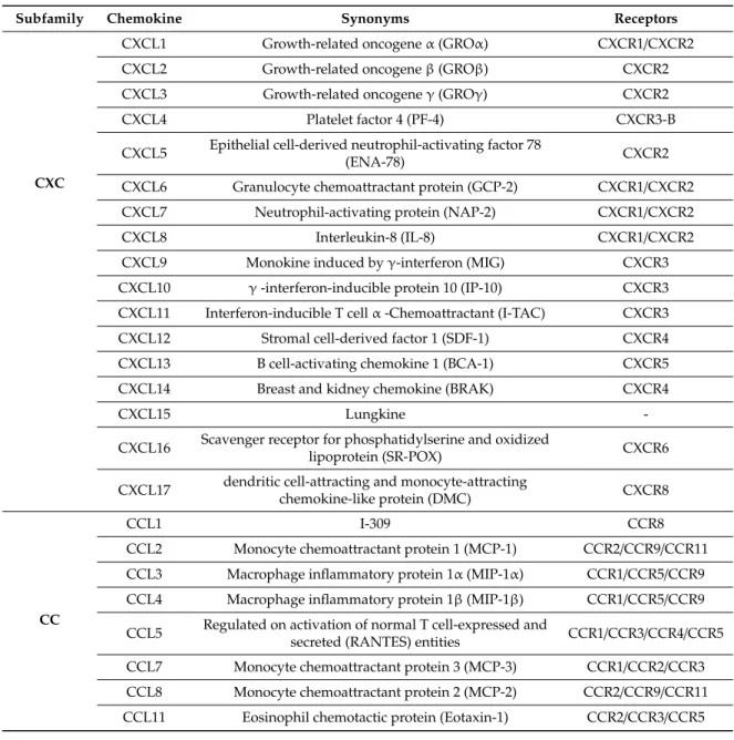
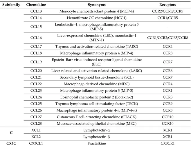
The chemokine superfamily has expanded rapidly after the initial identification of secreted platelet factor 4 (PF4/CXCL4) [71] in 1977. Subsequent studies have identified more than 50 chemokines, as well as 20 chemokine receptors [72]. The majority of human chemokine genes are clustered on chromosomes 4 and 17. CXC chemokines can be found at chromosomal location 4q12-21, whereas most of the CC chemokines are located at 17q11-21 [73]. This suggests a rapid evolution by repeated gene duplications [74]. All chemokines share a very similar tertiary structure [75], including a highly flexible N-terminal domain and a long rigid loop, which are essential for interacting with their respective receptors [76], and a C-terminal α -helix. Typically, a given chemokine can bind to more than one receptor (Table 1) and, correspondingly, a number of different chemokines can be recognized by the same receptor [65]. Chemokines are secreted in response to inflammatory cytokines, and they selectively recruit monocytes, lymphocytes, and neutrophil-inducing chemotaxis by activating G-protein-coupled receptors (GPCRs) [77].
Table 1. Chemokines and their known receptors. Chemokine receptors, which belong to the superfamily of GPCRs, can bind to multiple chemokines, and certain chemokines can similarly bind to more than one receptor. Adapted from Zlotnik and Yoshie 2012 [65].
Subfamily Chemokine Synonyms Receptors
CXC
CXCL1 Growth-related oncogene
α(GRO
α) CXCR1/CXCR2
CXCL2 Growth-related oncogene
β(GROβ) CXCR2
CXCL3 Growth-related oncogene
γ(GROγ) CXCR2
CXCL4 Platelet factor 4 (PF-4) CXCR3-B
CXCL5 Epithelial cell-derived neutrophil-activating factor 78
(ENA-78) CXCR2
CXCL6 Granulocyte chemoattractant protein (GCP-2) CXCR1/CXCR2 CXCL7 Neutrophil-activating protein (NAP-2) CXCR1/CXCR2
CXCL8 Interleukin-8 (IL-8) CXCR1/CXCR2
CXCL9 Monokine induced by
γ-interferon (MIG)CXCR3 CXCL10
γ-interferon-inducible protein 10 (IP-10) CXCR3 CXCL11 Interferon-inducible T cell
α-Chemoattractant (I-TAC) CXCR3 CXCL12 Stromal cell-derived factor 1 (SDF-1) CXCR4 CXCL13 B cell-activating chemokine 1 (BCA-1) CXCR5
CXCL14 Breast and kidney chemokine (BRAK) CXCR4
CXCL15 Lungkine -
CXCL16 Scavenger receptor for phosphatidylserine and oxidized
lipoprotein (SR-POX) CXCR6
CXCL17 dendritic cell-attracting and monocyte-attracting
chemokine-like protein (DMC) CXCR8
CC
CCL1 I-309 CCR8
CCL2 Monocyte chemoattractant protein 1 (MCP-1) CCR2/CCR9/CCR11 CCL3 Macrophage inflammatory protein 1
α(MIP-1
α) CCR1/CCR5/CCR9 CCL4 Macrophage inflammatory protein 1β (MIP-1β) CCR1/CCR5/CCR9 CCL5 Regulated on activation of normal T cell-expressed and
secreted (RANTES) entities CCR1/CCR3/CCR4/CCR5
CCL7 Monocyte chemoattractant protein 3 (MCP-3) CCR1/CCR2/CCR3
CCL8 Monocyte chemoattractant protein 2 (MCP-2) CCR2/CCR9/CCR11
CCL11 Eosinophil chemotactic protein (Eotaxin-1) CCR2/CCR3/CCR5
Table 1. Cont.
Subfamily Chemokine Synonyms Receptors
CCL13 Monocyte chemoattractant protein 4 (MCP-4) CCR2/CCR3/CCR5
CCL14 Hemofiltrate CC chemokine (HCC1) CCR1/CCR5
CCL15 Leukotactin-1, macrophage inflammatory protein 5
(MIP-5) -
CCL16 Liver-expressed chemokine (LEC), monotactin-1
(MTN-1) CCR1/CCR2/CCR5/CCR8
CCL17 Thymus and activation-related chemokine (TARC) CCR4 CCL18 Macrophage inflammatory protein 4 (MIP-4) CCR8 CCL19 Epstein–Barr virus-induced receptor ligand chemokine
(ELC) CCR7
CCL20 Liver-related and activation-related chemokine (LARC) CCR6 CCL21 Secondary lymphoid tissue chemokine (SCL) CCR7
CCL22 Macrophage-derived chemokine (MDC) CCR4
CCL23 Macrophage inflammatory protein 3 (MIP-3) CCR1 CCL24 Eosinophil chemotactic protein 2 (Eotaxin-2) CCR3 CCL25 Thymus lymphoma cell-stimulating factor (TECK) CCR9 CCL26 Macrophage inflammatory protein 4-
α(MIP-4-
α) CCR3 CCL27 Cutaneous T cell-attracting chemokine (CTACK) CCR10 CCL28 Mucosae-associated epithelial chemokine (MEC) CCR10
C
XCL1 Lymphotactin-α XCR1
XCL2 Lymphotactin-
βXCR1
CX3C