P HOTOCHROMIC
G- PROTEIN COUPLED RECEPTOR LIGANDS
Dissertation
zur Erlangung des Doktorgrades der Naturwissenschaften (Dr. rer. nat.)
an der Fakultät für Chemie und Pharmazie der Universität Regensburg
vorgelegt von Daniel Lachmann
aus Kelheim
2019
Der experimentelle Teil der vorliegenden Arbeit wurde in der Zeit von Oktober 2015 bis Dezember 2018 unter der Betreuung von Prof. Dr. Burkhard König am Institut für Organische Chemie der Universität Regensburg durchgeführt.
Diese Arbeit wurde angeleitet von: Prof. Dr. Burkhard König Promotionsgesuch eingereicht am: 16.01.2019
Promotionskolloquium am: 01.03.2019
Prüfungsausschuss
Vorsitzende: Prof. Dr. Julia Rehbein
1. Gutachter: Prof. Dr. Burkhard König
2. Gutachter: Prof. Dr. Joachim Wegener
3. Prüfer: Prof. Dr. Frank-Michael Matysik
M EINEN E LTERN & K ERSTIN
Table of contents
Fulgimides in biological applications 1
1. Introduction 1
2. Fulgimides as photoswitches in biological applications 5
3. Conclusion 10
4. Literature 11
1. Photochromic Dopamine Receptor Ligands based on Dithienylethenes
and Fulgides 15
1. Introduction 16
2. Results and Discussion 18
2.1 Synthesis - pharmacophoric headgroups 18
2.2 Synthesis - maleimides and cyclopentenes 19
2.3 Photophysical properties - arylethenes 21
2.4 Synthesis - fulgides and fulgimides 23
2.5 Photophysical investigations - fulgimides 25
2.6 Biological investigations 26
3. Conclusions 29
4. Experimental section 29
4.1 Synthesis 30
4.2 Assays 58
5. Literature 61
6. Supporting Information 64
2. Photochromic Peptidic NPY Y 4 -Receptor Ligands 89
1. Introduction 90
2. Discussion 91
2.1 Synthesis 91
2.2 Photopyhsical properties 93
2.3 Biological investigations 97
3. Conclusion 99
4.Experimental Section 99
4.1 General Conditions 99
4.2 Synthesis procedures 100
4.3 Assays 110
5. Literature 112
6. Supporting information 114
6.4 Biological characterization 119
3. Covalent binding photochromic GPCR-Ligands for single molecule
spectroscopy 129
1. Introduction 130
2. ß
2-Adrenergic receptor 131
2.1 Molecular docking studies 131
2.2 Synthesis of the photochromic ß
2-AR ligands 133
2.3 Photophysical investigations 136
2.4 Biological investigations 137
2.5 Single molecule spectroscopy 138
3. µ-Opioid receptor 140
3.1 Docking studies towards the µOR 140
3.2 Synthesis of the azopyrazole based fentanyl derivatives 142
3.3 Photophysical investigations 146
3.4 Biological investigations 148
4. Conclusion and Outlook – ß
2-AR and µOR 149
5. Experimental section 150
6. Literature 185
7.Supporting information 188
4. Appendix
1. Abbreviations 206
2. Danksagung 208
3. Curiculum Vitae 209
I NTRODUCTION
Fulgimides in biological applications
Parts of this chapter have been published as:
D. Lachmann, R. Lahmy, B. König, Eur. J. Org. Chem, Minireview
1. Introduction
Within the field of photopharmacology, photoswitches have drawn increasing attention over the past years. Adjacent to photocaging 1 , which is an irreversible process, the common reversible photoswitches, which are used in biological applications are mainly azobenzenes and diarylethenes (DTEs). 2,3 In addition, molecules like spiropyranes 4 , hemithioindigos 5 , donor-acceptor Stenhouse adducts (DASAs) 6 and fulgides 7,8 were also used for the reversible modulation of biological targets by light. Photoresponsive molecules can be divided into two groups: the thermally bistable switches (T-type, e.g. azobenzenes, hemithioindigos) and the thermally stable ones (P-type, e.g. diarylethenes, fulgides). Fulgimides are the imide derivatives of fulgides and were mainly used in optical data storage 9 , molecular computing 10 and photomechanical materials. 11 However, beyond applications in material science, fulgimides are also well suited for biological applications. 7,12,13 Their stability in conditions typical for biological assays was proven by Lees et al. with the development of highly polar indolyl fulgimides, which switch reversibly in sodium phosphate buffer. 14
Fulgimides show excellent photochemical properties: High photostationary states (PSS) and reversible toggling between both photoisomers without degradation. In general, the photochromism occurs between the colourless O-isomers and the coloured C-isomer in a conrotatory electrocyclization. The O-isomer shows E/Z isomerization depending on the substitution pattern of the 1,3,5-hexatrien-system. The thermal stability reveals from the substitution of the methylene hydrogen atoms by methyl groups. 15 Introducing a more sterically demanding group (e.g. isopropyl) at position R 1 limits the isomerization to the O
E/C- isomerization (Scheme 1). 16
Scheme 1. Photoisomerization of fulgides/fulgimides with UV or visible light. When R
1is replaced by an isopropyl group, the switching is confined to the O
E- and C-isomer interconversion.
Table 1 summarizes the mainly used heteroaromatics for the fulgide and fulgimide synthesis
resulting in different photochromic properties. The most extensive research was done on
thiophenyl-, furyl- and indolyl fulgides and fulgimides.
Table 1. Characteristic wavelengths and photostationary states of fulgides and fulgimides comprising different heteroaromatic moieties.
[a] Amount of closed isomer at the best fitting irradiation wavelength. [b] R
1position is replaced by a methyl group; X = O, see Scheme 1; solvent toluene, irradiation wavelength 366 nm except for the indole derivative, 405 nm was used.
Electron rich heteroaromatic moieties 27,28,29 or a dicyanomethylene modified anhydride 26 as part of the fulgimide structure cause a bathochromic shift of the absorption spectrum, which is beneficial for biological applications as longer wavelengths can be used to initiate the isomerization (Figure 1). Indolyl fulgimides, for example, can be photoisomerized by blue and green light. Furthermore, their photostationary states are much higher due to a better separation of the absorption bands of the O- and C-isomer. Figure 1 depicts exemplarily the bathochromic shift of the absorption spectra of indolyl fulgide 2 (right) in comparison to the benzothiophenyl fulgide 1 (left).
Figure 1. Comparison of the UV/VIS spectra (c = 10
-4M in DMSO) of the benzothiophene fulgide 1 (left) and the idolyl fulgide 2 (right). The spectrum of the indolyl fulgide 2 exhibits a bathochromic shift compared to the benzothiophene derivative 1.
Fulgide/fulgimide Heteroaromatic moiety
l
max[nm]
(O
E/Z-isomers)
l
max[nm]
(C-isomer)
PSS
[a]QY
[b]O
Eà C
Furane
16,12333-364 472-519 96-98% 0.18
Thiophene
16,17,18272-339 514-532 51-92% 0.13
Pyrrole
19364-389 584-642 30-60% 0.20
Benzofurane
20,21,22,23330-387 488-511 43% 0.17
Benzothiophene
7,24,25307-328 473-567 45-70% 0.39
Indole
7,13,26360-481 543-606 19-86% 0.045
Upon isomerization, the change in electronic properties and flexibility is accompanied by a change in geometry. Whereas the C-isomer is almost planar, the O
Eisomer is twisted and sterically more demanding (Figure 2).
a) b)
Figure 2. Topology and flexibility of indolyl fulgimide 3. A) Structure of the fulgimide 3. b) Left: sterically more demanding O
E-isomer superimposed to the more planar C-isomer; Right: change in lateral steric demand, frontview. The geometry of the structures was optimized using Gaussian09 at the B3LYP/6- 31G(d) level.
30Table 2 summarizes the photochemical parameters, essential for biological applications of photoswitches in general. Fulgimides are very promising candidates as the most photophysical criteria are perfectly fulfilled.
Table 2. Photophysical properties of different photoswitches: Azobenzenes
31,32, hemithioindigos
5,33, spiropyrans
34-37, DASAs
6,38,39, diarylethenes
32,40-42, fulgimides
7,36,26.
Property Azobenzenes Hemithioindigos Spiropyrans DASAs Diarylethenes Fulgimides Thermal
stability (thermal half-life)
[a]- (days)
- (hours)
- (hours)
-
(minutes) + +
Fatigue resistance
(aqueous solution)
[b]+ + + - +
[e]+
l
max(O-isomer/
E-isomer)
[c]310-440 nm 480-514 nm 320-380 nm 450-700 nm 230-300 nm 270-481 nm
l
max(C-isomer/
Z-isomer)
[c]420-900 nm 400-415 nm 440-660 nm UV 530-980 nm 470-825 nm
Switching effect
conformation, dipole moment
conformation, dipole moment
conformation, polarity
geometry, polarity
rigidity, electronics
rigidity, electronics
Mechanism E/Z Z/E cyclization/ring
opening
cyclization/ring opening
cyclization/ring opening
cyclization/ring opening [a] + à thermally stable, - à thermally non-stable. [b] + à toggling between the isomers without degradation in aqueous solution, - à no reversible switching in aqueous solution. [c] Range of irradiation wavelength to obtain the O à C/E à Z or the C à O/Z à E photoisomer. [d] Very strong depending on the substitution pattern.
The key step of the fulgide synthesis, the Stobbe condensation, affords a mixture of the syn- and anti-lactone isomers or E/Z halfesters depending on the heteroaromatic moiety that is
N O
N O
H
2N
3
installed. 7,16 The ratio depends mainly on the sterically demand of the acyl residue. In addition, the heteroaromatic moiety also influences the reaction process whereby a strongly electron- donating system lowers the reactivity. 43,44 Scheme 2 shows the synthesis of the thiophene fulgimide E-10 based on a general fulgide and fulgimide procedure. 7
Scheme 2. Synthesis of the thiophene fulgimide E-10: (a) Stobbe condensation of 4 and 5 forming a mixture of lactones syn/anti-6. (b) Saponification of 6 forming the diacid E-7. (c) Anhydride formation of E-7 (d) Imide formation of fulgide E-8 and amine 9.
7Nevertheless, in the last years slightly improvements of the synthesis were achieved. 17,45,46 Furthermore, Kiji et al. reported a Pd-catalyzed carbonylation synthesis yielding fulgides and the corresponding fulgenic acids of substituted 1,4-butynediols. This method is suitable for the synthesis of sterically demanding fulgides, but does not work for the synthesis of fulgides with strongly electron-donating aryl groups e.g. indolyl fulgimide 16. 44,47
The aim of this microreview is to summarize the biological applications of fulgimides over the last 30 years. The first reports focused on the modification of an enzyme or protein whereas recent publications discuss modifications of ligands that binds to a receptor or protein.
2. Fulgimides as photoswitches in biological applications
The first incorporation of fulgides in biological structures was reported by Bäuerle et al. 48 The
carbohydrate-binding protein Concanavalin A (ConA) was chemically modified into a
thiophenefulgide to control the binding of a D-mannopyranoside by light. The thiophene fulgide
was functionalized by a N-hydroxysuccinimide (NHS) ester, which was supposed to react with
lysine residues of the ConA (Scheme 3). The photoregulated association of 4-nitrophenyl-α-
D-mannopyranoside towards the fulgide in its open and closed isomeric state, respectively,
was investigated by the determination of association constants. The highest difference was
achieved when 9 fulgides were connected to the protein.
Further studies on an esterification reaction, catalyzed by the enzyme α-Chymotrypsin followed the same strategy for a photoresponsive modification of proteins. Structural changes of the protein were observed, but only moderate differences upon the interconversion of the two photoisomers.
a)
b)
Scheme 3. a) Schematic representation of the photostimulated “on” – “off” activities of an enzyme by covalent modification of a protein with fulgides (O = open, C = closed). b) Reaction of lysine residues of α-Chymotrypsin with the NHS ester derivatized fulgimide 12.
A possible explanation for those slight differences is the marginal conformational perturbation of the protein backbone upon photoisomerization. As the fulgide-modified α-Chymotrypsin system did not work in aqueous solution, a bioimprinted version of the fulgide-modified α- Chymotrypsin was used to record the esterification rates of N-acetyl-L-phenylalanine and ethanol in cyclohexane. Online switching experiments, which showed an increase or decrease of the esterification rate, were done successfully. Finally, they could show that the bioimprinted protein revealed enhanced biocatalytic activity in an organic solvent but the switching efficiency remained moderate.
A polarity dependent indolyl fulgimide that switches fluorescence in living cells was developed
(Scheme 4) by the group of Rentzepis. 49 The fulgimide 13 only exhibited strong fluorescence
in its polar, ring closed isomer, while the non-polar, open form showed no fluorescence.
Scheme 4. Structure of the open and closed isomer of indolyl fulgimide 13. The closed isomer emits at 630 nm after excitation at 550 nm.
Ultrafast time resolved spectroscopy for the transformation of the polar (closed isomer) to the non-polar (open isomer) form was measured to record kinetics and intermediate spectra. The obtained data suggested, that the reversible switching between the two states is rather in the picosecond time scale compared to the far slower diffusion controlled rates of most chemical and biological reactions. The indolyl fulgimide 13 could be used as an intracellular chemical/molecular sensor to investigate local changes in living cells, such as pH and viscosity. The cell experiments showed that the fulgimide 13 enters the living cell and associates with internal membranous organelles, especially with mitochondria. Seven performed cycles showed the stability of fulgimide 13 within the living cell.
In more recent publications, fulgimides are often used to overcome specific limitations of the photochemical properties of diarylethenes. Some cyclopentene-dithienylethenes derivatives showed degradation after a few isomerizations and dithienylmaleimides do not switch reversibly in aqueous buffer solutions. 7 Fulgimides typically show high fatigue resistance and high PSS, dependent on the heteroaromatic moiety and substitution of the 1,3,5-hexatrien- system.
Photoresponsive histone deacetylase (HDAC) inhibitors, based on thermally stable
diarylethenes and fulgimides were developed. 12 The enzyme plays a role in cancer formation
and catalyzes the deacetylation of lysine residues from acetylated lysine residues. First
approaches were conducted on dithienylethenes (DTEs) and dithienylmaleimides, which were
functionalized by hydroxamic acids binding to zinc dependent HDACs. As the photochemical
properties of the cyclopentene-DTEs and the dithienylmaleimides showed drastic limitations,
the photochromic scaffold was replaced by a fulgimide. The fulgimide derivatives 14 and 15,
containing the hydroxamic acids, showed excellent properties exhibiting high photostationary
states and good fatigue resistance (Scheme 5).
Scheme 5. Structures of the furylfulgimide based HDAC inhibitors 14 and 15 (right). Exemplarily IC
50values of compound 14 and 15 at the hHDAC6.
The IC 50 values of the respective isomers of fulgimides 14 and 15 were determined and for the hHDAC6 inhibition, a 3-fold difference between the photoisomers of compound 14 could be obtained. In order to explain the in vitro activity of the photochromic inhibitors, docking on different classes of HDACs was performed. While the docking studies rationalized the potency well, no explanation for the lack of selectivity between the open and closed photoisomers could be derived.
First investigations on fulgides, embedded into dopaminergic G protein-coupled receptor ligands (GPCRs) were performed by König and coworkers. 7 Fulgides, dithienylethens and dithienylmaleimides were incorporated in highly potent and selective dopamine D 2S receptor ligands, for instance 1,4-disubstituted aromatic- and hydroxybenzoxazinone piperazines. The obtained photochromic ligands are biochemical tools and are useful for the investigation of the receptor’s function or dynamics. Different fulgimides were synthesized comprising benzothiophene, thiophene and indole heteroaromatic moieties resulting in different photochromic properties. Particularly, the indolyl fulgimides showed a red shift in the absorption spectra, high PSS and could be reversibly switched several times in aqueous buffer. The biological investigation was targeted towards the activation of the dopamine D 2S
receptor and revealed good agonistic activity observed for the G-protein mediated signaling and weak arrestin recruitment. Compounds with isomer-specific activities were subjected to a IP-One accumulation assay. At a concentration of 1 nM a cyclopentene-DTE derivative showed 11-fold difference between the open and closed state and the fulgimide 16 was discovered as an alternative photoswitch with an inverse activation profile exhibiting four-fold difference between the O
E-16 and the C-16 state (Scheme 6).
Compd. IC
50hHDAC6
O-14 1.8 ± 0.5
C-14 6.1 ± 1.7
O-15 0.047 ± 0.032
C-15 0.075 ± 0.047
Scheme 6. The hydroxybenzoxazinone piperazine substituted fulgimide 16 was reversibly switched with light of 400 nm and 528 nm. The E
maxvalues were determined in comparison to the reference quinpirole at a concentration of 1 nM.
As fulgimide 16 fulfilled most of the requirements for an application in biological systems, it represents a promising tool for the regulation of the pharmacologically important dopamine D 2S receptor.
Simeth et al. attempted to improve dithienylmaleimide based photocontrolable inhibitors for sirtuins by introducing N-alkylated indolyl fulgimides. 13 As the dithienylmaleimides were not photoisomerizable in aqueous solution, indolyl fulgimides with improved photochromic properties were used. In addition, a bathochromic shift in the absorption profile caused by the indolyl moiety is beneficial for potential applications in biological systems. Different substitution patterns of the heteroaromatic moiety affected the synthesis yields and the photochemical properties. Again, the Stobbe condensation was the limiting reaction step. For steric reasons, also the fulgimide formation occurred with low yields below 10%. The synthesized fulgimide- derivatives are addressable with purple (400 nm) and orange (590 nm) light and showed fluorescence when irradiated with light of 400 nm. The fulgimides could be toggled between their open and closed state several times without significant loss of responsiveness. Three human sirtuin isoforms (hSirt1-3) were treated with two different fulgimide derivatives in a fluorescence-based ZMAL assay. The closed isomer was generated during the assay applying a 96-well plate LED irradiation setup. All compounds inhibited hSirt3. One derivative showed an IC 50 value of 19.9 µM in its open isomer and 1.5-fold lower inhibition in its closed state. In summary, the photochromic properties could be improved as desired, but compared to the previously reported maleimides partially at the expense of inhibitory activity and isomer specificity.
Recently, photoresponsive dimeric peptides were developed to further investigate the G
protein-coupled neuropeptide Y Y 4 receptor (NPY Y 4 receptor). 22 The NPY Y 4 receptor is
targeted by pancreatic polypeptide, a homologue of NPY. Selective Y 4 R agonists were
suggested as potential therapeutics for the treatment of obesity. Highly potent dimeric peptidic
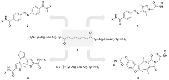
Y 4 R agonists, constituted by two pentapeptide moieties connected through an aliphatic linker, represent an interesting class of Y 4 R ligands. Based on this compound class, photoresponsive Y 4 R ligands, containing an azobenzene, azopyrazole, diethienylethene or a fulgimide chromophore as linker were synthesized to explore structural requirements of such Y 4 R agonists on Y 4 R binding (Scheme 7).
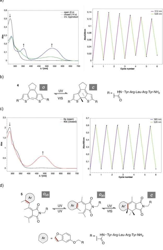
Scheme 7. The structure of the benzofuryl fulgimide based dimeric peptidic NPY Y
4receptor ligand (17) is shown.
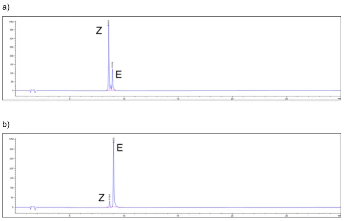
The synthesized Y 4 R ligands, containing a non-aliphatic rigid photochromic linker, showed an efficient and reversible switching in aqueous buffer and exhibited high Y 4 R affinity. This demonstrated that the replacement of the highly flexible aliphatic linker by a considerably less flexible photochromic linker was well tolerated with respect to Y 4 R binding. Differences in Y 4 R affinity and activity between the individual photoisomers, varying in spatial orientation and flexibility, were marginal suggesting that the linking element in the dimeric ligands is less important for the adaptation of high-affinity binding modes at the receptor.
3. Conclusion
The photochemical properties of indolyl-, thiophene- and furane- fulgides are well-suited for
applications in the photomodulation of biological properties. Their photochemical reversibility,
stability and absorption wavelength range is in many cases superior to azobenzenes or
diethenylethenes. However, the number of applications of fulgides and fulgimides is still small
and one reason for this is their challenging synthesis. The success of the Stobbe condensation
and the fulgimide formation depend on the amine that is used and partially on the substitution
pattern of the hexatrien system. More predictable and more general synthetic routes to
functionalized fulgides and fulgimides are clearly in demand to provide this interesting class of
photochromic molecules for broader applications in life science.
4. Literature
1 P. Klán, T. Šolomek, C. G. Bochet, A. Blanc, R. Givens, M. Rubina, V. Popik, A.
Kostikov, J. Wirz, Chem. Rev. 2013, 113, 119
2 W. C. Lin, M. C. Tsai, R. Rajappa, R. H. Kramer, J. Am. Chem. Soc. 2018, 140, 7445.
3 B. Reisinger, N. Kuzmanovic, P. Löffler, R. Merkl, B. König, R. Sterner, Angew. Chem.
Int. Ed., 2014, 126, 606.
4 T. Hirakura, Y. Nomura, Y. Aoyama, K. Akiyoshi, Biomacromolecules 2004 , 5 , 1804.
5 S. Kitzig, M. Thilemann, T. Cordes, K. Rück-Braun, ChemPhysChem 2016, 17, 1252.
6 M. M. Lerch, W. Szymanski, B. L. Feringa, Chem. Soc. Rev. 2018, 47, 1910.
7 D. Lachmann, C. Studte, B. Männel, H. Harald, P. Gmeiner, B. König, Chem. Eur. J.
2017, 23, 13423.
8 I. Willner, M. Lion-Dagan, S. Rubin, J. Wonner, F. Effenberger, P. Bäuerle, Photochem.
Photobiol. 2004, 5, 169.
9 M. Seibold, M. Handschuh, H. Port, H. C. Wolf, J. Luminescence 1997, 72-74, 454.
10 P. Remon, M. Bälter, S. Li, J. Andreasson, U. Pischel, J. Am. Chem. Soc. 2011, 133, 20742.
11 T. Kim, L. Zhu, R.O. Al-Kaysi, Ch. J. Bardeen, Chem. Phys. Chem., 2014 15, 400.
12 D. Wutz, D. Gluhacevic, A. Chakrabarti, K. Schmidtkunz, D. Robaa, F. Erdmann, C.
Romier, W. Sippl, M. Jung, B. König, Org. Biomol. Chem. 2017, 15, 4882.
13 N. A. Simeth, L.-M. Altmann, N. Wössner, E. Bauer, M. Jung, B. König, J. Org. Chem.
2018, 83, 7919.
14 X. Chen, N.I. Islamova, P.S. Garcia, J.A. DiGirolamo, E.J. Lees, J. Org. Chem. 2009, 74, 6777.
15 J.P. Darcy, G.H. Heller, P.J. Strydom, J. Whittall, J. Chem. Soc., P. Trans. 1 1981, 202.
16 Strübe F., Siewertsen R., Sönnichsen D.R., Renth T., Temps F., Mattay F., Eur. J. Org.
Chem., 2011, 10, 1947.
17 B. Otto, K. Rück-Braun, Eur. J. Org. Chem. 2003, 13, 2409
18 K. Ulrich, H. Port, H. C. Wolf, J. Wonner, F. Effenberger, H.-D. Ilge, Chem. Phys. 1991, 2, 311
19 S. A. Harris, H. G. Heller, S. N. Oliver, J. Chem. Soc. Perkin Trans 1, 1991, 3259 20 V. P. Rybalkin, N. I. Makarova, S. Yu. Pluzhnikova, L. L. Popova, A. V. Metelitsa, V. A.
Bren, V. I. Minkin, Russ. Chem. Bull., Int. Ed. 2014, 63, 1780 21 F. Strübe, S. Rath, J. Mattay, Eur. J. Org. Chem. 2011, 24, 4645
22 D. Lachmann, A. Konieczny, M. Keller, B. König, Org. Biomol. Chem. 2019, submitted 23 R. Siewertsen, F. Strübe, J. Mattay, F. Renth, F. Temps, Phys. Chem. Phys. 2011, 13,
3800
24 S. I. Luyksaar, V. A. Migulin, B. V. Nabatov, M. M. Krayushkin, Russ. Chem. Bull., Int.
Ed. 2010, 59, 446
25 M. Kose, E. Orhan, J. Photochem. Photobiol. A 2006, 177, 170
26 A. Liang, A. S. Dvornikov, P. M. Rentzepis, J. Mater. Chem. 2003, 13, 286.
27 Y. Yokoyama, T. Sagisaka, Y. Mizuno, Y. Yokoyama, Chem. Lett. 1996, 587–588.
28 L. Yu, Y. Ming, W. Zhao, M. Fan, J. Photochem. Photobiol. A 1992, 68, 309–317.
29 Y. Yokoyama, T. Tanaka, T. Yamane, Y. Kurita, Chem. Lett. 1991, 7, 1125–1128.
30 M. J., G. W. Trucks, H. B. Schlegel, G. E. Scuseria, M. A. Robb, J. R. Cheeseman, G.
Scalmani, V. Barone, B. Mennucci, G. A. Petersson, H. Nakatsuji, M. Caricato, X. Li, H. P. Hratchian, A. F. Izmaylov, J. Bloino, G. Zheng, J. L. Sonnenberg, M. Hada, M.
Ehara, K. Toyota, R. Fukuda, J. Hasegawa, M. Ishida, T. Nakajima, Y. Honda, O. Kitao, H. Nakai, T. Vreven, J. A. Montgomery, Jr., J. E. Peralta, F. Ogliaro, M. Bearpark, J. J.
Heyd, E. Brothers, K. N. Kudin, V. N. Staroverov, R. Kobayashi, J. Normand, K.
Raghavachari, A. Rendell, J. C. Burant, S. S. Iyengar, J. Tomasi, M. Cossi, N. Rega, J. M. Millam, M. Klene, J. E. Knox, J. B. Cross, V. Bakken, C. Adamo, J. Jaramillo, R.
Gomperts, R. E. Stratmann, O. Yazyev, A. J. Austin, R. Cammi, C. Pomelli, J. W.
Ochterski, R. L. Martin, K. Morokuma, V. G. Zakrzewski, G. A. Voth, P. Salvador, J. J.
Dannenberg, S. Dapprich, A. D. Daniels, J. Farkas; Foresman, J. V. B.; Ortiz, J.
Cioslowski, and D. J. Fox. 2009. 'Gaussian 09, revision B.01', Gaussian, Inc.:
Wallingford, CT.
31 K. Hüll, J. Morstein, D. Trauner, Chem. Rev. 2018, 118, 10710 32 D. Bléger, S. Hecht, Angew. Chem. Int. Ed. 2015, 54, 11338
33 W. Szymanski, J. M. Bierle, H. A. V. Kistemaker, W. A. Velema, B. L. Feringa, Chem.
Rev. 2013, 113, 6114
34 R. Klajn, Chem. Soc. Rev. 2014, 43, 148
35 M. Hammarson, J. R. Nilson, S. Li, T. Beke-Somfai, J. Andréasson, J. Phys. Chem. B 2013, 117, 13561
36 V. I. Minkin, in Molecular Switches, Wiley-VCH Verlag GmbH & Co. KGaA, 2011, pp.
37-80.
37 T. Halbritter, C. Kaiser, J. Wachtveitl, A. Heckel, J. Org. Chem. 2017, 82, 8040
38 J. R. Hemmer, S. O. Poelma, N. Treat, Z. A. Page, N. D. Dolinski, Y. J. Diaz, W.
Tomlinson, K. D. Clark, J. P. Hooper, C. Hawker, J. R. de Alaniz, J. Am. Chem. Soc.
2016, 138, 13960
39 J. D. Harris, M. J. Moran, I. Aprahamian, PNAS 2018, 115, 9414 40 M. Irie, Chem. Rev. 2000, 100, 1685
41 S. Kobatake, M. Irie, Ann. Rep. Prog. Chem. Sect. C. 2003, 99, 277
42 K. Matsuda, M. Irie, Photochem. Photobiol. C. 2004, 5, 169
43 J. Kiji, H. Kitamura, Y. Yokoyama, S. Kubota, Y. Kurita, Bull. Chem. Soc. Jpn. 1995, 68, 616.
44 S. Uchida, Y. Yokoymama, J. Kiji, T. Okano, H. Kitamura, Bull. Chem. Soc. Jpn. 1995, 68, 2961.
45 C. J. Thomas, M. A. Wolak, R. R. Birge, W. J. Lees, J. Org. Chem. 2001, 66, 1914 46 K. K. Krawczyk, D. Madej, J. K. Maurin, Z. Czarnocki, C. R. Chimie 2012, 15, 384 47 J. Kiji, T. Okano, H. Kitamura, Y. Yokoyama, S. Kubota, Y. Kurita, Bull. Chem. Soc.
Jpn. 1995, 68, 616.
48 I. Willner, S. Rubin, J. Wonner, F. Effenberger, P. Bäuerle, J. Am. Chem. Soc. 1992, 114, 3151
49 I. Willner, M. Lion-Dagan, S. Rubin, J. Wonner, F. Effenberger, P. Bäuerle, Photochem.
Photobiol. 1994, 59, 491
C HAPTER 1
1. Photochromic Dopamine Receptor Ligands based on Dithienylethenes and Fulgides
This chapter has been published as:
D. Lachmann, C. Studte, B. Männel, H. Hübner, P. Gmeiner, B. König, Chem. Eur. J. 2017, 23, 13423.
DL synthesized compounds 25, 32, 34, 36, 37 – 52 and performed the corresponding photochemical
measurements and wrote the manuscript. CS synthesized compounds 21 – 24 and 26 – 31 and
performed the corresponding photochemical measurements. BM synthesized compounds 2 – 17. HH
did the biological investigations. BK supervised the project and is corresponding author.
1. Introduction
G-protein coupled receptors (GPCRs) are a vibrant field of research because their dysfunction is linked to numerous diseases like central nervous system (CNS) disorders, cancer or inflammatory diseases. 1a-c Therefore, more than 30% of the approved drugs target the GPCR family. Focusing on the dopamine receptors several CNS disorders like schizophrenia, drug addiction, Parkinson’s and Huntington’s disease are linked to their dysfunction. 2 Privileged structures targeting the dopamine D 2 -like receptors are derivatives of 1,4-disubstituted aromatic piperazines (1,4-DAPs), hydroxybenzoxazinone substituted piperazines as well as conformationally restricted dopamine analogs involving aminoindanes. The synthesis of D 2 , D 3
and D 4 receptor agonists and antagonists with individual subtype selectivity or functional selectivity (biased signaling) has received considerable attention, in recent years. 3a-f Although a better understanding of the mechanism of GPCR-promoted drug action was achieved, there is still a need to develop selective molecular tools to obtain more insight into the dynamics or receptors function.
Photopharmacology can address this issue in a non-invasive way with high spatial and temporal precision. The importance of this concept is revealed by the increasing number of photochromic enzyme inhibitors, 4a-f photochromic ligands for receptors and ion channels, 5a-e photoswitchable antibiotics, 6a-b photo-switches, which are applicable in vivo using visible- light, 7a-c and recently even photochromic ligands, which change their intrinsic activity. 8 Recently, photochromic azobenzol based opioids 5c and dopamine receptor ligands, embedded in a phenethyl-propyl-hydroxytetraline (PPHT) structure, were developed by Trauner et al. 9 Azobenzenes as well as dithienylethenes (DTEs) and fulgides convert light-induced between two isomers, which differ for the azobenzenes in geometry and dipole moment and for the DTEs in conformational flexibility and electronic properties. 10 Both classes of switches show a high fatigue resistance, but only the open and closed form of the DTEs and fulgides are thermally stable, which makes them interesting candidates for photopharmaceuticals. 11a-b Especially fulgides are very interesting for biological applications, because they show high fatigue resistance and are mostly water soluble and switchable in aqueous buffer solutions (Scheme 1). 12a-b
a)
S S R
2R
1UV
VIS R
1S S R
2open (propeller shape)
closed (rigid)
H N O O
=
=
Cyclopentene-DTE
Dithienylmaleimide
b)
Scheme 1. a) Switching principle of dithienylethene based switches. b) Photochromic fulgides/fulgimides, depicted in three forms after irradiation with UV light.
Two major strategies are commonly applied to introduce a photoswitchable moiety into a bioactive compound: the chromophore can either be embedded into the structure of a pharmacophore or attached to one or two pharmacophores via a suitable linker. 10,13 In each case, it is desirable that the light induced switching between the two isomers results in a distinct difference in affinity or intrinsic activity. This would allow the photocontrol of biological functions and offer the possibility to investigate drug targets like GPCRs very precisely.
We envisioned that a formal exchange of a structural appendage of known dopamine ligands by a photoswitchable unit would lead to potent photochromic dopaminergic ligands (Figure 1).
Herein, we discuss the synthesis, photophysical characterization and biological evaluation of photochromic dopamine receptor ligands based on dithienylethene- and fulgide-type scaffolds.
Figure 1. Structures of known dopamine ligands: 1,4-DAP FAUC 346
14, aminoindane FAUC 185
15and benzoxazinone derivative
161, where the blue coloured moieties are replaced by a photochromic diarylmaleimide, dithienylethene or fulgide.
R
3X O
O Ar
X O
O R
3X O
O R
3Ar Ar'
Z-isomer (open)
C-isomer (closed) E-isomer
(open) UV
UV
UV VIS
X = O (Fulgide) X = NR (Fulgimide) R
3= methyl, isopropyl
N
NH O
Fe
FAUC 185 N
N
O
H N
O S
FAUC 346
O HN HO
O
N H
O O
2
1
2. Results and Discussion
Due to their thermal stability, we investigated DTEs first. Within this classes two kind of photoswitches were used to synthesize photoresponsive dopamine ligands: 1) the cyclopentene based switches and 2) the diarylmaleic anhydrides. Both types of DTEs have been successfully applied in photopharmacology. [21] Furthermore, the isomerization wavelength for the chromophores can be bathochromically shifted, reducing cell damage otherwise caused by high energy light. 11a The photoswitches differ in their attachment mode to the pharmacological headgroups. Introducing two different sterically demanding photochromic groups to the pharmacological headgroups allows us to investigate the importance of the size of the heterocyclic appendage for the affinity and subtype selectivity.
2.1 Synthesis - pharmacophoric headgroups
Privileged structures for aminergic GPCR ligands are 1,4-DAPs 3, 5, 7 and 9, aminoindanes 11 and 13 and the benzoxazinon piperazine 17. A coupling reaction between the pharmacophoric head groups and the photoswitches gave the target structures. The 2- methoxy- and 2-methylthiophenylpiperazine building blocks were synthesized as described in literature. 17 Commercially available 1-(2-methoxyphenyl)- and 1-(2-(methylthio)phenyl)- piperazine were alkylated with N-(4-bromobutyl)- or N-(2-bromoethyl)phthalimide to give intermediates 2, 4, 6 and 8 in good yields. Hydrazinolyses of the respective phthalimides yielded building blocks 3, 5, 7, and 9. The same protocol was used to obtain hydroxybenzoxazinone substituted piperazine 17. The synthesis of precursor 14 is described in the supporting information (SI1). Alkylation of 14 with N-(2-bromoethyl)phthalimide gave compound 15. Subsequent hydrazinolysis and acid mediated cleavage of the benzyl protecting groups yielded compound 17. Building blocks 11 and 13 were readily accessible following a two-step literature procedure. 18
2-Propylaminoindane oxalate was reacted with 4-bromobutyronitrile, respectively 2- bromoacetonitrile to afford compounds 10 and 12. Reduction of the nitrile group by LiAlH 4 yielded compounds 11 and 13 (Scheme 2).
Headgroups A:
N Y
N N
n
O
O N
Y
NH
Y = O, S
N Y
N NH
2n
3 (Y = O, n = 2) 5 (Y = O, n = 4) 7 (Y = S, n = 2) 9 (Y = S, n = 4)
a b
2 (Y = O, n = 2)
4 (Y = O, n = 4)
6 (Y = S, n = 2)
8 (Y = S, n = 4)
Headgroups B:
Headgroup C:
Scheme 2. Synthesis of pharmacophore building blocks, headgroups A, B and C. Reagents and conditions: (a) N-(4-bromobutyl)- or N-(2-bromo-ethyl)phthalimid, K
2CO
3, KI, MeCN, 75 °C, 5–16 h, 75–
90%. (b) Hydrazine hydrate, EtOH, 75 °C, 2 h, then 2 M HCl, EtOH, 75 °C, 2 h, 60–85%. (c) MsOH, toluene, 100°C, 2 h, 89%. (d) 4-bromobutyronitrile or 2-bromoacetonitrile, K
2CO
3, KI, MeCN, 75 °C, 16 h 88%. e) LiAlH
4, Et
2O, 0 °C à r.t., 1 h, 71–90%.
2.2 Synthesis - maleimides and cyclopentenes
The DTE and diarylmaleic based pharmacophores, containing the 1,4-DAP, were used with two different spacer lengths. Depending on the DTEs, two different strategies were applied to combine photoswitch 18 with headgroups 3, 5, 11, 13, 17 and cyclopentene-DTE 20 with the pharmacophores 3, 5, 7, 9, 11, 13 and 17. In case of the diarylmaleic anhydride 18, the coupling was performed with the core of the DTE, while the cyclopentene based DTE 20 was coupled with one of the thiophene moieties. Diarylmaleimide 18 was easily accessible according to literature as well as the precursor 19, which was then carboxylated on only one thiophene moiety to obtain 20. 19,20a-b The syntheses of the diarylmaleic anhydride based photochromic ligands 21-25 were performed under slightly basic conditions using potassium carbonate as base and are outlined in Scheme 3.
Scheme 3. Syntheses of the diarylmaleimide based photochromic ligands 21 – 25.
NH N
N NH
2 nN
n
11 (n = 1) 13 (n = 3)
d e
10 (n = 1) 12 (n = 3)
*oxalate
NH N
BnO O
BnN O
14
N
N N
O HN BnO
O
N
N NH
2O
’RN
’RO O
O
15 O 16 (R’ = Bn)
17 (R’ = H)
a b
c
S S Cl
Cl
N O
O R
S S Cl
Cl
O O
O
K
2CO
3CH
2Cl
2or DMF
21 (amine A, 3 , 68%) 22 (amine A, 5 , 80%) 23 (amine B, 11 , 15%) 24 (amine B, 13 , 86%) 25 (amine C, 17 , 9%) R -NH
2(amines A, B or C)
18
Standard peptide coupling conditions using HBTU and N,N-diisopropylethylamine were applied to synthesize the photochromic ligands 26 - 32 (Scheme 4).
Scheme 4. Syntheses of the cyclopentene-DTE based photochromic ligands 26 - 32.
Following these two synthetic strategies, we were able to readily obtain various photochromic ligands in just one step. A stock solution for the biological testing was prepared with dimethyl sulfoxide as solvent. Dilutions thereof were used to study the photophysical properties. Even though it is reported that diarylmaleimides cannot be reversibly toggled between their two photoisomers in polar solvents due to a twisted intramolecular charge transfer (TICT), we observed a reversible photoisomerization in dimethyl sulfoxide for all photochromic ligands based on diarylmaleimides. 21a-c
S S Cl
H N
S S Cl O
O
HBTU, DIPEA HO
R -NH
2(amines A, B or C)
26 (amine A, 3 , 38%) 27 (amine A, 5 , 19%) 28 (amine A, 7 , 29%) 29 (amine A, 9 , 26%) 30 (amine B, 11 , 26%) 31 (amine B, 13 , 70%) 32 (amine C, 17 , 7%) 20
CH
2Cl
2or DMF R
2.3 Photophysical properties - arylethenes
The photophysical properties of the photochromic dopamine receptor ligands 21-25 and 26- 32 were investigated by absorption spectroscopy. Therefore, the dissolved compounds were irradiated with UV-light (312 or 400 nm) which resulted in a color change of the solution accompanied with new absorption maxima, characteristic for each of the photochromic ligands.
The resulting isosbestic points indicate a clean two-component switching. In addition, the PSS was determined by HPLC measurements. All photophysical properties of the compounds 21- 25 and 26-32 are summarized in Table 1.
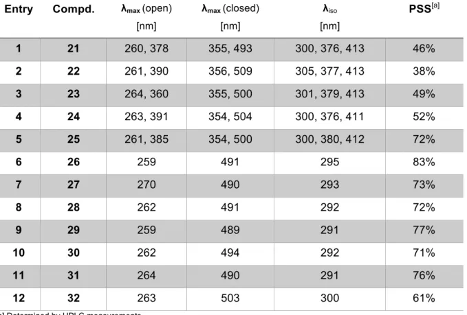
Table 1. UV-Vis data of the open and closed form of the synthesized ligands 21-25 and 26-32 (50 or 100 µM in DMSO) after irradiation with λ = 312 or 410 nm.
[a] Determined by HPLC measurements.
Entries 1-5 represent the photophysical characterization of the diarylmaleimide based photochromic ligands 21, 22, 23, 24 and 25, whereas entries 6-12 summarize the cyclopentene based photochromic ligands 26-32. A slightly bathochromic shift is observed for the new arising absorption maxima corresponding to the closed photoisomers of the diarylmaleimides 21-25, which is characteristic for this class of DTEs. 11a To our surprise, the photoconversion of the diarylmaleimides 21, 22, 23 and 24 (excluded 25) is not as efficient as for the cyclopentene ligands 26-32. Compared to a recently reported PSS of 94% for a diarylmaleimide photoswitch, with a free maleimide core and a phenyl substitution pattern on the thiophene moieties, 4a the substitution on the nitrogen atom of the maleimide core with the pharmacophores dramatically decreases the efficacy of the photoisomerization. In contrast, the cyclopentenes 26-32 exhibit
Entry Compd. λ
max(open) [nm]
λ
max(closed) [nm]
λ
iso[nm]
PSS [a]
1 21 260, 378 355, 493 300, 376, 413 46%
2 22 261, 390 356, 509 305, 377, 413 38%
3 23 264, 360 355, 500 301, 379, 413 49%
4 24 263, 391 354, 504 300, 376, 411 52%
5 25 261, 385 354, 500 300, 380, 412 72%
6 26 259 491 295 83%
7 27 270 490 293 73%
8 28 262 491 292 72%
9 29 259 489 291 77%
10 30 262 494 292 71%
11 31 264 490 291 76%
12 32 263 503 300 61%
sufficiently high photoconversions shown by PSS ranging from 61-83%. Surprisingly, the cycle performance exhibits a better fatigue resistance for the maleimide-based switches 21-25.
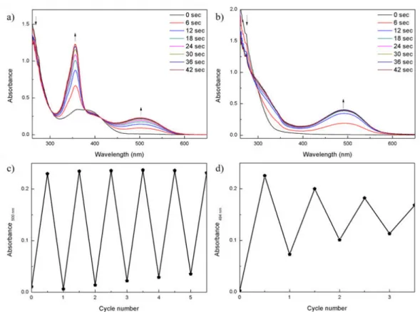
Figure 2 exemplary shows the UV-Vis absorption spectra and the cycle performance for the diarylmaleimide based ligand 23 and for the cyclopentene based ligand 30.
Figure 2. Comparison of the photochromic properties of diarylmaleimide based ligand 23 (a, c) (100 µM in DMSO) with the cyclopentene based ligand 30 (b, d) (100 µM in DMSO); a) and b) represent the UV-Vis absorption spectra upon continuous irradiation with light of λ = 312 nm (Herolab, 6 W), respectively; c) and d) represent the cycle performance using λ = 312 nm (Herolab, 6 W) for the ring closing and λ = 530 nm (green LED, 2.5 W) for the ring opening reaction, respectively: a) UV-Vis absorption spectra of ligand 23; b) UV-Vis absorption spectra of ligand 30; arrows indicate the characteristic changes in the absorption spectra; c) repetitive switching cycles at 500 nm of ligand 23; d) repetitive switching cycles at 494 nm of ligand 30.
The arrows indicate the characteristic changes in the spectra upon continuous irradiation with light of λ = 312 nm (Herolab, 6 W), accompanied by a characteristic color change of the samples. The cycle performance showed that ligand 23 is stable over at least six switching cycles, whereas a fast degradation of cyclopentene 30 was observed.
Further investigations by continuous irradiation with UV light (λ = 312 nm) revealed the
formation of an irreversible by-product 33 (Scheme 5). The phenomenon of this annulated
isomer has been reported previously, but the mechanism of the by-product formation, which is
related to the substitution pattern of the DTEs is not yet fully understood. 22a-c
Scheme 5. Formation of the irreversible by-product 33 of the ligand 30 (R = 11) upon continuous UV irradiation.
The photoisomerization can be monitored by UV-Vis absorption spectroscopy and is depicted in Figure SI2a-c. By irradiating the dissolved compound 30 with λ = 312 nm (Herolab, 6 W) one new maximum is arising at 494 nm, which is decreasing upon further UV irradiation. A new absorption maximum is observed, hypsochromically shifted to 360 nm, which is attributed to the irreversible occurring by-product 33. The HPLC traces in Figure SI2d show that upon reaching the PSS, approximately 55% of the by-product has already been formed and its formation is completed after 192 sec. Similar to the results by Hecht et al. 22a we could confirm the structure of 33 with 2D-NMR spectroscopy experiments (see SI3, Scheme SI3-1 and SI3- 2).
2.4 Synthesis - fulgides and fulgimides
In a second series of dopamine receptor ligands, fulgides were synthesized, promising reversible switching in aqueous solution 12a-b and good fatigue resistance. The synthesis of the benzothiophene fulgide 37 was performed according to the experimental protocol of Stobbe e.
al. and Mattay et al. 23a-b The challenging step, the Stobbe condensation, was accomplished by enolization of isopropylidenesuccinate and subsequent reaction with 34. This reaction results in a mixture of syn/anti lactones 36 whereby the syn-isomer 36 is the favoured one due to the isopropylgroup of 34. The diacids were obtained after an elimination reaction and subsequent saponification by aqueous potassium hydroxide solution. For the anhydride formation, the crude diacids were treated with acetyl chloride to form the fulgide 37E (Scheme 6).
Scheme 6. Benzothiophene fulgide 37 synthesis according Stobbe et al.; Reagents and conditions: (a) Isopropylidenesuccinate, LDA, THF, -78 °C à r.t., 48 h, 16%. b) Step 1: KOH, EtOH, H
2O, 70 °C, 24 h;
Step 2: AcCl, CH
2Cl
2, 40 °C, 20 h, 87%.
S S NH R
O Cl
312 nm 530 nm
S S
Cl NH R
O S S
O NH R Cl
312 nm
30 open
30 closed
33 by-product
S O
S O
O
COOEt S O
O
O
syn/anti- 36 37E
34
a b
The indolyl-isopropyl fulgide 38, 5-methylthiophene-isopropyl fulgide 39, benzothiophene methyl fulgide 40 and indolylmethyl fulgide 41 were synthesized according to literature. [24-26]
Only the E-isomers were isolated and used for fulgimide formation.
The target fulgimides were prepared in a two-step reaction. First, the succinamic acids 42, 44, 47, 49 and 51 were obtained by heating the amines 7, 11 and 17 with the fulgides 37 - 41 in chloroform or DMF. Second the imides were formed by an active ester method with DCC, HOBt and DIPEA or MSNT and Me-imidazole to obtain the desired fulgimides 43, 45, 46, 48, 50 and 52 (Scheme 7).
Scheme 7. Fulgimide synthesis with fulgide 37 (Ar = benzothiophene, R
3= isopropyl), 38 (Ar = indole, R
3= isopropyl), 39 (Ar = 5-methyl-thiophene, R
3= isopropyl), 40 (Ar = benzothiophene, R
3= methyl), 41 (Ar = indole, R
3= methyl) and amines 7, 11, 17. Reaction conditions: a) amine A, B or C, CHCl
3or DMF, 60 °C, 4-48h; b) DCC, HOBt, DIPEA or MSNT, Me-imidazole, CHCl
3or DMF, 25 °C, 24-48h.
The fulgimide 46 was formed after heating in CHCl 3 for 24 h without adding coupling reagents.
No fulgimide could be obtained with amine 9 and 13.
O
aO
O R
3Ar N
O
O R
3Ar R
37 - 41
COOH R
3O
Ar NH R
42
(amine A, 7, fulgide 37)44
(amine A, 7, fulgide 38)47
(amine B, 11, fulgide 40)49
(amine B, 11, fulgide 41)51
(amine C, 17, fulgide 41)43
(amine 7, fulgide 37, 74%)45
(amine 7, fulgide 38, 25%)46
(amine 11, fulgide 39, 10%)48
(amine 11, fulgide 40, 9%)50
(amine 11, fulgide 41, 14%)52
(amine 17, fulgide 41, 10%) b2.5 Photophysical investigations - fulgimides
Applications of photoswitches in biological assays require their solubility in aqueous buffer.
Hence, the photoisomerization of the fulgimides 45, 50 and 52 were carried out in polar solvents to obtain UV/Vis absorption spectra. Table 2 summarizes the photostationary states (PSS), isosbestic points as well as the absorption maxima of 43 and 45, determined in DMSO.
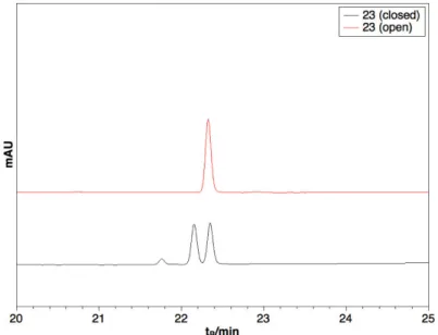
The method for the PSS determination by HPLC is exemplarily shown for compound 48 in the SI4. The indolyl fulgimides 45, 50 and 52 were also measured in a 10% DMSO – tris-buffer solution (pH = 7.4) (Table 2, Entry 3,7 and 9), whereby a hypsochromic shift of 18 nm of the absorption maximum in the visible light range was observed.
Table 2. Photochemical properties of fulgimides 43-52.
[a] Closing of the open E-isomers with UV light, PSS determined by HPLC measurements. [b] Opening of the closed mixture with visible light, PSS determined by HPLC measurements. [c] Solvent: DMSO. [d] Solvent: Tris-buffer + 10% DMSO.
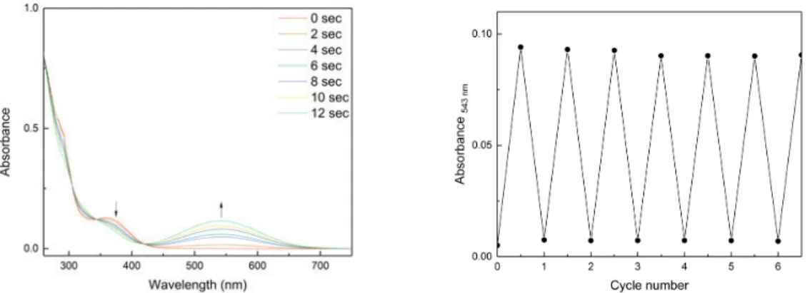
The UV/Vis absorption spectra changes are exemplarily shown for compound 45 in Figure 3 upon irradiation with 410 nm light in periods of 2 sec. New absorption bands arise at 543 nm and 330 nm, the band at 360 nm decreases and belongs to the open isomer. The opening of the closed photoisomers was accomplished by irradiation with light of 505 or 530 nm (1-2 min) for 43, 45, 46, 48, 50 and 52.
Entry Compd. λ
max(open) [nm]
λ
max(closed) [nm]
λ
iso[nm]
PSS [a]
OàC (Z:E:C)
PSS [b]
CàO (Z:E)
1 43 [c] 307 473 290, 342, 347 0:30:70 -
2 45 [c] 360 543 310, 344, 420 0:20:80 -
3 45 [d] 356 525 305, 337, 417 0:19:81 -
4 46 [c] 272 532 360 0:16:84 -
5 48 [c] 328 486 315, 357 19:36:45 19:81
6 50 [c] 370 564 306, 436 6:16:78 6:94
7 50 [d] 370 325, 546 475 4:18:78 4:96
8 52 [c] 363 563 302, 325, 430 4:12:84 4:96
9 52 [d] 279, 366 300, 545 300, 320, 409 4:10:86 4:96
Figure 3. Left) the photochromic properties of a solution of 45 (50 µM in DMSO). The slightly yellow open form turned purple after 12 sec irradiation with 410 nm. The absorption spectra showed a new absorption band at 543 nm. Right) Repetitive switching of 45 is depicted on the right side.
The cycle performances of compounds 43, 45, 46, 48, 50 and 52 were studied by alternating irradiation at the appropriate wavelengths (43 à 312 close nm, 505 open nm; 45 à 400 close nm, 530 open nm; 46, 48 à 365 close nm, 530 open nm; 50, 52 à 400 close nm, 530 open nm). The compounds showed good fatigue resistance and only little degradation was observed.
Exemplarily, the cycle performance of compound 45 is depicted in Figure 3.
2.6 Biological investigations
The biological investigation of all test compounds was targeted towards their ability to activate the dopamine D 2S receptor. Our studies were directed to the identification of compound pairs exhibiting different activation profiles for the open and the closed photostationary state.
Therefore, we applied screening assays for G-protein mediated signaling and arrestin recruitment at a fixed concentration (10 µM). The G-protein pathway was investigated utilizing the IP-One ® assay (Cisbio) with HEK 293T cells transiently co-expressing D 2S and the hybrid G-protein Gaqi5HA while ß-arrestin-2 recruitment was determined applying the PathHunther ® assay (DiscoverX). All activation data are summarized in Table 3, SI5 and SI6 in comparison to the reference ligand quinpirole. In the arrestin assay, the cyclopentene DTEs 26-30, 32, the diarylmaleimides 21-23, 25 and the fulgimides 43, 45, 46, 48, 50 and 52 showed E max values less than 15%. Only DTE 31 and the maleimide 24 both bearing the indanylamine moiety and a 4-atom spacer between the pharmacophoric headgroup and the photochromic entity (13) showed Emax values of 70% (31-open), 76% (31-closed), 33% (24-open), and 37% (24- closed), respectively.
Additional measurement of arrestin recruitment for those compounds at 100 nM revealed
Emax values less than 10% and no differences in the activation pattern of the open and closed
conformation. Whereas weak arrestin recruitment was observed, the determination of the G-
protein mediated signaling revealed agonist properties with efficacies in the range of 40 to 95%
for all test compounds (Figure SI5, Table SI5-1, SI5-2). More detailed measurements at 100 nM and 1 nM showed a dose-dependent range of efficacies allowing a precise evaluation of the open and closed conformations of each photochromic ligand. Within the series of maleimides, the indanylamine 24 showed the highest Emax values of 95% for both conformers, which is similar to the rank order of agonist effects derived from the arrestin assay, but without any difference between the open and the closed form. This observation can be confirmed by the results for the cylopentene DTEs, where 31 (indanylamine with headgroup 13) showed an Emax value of 95% for the open and the closed conformation, respectively.
Clear differences in Emax between both photochromic states could be observed for the methyl ether substituted phenylpiperazines 27 and 29 both carrying a 4-carbon spacer and only differing in the heteroatom of the ether group (O for 27, S for 29). For 29, Emax values of 77%
(10 µM), 80% (100 nM) and 32% (1 nM) have been determined for the open state, while efficacies of 70% (10 µM), 47% (100 nM) and 3% (1 nM) could be measured for the closed form (Table 3, Figure 4). This data indicate a 11-fold more efficient receptor activation by the open conformer compared to the closed derivative at low concentration (1 nM). In contrast, the methoxyphenylpiperazine 27 showed stronger receptor activation profile in the closed state compared to the open derivative, when a 3-fold increase of efficacy was determined for 27- closed at 100 nM (closed-open: 49%-16% at 100 nM, 15%-11% at 1 nM). A similar selectivity profile could be detected for the indolyl fulgimides 45 and 52. The methylthiophenylpiperazine 45-closed showed a 2-fold increase of efficacy at 100 nM compared to the open state, while the benzoxazinone 52-closed revealed a 4-fold improve of activity at 1 nM (for 45: closed- open: 45%-25% at 100 nM, 8%-5% at 1 nM, for 52: closed-open: 47%-48% at 100 nM, 40%- 10% at 1 nM).
To learn more about the mechanistic relations of these selective photoswitchable ligands, we investigated the binding affinities of the most promising compounds 27-open, 27-closed, 29- open, 29-closed, 45-open, 45-closed, and 52-(E)-open, 52-closed at the dopamine D 2S
receptor in a competition binding experiment. As all compounds displayed K i values between
9 and 17 nM (Table SI6), the state-selective intrinsic activity appears to be controlled by the
individual ability to preferentially stabilize the active state of the receptor rather than by different
binding affinities.
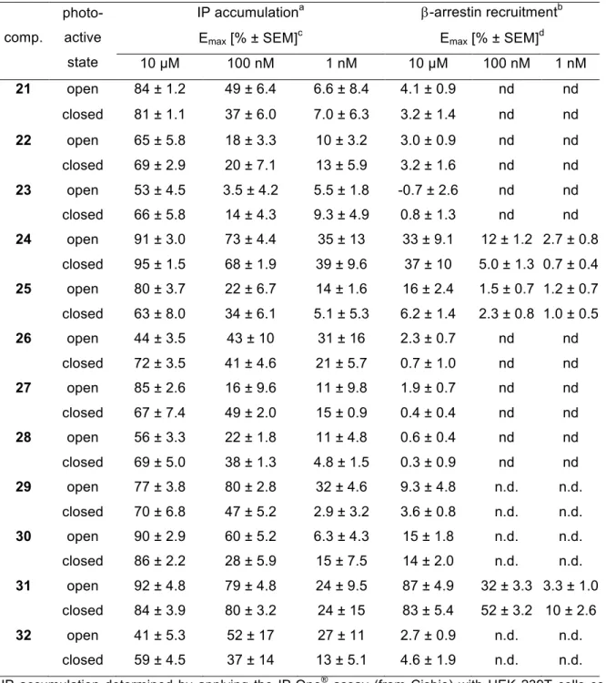
Table 3. Functional screening of the most promising photoactive ligands 27, 29, 45 and 52 for G-protein mediated activation of the dopamine D
2Sreceptor applying an IP accumulation assay
[a].
[a] IP accumulation determined by applying the IP-One
®assay (from Cisbio) with HEK 239T cells co-transfected with the cDNA of the dopamine D
2Sreceptor and that of the hybrid G-protein Gaqi5HA. [b] Emax value ± S.E.M. derived from 4 to 8 individual experiments each done in quadruplicate relative to the maximum effect of quinpirole. [c] The open isomer describes only the E-isomer.
An overall analysis of the data confers that the photoswitches 29 and 52 are most promising biochemical tools for the regulation of the pharmacologically important dopamine D 2S receptor.
The potency of the more active isomers is comparable to the activation power of the reference dopaminergic agent quinpirole. The compounds require a very low dose of 1nM to exhibit a degree of receptor activation that strongly depends on the photostationary state of the ligand.
Hence, the presence of the cyclopentene-DTE based photochromic ligands 29 in the open state induces an 11-fold higher G protein-promoted signaling than the existence of 29-open in the same concentration. As a complementary pair of photochromic ligands, the fulgimide 52 shows four-fold higher D 2S receptor activation in the closed state compared to 52-open (Figure 4).
Entry Compd. Photoactive
state 10 [µM]
Emax [% ± SEM]
[b]100 [nM] 1 [nM]
1 27 open 85 ± 2.6 16 ± 9.6 11 ± 9.8
2 27 closed 67 ± 7.4 49 ± 2.0 15 ± 0.9
3 29 open 77 ± 3.8 80 ± 2.8 32 ± 4.6
4 29 closed 70 ± 6.8 47 ± 5.2 2.9 ± 3.2
5 45 open 68 ± 2.8 25 ± 5.3 5.6 ± 3.2
6 45 closed 62 ± 2.4 45 ± 7.8 7.7 ± 2.6
7 52 [c] open 58 ± 6.9 48 ± 8.5 10 ± 3.8
8 52 [c] closed 69 ± 6.6 47 ± 4.2 40 ± 4.6
Figure 4. Activation of G-protein mediated receptor signaling determined by an IP accumulation assay using D
2s and the G-protein hybrid Gaqi5HA. Accumulation of IP for the most promising photoswitchable ligands 29 and 52 in comparison to the reference quinpirole all determined at 1 nM. The DTE 29 shows an 11-fold improved activity for the open state (dark blue) than the closed state (light blue). In contrary, fulgimide 52 is 4-fold less active in the open conformation than for the closed one. For comparison, receptor activation induced by 1 nM of quinpirole (striped) reveals an efficacy of 28 % (derived from the dose-response curve, Figure SI4). Bars represent average efficacy [%±S.E.M.] derived from 4 to 8 individual experiments each done in quadruplicates.
3. Conclusions
We succeeded in synthesizing DTE and fulgide based photochromic dopamine receptor ligands. The maleimides could not be switched in aqueous solution and the cyclopentene- DTEs had a low fatigue resistance, whereas the fulgimides showed excellent photophysical properties in aqueous solution. At a concentration of 1 nM, the cyclopentene-DTE 29-open showed a more than 10-fold higher activation of D 2S , a pharmacologically important G protein- coupled receptor, than its photochromic congener 29-closed. Interestingly, the fulgimide-based pair 52-open/52-closed could be discovered as an alternative photoswitch with inverse activation properties exhibiting four-fold higher activity in the closed state. Further studies on the optimization of GPCR-regulating photoswitches and biological investigations including reversible, light-induced control of photochromic ligands when bound to the receptor are in progress.
4. Experimental section
Starting materials were purchased from commercial suppliers and used without any further purification. Solvents were used in p.a. quality and dried according to common procedures, if necessary. Dry nitrogen was used as inert gas atmosphere. Thin-layer chromatography (TLC) for reaction monitoring was performed with alumina plates coated with Merck silica gel 60 F 254 (layer thickness: 0.2 mm) and analyzed under UV-light (254 nm). Flash column chromatography was performed with Sigma Aldrich MN silica gel 60M (0.040-0.063 mm, 230-
0 2 5 5 0 7 5 1 0 0
IP accumulation [% rel. quinpirole]
q u i n p i r o l e 2 9
o p e n 2 9 c l o s e d
5 2 o p e n
5 2 c l o s e d o p e n s t a t e c l o s e d s t a t e