R E S E A R C H Open Access
Re-irradiating spinal column metastases using IMRT and VMAT with and without flattening filter - a treatment planning study
Barbara Dobler*, Amine Khemissi, Tina Obermeier, Matthias G. Hautmann, Zaira Katsilieri and Oliver Kölbl
Abstract
Background:The aim of this study was to investigate the potential of the flattening filter free (FFF) mode of a linear accelerator for intensity modulated radiation therapy (IMRT) and volumetric modulated arc therapy (VMAT) for patients with in-field recurrence of vertebral metastases.
Methods:An Elekta Synergy Linac with Agility™head is used to simulate the treatment of ten patients with locally recurrent spinal column metastases. Four plans were generated for each patient treating the vertebrae sparing the spinal cord: Dual arc VMAT and nine field step and shoot IMRT each with and without flattening filter. Plan quality was assessed considering target coverage and sparing of the spinal cord and normal tissue. All plans were verified by a 2D-ionisation-chamber-array, peripheral doses were measured and compared to calculations. Delivery times were measured and compared. The Wilcoxon test was used for statistical analysis with a significance level of 0.05.
Results:Target coverage, homogeneity index and conformity index were comparable for both flat and flattening filter free beams. The volume of the spinal cord receiving the allowed maximum dose to keep the risk of radiation myelopathy at 0 % was at the same time significantly reduced to below the clinically relevant 1 ccm using FFF mode. In addition the mean dose deposited in the surrounding healthy tissue was significantly reduced in the FFF mode. All four techniques showed equally good gamma scores for plan verification. FFF plans required considerably more MU per fraction dose. Regardless of the large number of MU, out-of-field point dose was significantly lower for FFF plans, with an average reduction of 33 % and mean delivery time was significantly reduced by 22 % using FFF beams.
When compared to IMRT FF, VMAT FFF offered even a reduction of 71 % in delivery time and 45 % in peripheral dose.
Conclusions:FFF plans showed a significant improvement in sparing of normal tissue and the spinal cord, keeping target coverage and homogeneity comparable. In addition, delivery times were significantly reduced for FFF treatments, minimizing intrafractional motion as well as strain for the patient. Shortest delivery times were achieved using VMAT FFF.
For radiotherapy of spinal column metastases VMAT FFF may therefore be considered the preferable treatment option for the combination of Elekta Synergy Linacs and Oncentra® External Beam v4.5 treatment planning system.
* Correspondence:barbara.dobler@ukr.de
Department of Radiotherapy, Regensburg University Medical Center, Regensburg, Germany
© 2016 Dobler et al.Open AccessThis article is distributed under the terms of the Creative Commons Attribution 4.0 International License (http://creativecommons.org/licenses/by/4.0/), which permits unrestricted use, distribution, and reproduction in any medium, provided you give appropriate credit to the original author(s) and the source, provide a link to the Creative Commons license, and indicate if changes were made. The Creative Commons Public Domain Dedication waiver (http://creativecommons.org/publicdomain/zero/1.0/) applies to the data made available in this article, unless otherwise stated.
Background
The flattening filter in the treatment head of a linear ac- celerator allows achieving a homogeneous dose profile for photon beams. In the past this was necessary for 2D radiation therapy planning and facilitated forward 3D ra- diation therapy planning. The use of flattening filters im- plies, however, also certain disadvantages: The flattening filter reduces photon fluence, leading to lower dose rates and prolonged beam-on times, and increases scatter dose produced in the treatment head [1–3]. Through the development of intensity modulated radiation therapy (IMRT) and the associated inverse treatment planning, a homogeneous beam profile is no longer necessary. The latest development in the technology of linear accelera- tors is therefore the opportunity to irradiate patients without a flattening filter in the beam path to increase dose rate and reduce beam-on times as well as out-of- field doses [1]. The physical properties of flattening filter free beams have been subject to a wide number of inves- tigations during the last years [4–14]. Several treatment planning studies have been published to assess the clin- ical value of FFF beams for patient treatment, most of them using Varian linear accelerators [15–23]. The flat- tening filter free technique became commercially avail- able for Elekta linear accelerators in 2013, therefore only a few planning studies are available for Elekta up to now, most of them conducted in the treatment planning system Monaco for stereotactic treatments [24–27].
Only a few planning studies also consider the benefit of a potential reduction in peripheral dose [2, 25].
Since patients with spinal metastases suffer from enor- mous pain, a reduction in delivery time would mean less emotional strain for the patients as well as reduced risk for intrafractional motion [28]. A reduction in peripheral dose leads in general to a reduction in normal tissue complication probability. The purpose of the project presented here was therefore to investigate the benefit of flattening filter free beams in the re-irradiation of spinal column metastases considering plan quality, total deliv- ery time and peripheral dose.
Methods Patients
CT data of ten patients with spinal column metastases who had previously been treated with 3D-CRT were se- lected from our treatment database. These data had pre- viously been used for another study with identical delineation of the planning target volume (PTV) and or- gans at risk (OAR) and dose prescription [29]. The PTV of the first course (pre-irradiation) consisted of the 1–5 thoracic vertebrae including the spinal cord. For this study it was assumed that the patients have an in-field recurrence and the whole vertebra region including the spinal canal was pretreated with a dose of 10 × 3 Gy in
the first course. For the second course considered in this planning study, the CTV consists of one to five whole vertebrae including the vertebral body, the vertebral arch, the transverse processes and the spinous process.
The spinal canal is excluded from the CTV and consid- ered as organ at risk. The PTV is defined as CTV ex- tended by a 3 mm margin in each direction excluding the spinal canal from this expansion. Volumes of the PTV ranged from 101 to 388 ccm, with a cranio-caudal extension ranging from 4 to 11.5 cm. Prescription for the second course is 6 × 4 Gy average dose to the PTV.
To avoid myelopathy the dose to the spinal cord was re- stricted to 18 Gy due to the exposure in the first series.
The restriction to 18 Gy was calculated based on the risk score model of Nieder et al. [30, 31] who found no incidence of radiation myelopathy after a total biologic- ally effective dose BED of 120 Gy2, if the interval be- tween radiation courses was at least 6 months and the BED of each course was not higher than 98 Gy2. Theα/
βvalue for spinal cord was hereby assumed to be 2 Gy [30, 31]. The biologically effective dose is defined as BED = n ∙ d ∙ (1 + d/(α/β)) according to Fowler [32], where n is the number of fractions, d the dose per frac- tion and αand βthe coefficients of the linear quadratic cell model. Since the BED of the first course considered in our study was 75 Gy2, the remaining maximum BED for the second course was 45 Gy2corresponding to 6 × 3 Gy to assure a total maximum BED of 120 Gy2, i.e. a 0 % risk of radiation myelopathy. Details about the calculation are de- scribed in Groeger et al. [29].
Linear accelerator and treatment planning system Treatment planning is performed with Oncentra® Exter- nal Beam v4.5 (Nucletron, an Elekta Company) for a Synergy linear accelerator with Agility™head (Elekta AB, Stockholm, Sweden) and six MV photons with flattening filter (FF) or without (FFF). The FFF beams were energy-matched to the FF beams as it is common for Elekta accelerators [12, 13, 33]. The multi leaf collimator consists of 80 leaf pairs of 5 mm width at isocenter. The maximum nominal dose rate is 500 MU/min in FF Mode and 1700 MU/min in FFF mode. Beam profiles, depth doses and dose output were found to be stable for 4 MU and larger in both irradiation modes as also reported by Akino [14]. Verification of the linac model in Oncentra by collapsed cone dose calculations of per- centage depth doses, profiles and output factors was within specifications of Oncentra®, i.e. 3 % of calibration dose in the dose plateau and 3 mm distance deviation to correct dose value in sharp dose gradients, for both FF and FFF. The accuracy of the collapsed cone dose calcu- lation algorithm implemented in Oncentra has previ- ously been reported to be at least as high for FFF beams as for FF beams [34].
Treatment planning
In total four treatment plans were created for each pa- tient, using two different treatment techniques IMRT and volumetric modulated arc therapy (VMAT) and two different irradiation modes with and without flattening filter. In the following the plans are referred to as IMRT FF, IMRT FFF, VMAT FF and VMAT FFF. The IMRT plans consist of nine equispaced beams, minimal seg- ment size was 9 cm2, maximal number of segments allowed was 70. Minimal number of monitor units per segment is four due to the determined stability of the beam for 4 MU and higher. The VMAT plans consist of two full rotations with gantry spacing between two con- trol points of 4°. Collimator angles ranged from 0 to 45°
for both techniques. Identical dose volume objectives (DVO) and weights were used for optimization of all plans (Table 1). Suitable DVO and weights were deter- mined creating plans in FF mode which met the goals and then transferred to the FFF plans. All plans were ac- cepted for treatment by a specialized radiation oncologist.
Dosimetry
For verification all 40 plans were transferred to a CT scan of the MatriXX Evolution™ 2D-ionisationchamber-array (IBA Dosimetry, Schwarzenbruck, Germany) set up in be- tween slabs of a RW3 phantom (PTW, Freiburg, Germany) for measurement in a coronal plane [35, 36].
The 2D-ionisationchamber-array MatriXX Evolution™
consists of 1020 vented pixel ionisationchambers arranged in a square of 24.4 cm × 24.4 cm with a center-to-center distance of 0.76 cm, the chamber size is 0.45 cm diameter and 0.5 cm height, the active volume is 0.08 cm3, and RW3 is used as backscatter material. RW3 consists of white polystyrene and is dosimetrically water-equivalent for photons in the range of60Co to 25 MV. The isocenter was placed such that the measurement plane intersected both the PTV and the spinal cord in order to verify the dose to the PTV and the dose to the organ at risk at the
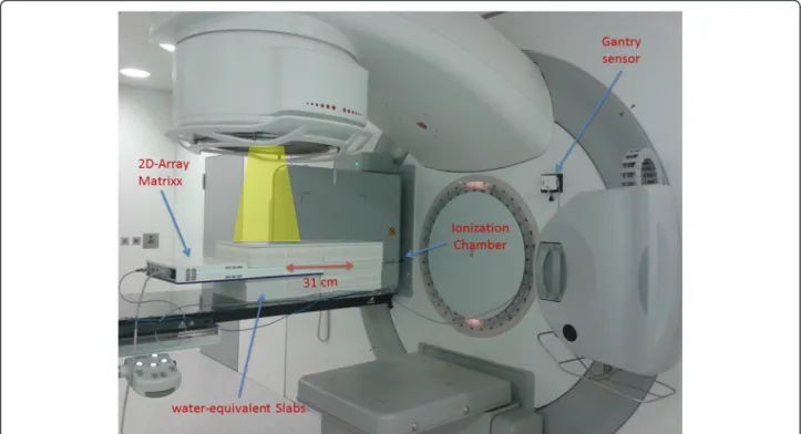
same time. This is possible considering the diameter of the spinal cord of around 1.5 cm, the chamber distance of 0.76 cm and the active chamber volume of 0.08 cm3. It should be mentioned, however, that the measurement of very steep gradients between PTV and spinal cord is lim- ited by the chamber distance. The dose in the measure- ment plane was calculated with a dose grid resolution of 0.15 cm, all other parameters were kept identical to the patient plan. The plans were then delivered to the phan- tom. Measurements were corrected for angular dependen- cies and couch attenuation in the steering and evaluation software OmniPro I’mRT v.1.7a (IBA Dosimetry, Schwar- zenbruck, Germany). In addition a point dose measure- ment was performed in the same coronal plane but 31 cm cranial of the isocenter using a 0.3 ccm PTW ionization chamber to assess peripheral dose. The dose at the point of measurement was calculated in Oncentra on the CT scan of the phantom. The complete measurement setup is shown in Fig. 1.
Efficiency
Delivery times were measured from first beam on to last beam off to assess the achievable reduction in delivery time. In addition the number of required monitor units (MU) per Gray was compared.
Evaluation
Plan quality was assessed by analysis of the dose vol- ume histogram (DVH) with respect to target cover- age, dose homogeneity and conformity, dose to the spinal cord and normal tissue. Target coverage was represented by the volume of the PTV covered by 95 % of the prescription dose (V95%). The homogen- eity index was defined as HI := (D1% - D99%)/D50%, the conformity index according to Paddick et al. [37]
as CI := V95%2
/(TV ⋅ PIV). Here TV means the vol- ume of the PTV, PIV the total volume covered by 95 % of the prescription dose. To assess the sparing of the spinal cord as described in the treatment goals, the volume V75% covered by 75 % of the prescription dose i.e. 18 Gy was recorded. The value of 18 Gy was calculated as residual tolerance dose due to the ex- posure in the first series. According to ICRU report 50 “a significant tissue volume must be irradiated for the dose level to be reported as maximum” [38].
ICRU report 80 suggests that D2% may be an appro- priate value if the whole structure is delineated [39], which corresponds to a volume of about 1 ccm which is also commonly used in the literature [40]. The me- dian dose D50% of the normal tissue, which is defined as the patient body excluding the PTV, is listed as a measure of low dose in the periphery.
For evaluation of plan verifications gamma indices as defined by Low et al. [41] were calculated with a dose Table 1Dose Volume Objectives (DVO)
Organ Type DVO relative
weight
PTV target uniform dose 24.0 Gy 7000
minimum dose 23.5 Gy 7000 maximum dose 24.5 Gy 7000 spinal canal organ at risk maximum dose 18.0 Gy 750 spinal cord organ at risk maximum dose 16.0 Gy 1000 normal
tissue
organ at risk maximum dose 24.5 Gy 5000 surrounding dose fall off from 24.0 to 4.8 Gy in 5.0 cm
5000
Identical dose volume objectives (DVO) and weights were used for optimization of all plans
tolerance of 3 % of the maximum dose and 3 mm dis- tance to agreement. Dose calculations are considered ac- ceptable if at least 95 % of the pixels with a dose value of≥10 % of the maximum dose have a gamma value≤1 as recommended by the AAPM TG119 [42, 43]. Ionch- amber point dose measurements and corresponding cal- culations 31 cm cranial of the isocenter were compared for the two irradiation modes to assess peripheral dose.
The Wilcoxon test implemented in IBM SPSS® Statis- tics 23.0 (IBM Corporation) was used for statistical ana- lysis with a significance level of 0.05 of a) the FF mode versus the FFF mode separated by the planning tech- niques IMRT and VMAT and b) IMRT versus VMAT separated by the irradiation mode FF and FFF. For the assessment of peripheral dose and delivery time, the Wilcoxon test was also performed for all plans in FF mode versus FFF mode.
Results
Since the main subject of the study was the compari- son of the two irradiation modes FF and FFF, details about statistical significance are listed in the tables for these Wilcoxon tests. Differences between IMRT and VMAT are mentioned in the text but not listed in detail in the tables for the sake of clarity.
Plan quality
Analysis of the dose volume parameters listed in detail in Table 2 shows slightly higher plan quality for both IMRT and VMAT, if flattening filter free beams were used: the volume of the spinal cord receiving the allowed maximum dose as well as the dose to the normal tissue could be significantly reduced keeping target coverage, homogeneity and conformity at the same level. The vol- ume of the spinal cord receiving 18 Gy was kept well below the clinically relevant volume of 1 ccm in all cases if FFF mode was used but exceeded 1 ccm in 50 % of the cases for FF mode, with a maximum volume of 2.2 ccm.
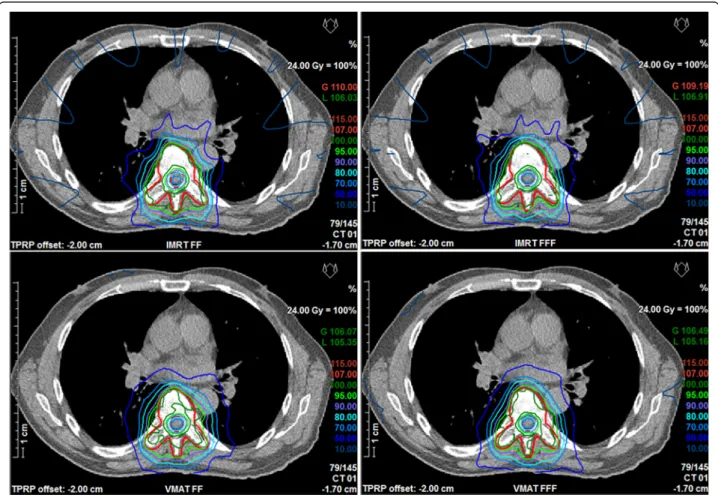
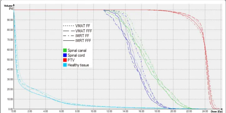
Comparison of VMAT versus IMRT also showed signifi- cant differences in target coverage and homogeneity as well as sparing of the spinal cord and normal tissue. Best target coverage and homogeneity were achieved with VMAT FFF, whereas lowest doses to the spinal cord and normal tissue were achieved with IMRT FFF. A compari- son of dose distributions and dose volume histograms is shown in Figs. 2 and 3 for a typical case.
Dosimetry
All 40 plans passed the gamma evaluation. Passing rates were similar for both techniques and showed no signifi- cant difference between FF and FFF or IMRT and VMAT. Details are listed in Table 3.
Fig. 1Measurement setup at the linac. The 2D-ionisationchamber-array MatriXX EvolutionTMwas set up between slabs of RW3 and centered at the isocenter of the linac. The setup was extended cranially by additional slabs of RW3 of the same height with a 0.3 ccm ionisation chamber positioned 31 cm cranially of the isocenter
Out-of-field point doses measured at 31 cm from the isocenter were significantly reduced for FFF as compared to FF. Averaged over all plans a dose reduction of 33.2 % ± 5.4 % of the local dose was achieved. The lowest out-of-field point dose was found for VMAT FFF, which was significantly lower than for IMRT FFF (p< 0.01).
Calculation of the peripheral dose at the point of meas- urement in the phantom in Oncentra showed no signifi- cant differences between the FF and FFF. Comparison of calculated versus measured peripheral doses showed a
mean local dose deviation of - 33.6 % ± 16.6 % for flat beams and −2.0 % ± 29.4 % for FFF beams. Details are listed in Table 3.
Efficiency
FFF Plans required significantly more MU per fraction dose for both IMRT and VMAT, whereas no significant difference could be found between IMRT and VMAT in both irradiation modes. Details are listed in Table 3.
Table 2Comparison of plan quality
IMRT VMAT
Parameter FF FFF p FF FFF p
PTV V95%(%) 86.4 ± 1.7 88.4 ± 1.7 <0.01 91.0 ± 1.5 91.7 ± 1.3 0.14
HI 0.27 ± 0.02 0.26 ± 0.02 0.06 0.23 ± 0.02 0.23 ± 0.02 0.96
CI 0.65 ± 0.04 0.66 ± 0.04 0.17 0.65 ± 0.04 0.66 ± 0.04 0.51
Spinal Cord D50% 15.9 ± 0.6 15.1 ± 0.5 <0.01 15.6 ± 0.3 15.0 ± 0.4 0.01
V75%(ccm) 0.9 ± 0.3 0.5 ± 0.1 <0.01 1.0 ± 0.5 0.7 ± 0.2 0.01
Normal Tissue D50% 0.8 ± 0.4 0.6 ± 0.4 <0.01 1.1 ± 0.7 0.8 ± 0.6 <0.01
Mean values and standard deviation of the dose volume parameters for the four different planning techniques averaged over all patients. Dose values are given in Gy.P-values for comparison of FF and FFF are calculated separately for IMRT and VMAT. Bold values indicate significantly superior values
Fig. 2Comparison of dose distributions. Comparison of dose distributions in one transversal slice for a representative case. Top IMRT, bottom VMAT, left FF, right FFF
Delivery time was significantly reduced for FFF beams in both treatment techniques. Averaged over all patients the delivery time was reduced from 557 to 438 s for IMRT and 216 s to 163 s for VMAT, which corresponds to a reduction of 21 % ± 4 % for IMRT and 24 % ± 5 % for VMAT. The lowest delivery time was found for VMAT FFF, which was significantly lower than for IMRT FFF (p< 0.01). Details about delivery times are listed in Table 3.
Discussion
The aim of this study was to investigate the potential of the flattening filter free (FFF) mode of a linear acceler- ator for intensity modulated radiation therapy (IMRT) and volumetric modulated arc therapy (VMAT) for pa- tients with in-field recurrence of vertebral metastases.
The data show significant advantages for FFF beams as compared to FF beams in terms of plan quality as well
as delivery time and peripheral dose: FFF offered a sig- nificant improvement in the sparing of normal tissue and the spinal cord, which could be of importance, min- imizing the risk of radiation myelopathy, keeping target coverage and homogeneity at the same level. According to the literature [31], the risk for radiation myelopathy is 0 % if the BED of each series is≤98 Gy2,the interval be- tween the series is at least 6 months, and the total BED of all series is≤120 Gy2. The risk increases to 3 % for an upper limit of the total BED of 135.5 Gy2. In the sce- nario presented here this means that the dose to the spinal cord has to be restricted to 18 Gy to assure a 0 % risk of myelopathy. The volume of the spinal cord re- ceiving 18 Gy was kept well below the clinically relevant volume of 1 ccm in all cases if FFF mode was used, but exceeded 1 ccm in 50 % of the cases, when FF mode was used. The reason for the better sparing of the spinal cord may be found in the shape of the dose profiles for beams
Fig. 3Comparison of dose volume histograms. Comparison of dose volume histograms for the case of Fig. 2
Table 3Comparison of plan delivery and evaluation of dosimetry
IMRT VMAT
Parameter FF FFF p FF FFF p
Delivery time (s) 557 ± 41 438 ± 23 <0.01 216 ± 15 163 ± 5 <0.01
Monitor units per Gy 381 ± 25 448 ± 43 <0.01 391 ± 29 429 ± 37 0.02
Measured peripheral dose (mGy) 8.6 ± 1.4 5.8 ± 1.2 <0.01 7.0 ± 1.0 4.8 ± 1.0 <0.01
Calculated peripheral dose (mGy) 4.7 ± 1.3 4.6 ± 1.5 0.51 5.7 ± 1.4 5.6 ± 1.4 0.24
Passing rate ofγ(%) 97.6 ± 1.1 97.4 ± 1.2 0.51 98.1 ± 1.1 97.9 ± 1.0 0.37
Mean values and standard deviation of delivery time, monitor units, measured and calculated peripheral doses and result of the gamma evaluation for the four different planning techniques averaged over all ten patients.P-values for comparison of FF and FFF are calculated separately for IMRT and VMAT. Bold values indicate significantly superior values
with the central part blocked: For the peak formed pro- file of the FFF beams the gradient towards the blocked center is slightly steeper than of FF beams. The lower dose to the normal tissue in the periphery can be ex- plained by reduced head scatter if the flattening filter is removed. In spite of increased MU in the FFF mode, de- livery time and peripheral dose exposure are reduced, which means less strain for the patient and possibly re- duced risk of normal tissue complications. When com- pared to IMRT FF, VMAT FFF offered a reduction of 71 % in delivery time and 45 % peripheral dose. Accord- ing to Ma et al. [28] the critical time to keep target mo- tion within 1 mm of translation or 1° of rotational deviation is 5.9 min (354 s) when patients are immobi- lized in a vacuum cushion. This was achieved in all cases of our study for VMAT only. In summary VMAT FFF may therefore be considered the preferable treatment option for radiotherapy of spinal column metastases which the combination of Elekta Synergy linacs with Agility™head and the treatment planning system Oncen- tra® External Beam v4.5.
Concerning plan quality there is a broad variety in the studies published up to now: For small targets with low modulation, as they are common in stereotactic treat- ments, plan quality was mostly found to be comparable [19, 26]. For larger targets, one group reported similar target coverage at significantly reduced doses to organs at risk for FFF beams [15, 21] which is in concordance with our findings. Other studies found superior plan quality for flat beams [20, 44, 45]. The only planning study dealing with irradiation of vertebrae with FFF beams has to our knowledge been published by Ong et al. for RapidArc plans created in the Eclipse planning system for Varian linear accelerators [23]. In their study different fractionation schemes of single doses ranging from 9 to 16 Gy were used and plans were normalized with respect to acceptable dose to the spinal cord. Ong et al. found slightly increased target dose for FFF as compared to FF, which is in concordance with the results of our study presented here. However, photon energies used in their study differed between FF (6 MV) and FFF (10 MV), which might bias the results, whereas we used the same energy for both irradiation modes in our study. Validation of the dose calcula- tion by measurements in the study of Ong et al.
showed good agreement for a region receiving more than 20 % of the prescription dose, evaluation of per- ipheral low dose was not performed. Reduction in delivery time was more pronounced in the study of Ong et al., with a higher mean for FF (402 s vs 216 s) and a comparable mean for FFF (168 s versus 163 s), since the benefit of higher dose rates is more pronounced for higher fraction doses as they are used in the study of Ong.
Reduction in out-of-field dose has been reported pre- viously in the context of Monte Carlo simulations of FFF beams for both Elekta [13] and Varian [46]. The authors emphasize, however, that a reduction of peripheral dose in patient treatments “may not always be achievable” due to dependence on many parameters [46]. Most of the planning studies published up to now did not take peripheral dose measurements into account. Spruijt et al. [20] performed a planning study for breast cancer and created one phantom case for out-of-field dose mea- surements 0.3 cm to 3.1 cm from the field edge. They found an average reduction in out-of-field dose of 10 %.
Comparison to dose calculations in this region showed, however, an underestimation by the treatment planning system Eclipse of 26 % to 85 % as compared to measure- ments. Absolute dose values are not reported. Because of the uncertainties in the dose calculation the authors abandoned evaluation of dose volume parameters of the contralateral breast in their planning study. Kragl et al.
[2] measured peripheral dose for four sample cases, one 7-field phantom plan, one lung SBRT, one prostate IMRT and one head and neck IMRT case and found a substantial local dose reduction at around 20 cm from the field edge. Because only one case per technique was considered, no conclusion with statistical significance could be drawn. The results of our study evaluated for a larger number of cases support their findings with statis- tical evidence. As Kragl et al. pointed out, peripheral doses are difficult to calculate correctly with a high ac- curacy, and should therefore be determined by measure- ments or Monte Carlo simulations. Therefore they did not compare the peripheral dose measurements with calculations. Comparison of calculated versus measured peripheral doses in our study showed significantly higher agreement between calculated and measured peripheral doses for FFF beams than for FF beams. The calculation in Oncentra did not reveal the actual reduction of per- ipheral dose which is possible to achieve by the use of FFF due to a systematic underestimation of the periph- eral dose for FF beams, which was not observed for FFF beams. Thus, the advantage of FFF determined in the calculated DVH of the normal tissue is even underesti- mated by the dose calculation in Oncentra.
Conclusions
For the combination of an Elekta Synergy linac with Agil- ity™ head and the treatment planning system Oncentra®
External Beam v4.5 the use of flattening filter free beams in re-irradiation of spinal column metastases allows better sparing of the spinal cord, minimizing the risk of radiation myelopathy, without compromising target coverage and homogeneity for both IMRT and VMAT. Delivery time is significantly lower as compared to flat beams, which may reduce intrafractional movement and emotional strain to
the patient. When compared to IMRT FF, VMAT FFF offered a reduction of 71 % in delivery time and 45 % in peripheral dose. For radiotherapy of spinal column metas- tases VMAT FFF may therefore be considered the prefera- ble treatment option for the combination of Elekta Synergy linacs with Agility™head and the treatment plan- ning system Oncentra® External Beam v4.5.
Abbreviations
CI:conformity index; ccm: cubic centimeter; DVH: dose volume histogram;
DVO: dose volume objective; Dx: dose to volume x of the structure;
FF: flattening filter; FFF: flattening filter free; HI: homogeneity index;
IMRT: intensity modulated radiation therapy; min: minutes; PTV: planning target volume; PIV: total volume of the 95 % isodose; s: seconds; TV: volume of the PTV; VMAT: volumetric modulated arc therapy; Vx%: volume covered by x% of the prescription dose.
Competing interests
The department has research cooperation with Elekta GmbH Hamburg. The authors state there are no conflicts of interest.
Authors' contributions
BD and AK designed the concept of the study. MGH selected the patient collective and contoured the target volume and organs at risk. AK and ZK created the treatment plans. MGH was responsible for the clinical evaluation of the treatment plans. AK and TO performed dose measurements. TO calculated the peripheral doses. BD evaluated the results and performed statistical analysis. BD drafted the manuscript, AK and MGH helped to draft the manuscript. All authors read and approved the final manuscript.
Acknowledgements
This study was funded by the Bavarian State Ministry of the Environment and Consumer Protection.
Received: 29 September 2015 Accepted: 18 February 2016
References
1. Georg D, Knoos T, McClean B. Current status and future perspective of flattening filter free photon beams. Med Phys. 2011;38(3):1280–93.
2. Kragl G, Baier F, Lutz S, Albrich D, Dalaryd M, Kroupa B, et al. Flattening filter free beams in SBRT and IMRT: dosimetric assessment of peripheral doses. Z Med Phys. 2011;21(2):91–101. doi:10.1016/j.zemedi.2010.07.003.
3. Jank J, Kragl G, Georg D. Impact of a flattening filter free linear accelerator on structural shielding design. Z Med Phys. 2014;24(1):38–48. doi:10.1016/j.
zemedi.2013.05.002.
4. Ponisch F, Titt U, Vassiliev ON, Kry SF, Mohan R. Properties of unflattened photon beams shaped by a multileaf collimator. Med Phys.
2006;33(6):1738–46.
5. Titt U, Vassiliev ON, Ponisch F, Dong L, Liu H, Mohan R. A flattening filter free photon treatment concept evaluation with Monte Carlo. Med Phys.
2006;33(6):1595–602.
6. Vassiliev ON, Titt U, Ponisch F, Kry SF, Mohan R, Gillin MT. Dosimetric properties of photon beams from a flattening filter free clinical accelerator.
Phys Med Biol. 2006;51(7):1907–17. doi:10.1088/0031-9155/51/7/019.
7. Mesbahi A, Mehnati P, Keshtkar A, Farajollahi A. Dosimetric properties of a flattening filter-free 6-MV photon beam: a Monte Carlo study. Radiat Med.
2007;25(7):315–24. doi:10.1007/s11604-007-0142-6.
8. Kry SF, Titt U, Ponisch F, Vassiliev ON, Salehpour M, Gillin M, et al. Reduced neutron production through use of a flattening-filter-free accelerator. Int J Radiat Oncol Biol Phys. 2007;68(4):1260–4. doi:10.1016/j.ijrobp.2007.04.002.
9. Parsai EI, Pearson D, Kvale T. Consequences of removing the flattening filter from linear accelerators in generating high dose rate photon beams for clinical applications: A Monte Carlo study verified by measurement. Nucl Inst Methods Phys Res B-Beam Interactions with Materials and Atoms. 2007;
26(1–2):755–9. doi:10.1016/j.nimb.2007.03.020.
10. Cashmore J. The characterization of unflattened photon beams from a 6 MV linear accelerator. Phys Med Biol. 2008;53(7):1933–46. doi:10.1088/0031- 9155/53/7/009.
11. Kry SF, Howell RM, Titt U, Salehpour M, Mohan R, Vassiliev ON. Energy spectra, sources, and shielding considerations for neutrons generated by a flattening filter-free Clinac. Med Phys. 2008;35(5):1906–11.
12. Kragl G, af Wetterstedt S, Knausl B, Lind M, McCavana P, Knoos T, et al.
Dosimetric characteristics of 6 and 10MV unflattened photon beams.
Radiother Oncol. 2009;93(1):141–6. doi:10.1016/j.radonc.2009.06.008.
13. Almberg SS, Frengen J, Lindmo T. Monte Carlo study of in-field and out-of-field dose distributions from a linear accelerator operating with and without a flattening-filter. Med Phys. 2012;39(8):5194–203.
doi:10.1118/1.4738963.
14. Akino Y, Ota S, Inoue S, Mizuno H, Sumida I, Yoshioka Y, et al. Characteristics of flattening filter free beams at low monitor unit settings. Med Phys. 2013;
40(11):112101. doi:10.1118/1.4824920.
15. Nicolini G, Ghosh-Laskar S, Shrivastava SK, Banerjee S, Chaudhary S, Agarwal JP, et al. Volumetric modulation arc radiotherapy with flattening filter-free beams compared with static gantry IMRT and 3D conformal radiotherapy for advanced esophageal cancer: a feasibility study. Int J Radiat Oncol Biol Phys.
2012;84(2):553–60. doi:10.1016/j.ijrobp.2011.12.041.
16. Alongi F, Cozzi L, Arcangeli S, Iftode C, Comito T, Villa E, et al. Linac based SBRT for prostate cancer in 5 fractions with VMAT and flattening filter free beams: preliminary report of a phase II study. Radiat Oncol. 2013;8(1):171.
doi:10.1186/1748-717X-8-171.
17. Shi LW, Lai YQ, Lin Q, Ha HM, Fu LR. The Effect of Flattening Filter Free on Three-dimensional Conformal Radiation Therapy (3D-CRT), Intensity- Modulated Radiation Therapy (IMRT), and Volumetric Modulated Arc Therapy (VMAT) Plans for Metastatic Brain Tumors from Non-small Cell Lung Cancer. Health Phys. 2015;109(1):1–9. doi:10.1097/HP.
0000000000000278.
18. Anchineyan P, Mani GK, Amalraj J, Karthik B, Anbumani S. Use of flattening filter-free photon beams in treating medulloblastoma: a dosimetric evaluation. ISRN Oncol. 2014;2014:769698. doi:10.1155/2014/769698.
19. Hrbacek J, Lang S, Graydon SN, Klock S, Riesterer O. Dosimetric comparison of flattened and unflattened beams for stereotactic ablative radiotherapy of stage I non-small cell lung cancer. Med Phys. 2014;41(3):031709. doi:10.
1118/1.4866231.
20. Spruijt KH, Dahele M, Cuijpers JP, Jeulink M, Rietveld D, Slotman BJ, et al.
Flattening filter free vs flattened beams for breast irradiation. Int J Radiat Oncol Biol Phys. 2013;85(2):506–13. doi:10.1016/j.ijrobp.2012.03.040.
21. Subramaniam S, Thirumalaiswamy S, Srinivas C, Gandhi GA, Kathirvel M, Kumar KK, et al. Chest wall radiotherapy with volumetric modulated arcs and the potential role of flattening filter free photon beams. Strahlenther Onkol. 2012;188(6):484–90. doi:10.1007/s00066-012-0075-6.
22. Zhuang M, Zhang T, Chen Z, Lin Z, Li D, Peng X, et al. Volumetric modulation arc radiotherapy with flattening filter-free beams compared with conventional beams for nasopharyngeal carcinoma: a feasibility study.
Chin J Cancer. 2013;32(7):397–402. doi:10.5732/cjc.012.10182.
23. Ong CL, Verbakel WF, Dahele M, Cuijpers JP, Slotman BJ, Senan S. Fast arc delivery for stereotactic body radiotherapy of vertebral and lung tumors. Int J Radiat Oncol Biol Phys. 2012;83(1):e137–43. doi:10.1016/j.ijrobp.2011.12.014.
24. Boda-Heggemann J, Mai S, Fleckenstein J, Siebenlist K, Simeonova A, Ehmann M, et al. Flattening-filter-free intensity modulated breath-hold image-guided SABR (Stereotactic ABlative Radiotherapy) can be applied in a 15-min treatment slot. Radiother Oncol. 2013;109(3):505–9. doi:10.1016/j.
radonc.2013.09.014.
25. Murray LJ, Thompson CM, Lilley J, Cosgrove V, Franks K, Sebag-Montefiore D, et al. Radiation-induced second primary cancer risks from modern external beam radiotherapy for early prostate cancer: impact of stereotactic ablative radiotherapy (SABR), volumetric modulated arc therapy (VMAT) and flattening filter free (FFF) radiotherapy. Phys Med Biol. 2015;60(3):1237–57. doi:10.1088/
0031-9155/60/3/1237.
26. Stieler F, Fleckenstein J, Simeonova A, Wenz F, Lohr F. Intensity modulated radiosurgery of brain metastases with flattening filter-free beams. Radiother Oncol. 2013;109(3):448–51. doi:10.1016/j.radonc.2013.10.017.
27. Lechner W, Kragl G, Georg D. Evaluation of treatment plan quality of IMRT and VMAT with and without flattening filter using Pareto optimal fronts. Radiother Oncol. 2013;109(3):437–41. doi:10.1016/j.
radonc.2013.09.020.
28. Ma L, Sahgal A, Hossain S, Chuang C, Descovich M, Huang K, et al.
Nonrandom intrafraction target motions and general strategy for correction of spine stereotactic body radiotherapy. Int J Radiat Oncol Biol Phys. 2009;
75(4):1261–5. doi:10.1016/j.ijrobp.2009.04.027.
29. Groger C, Hautmann MG, Loeschel R, Repp N, Kolbl O, Dobler B.
Re-irradiation of spinal column metastases by IMRT: impact of setup errors on the dose distribution. Radiat Oncol. 2013;8:269. doi:10.1186/
1748-717X-8-269.
30. Nieder C, Grosu AL, Andratschke NH, Molls M. Proposal of human spinal cord reirradiation dose based on collection of data from 40 patients. Int J Radiat Oncol Biol Phys. 2005;61(3):851–5. doi:10.1016/j.ijrobp.2004.06.016.
31. Nieder C, Grosu AL, Andratschke NH, Molls M. Update of human spinal cord reirradiation tolerance based on additional data from 38 patients. Int J Radiat Oncol Biol Phys. 2006;66(5):1446–9. doi:10.1016/j.ijrobp.2006.07.1383.
32. Fowler JF. The linear-quadratic formula and progress in fractionated radiotherapy. Br J Radiol. 1989;62(740):679–94. doi:10.1259/0007-1285- 62-740-679.
33. Paynter D, Weston SJ, Cosgrove VP, Evans JA, Thwaites DI. Beam characteristics of energy-matched flattening filter free beams. Med Phys.
2014;41(5):052103. doi:10.1118/1.4871615.
34. Kragl G, Albrich D, Georg D. Radiation therapy with unflattened photon beams: dosimetric accuracy of advanced dose calculation algorithms.
Radiother Oncol. 2011;100(3):417–23. doi:10.1016/j.radonc.2011.09.001.
35. Dobler B, Groeger C, Treutwein M, Alvarez-Moret J, Goetzfried T, Weidner K, et al. Commissioning of volumetric modulated arc therapy (VMAT) in a dual-vendor environment. Radiother Oncol. 2011;99(1):86–9. doi:10.1016/j.
radonc.2011.01.024.
36. Dobler B, Streck N, Klein E, Loeschel R, Haertl P, Koelbl O. Hybrid plan verification for intensity-modulated radiation therapy (IMRT) using the 2D ionization chamber array I'mRT MatriXX–a feasibility study. Phys Med Biol.
2010;55(2):N39–55. doi:10.1088/0031-9155/55/2/N02.
37. Paddick I. A simple scoring ratio to index the conformity of radiosurgical treatment plans. Technical note. J Neurosurg. 2000;93 Suppl 3:219–22.
doi:10.3171/jns.2000.93.supplement3.0219.
38. International Commission On Radiation Units And Measurements.
Prescribing, Recording and Reporting Photon Beam Therapy. Bethesda, Maryland, USA: ICRU; 1993. Report No. 50. ISBN 0-913394-48-3.
39. International Commission On Radiation Units And Measurements.
Prescribing, Recording, and Reporting Photon-Beam Intensity-Modulated Radiation Therapy (IMRT); Report 83. Journal of the ICRU. 2010;10(1).
doi:10.1093/jicru/ndq002.
40. Zhen H, Nelms BE, Tome WA. On the use of biomathematical models in patient-specific IMRT dose QA. Med Phys. 2013;40(7):071702. doi:10.
1118/1.4805105.
41. Low DA, Harms WB, Mutic S, Purdy JA. A technique for the quantitative evaluation of dose distributions. Med Phys. 1998;25(5):656–61.
42. Ezzell GA, Burmeister JW, Dogan N, LoSasso TJ, Mechalakos JG, Mihailidis D, et al. IMRT commissioning: multiple institution planning and dosimetry comparisons, a report from AAPM Task Group 119. Med Phys. 2009;36(11):5359–73.
43. Ezzell GA, Galvin JM, Low D, Palta JR, Rosen I, Sharpe MB, et al. Guidance document on delivery, treatment planning, and clinical implementation of IMRT: report of the IMRT Subcommittee of the AAPM Radiation Therapy Committee. Med Phys. 2003;30(8):2089–115.
44. Zhuang M, Huang L, Zhu D, Peng X, Lin Z. Reirradiation of nasopharyngeal carcinoma focusing on volumetric modulated arcs with flattening filter-free beams. Br J Radiol. 2015;88(1052):20140837. doi:10.1259/bjr.20140837.
45. Zhuang M, Zhang T, Chen Z, Lin Z, Li D, Peng X, et al. Advanced nasopharyngeal carcinoma radiotherapy with volumetric modulated arcs and the potential role of flattening filter-free beams. Radiat Oncol. 2013;8:
120. doi:10.1186/1748-717X-8-120.
46. Kry SF, Vassiliev ON, Mohan R. Out-of-field photon dose following removal of the flattening filter from a medical accelerator. Phys Med Biol. 2010;55(8):
2155–66. doi:10.1088/0031-9155/55/8/003.
• We accept pre-submission inquiries
• Our selector tool helps you to find the most relevant journal
• We provide round the clock customer support
• Convenient online submission
• Thorough peer review
• Inclusion in PubMed and all major indexing services
• Maximum visibility for your research Submit your manuscript at
www.biomedcentral.com/submit
Submit your next manuscript to BioMed Central and we will help you at every step: