Eur. J. Clin. Chem. Clin. Biochem.
Vol. 32, 1994, pp. 915-921
© 1994 Walter de Gruyter & Co.
Berlin · New York
Acute Intermittent Porphyria: Diagnostic Conundrums
By W. S. Wassif
1, A. C. Deacon
2, Ylva Flodenis
3, S. TliunelP and T. J. Peters*
1
Department ofCIinical Biochemistry, King's College School of Mediane and Dentistry, London, UK
2
Department ofCIinical Chemistry, Northwick Park Hospital, Harrow, Middlesex, UK
3
National Porphyrias Service, St G ran i Hospital, Stockholm, Sweden
(Received June 6/September 20, 1994)
Summary: Acute intermittent porphyria is a genetic disorder of haem biosynthesis caused by defects in the gene encoding hydroxymethylbilane synthase on the long arm of chromosome 11. Every effort should be made to identify gene carriers amongst the relatives of patients known to have acute intermittent porphyria s they are at risk of developing potentially fatal neurogenic attacks if exposed to precipitating factors. Erythrocyte hydroxymethylbilane synthase activity was determined in 46 members of two large well characterised families by assaying enzyme activity by both high performance liquid chromatography (HPLC) and fluorimetric assays. Additionally, hydroxy- methylbilane synthase immunoreactivity was determined by a sandwich-type ELISA. Statistically significant corre- lations were observed between erythrocyte hydroxymethylbilane synthase activity assayed by HPLC and by the fluorimetric assay, and enzyme protein concentration (r = 0.85, p < 0.001 and r = 0.80, p < 0.001, respectively).
The assay of hydroxymethylbilane synthase immunoreactive concentration in erythrocytes was useful in excluding acute intermittent porphyria in one patient in whom unequivocal assignment of porphyric Status was not possible by assaying enzyme activity alone. Erythrocyte hydroxymethylbilane synthase activity assayed by HPLC and fluo- rimetry showed approximately equal diagnostic performances, both giving rise to a dichotomic distribution of values, with overlap zones of 6% (1/16) and 22% (2/9), respectively, at the "cut off' applied.
Introduction should be made to identify gene carriers amongst the , . . i j · relatives of patients known to have acute intermittent Acute intermittent porphyria is an autosomal dominant . ^ disorder of haem biosynthesis in which over-production ofhaemprec rsofsisassociatedwithcharactensticchn- / Λ . . . . ., -, . j ... - . . ,. ,. asymptomatic gene camers. Furthermore, normal por- , " "
tf : . . , . , , j . porphyria (5). Normal haem precursors usually occur m
r J^ '
r_
F. , ' , · 11 .r i u· * · phynn excretion may occur in known patients with acute ical features and potentially fatal neuropsychiatnc syn- . .
t. _ . . . . , ,_.
j /ι ο\" Ά * · * ·«. * u · · A u intermittent porphyna dunng the remission penods (6).
drome (l, 2). Acute intermittent porphyna is caused by , / , , „ ., , . . defects in the gene eneodmg hydroxymethylbilane syn-
Low^hrocyte hydroxymethylb ane synthase activaty thase (EC 4.3.1.8),') also known s poiphobilinogen de-
1S^c.fically diagnost,c of acute mtermittent porphyna aminase (3). Hydroxymethylbilane synthase is a cyto- &>
7>
and remains the most effectiv6 method for detect- solic enzyme and has reduced activity in abnormal gene
ing g
ene carriers(
8»
9>- Nevertheless, unequivocal s-
„
o. ,
0 A^ signment confirming or refuting the diagnosis of acute camers \y·,. *·/·
intermittent porphyria cannot be achieved among some It is important to investigate asymptomatic individuals
rei
atives of a known patient with acute intermittent por- for latent or subclinical poφhyria and every eifort
phyria due to the overiap inenzyme activities between
affected patients and normal subjects (8—10).
) Enzyme:
Wrtwtl Q0„
ΛΓηι,
Λκ;ΐίηο«ι»η Α* Molecular heterogeneity in acute intermittent porphyria
Hydfoxymethylbilane synthase also known s porpnobilmogen de- ° J \ ^aminase (BC 4.3.1.8) 00
and ^e large number of pomt mutations (12—14)
Eur. J. Clin. Chem, Clin. Biochem. / Vol. 32,1994 / No. 12
916
Wassif et al.: Acute intermittent porphyria: Diagnostic conundrumsdetected so far, make genetic studies in affected families
an impractical Option at the present time. None of the mutations so far identified has been found to occur more frequently than any of the others, and many of these mutations appear to be unique to one family. Addition- ally, all polymorphic sites are clustered within a small fragment (1.5 X l O
3bases in the first intron) of the hy- droxymethylbilane synthase gene and thus limiting the usefulness of restriction fragment length polymorphisms in identifying and differentiating the mutant allele frorn the normal one (15). However, restriction fragment length polymoφhisms have been successfully used for haplotype analysis and heterozygote canier detection in specific families with acute intermittent porphyria (16,
17).
In this study, the level of hydroxymethylbilane synthase gene expression in erythrocytes was determined in two large well characterised families by assaying hydroxy- methylbilane synthase activity by both high performance liquid chroniatography (HPLC) of the reaction products (18), and by a fluorimetric assay (19). In addition, im- munoreactive enzyme was assayed by ELISA (20). The aim of this study was to assess the performance of as- says measuring hydroxymethylbilane synthase activity, to document the degree of overlap in enzyme activities between gene camers and normal subjects using dif- ferent methods, and secondly, to investigate whether de- termination of hydroxymethylbilane synthase immuno- reactivity and specific enzyme activities have a better diagnostic performance in individuals with equivocal hydroxymethylbilane synthase activities.
Patients and Methods Patient details
We studied 46 members from two large well characterised families:
33 members aged 2 to 70 years (mean ± SEM, 34.5 ± 3.4 years) (19 females and 14 males) were investigated from family l (fig.
1), and 13 individuals aged 5 to 50 years (mean ± SEM, 28.2
± 4.4 years) (7 females and 6 males) from family 2 (flg. 2) were studied.
Urinary and faecal porphyrin quantification
Urinary and faecal porphyrins were identified and quantified by high performance liquid chromatography (HPLC) according to the method of Lim & Peters (21). If acute porphyria was suspected, urinary 5-aminolaevulinic aeid and pprphobilinogen excretion were quantified after prior purification by anion-exchange chromatogra- phy (22). · r
Erythrocyte hydroxymethylbilane synthase activity Hydroxymethylbilane synthase activity was assayed by the HPLC method of Wright & Lim (18) using fresh heparinised erythrocytes.
The enzyme activity was expressed s uroporphyrin, μηιοΐ/h · l erythrocytes. The between^run coefficient of Variation was 8% at a level of 25 uroporphyrin, μηιοΐ/h · l erythrocytes. Hydroxymeth^
ylbilane synthase activity was also determined by the fluorimetric assay of Magnussen et al. (19) with the assay of haemoglobin in the haemolysate (23). Enzyme activity was related to haemoglobin concentration and expressed s pkat/g Hb. The between-run coeffi- cients of Variation of the fluorimetric assay were 2.1% and 5.4%
for healthy and porphyric subjects, respectively. The lower refer- ence limit applied was 90 pkat/g Hb, representing a diagnostic sen- sitivity of 97% and a specificity of 85%. Non-^gene carriers do not exhibit erythrocyte hydroxymethylbilane synthase activities below 70 pkat/g Hb, a value representing a "cut off' limit giving a sensi- tivity of 100%, but a low specificity of 47% (24).
Erythrocyte hydroxymethylbilane synthase concentration
Hydroxymethylbilane synthase concentration was assayed by a sandwich-type ELISA with monospecific polyclonal antiserum (IgG) raised against hydroxymethylbilane synthase, s previously described (20). Haemoglobin concentration (absorbance read at 410 nm) of the samples was determined and hydroxymethylbilane synthase concentration was expressed relative to haemoglobin con- centration s μg/g Hb. Erythrocytes hydroxymethylbilane synthase specific activity was determined by dividing enzyme activity with enzyme protein concentration, and expressed s nkat/g enzyme protein (20). A lower reference limit of 110 μg/g Hb was used.
This "cut off' Value gives a diagnostic sensitivity of 85% and spec- ificity of 85% (24).
Further blood analyses
All patients had assays for f ll blood count, white cell differential and subset assays. Biochemical profile including renal and liver function and toxicity tests was carried-out on the DAX-48 multi- channel analyser (Bayer Diagnostics, Basingstoke, UK) by Stan- dard methods.
1 23 4 5 6 7 8 9 10 l 11 12 113 14 15\16 17 181 19 20 18 19 20 21 IV
Fig. l Pedigree of family l showing autosomal dominant inheri- tigated are represented by hatched Symbols (m ©). Patient 111:8, in tance of acute intermittent porphyria. Affected individuals are rep- whom definitive porphyric Status was not possible, is represented resented by black Symbols (H o), unaffected individuals are de- by half-filled Symbol (3). \ indicates propositus.
picted s open Symbols (D O), while members who were not inves- > ;
Eur. J. Clin. Chem. Clin. Biochem. / Vol. 32, 1994/No. 12
» Ώ-Γ&
Fig. 2 Pedigree of family 2 showing autosomal dominant inheri- tance of acute intermittent porphyria. Affected individuals are rep- resented by black Symbols (D 0), unaffected individuals are de- picted s open Symbols (G O), while members who were not inves- tigated are represented by hatched Symbols (@ ©). \ indicates pro- positus.
Results
In the two families the propositus (patient III: 15, fig. 1;
patient 111:2, fig. 2) presented at the age of 34 and 27 years, respectively, with a history of dieting prior to hos- pital admission. Both index patients were admitted to hospital with severe abdominal pain, hypertension, vom- iting and constipation and had elevated urinary excretion of 5-aminolaevulinic acid, porphobilinogen, raised uri- nary porphyrin and normal faecal porphyrin (tab. 1). The decreased red cell hydroxymethylbilane synthase activ- ity in both patients (assayed by HPLC, tab. 1) was con- sistent with the diagnosis of acute intermittent por- phyria.
Assay of erythrocyte hydroxymethylbilane synthase ac- tivity (by the HPLC technique) in 33 members of family l (fig. 1) identified 11 (33%) individuals s gene carri- ers. Twenty one family members did not inherit the gene defect. In one individual (patient 111:8, fig. 1) unequivo- cal assignment confirming or reiuting the diagnosis of acute intermittent porphyria was not possible by assay of hydroxymethylbilane synthase activity alone due to the overlap in enzyme activities between affected pa- tients and normal subjects (8—10). Of 13 members of family 2 (fig. 2) 4 (31%) patients were identified s gene carriers. There was no overlap in enzyme activity in family 2 and unequivocal assignment excluding acute intermittent porphyria was possible in the other 9 family members. All gene carriers in both families, with the exception of the two index patients, had urinary and fae- cal porphyrins and urinary 5-aminolaevulinic acid and porphobilinogen within the reference ranges. Of 15 gene carriers, only 2 (13%) patients (patient 111:15, fig. 1; pa- tient 111:2, fig. 2) developed attacks of acute intermit- tent porphyria.
To charaeterize further the defect in acute intermittent porphyria, and establish a definitive diagnosis in the subjects with equivocal hydroxymethylbilane synthase activity, 22 members (11 from each family) were sub- jected to further investigations including the measure- ment of hydroxymethylbilane synthase immunoreactive
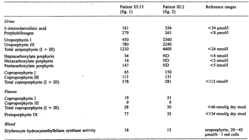
Tab. l Urinary 5-aminolaevulinic acid and porphobilinogen excretion, urinary and faecal porphyrins pattern and erythrocyte hydroxymethylbilane synthase activity in the two index patients.
Hydroxymethylbilane synthase activity was assayed by the method of Wright &. Lim (\%).
Patient III: 15 (fig. 1)
Patient 111:2 («g. 2)
Reference ranges Urine
5-Aminolaevulinic acid Porphobilinogen Uroporphyrin I Uroporphyrin III
Total uropprphyrin (I + HI) Heptacarboxylate porphyrin Hexacarboxylate porphyrin Pentacarb xylate poiphyrih Coproporphyrin I
Coproporphyrin III
Total Coproporphyrin (I + ΙΠ) Faeces
Coproporphyrin I Coproporphyrin III
Total Coproporphyrin (I 4- ΙΠ) Protoporphyrin IX
Blood
Erythrocyte hydroxymethylbilane synthase activity
279161 450780 1230 3414 147 11365 178
199 28 77
16
356261 2360 22404600 NDND ND 150131 281
219 30 32
13
<34 μηιοΐ/ΐ
<8 μηιοΙ/1
<24 nmol/1
<4 nmol/l
<3 nmol/1
<5 nmol/1
< 115 nmol/1
<46 nmol/g dry stool
<134 nmol/g dry stool
uroporphyrin, 20—42 μηιοΐ/h · l red cclls
Eur. J. Clin. Chem. Clin. Biochem. /Vol. 32,1994 / No. 12
918
Wassif et al.: Acute intermittent porphyria: Diagnostic conundrumsconcentration. Additionally, enzyme activity was inea- sured by the independent fluorimetric assay described by Magnussen et al. (19) and specific enzyme activity was determined. Of the 22 subjects 8 had been classified s gene carriers by the HPLC method, 13 were normal and not at risk of acute intermittent porphyria, and in one member porphyric Status was questionable s eryth- rocyte hydroxymethylbilane synthase activity was equivocal s shown in table 2. Seven out of eight in- dividuals diagnosed s gene carriers by the HPLC method were identified s such by the fluorimetric assay.
One individual given the diagnosis of acute intermittent porphyria by the HPLC method, exhibited a value just above the reference value applied when erythrocyte hy- droxymethylbilane synthase activity was assayed by the fluorimetric assay, s did the individual exhibiting equivocal result in the HPLC assay. Thus both assays show similar diagnostic efficiency and give rise to a rel- atively clear-cut dichotomic distribution of gene carriers and non-carriers (flg. 3).
It is clear from table 2 that all gene carriers had hydro- xymethylbilane synthase concentrations below 110 μg/g Hb. The measurement of hydroxymethylbilane synthase concentration in erythrocytes was usefiil in excl ding aeute intermittent porphyria in patient 111:8 (fainily l, flg. l, tab. 2) who had equivocal enzyme activity using the HPLC assay but had hydroxymethylbilane synthase concentration of 125 μ^/g Hb. However, there was one overlap in hydroxymethylbilane synthase concentration in a normal subjeet (subject 111:8, family 2, flg. 2, tab.
2) who did not inherit the gene defect but had hydroxy- methylbilane synthase concentration of 90 μξ/g Hb. As shown in table 2, hydroxymethylbilane synthase specific actiivty was not helpful in identifying patients at risk of acute intermittent porphyria and there was no clear Separation in enzyme specific activity between gene car- riers and normal subjects. Indeed, there was no correla- tion between hydroxymethylbilane synthase specific ac^
tivity and both enzyme activity and enzyme concentra- tion (r = 0.42, p = 0.06 and r = 0.14, p = 0.52, respec-
Tab. 2 Erythrocyte hydroxymethylbilane synthase activity, concentration and specific activity in the two families with acute intermittent porphyria.
Family 1 (fig. 1) Patient 11:5 Patient 11:7 Patient III: 13 Patient III: 15*
Patient 111:16 Patient 11:8 Patient 111:7 Patient III: 11 Patient 111:19 Patient 111:20 Patient 111:8 Family 2 (fig. 2) Patient 111:2*
Patient 111:6 Patient IV:2 Patient 11:5 Patient 11:9 Patient 111:3 Patient 111:4 Patient 111:7 Patient 111:8 Patient 111:9 Patient IV: 1 Reference Range
Age(years)
6364 38 3634 5837 4031 21 39
2824 435 4926 2814 3010 10
Gen-der
c?c?
99 c?9
?9
9 9 9 9 9
c?
ci 9
<?9
c?<?
9 9
Genecarrier
YesYes YesYes YesNo NoNo NoNo Possibly YesYes NoYes NoNo NoNo NoNo No
Hydroxy- methylbilane synthase activity (Wright & Lim, 1983)
(uroporphyrin, μπιοΐ/h · 1 red cells) 1511
1616 259 2823 2824 21
1318 3616 3825 2429 2328 24 20-42
Hydroxy- methylbilane synthase activity (Magnussen et al., 1974)
(pkat/g Hb)
7287 7986 13187 142117 137156 96
9165 13854 183130 113123 14595 95
>90
Hydroxy- methylbilane concen- tration
(μ§/8 Hb)
10689 10499 100151 151181 168157 125
9297 14869 209145 127125 15590 118
>110
Hydroxy- methylbilane synthase specific activity (nkat/g)
806823 801822 866865 782776 872930 786
708944 782933 874894 905966 1050934 808
* indicates propositus t *
Eur. J. Clin. Chem. Clin. Biochem. /Vol. 32, 1994/ No. 12
so-
10-
oo 8
2001
150
100 OO
0J
8 8
o
50 0 CP 00
Family 1 Family 2 Family 1 Family 2
So
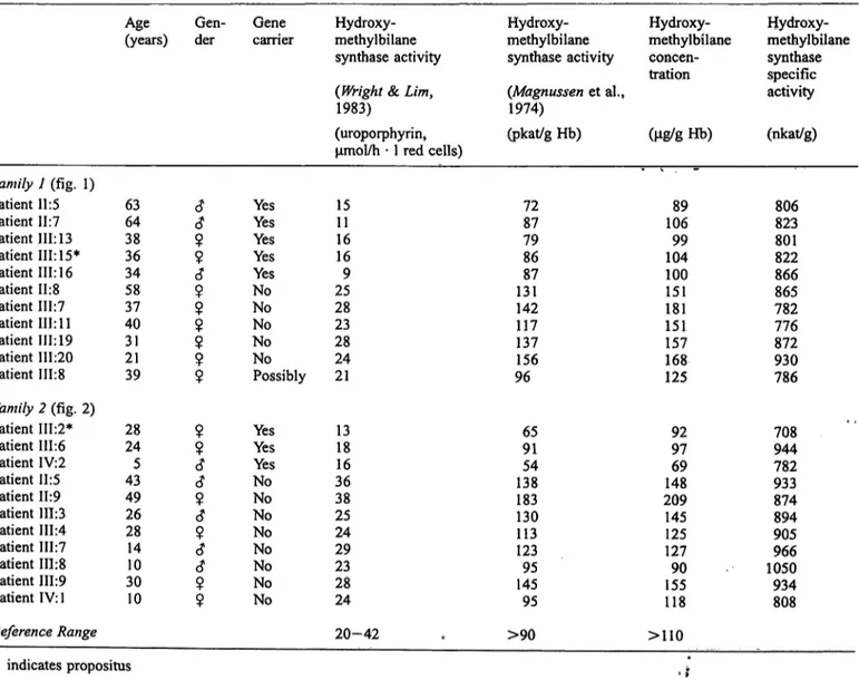
Family 1 Family 2 Fig. 3 Erythrocyte hydroxymethylbilane synthase activity and
concentration in two families with acute intermittent porphyria.
Hydroxymethylbilane synthase activities were determined by the HPLC method of Wright & Lim18 and the fluorimetric method of
Magmissen et al.19. Carrier Status was arbitrarily assigned using the HPLC assay data. The lower limits of the reference ranges of various assays are represented with solid lines. Gene carriers (o), non carriers (O) and equivocal Status ( ). / indicates propositus.
tively). In contrast, statistically significant correlations were observed between erythrocyte hydroxymethylbi- lane synthase activity assayed by the method of Wright & Lim and enzyme activity determined by the method of Magnussen et al., and enzyme concentration (r = 0.85, p < 0.001 and r = 0.80, p < 0.001 respec- tively, n = 22). A highly significant correlation was also observed between erythrocyte hydroxymethylbilane synthase activity assayed according to the method of Magnussen et al. and hydroxymethylbilane synthase concentration (r = 0.96, p < 0.001, n = 22).
All members investigäted from both families had normal biochemical profiles and haematological indices except 2 gene carriers from fämily 2 (patients 111:6 and IV:2, fig. 2) who had iron deficiency anaemia and a repeat hydroxymethylbilane synthase activity was performed after iron replacement therapy.
Discussion
The elevated urinary excretion of 5-aijiinolaevulinic acid and porphobilinogen in both index patients is consistent with the diägiiosis of one of the acute pörphyrias (i. e., acute intermittent porphyria, variegate porphyria and he- reditary coproporphyria). The raised urinary porphyrin content (tab. 1) has probably arisen by non-enzymic po- lymerization of po hobilinogen in the urine to form a mixture of uropo hyrin isomers. The normal faecal por- phyrin excretion in both patients indicates that variegate porphyria and hereditary coproporphyria are highly uft- likely, whereas the decreased red cell hydroxymethylbi-
lane synthase activity (tab. 1) is consistent with acute intermittent porphyria.
The dominant mode of inheritance, the occurrence of asymptomatic gene carriers and the risk of developing potentially fatal attacks if exposed to a wide ränge of common precipitating factors, make it essential to ex- clude or confirm the diagnosis of acute intermittent por- phyria in all relatives whenever the diagnosis has been made in one member of the fämily (2, 7). Patients with acute intermittent porphyria are at risk of developing potentially fatal neurogenic attacks if exposed to exoge- nous precipitating factors including a wide ränge of commonly prescribed drugs (25), alcohol, fasting, stress, hormones (2, 26) and, interestingly, if they continue to smoke (27). In our study, the index patients in both fam- ilies had acute attacks of porphyria following low calo- rie diet for weight reduction.
In our haads the HPLC and fluorimetric assays for erythrocyte hydroxymethylbilane synthase activity showed about the same diagnostic efficiency at the "cut off" levels applied, 8 and 7 individuals, respectively, diagnosed äs gene carriers. However, it is clear that, whichever assay is used, there was a small overlap be- tween enzyme activities in normal subjects and gene carriers in our study äs in previous studies (8—10). It is well established that a definitive assignment of porph- yric Status cannot always be made in all relatives, even when porphobilinogen excretion, 5-aminolaevulinic acid synthase activity (28) and gene dosage effect (9) are taken into account. Lamon et al. demonstrated that pedi- gree analysis with respect to Üie frequency distribution
Eur. J. Clin. Chem. Clin. Biochem. / Vol. 32,1994 / No. 12
920
Wassif et al.: Acutc intermittent pörphyria: Diagnostic conundrurnsof hydroxymethylbilane synthase enzyme activities in family members was successful in establishing porph- yric Status in 50% of those subjects with equivocal en- zyme activity (9). It is noteworthy that the existence of a variant of acute intermittent po hy ia has been reported where enzyme defect is not expressed in erythrocytes (29), in such families the measurement of erythrocyte hydroxymethylbilane synthase activity is unhelpful.
In this study, the measurement of erythrocyte hydroxy- methylbilane synthase immunoreactive concentration was useful in excluding the diagnosis of acute intermit- tent pörphyria in one member who had equivocal hydro- xymethylbilane synthase activity. The estimatiorl of en- zyme specific activity was not useful in identifying gene carriers with no clear Separation between normal sub- jects and gene carriers. This is hardly surprising since there is a good correlation between hydroxymethylbi- lane synthase activity and enzyme protein concentration.
This indicates that in these two families there is either no gene product or that the protein produced is neither
catalytically active nor recognised by the anti-serum used in the ELISA, i. e., we are dealing with a form of acute intermittent pörphyria in whicfa no cross reacting immunoreactive material (CRIM) is iproduced (GRIM negative). Since hydroxymethylbilane synthase activity decreases äs red cells age any shift in their age distribu- tion will be reflected in the enzyme activity (5), a repeat of hydroxymethylbilane synthase activity was essential in the two suspected gene carriers after the correction of the haematological abnormälities.
Acknowledgements
We are grateful to Dr Hughes, Department of Medicirie, Pembury Hospital and to Dr B. Hoffbrand, Department of Medicine, The Whittington Hospital for referring the index patients. We wish to thank Dr Ruth Ayling and Dr Roy Sherwood, Department of Clin- ical Biochemistry, King's College Hospital for their critical com- ments on the manuscri.pt, Dr Richard Fink and Dr Nick Walcot, West Middlesex University Hospital for their advice and Dr E.
Baylis and Mrs A. Hayward, Department of Clinical Chemistry, Kent and Sussex Hospital for their assistance in sample collection.
References
1. Waldenstrom, J. (1937) Studien über Porphyrie. Acta Med.
Scand. Suppl. 82, 1.
2. Kappas, A., Sassa, S., Galbraith, R. A. & Nordman, Y. (1989) The porphyria. In: The Metabolie Basis oflnherited Diseases, 6th edn. (Scriver, C. R., Beaudet, A. L., Sly, W. S. & Valle, D., eds.) pp. 1305-1366, McGraw-Hill Book Company, New York.
3. Meisler, M., Wanner, L, Eddy, R. E. & Shows, T. B. (1980) The UPS locus encoding uroporphyrinogen I synthase is lo- cated in human chromosome 11. Biochem. Biophys. Res.
Cornmun. 95, 170-176.
4. Poulos, V, Blake, C., McManus, J. & Rossi, E. (1991) Haem biosynthetic enzymes. Clin. Biochem. Revs. 12, 31—33.
5. Eider, G. H., Smith, S. G. & Jane Smyth, S. (1990) Laboratory investigation of the porphyrias. Ann. Clin. Biochem. 27, 395-412.
6. Doss, V. M. & Tiepermann, R. V. (1978) Uroporphyrinogen- Synthase in Erythrocyten bei akuter intermittierender Porphy- rie: Neue pathobiochemische Aspekte. J. Clin. Chem. Clin.
Biochem. 16, 111-118.
7. Kappas, A., Sassa, S. & Anderson, K. (1983) The porphyria.
In: The Metabolie Basis oflnherited Diseases, 5th edn. (Stan- bury, J. B., Wyngaarden, J. B., Fredrickson, D. S., Goldstein, J. L. & Brown, M. S., eds.) pp. 1301-1384, McGraw-Hill Book Company, New York.
8. Pierach, C. A., Weimer, M. K., Cardinal, R. A., Bossenmaier, I. C. & Bloomer, J. R. (1987) Red blood cell porphobilinogen deaminase in the evaluation of acute intermittent porphyria. J.
Am. Med. Ass. 257, 60-61.
9. Lamon, J. M., Frykholm, B. G. & Tschudy, D. P. (1979) Family evaluations in acute intermittent porphyria using red cell uro- porphyrinogen I synthase. J. Med. Genet. 16, 134-139.
10. Bonaiti-Pellie, C., Phung, L. & Nordmann, Y. (1984) Recur- ., rence risk estimation of acute intermittent porphyria based on analysis of porphobilinogen deaminase activity: A Bayesian approach. Am. J. Med. Genet. 19, 755-762.
11. Desnick, R. J., Ostasiewicz, L. T., Tishler, P. A. & Mustajoki, P. (1985) Acute intermittent porphyria: Characterization of a novel mutation in the structural gene for porphobilinogen de^
aminase. J. Clin. Invest. 76, 865-874.
12. Grandchamp, B., Picat, C., Mignotte, V., Wilson, J. H. R, Te Velde, K., Sankuyl, L.„ Romeo, P. H., Goossens, M. & Nord- marin, Y. (1989) Tissue specific spljcing mutation ifi acute in- termittent porphyria. Proc. Natl. Acad. Sei. USA 86, 661-664.
13. Scobie, G. A., Urquhart, A. J., Eider, G. H., Kalsheker, N. A., LIewellyn, D. H., Smyth, J. & Harrisons, P. R. (1990) Acute intermittent porphyria caüsed by a C —»T mutation that pro- duces a stop codon in the porphobilinogen deaminase gene.
Hum. Genet. 85, 631-634.
14. Delfau, M. H., Picat, C., De RopijVF. W. M., Hamer, K., Bo- gard, M., Wilson, J. H. P, Deybach, J. C., Nordmann, Y. &
Grandchamp, B. (1991) Two different point mutations G to A in exon 10 of the porphobilinogen deaminase gene are respon- sible for acute intermittent porphyria. J. Clin. Invest. 86>
1511-1516.
15. Lee, J-S. & Anvret, M. (1991) Identification of the most com- mon mutation within the porphobilinogen deaminase gene in Swedish patients with acute intermittent porphyria. Proc. Natl.
Acad. Sei. USA 88, 10912-10915.
16. LIewellyn, D. H., Eider, G. H., Kalsheker, N. A., Marsh, O.
W. M., Harrison, P. R., Grandchamp, B., Picat, C., Nordmann, Y, Romeo, P. H. & Grandchamp, B. (1987) DNA polymor- phism of human porphobilinogen deaminase gene in acute in- termittent porphyria. Lancet //, 706-708.
17. Lee, J-S., Anvret, M., Lindsten, J., Lannfelt, L.·, Gellerfors, P, Wetterberg, L., Floderus, Y. & Thunell, S; (1988) DNA polymorphisms within the porphobilinogen deaminase gene in two Swedish families with acute intermittent porphyria. Hum.
Genet. 79, 379-381.
18. Wright, D. J. & Lim, C. K. (1983) Siinültaneous detefmination of hydroxymethylbilane synthase and uroporphyrinogen III synthase in eiythrocytes by high performance liquid chroma- tography. Biochem. J. 213, 85-88.
19. Mägnussen, C. R., Levine, J. B., Doherty, J. M., Cheesman, J.
O. & Tschudy, D. P. (1974) A red cell enzyme method for the diagnosis of acute intermittent porphyria. Blood 44, 857-868.
20. Lannfelt, L., Wetterberg, L., Lilius, L., Thunell, S. & Geller- fors, P. (1989) ELISA for measuring porphobilinogen deami- nase in human erythrocytes. Clin.^him. Acta 83, 227-238.
Eur. J. Clin. Gheni. Clin. Bioehem. / Vol. 32, 1994/No. 12
21. Lim, C. K. & Peters, T. J. (1984) Urine and faecal porphyrin 27. Lip, G. Y. H., McColl, K. E. L., Goldberg, A. & Moore, M.
profiles by reversed-phase high performance Chromatograph^ R. (1991) Smoking and recurrent attacks of acute intermittent in the porphyria. Clin. Chim. Acta 139, 55-63. no P?1^?1?; ^5\MeKd; L 3\^2' ™ n n * n MU 22. Mauzerall, D. & Granick, S. (1956) The occurrence and deter- 28· ^cCol! K. *' L" Moor,e' *?' R-;Th™P?on>G; G. f Goldberg,
. 4. - s · ι ι· · ·. « , .... · A. (1982) Screenmg for latent acute intermittent porphyna:
mmation of S-arnmolaevulmic acid and poiphobilmog^n m jhe value of measuring both leucocyte δ-aminolaevulinic acid unne. J. Biol. Chem. 219, 435-446. synthase and erythrocyte uropoφhyrinogen-l-synthase activi- 23. Johansson, L., Thunell, S. & Wetterberg, L. (1984) A filter tjes j Med Genet 19> 271-276.
paper dry blood spot procedure for acute intermittent porphyria 29. Mustajoki, P. & Tenhunen, R. (l 985) Variant of acute intermit- population screening by use of whole blood uroporphyrinogen- tent ροφ^πβ with normal erythrocyte uroporphyrinogen-1- 1-synthase assay. Clin. Chim. Acta 737, 317-331. synthase activity. Eur. J. Clin. Invest. 75, 281-284.
24. Lannfelt, L. (1990) Immunological determination of poφho-
bilinogen deaminase s a diagnostic measure in acute intermit- ^· ^· Wassif, M. p.
tent ΡοφηΥη&. J. Clin. Chem. Clin. Biochem. 28, 273-278. ^^^ °f C*™*1 B™h*™siry . n , ^
^c ** K/T> ο τν ι D /loooNr* ·*· Λ- Kmg's College School of Medicine and Dentistry 25. Moore, M. R. & Disler, P. B. (1988) Drug-sensitive diseases: Bessemer Road
Acute poφhyrias. Adverse Drug Reaction Bulletin 729, London SE5 9PJ 484-487. UK
26. Hindmarsh, J. T. (1986) The ροφί^παβ: Recent advances.
Clin. Chem. 32, 1255-1263.
Eur. J. Clin. Chem. Clin. Biochem. / Vol. 32,1994 / No. 12