–308G ) ) A and –1031T ) ) C tumor necrosis factor gene polymorphisms in Tunisian patients with coronary artery disease
Lakhdar Ghazouani
1, Sonia-Ben-Hadj Khalifa
1, Nesrine Abboud
1, Faouzi Addad
2, Ali Ben Khalfallah
3, Nsiri Brahim
1, Mounira Mediouni
4, Wassim Y. Almawi
5and Touhami Mahjoub
1,*
1
Research Unit of Biology and Genetics of Cancer, Hematological and Autoimmune Diseases, Faculty of Pharmacy of Monastir, Monastir, Tunisia
2
Fattouma Bourguiba Hospital Center, Monastir, Tunisia
3
Menzel Bourguiba Hospital Center, Bizerte, Tunisia
4
Hospital Center of Jendouba, Jendouba, Tunisia
5
College of Medicine and Medical Sciences, Arabian Gulf University, Manama, Bahrain
Abstract
Background:
Recent research has shown that inflam- mation plays a key role in coronary artery disease (CAD) and other manifestations of atherosclerosis.
Several lines of evidence support a key role for tumor necrosis factor-a (TNF-a), a potent immunomodulator and pro-inflammatory cytokine, in the development of atherosclerosis and in complications of CAD.
Methods:
We investigated the possible association between CAD and the
TNFgene promoter poly- morphisms –308G)A and –1031T)C in a Tunisian population. We compared the distribution of these polymorphisms between 418 patients with CAD and 406 healthy controls using polymerase chain reaction restriction fragment length-polymorphism analysis.
Results:
The frequency of the TNF-a –308A allele in the control group was similar to that observed in CAD patients
wp
s0.78; odds ratio (OR)
s1.15; 95% confi- dence interval (CI)
s0.86–1.55
x, but higher than those described in other Europeans, such as in the French, Finnish and Spanish. Concerning the TNF-a –1031T/C polymorphism, the same distribution was observed between patients with CAD and controls (p
s0.12;
OR
s1.27; 95% CI
s0.94–1.72). In addition, the geno- type and allele frequencies of control individuals were comparable to those previously reported in healthy Tunisian controls and other ethnic groups. Haplotype analysis (TNF-a –308G
)A and –1031T
)C) demon- strated no significant association between
TNFhaplo- types and CAD.
*Corresponding author: Prof. Touhami Mahjoub, Research Unit of Biology and Genetics of Cancer, Hematological and Autoimmune Diseases, Faculty of Pharmacy of Monastir, Monastir 5000, Tunisia
E-mail: mahjoubtouhami@yahoo.fr
Received February 24, 2009; accepted July 11, 2009
Conclusions:
We conclude that
TNFpromoter gene polymorphisms at position –308G
)A and –1031T
)C do not play a major role in the pathogenesis of CAD in the Tunisian population.
Clin Chem Lab Med 2009;47:1247–51.
Keywords:
atherosclerosis; coronary artery disease;
polymorphisms; tumor necrosis factor.
Introduction
Cardiovascular disease, including coronary artery dis- ease (CAD), is the leading cause of death in most industrialized nations and will soon be the leading cause of death in the developing world (1, 2). CAD is a chronic inflammatory process in which pro-inflam- matory cytokines, such as tumor necrosis factor-a (TNF-a) and interleukine-6 (IL-6) may play a crucial role in its pathogenesis (3, 4). In recent years, markers of subclinical inflammation and subclinical athero- sclerosis have been studied in order to try to under- stand how they predict adverse outcomes in patients with CAD (5). Several lines of evidence support a key role for TNF-a in the development of atherosclerosis and its complications (6). High concentrations of plasma-soluble TNF and its soluble receptors are con- sidered to play a central part in the inflammatory cas- cade that is a key feature in cardiovascular diseases, such as atherosclerosis (7, 8), CAD (9) and acute myo- cardial infarction (MI) (10, 11). Prospective studies suggest that elevated concentrations of TNF-a are independent predictors of first-time cardiovascular disease (12) and that it is also a marker for recurrent coronary events following a previous MI (7). Circulat- ing concentrations of TNF are increased in patients with ischemic artery disease (13), and TNF-a has been shown to reduce the bioavailability of nitric oxide (NO), a potent endothelium-derived vasodilator, in cultured endothelial cells (14). Furthermore, TNF-a concentrations correlate with progression of early carotid atherosclerosis (15). It also stimulates endo- thelial expression of adhesion molecules for mono- nuclear cells and induces endothelial apoptosis (16).
These processes are highly relevant to the initiation
and progression of atherosclerosis. While many fac-
tors can affect TNF-a production, genetic regulation
also plays a significant role. Among the many DNA
variants in the TNF-a gene, a G to A transition at the
–308 bp position (NcoI polymorphism) was shown to
be associated with increased promoter activity (17),
and increased plasma TNF-a concentrations (18) and
is considered to play an important pathogenic role
(19). We also chose to genotype TNF-a T-1031C on the basis of its association with increased TNF-a secretion, and its implication in susceptibility to sev- eral autoimmune diseases, such as systemic lupus erythematosus, insulin-dependent diabetes, and inflammatory bowel disease (20, 21).
The goal of the present study was to investigate the potential role played by two
TNFpromoter single nucleotide polymorphisms (SNPs) and the subse- quent risk of CAD in a population sample of 406 controls and 418 patients with CAD from whom infor- mation regarding most known risk factors for CAD had been obtained.
Subjects and methods
Patient and control groupsAll participants were genetically unrelated Tunisian subjects.
The study population included 418 patients with CAD (331 males and 87 females, mean age 58.1"12.0 years) who were seen at the Farhat Hached Hospital in Sousse, Tunisia.
Patients enrolled in our study had evidence of CAD docu- mented by coronary angiography (presence of one or more coronary arteries withG50% stenosis), prior cardiac bypass surgery or documented acute coronary syndrome. Diagnosis of MI was confirmed following review of the patients’
records using the World Health Organization (WHO) (22) cri- teria based on the diagnosis of chest pain and clinical symp- toms, increases in cardiac markers or electrocardiographic changes. None of the study patients had evidence of signif- icant atherosclerotic vascular diseases, auto immune dis- ease, cancer, renal or hepatic disease. The control group comprised 406 apparently healthy subjects (299 males and 107 females, mean age 56.7"14.1 years), undergoing pre- employment examination. Written informed consent was obtained from all participants after explanation of the goals and details of the study. The study was approved by the Ethics Committee of Sousse, and all institutional ethnic requirements were met.
During the interview, a standard questionnaire was used to carefully ascertain information on smoking habits, history of medication for hypertension, diabetes mellitus and hypercholesterolemia. Serum lipids wtriglycerides, total cholesterol, low-density lipoprotein (LDL)-cholesterol and high-density lipoprotein (HDL)-cholesterolx were measured.
For coronary risk factors, the following definitions were used: individuals were defined as hypertensive if their sys- tolic blood pressure wasG140 mm Hg and/or diastolic blood pressure wasG90 mm Hg on more than one occasion, or taking antihypertensive medication. Blood pressure (right arm) was measured twice, using mercury sphygmomanom- eter with participants in the sitting position following a 5- min rest; the mean of two readings measured 1 min apart was used. Body mass index (BMI) was calculated as weight/
height2(kg/m2). Individuals with a history of diabetes melli- tus or those receiving medication for diabetes were consid- ered to be diabetic. Smoking history was coded as ‘never’
or ‘current smoker’.
Gene analysis
Peripheral blood was collected, separated within 1 h and the samples maintained at –808C until analysis. Genomic DNA was extracted from blood leukocytes using the proteinase K/
salting-out method. TNF-agene polymorphisms –308G)A
and –1031T)C were determined using polymerase chain reaction-restriction fragment-length polymorphism analysis.
The DNA was amplified using the following primers: for TNF-a –308G)A: sense 59-GAG GCA ATA GGT TTT GAG GGC CAT-39, antisense 59-GGG ACA CAC AAG CAT CAA G-39; for TNF-a–1031T)C: sense 59-TATGTGATGGACTCAC- CAGGT-39, anti-sense 59-CCTCTACATGGCCCTGTCTT-39;
Genotype determination was made after restriction enzyme digestion (NcoI for TNF-a –308G)A and BbsI for TNF-a –1031T)C). Digested fragments were separated by electro- phoresis on 3% ethidium bromide-staining agarose gels, and were visualized by UV transillumination.
Statistical analysis
Statistical analysis was performed with SPSS version 13.0 statistics software (SPSS, Chicago, IL, USA). Comparisons of categorical variables were performed with the Pearson x2-test. Continuous variables were compared with Student’s t-test. Data were expressed as mean"standard deviation (SD) (continuous variables) or as percentages of the total (categoric variables). The overall power (69.2%) was calcu- lated as the average power over the two TNF-aSNPs that were genotyped (Genetic Power Calculator; SGDP Statistical Genetics Group). Allele frequencies were calculated using the gene-counting method, and both polymorphisms were tested for Hardy-Weinberg equilibrium using thex2good- ness-of-fit-test with HPlus 2.5 software. Comparisons of the genotype and allele frequencies of the gene between cases and controls were performed using the Pearsonx2-test and Fisher’s exact tests. The degree of linkage disequilibrium (LD) between polymorphisms was assessed using the The- sias software (http://genecanvas.ecgene.net) (23). For all analyses, odds ratios (OR) and their 95% confidence intervals (CI) were calculated assuming an additive effect of alleles.
The significance level was p-0.05.
Results
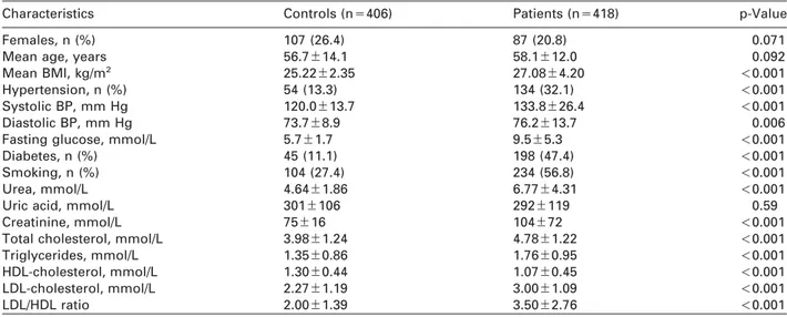
Study subjectsTable 1 shows the demographics and clinical bio- chemistry parameters of individuals in the CAD and control groups. CAD cases and controls were matched for age and gender, but typical differences for several conventional risk factors for CAD including habitual smoking, hypertension, diabetes mellitus, and BMI were observed (all p
-0.001). The CAD group showed higher concentrations of triglyceride, total cholesterol, LDL-cholesterol, and lower concentra- tions of HDL-cholesterol compared with the control group. We also found a statistically significant differ- ence between cases and controls with respect to systolic and diastolic blood pressure, uric acid, urea and creatinine.
TNF-agenotyping
The association of G-308A and T-1031C
TNFpoly-
morphisms with CAD risk was examined. There was
no significant departure from Hardy-Weinberg equi-
librium among participants in the genotype frequency
distributions of the two polymorphisms. TNF-a –308A
allele frequency was 19.6 in CAD patients and 19.0 in
healthy controls (p
s0.78; OR
s1.15; 95% CI
s0.86–
Table 1 Clinical characteristics of patients with coronary artery disease and controls.
Characteristics Controls (ns406) Patients (ns418) p-Value
Females, n (%) 107 (26.4) 87 (20.8) 0.071
Mean age, years 56.7"14.1 58.1"12.0 0.092
Mean BMI, kg/m2 25.22"2.35 27.08"4.20 -0.001
Hypertension, n (%) 54 (13.3) 134 (32.1) -0.001
Systolic BP, mm Hg 120.0"13.7 133.8"26.4 -0.001
Diastolic BP, mm Hg 73.7"8.9 76.2"13.7 0.006
Fasting glucose, mmol/L 5.7"1.7 9.5"5.3 -0.001
Diabetes, n (%) 45 (11.1) 198 (47.4) -0.001
Smoking, n (%) 104 (27.4) 234 (56.8) -0.001
Urea, mmol/L 4.64"1.86 6.77"4.31 -0.001
Uric acid, mmol/L 301"106 292"119 0.59
Creatinine, mmol/L 75"16 104"72 -0.001
Total cholesterol, mmol/L 3.98"1.24 4.78"1.22 -0.001
Triglycerides, mmol/L 1.35"0.86 1.76"0.95 -0.001
HDL-cholesterol, mmol/L 1.30"0.44 1.07"0.45 -0.001
LDL-cholesterol, mmol/L 2.27"1.19 3.00"1.09 -0.001
LDL/HDL ratio 2.00"1.39 3.50"2.76 -0.001
For continuous variables, mean and SD are given. BP, blood pressure.
Table 2 TNF-aG-308A and T-1031C genotypic and allelic distributions.
SNP Genotype frequency, % pa Allele frequency, % pa
G/G G/A A/A G A
G-308A 0.43 0.78
Controlsb 267 (65.8)b 124 (30.5) 15 (3.7) 81.0 19.0
Patientsb 265 (63.4) 142 (34.0) 11 (2.6) 80.4 19.6
T/T T/C C/C T C
T-1031C 0.25 0.12
Controlsb 284 (70.0) 111 (27.3) 11 (2.7) 83.6 16.4
Patientsb 270 (64.6) 134 (32.1) 14 (3.3) 80.6 19.4
aPearson’sx2-test;bnumber (% of total). SNP, single nucleotide polymorphism.
Table 3 TNF-aG-308A and T-1031C haplotype distribution.
Haplotype Controls Patients OR (95% CI)
Common haplotypes
–308G/–1031T 0.66 0.62 1a
–308A/–1031T 0.17 0.18 1.09 (0.82–1.45) ps0.54
–308G/–1031C 0.14 0.17 1.26 (0.95–1.67) ps0.1
Uncommon haplotypes
–308A/–1031C 0.01 0.01 1.01 (0.24–4.15) ps0.98
aHaplotype frequency determined by the maximum likelihood method; dfs3;x2s2.05; ps0.56. OR, odds ratio; CI, confidence interval.
1.55). Similarly, TNF-a –1031C allele frequency was 19.4 in CAD patients and 16.4 in healthy controls (p
s0.12; OR
s1.27; 95% CI
s0.94–1.72) (Table 2).
Based on the calculated OR, the calculated power of –308G/A (66.49%) and –1031T/C (71.79%) was obtained, which translated to an overall power of 69.14%. In this study, the frequency of
TNFG-308A (p
s0.43) and
TNFT-1031C (p
s0.25) genotypes were not significantly different between patients with CAD and healthy controls (Table 2).
Haplotype distribution
Table 3 shows the two-locus
TNFhaplotype analysis stratified by study subjects. Of the four possible
TNFhaplotypes, none was associated with CAD. The
‘double-mutant’ haplotype (–308A/–1031C) was un- common, and was present at very low frequencies in both controls and patients. Using the –308G/–1031T haplotype as reference, no significant association between the two remaining common haplotypes and CAD was observed.
Discussion
Previous cardiovascular studies have investigated the
association between several SNPs located in the pro-
moter region of the TNF-a gene and CAD or athero-
sclerosis (24–27), but conclusive data regarding the
role of TNF-a genotypes in CAD pathogenesis is need-
ed. The present study investigated the association between two biallelic polymorphisms of the TNF-a gene and their relation to CAD in an Arab African pop- ulation. The polymorphisms studied were two com- mon substitutions in the 59-flanking region.
No differences were found in the distribution of allelic or genotypic frequencies of TNF-a G-308A and TNF-a T-1031C polymorphisms between patients with CAD and healthy controls. In our hands, the frequency of the TNF-a –308A allele in the control group was similar to that observed in CAD patients. The lack of association between –308 TNF-a polymorphism and CAD is consistent with other studies (24, 28), and con- firm the results of Francis et al. (29) who found no association between TNF-a –308A allele and clinically or angiographically-documented coronary stenosis.
Our data confirm some previous studies that investi- gated Caucasian populations and failed to demon- strate that this locus could be a marker for greater risk of CAD. The same studies affirm that the allelic dis- tribution is different according to the geographical ori- gin of the study group (ranging from 24% to 12% for the rare allele) (25, 30). However, in contrast to other published data (26, 31), this may be due to different genetic and environmental risk factors or to different selection criteria of CAD patients.
Concerning the TNF-a –1031T/C polymorphism, the same distribution was observed between CAD patients and controls. The frequency of the –1031C allele was similar to that reported in a case control study on 1213 post-MI patients and 1561 healthy con- trols (32). In addition, the genotype and allele fre- quencies of control individuals were comparable to those previously reported for healthy Tunisian con- trols (33), which were similar to frequencies estab- lished for other ethnic groups, including healthy Swedish (32) and pan-Brazilian (34) individuals. The association between the two TNF-a loci and CAD was also studied by haplotype analysis. Of the possible four haplotypes, the ‘‘doublemutant’’ –308A/–1031C haplotype was under-represented in both groups, and no significant preferential association with incident CAD was seen.
In conclusion, our study demonstrates that neither –308G
)A nor –1031T
)C TNF-a gene polymorphisms are significantly associated with CAD among Tuni- sians, even before controlling for potential confound- ers. However, our study has some limitations. First, we were not able to measure plasma TNF-a concen- trations in patients and controls. It was also under- powered; a total of 636 (–308G/A) or 517 (–1031T/C) patients (compared to the current 418 patients) would have been needed for detection with 80% power. In addition, our study was limited to a specific ethnic group (North African Tunisian Arabs), thereby neces- sitating follow-up studies in patients from other ethnic groups. Another limitation includes the selection of the control group; while deemed healthy based on the routine pre-employment health examination, the pos- sibility that some of the controls that were included had asymptomatic stenosis needs follow-up. Also, the potential linkage of the TNF-a promoter polymor-
phisms studied with other TNF-a gene variants, or possibly polymorphisms in nearby genes (HLA region) cannot be excluded.
References
1. Mackay J, Mensah G. Atlas of heart disease and stroke.
World Healh Organization (Organisation Mondiale de la Sante´), 2005.
2. American Artery Association. Artery disease and stroke statistics 2003 update. Dallas, TX: American Artery Asso- ciation; 2002.
3. Libby P, Simon DI. Inflammation and thrombosis: the clot thickens. Circulation 2001;103:1718–20.
4. Beattie JH, Kwun IS. Is zinc deficiency a risk factor for atherosclerosis? Br J Nutr 2004;91:177–81.
5. Hansson GK. Inflammation, atherosclerosis, and coro- nary artery disease. N Engl J Med 2005;352:1685–95.
6. Bonnet J. Atherosclerosis. EMC-Cardiologie Ange´iologie 2005;2:436–58.
7. Ridker PM, Rifai N, Pfeffer M, Sacks F, Lepage S, Braun- wald E. Elevation of tumor necrosis factor-alpha and increased risk of recurrent coronary events after myo- cardial infarction. Circulation 2000;101:2149–53.
8. Valgimigli M, Ceconi C, Malagutti P, Merli E, Soukho- movskaia O, Francolini G, et al. Tumor necrosis factor- alpha receptor 1 is a major predictor of mortality and new-onset artery failure in patients with acute myocar- dial infarction: the cytokine-activation and long-term prognosis in myocardial infarction (C-ALPHA) study.
Circulation 2005;111:863–70.
9. Seiler C, Pohl T, Billinger M, Meier B. Tumour necrosis factor alpha concentration and collateral flow in patients with coronary artery disease and normal systolic left ventricular function. Heart 2003;89:96–7.
10. Maury CP, Teppo AM. Circulating tumour necrosis factor-alpha (cachectin) in myocardial infarction. J Intern Med 1989;225:333–6.
11. Basaran Y, Basaran MM, Babacan KF, Ener B, Okay T, Go¨k H, et al. Serum tumor necrosis factor levels in acute myocardial infarction and unstable angina pectoris.
Angiology 1993;44:332–7.
12. Cesari M, Penninx BW, Newman AB, Kritchevsky SB, Nicklas BJ, Sutton-Tyrrell K, et al. Inflammatory markers and onset of cardiovascular events: results from the Health ABC Study. Circulation 2003;108:2317–22.
13. Tentolouris C, Tousoulis D, Antoniades C, Bosinakou E, Kotsopoulou M, Trikas A, et al. Endothelial function and proinflammatory cytokines in patients with ischemic artery disease and dilated cardiomyopathy. Int J Cardiol 2004;94:301–5.
14. Kim F, Gallis B, Corson MA. TNF-alpha inhibits flow and insulin signalling leading to NO production in aortic endothelial cells. Am J Physiol Cell-Ph 2001;280:1057–
65.
15. Skoog T, Dichtl W, Boquist S, Skoglund-Andersson C, Karpe F, Tang R, et al. Plasma tumour necrosis factor-a and early carotid atherosclerosis in healthy middle-aged men. Eur Artery J 2002;23:376–83.
16. Paleolog EM, Delasalle SA, Buurman WA, Feldmann M.
Functional activities of receptors for tumor necrosis fac- tor-alpha on human vascular endothelial cells. Blood 1994;84:2578–90.
17. Wilson AG, Symons JA, McDowell TL, McDevitt HO, Duff GW. Effects of a polymorphism in the human tumor necrosis factor alpha promoter on transcriptional acti- vation. Proc Natl Acad Sci USA 1997;94:3195–99.
18. Pai JK, Pischon T, Ma J, Manson JE, Hankinson SE, Jos- hipura K, et al. Inflammatory markers and the risk of
coronary heart disease in men and women. N Engl J Med 2004;351:2599–610.
19. Vendrell J, Fernandez-Real JM, Gutierrez C, Zamora A, Simon I, Bardaji A, et al. A polymorphism in the pro- moter of the tumor necrosis factor-alpha gene (y308) is associated with coronary heart disease in type 2 diabetic patients. Atherosclerosis 2003;167:257–64.
20. Lee EB, Kim JY, Lee YJ, Park MH, Song YW. TNF and TNF receptor polymorphisms in Korean Behcet’s disease patients. Hum Immunol 2003;64:614–20.
21. Asghar T, Yoshida S, Kennedy S, Negoro K, Zhuo W, Hamana S, et al. The tumor necrosis factor promoter T-1031C polymorphism is associated with decreased risk of endometriosis in a Japanese population. Hum Reprod 2004;19:2509–14.
22. Report of the Joint International Society and Federation of Cardiology/World Health Organization Task Force on Standardization of Clinical Nomenclature. Nomenclature and criteria for diagnosis of ischemic artery disease.
Circulation 1979;59:607–8.
23. Tregouet DA, Escolano S, Tiret L, Mallet A, Golmard JL.
A new algorithm for haplotype-based association anal- ysis: the stochastic-EM algorithm. Ann Hum Genet 2004;
68:165–77.
24. Koch W, Kastrati A, Bo¨ttiger C, Mehilli J, von Beckerath N, Scho¨mig A. Interleukin-10 and tumor necrosis factor gene polymorphisms and risk of coronary artery disease and myocardial infarction. Atherosclerosis 2001;159:
137–44.
25. Herrmann SM, Ricard S, Nicaud V, Mallet C, Arveiler D, Evans A, et al. Polymorphisms of the tumour necrosis factor-a gene, coronary artery disease and obesity. Eur J Clin Invest 1998;92:14–8.
26. Keso T, Perola M, Laippala P, Ilvekoski E, Junnas TA, Mikkelsson J, et al. polymorphisms within the tumor necrosis factor locus and prevalence of coronary artery
disease in middleaged men. Atherosclerosis 2001;154:
691–7.
27. Vendrell J, Fernandez-Real JM, Gutierrez C, Zamora A, Simon I, Bardaji A, et al. A polymorphism in the pro- moter of the tumor necrosis factor-alpha gene (y308) is associated with coronary heart disease in type 2 diabetic patients. Atherosclerosis 2003;167:257–64.
28. Allen RA, Lee EM, Roberts DH, Park BK, Pirmohamed M.
Polymorphisms in the TNF-alpha and TNF-receptor genes in patients with coronary artery disease. Eur J Clin Invest 2001;31:843–51.
29. Francis SE, Camp NJ, Dewberry RM, Gunn J, Syrris P, Carter ND, et al. Interleukin-1 receptor antagonist gene polymorphism and coronary artery disease. Circulation 1999;99:861–6.
30. Wang XL, Oosterhof J. Tumour necrosis factor a G-308A polymorphism and risk for coronary artery disease. Clin Sci 2000;98:435–7.
31. Padovani JC, Pazin-Filho A, Simoes MV, Marin-Neto JA, Zago MA, Franco RF. Gene polymorphisms in the TNF locus and the risk of myocardial infarction. Thromb Res 2000;100:263–9.
32. Bennet AM, van Maarle MC, Hallqvist J, Morgenstern R, Frostegard J, Wiman B, et al. Association of TNFaserum levels and TNFA promoter polymorphisms with risk of myocardial infarction. Atherosclerosis 2006;187:408–14.
33. Kamoun M, Chelbi H, Houman MH, Lacheb J, Hamzaoui K. Tumor necrosis factor gene polymorphisms in Tuni- sian patients with Behcet’s disease. Hum Immunol 2007;68:201–5.
34. Atsuta Y, Ito LS, Oba-Shinjo SM, Uno M, Shinjo SK, Marie SK, et al. Associations of TNF-A-1031TT and –857TT genotypes with Helicobacter pylori seropositivity and gastric atrophy among Japanese Brazilians. Int J Clin Oncol 2006;11:140–5.